Regulation of ferroptosis by inter-organellar metabolic signaling network

Luna Zhang
#
,
Zefang Bimo Zhao
#
,
Weitao Li
#,*
,
Jiajun Zhu
*

*Correspondence to:
Weitao Li, State Key Laboratory of Molecular Oncology, School of Basic Medical Sciences, Tsinghua-Peking Center for Life Sciences, Tsinghua University, Beijing 100084, China.
E-mail: weitaol@mail.tsinghua.edu.cn
Jiajun Zhu, State Key Laboratory of Molecular Oncology, School of Basic Medical Sciences, Tsinghua-Peking Center for Life Sciences, Tsinghua University, Beijing 100084, China. E-mail: zhujiajun@tsinghua.edu.cn
Jiajun Zhu, State Key Laboratory of Molecular Oncology, School of Basic Medical Sciences, Tsinghua-Peking Center for Life Sciences, Tsinghua University, Beijing 100084, China. E-mail: zhujiajun@tsinghua.edu.cn
Ferroptosis Oxid Stress. 2026;2:202628. 10.70401/fos.2026.0038
Received: June 12, 2026Accepted: July 28, 2026Published: July 28, 2026
Abstract
Ferroptosis is a form of regulated cell death driven by iron-dependent lipid peroxidation, and is therefore intrinsically coupled to cellular redox metabolism. Progression of ferroptosis is regulated by an integrated network of metabolic signaling pathways that involve inter-organellar communication. In this Review, we synthesize recent progress in the field that has advanced our understanding of the metabolic determinants of ferroptosis. We discuss key metabolites which participate in the execution and prevention of ferroptosis, describe major signaling axes that impinge on regulating these metabolic activities to determine cellular ferroptosis sensitivity, and highlight how compartmentalized metabolism within and across distinct intracellular organelles collectively shape ferroptosis progression. Together, this Review aims to provide a framework that unifies metabolic state, signaling plasticity and organellar cooperation as a mechanistic basis of ferroptosis, underscoring its implications in the pathogenesis of various diseases including cancer.
Keywords
Cellular metabolism, metabolic signaling, subcellular compartments
1. Introduction
Ferroptosis is a form of regulated cell death characterized by iron-dependent lipid peroxidation, and is mechanistically distinct from other cell death modalities[1]. Since its conceptualization, ferroptosis has been increasingly recognized as a critical determinant in diverse pathophysiological contexts, such as embryonic development, neurodegeneration, ischemia–reperfusion injury, and cancer progression[2-5]. Ferroptosis is viewed as a metabolically regulated process determined by various aspects of the cellular reduction-oxidation (redox) state[6,7]. The progression of ferroptosis is promoted by both enzymatic and non-enzymatic reactions that produce and propagate lipid peroxides within cellular membranes[8-10]. On the other hand, ferroptosis is antagonized by multiple cellular antioxidant systems, which in turn are coupled with redox metabolism. As a result, ferroptotic activity is shaped by the intricate metabolic and signaling network that regulates the balance between lipid peroxidation and detoxification.
Furthermore, regulation of ferroptosis is spatially organized across intracellular compartments[11-13]. Although lipid peroxidation ultimately manifests at cellular membranes, execution of ferroptosis requires a coordinated interplay among multiple intracellular organelles that harbor metabolic pathways and act as signaling platforms. In this Review, we summarize recent progress towards understanding the metabolic determinants of ferroptosis. We highlight the signaling pathways that instruct metabolic activities, and emphasize the interplay among intracellular organelles that underlies ferroptosis regulation.
2. Metabolites Are Key Players in Ferroptosis
Ferroptosis is directly triggered by excessive phospholipid peroxidation. As a result, the progression of ferroptosis depends on a variety of metabolites that initiate and propagate lipid oxidation. On the other hand, cellular defense programs that restrain ferroptosis also rely on the availability of multiple metabolic molecules with antioxidant properties. Therefore, a plethora of intracellular metabolites have emerged as key determinants in ferroptosis regulation.
2.1 Polyunsaturated fatty acid (PUFA)
Peroxidation of phospholipids containing polyunsaturated fatty acyl chains (PUFA-PLs) can lead to ferroptosis. PUFA-PLs are exquisitely susceptible to oxidation due to the presence of chemically labile bis-allylic carbons within their unsaturated fatty acyl chains. In mammalian cells, fatty acyl chains derived from arachidonic acid (20:4) and adrenic acid (22:4) contain such bis-allylic moieties, and are potent substrates that sensitize membranes to peroxidation[6].
The abundance of cellular PUFA is dynamically regulated through multiple processes including their uptake, synthesis, membrane incorporation, storage, and degradation. Exogenous PUFAs are imported primarily via the scavenger receptor CD36 (Figure 1). For instance, in an acidic tumor microenvironment, cancer cells can increase CD36 expression and PUFA uptake, thereby enhancing ferroptosis susceptibility[14]. On the other hand, CD36-mediated PUFA import can also increase ferroptosis in tumor-infiltrating T cells and contribute to immunosuppression[15]. Beyond the context of cancer, the steroid hormone 17α-hydroxypregnenolone was reported to possess inhibitory activity on CD36 through binding to its receptor G protein-coupled receptor 56 (GPR56), thereby limiting PUFA import and ferroptotic cell death during hepatic injury[16]. In addition to direct uptake, arachidonic acid can be synthesized from linoleic acid through a pathway involving the enzymes fatty acid desaturase 2 (FADS2), elongation of very long-chain fatty acid protein 5 (ELOVL5), and fatty acid desaturase 1 (FADS1), all of which were shown to increase cellular ferroptosis sensitivity[17].
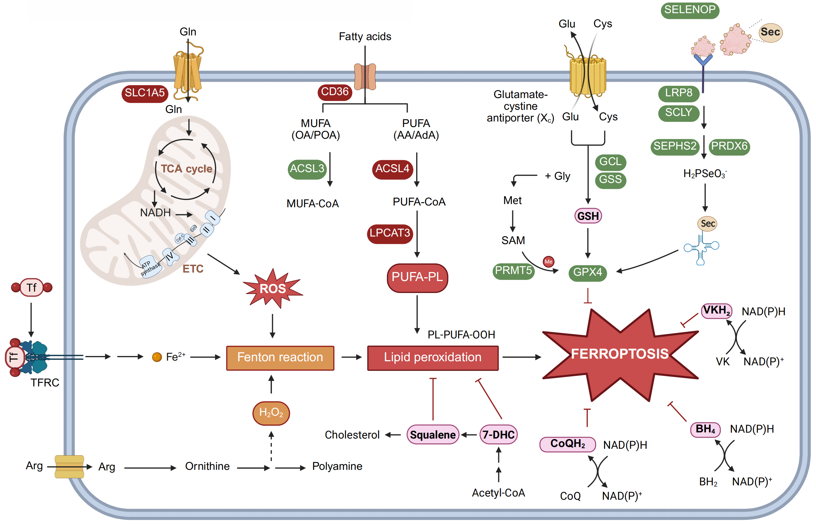
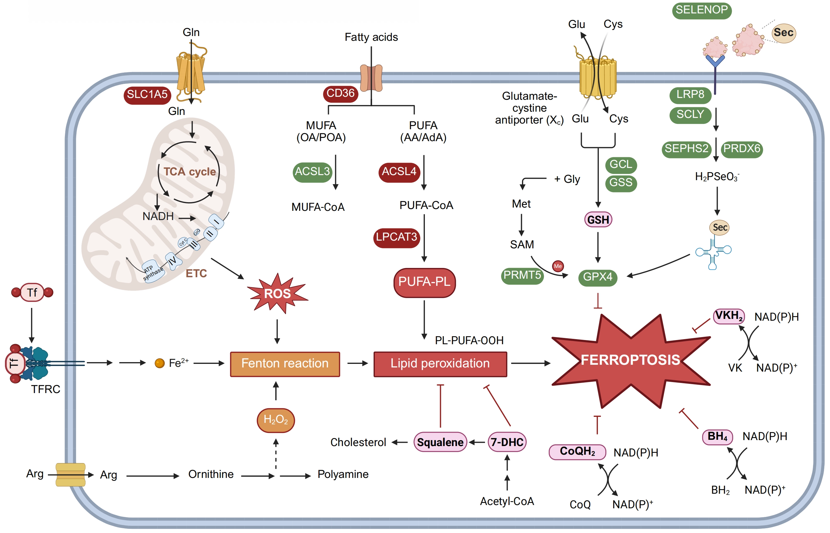
Figure 1. Metabolites are key players in ferroptosis. Ferroptosis sensitivity is determined by the balance between lipid peroxidation and antioxidant defense. SLC1A5-mediated glutamine uptake fuels the TCA cycle and mitochondrial respiration, thereby promoting ROS production and Fenton chemistry. TFRC-mediated iron uptake expands the labile iron pool, whereas pro-oxidant intermediates generated during polyamine biosynthesis further contribute to lipid peroxidation. CD36 supports the uptake of extracellular fatty acids. ACSL4 and LPCAT3 promote the generation of PUFA-containing phospholipids that are highly susceptible to peroxidation. In contrast, ACSL3 promotes MUFA incorporation into membrane phospholipids and suppresses lipid peroxidation. On the anti-ferroptotic side, xCT-dependent cystine uptake supports glutathione synthesis, and GPX4 detoxifies PLOOHs. LRP8-mediated SELENOP uptake supplies selenium for selenoprotein biosynthesis. Additional radical-trapping antioxidants, including CoQH2, BH4, VKH2, and 7-DHC, further inhibit ferroptosis. Created in BioRender. Zhao, B. (2026) https://BioRender.com/hdoxh5d. ROS: reactive oxygen species; TCA: tricarboxylic acid; TFRC: transferrin receptor; ACSL3: acyl-CoA synthetase long-chain family member 3; LPCAT3: lysophosphatidylcholine acyltransferase 3; PUFA: polyunsaturated fatty acid; MUFA: monounsaturated fatty acid; GPX4: glutathione peroxidase 4; LRP8: lipoprotein receptor-related protein 8; BH4: tetrahydrobiopterin; VKH2: vitamin K hydroquinone; 7-DHC: 7-dehydrocholesterol; SELENOP: selenoprotein P.
Following its acquisition, the acyl-CoA synthetase long-chain 4 (ACSL4) - Lysophosphatidylcholine acyltransferase 3 (LPCAT3) axis commits intracellular PUFA to a pro-ferroptosis fate by catalyzing the formation of PUFA-PL (Figure 1). In particular, ACSL4 activates arachidonic acid to arachidonoyl-CoA, and LPCAT3 esterifies it into membrane phospholipids, primarily forming phosphatidylethanolamine (PE)[18]. This esterification is essential because peroxidation of free PUFAs is insufficient to induce ferroptosis[19]. Notably, ACSL4 itself is dynamically regulated within a positive feedback loop. Initial lipid peroxides activate protein kinase C βII (PKCβII), which in turn phosphorylates and enhances the activity of ACSL4[20]. However, while loss of ACSL4 is strongly protective, it does not completely abrogate ferroptosis, indicating the existence of complementary pathways for PUFA-PL synthesis[21].
Membrane lipid composition is further determined by remodeling and repair mechanisms. For example, the Lands’ cycle enables dynamic replacement of fatty acyl chains, where the calcium-independent phospholipase iPLA2β (also known as PLA2G6) can hydrolyze and remove oxidized PUFA tails from phospholipids, thereby acting as a repair enzyme to attenuate ferroptosis[22]. As a consequence, genetic deficiency in iPLA2β is linked to pathological ferroptosis in human diseases, including Parkinson’s disease and placental dysfunction[23].
In addition to direct membrane incorporation, PUFAs can be diverted into storage as triacylglycerols (TAGs) within lipid droplets[24]. This sequestration acts as a buffering mechanism and limits substrate availability for peroxidation to protect against ferroptosis[25]. However, this process is reversible. Degradation of lipid droplets via lipolysis releases stored PUFAs into the more accessible metabolic pool and promotes ferroptosis. For instance, RAS-related in brain 7A (RAB7A)-mediated lipophagy in hepatocytes and adipose tissue triglyceride lipase (ATGL)-mediated lipolysis in breast cancer cells can both sensitize ferroptosis by increasing the supply of PUFAs for membrane incorporation and peroxidation[26].
Finally, PUFAs are subject to catabolism via mitochondrial β-oxidation, reducing their availability for phospholipid biosynthesis[6]. The enzyme 2,4-dienoyl-CoA reductase 1 (DECR1), essential for PUFA β-oxidation, is frequently upregulated in cancers such as prostate adenocarcinoma, which may represent an adaptive metabolic strategy to enhance cell survival against ferroptosis[27,28].
2.2 Monounsaturated fatty acid (MUFA)
In contrast to the pro-ferroptotic role of PUFAs, MUFAs, such as oleate or palmitoleate, have emerged as molecules that protect a wide range of cell types from cell death caused by ferroptosis inducers[29]. This protective effect is largely dependent on acyl-CoA synthetase long-chain family member 3 (ACSL3), which activates MUFAs to their CoA-thioester forms for subsequent incorporation into membrane phospholipids[29] (Figure 1).
Cells acquire MUFAs through both exogenous uptake and endogenous biosynthesis. For example, metastasizing melanoma cells can take up MUFA-enriched lipoproteins to overcome the oxidative stress associated with cancer progression[30]. Adipocyte-secreted oleate was shown to protect neighboring triple-negative breast cancer cells from ferroptosis in an ACSL3-dependent manner[30,31]. On the other hand, stearoyl-CoA desaturase 1 (SCD1) can generate MUFAs de novo from saturated precursors[32]. As a result, SCD1 inhibition was shown to decrease the cellular MUFA pool and sensitize cells to ferroptosis[33]. Similar to PUFAs, MUFAs can also be dynamically stored as TAGs in lipid droplets and mobilized under stress conditions. In clear cell renal cell carcinoma, hypoxia triggers the release of stored MUFAs via lipolysis and their subsequent incorporation into membranes, remodeling the cellular lipidome to inhibit ferroptosis[34].
While MUFA incorporation into phospholipids effectively displaces the PUFA-containing species and reduces membrane oxidation, its protective effect against ferroptosis is not fully explained by simple substrate competition[6]. It was shown that MUFA-containing phospholipids might also alter membrane biophysics to hinder radical propagation, activate protective signaling programs, or favor the formation of less toxic peroxidation products[35]. Thus, MUFAs establish a multi-faceted defense system, positioning MUFA metabolism as a critical determinant of the ferroptotic threshold and a promising target for therapeutic intervention.
2.3 Cholesterol
Unlike PUFA and MUFA, cholesterol metabolism plays a dual and context-dependent regulatory role in ferroptosis, acting in part through its biosynthetic intermediates and their derivatives[32]. For example, isopentenyl pyrophosphate (IPP) is required for maturation of the selenocysteine-specific tRNA, which is indispensable for translation of selenoproteins such as glutathione peroxidase 4 (GPX4). IPP also acts as an essential precursor for coenzyme Q (CoQ) biosynthesis in support of ferroptosis resistance[36]. As a result, pharmacological inhibition of the rate-limiting enzyme hydroxyl-methylglutaryl-CoA reductase (HMGCR) (e.g., by statins) in the mevalonate pathway that leads to IPP production was shown to sensitize cells to ferroptosis[37]. By contrast, inhibition of enzymes further down the cholesterol biosynthesis pathway, such as 7-dehydrocholesterol (7-DHC) reductase or squalene monooxygenase, can lead to the accumulation of intermediates including 7-DHC and squalene[38-40]. These cholesterol precursors function as highly effective lipophilic radical-trapping antioxidants (RTAs), directly scavenging lipid radicals and shielding cellular membranes from extensive peroxidation (Figure 1).
Cholesterol, as well as its oxidized derivatives such as 27-hydroxycholesterol (27-HC), can also act as signaling molecules in the tumor microenvironment and upregulate CD36 in tumor-infiltrating T cells. This in turn results in increased PUFA uptake by T cells, leading to their ferroptotic cell death[15]. Furthermore, chronic 27-HC exposure drives the selection of tumor cells with heightened lipid uptake and biosynthetic activity, thereby promoting tumorigenesis and metastasis[41]. In addition to these signaling effects, direct oxidation of cholesterol results in the formation of its hydroperoxide species that may further shape ferroptosis regulation[42]. The precise role of cholesterol peroxidation in initiating or propagating the ferroptotic lipid chain reaction remains an open question.
2.4 RTAs
A number of cellular RTAs make up a critical defense system against ferroptosis. These molecules function by donating a hydrogen atom to phospholipid peroxyl radicals (PLOO·), thereby converting them into less reactive lipid hydroperoxides and terminating the chain reaction of lipid peroxidation[19]. Multiple endogenous pathways support the generation and regeneration of these RTAs (Figure 1). For example, tetrahydrobiopterin (BH4) is a potent endogenous RTA that directly scavenges PLOO· to halt the peroxidation chain reaction. Its antioxidant activity is sustained through enzymatic recycling by dihydrofolate reductase (DHFR), which regenerates active BH4 from its oxidized form[43].
The FSP1-CoQ axis constitutes another key defense system. FSP1 reduces ubiquinone to ubiquinol, a lipophilic RTA that neutralizes PLOO· at the plasma membrane[44,45]. In parallel, dihydroorotate dehydrogenase (DHODH), located at the inner mitochondrial membrane, regenerates reduced CoQ within the mitochondrial compartment, thereby protecting mitochondria from ferroptotic damage[46]. In addition, FSP1 also participates in the non-canonical vitamin K cycle by reducing vitamin K to its active hydroquinone form (VKH2), which similarly functions as an RTA[47]. This vitamin K-based defense is further reinforced by the canonical vitamin K cycle, where vitamin K epoxide reductase complex subunit 1-like 1 (VKORC1L1) acts as a central reductase responsible for sustaining the VKH2 pool[48].
Vitamin E (also known as α-tocopherol) is another example of a lipophilic RTA, with particularly well-defined roles in physiological contexts such as T cell immunity[49,50]. It was recently shown that vitamin E delivery via circulating lipoproteins confers robust ferroptosis resistance across various cancer types[51]. In addition, vitamin A precursors, including retinol and retinal, function as direct RTAs, whereas the all-trans retinoic acid form of vitamin A primarily executes a transcriptional program via retinoic acid receptors to regulate ferroptosis[52].
2.5 Iron
Ferroptosis is conceptually defined by its dependence on iron, which fulfills multiple catalytic roles in the ferroptotic cascade. First, phospholipid hydroperoxides (PLOOHs), once accumulated, can engage labile intracellular iron to initiate the non-enzymatic Fenton chain reaction, which rapidly amplifies lipid peroxides (Figure 1). PLOOHs can also be reductively cleaved by ferrous ions (Fe2+) to generate highly reactive phospholipid alkoxyl radicals (PLO·), or react with ferric ions (Fe3+) to produce PLOO·[8]. As a result, this iron-catalyzed cycle (Fe2+ ⇌ Fe3+) continuously propagates radical-driven chain reactions[19].
In addition to non-enzymatic catalysis, iron also acts as an essential cofactor in enzyme-mediated lipid peroxidation. For instance, the lipoxygenase (LOX) family of iron-dependent dioxygenases plays an important role in ferroptosis regulation[9]. While lipoxygenase 5 (ALOX5) primarily oxidizes free arachidonic acid, ALOX12 and ALOX15 can directly oxidize esterified fatty acids in membrane phospholipids[53,54]. This activity is enhanced when ALOX15 complexes with phosphatidylethanolamine-binding protein 1 (PEBP1)[55]. The cytochrome P450 oxidoreductase (POR) system, another iron-dependent enzymatic complex, also contributes to peroxidation initiation[56]. While certain cytochrome P450 isoforms are capable of directly oxidizing fatty acids, the POR system and the related oxidoreductase cytochrome b5 (CYB5R1) reductase promote lipid peroxidation mainly by generating H2O2 to fuel Fenton chemistry[12].
Owing to the central role of iron in lipid peroxidation, cellular susceptibility to ferroptosis is largely governed by the size and reactivity of the labile iron pool (LIP). Most mammalian cells acquire iron via transferrin receptor (TFRC)-mediated endocytosis[57], during which Fe3+ is released from transferrin in the endosome and lysosome, and reduced to the soluble Fe2+ form by metalloreductases such as STEAP3. The divalent metal transporter DMT1 subsequently transports Fe2+ into the cytosol, thereby supplying the LIP[54]. Consistent with an essential role of endocytosis in supporting cellular LIP, lysosomal inhibitors were reported to suppress cysteine deprivation-induced ferroptosis[58].
The cellular LIP is further determined by other mechanisms. The iron-responsive element/iron-regulatory protein (IRE/IRP) system regulates LIP by post-transcriptionally coordinating the expression of key iron metabolism factors, including TFRC and ferritin[59-61]. Excess iron is sequestered within ferritin, a multimeric protein complex composed of ferritin light chain (FTL) and ferritin heavy chain (FTH1) subunits. By contrast, selective autophagic degradation of ferritin mediated by nuclear receptor coactivator 4 (NCOA4), a process known as ferritinophagy, releases stored iron and sensitizes cells to ferroptosis[62,63]. To prevent iron overload, excess ferrous iron can be exported out of cells via ferroportin (also known as SLC40A1)[64]. In the meantime, iron can be utilized for the biosynthesis of essential cofactors such as heme and iron-sulfur clusters (Fe-S)[65]. Iron liberation from heme through the catalytic activity of heme oxygenase-1 (HMOX1) also plays an important role in maintaining the cellular LIP[66].
2.6 Amino acid
The acquisition and metabolism of amino acids are determinant to ferroptosis regulation. Because cysteine is a rate-limiting substrate for glutathione (GSH) synthesis, its deprivation can lead to rapid accumulation of lipid hydroperoxides due to disruption of the GSH-GPX4 defense system[67] (Figure 1). As another sulfur-containing amino acid, methionine metabolism influences ferroptosis largely through S-adenosylmethionine (SAM)-dependent methylation reactions. SAM is required for the biosynthesis of CoQ[68]. As a result, methionine restriction or impaired SAM production compromises CoQ synthesis and sensitizes cells to ferroptosis[68]. Moreover, it was reported that stabilization of GPX4 is also regulated by SAM-dependent methylation. The protein arginine methyltransferase 5 (PRMT5) methylates GPX4, preventing its ubiquitination and subsequent proteasomal degradation[69]. Similar to the role of cysteine in GSH synthesis, glycine is another constituent of the GSH tripeptide[70]. Dietary restriction of glycine, particularly when combined with inhibition of the Fe-S biogenesis pathway and thus an increased LIP, promotes ferroptosis in experimental models of cancer[71]. In addition, serine can function as a precursor to support both cysteine and glycine de novo biosynthesis[72]. Serine is also a direct substrate for sphingolipid production, thereby influencing membrane architecture and the lipid landscape where peroxidation occurs[73,74].
By contrast, several other amino acids can function to promote ferroptosis. For example, glutamine facilitates ferroptosis through glutaminolysis to replenish the tricarboxylic acid (TCA) cycle[57], which enhances mitochondrial respiration and electron transport chain (ETC) activity, leading to increased reactive oxygen species (ROS) generation[75,76] (Figure 1). Moreover, arginine and its related metabolites, ornithine and polyamines, were shown to promote ferroptosis likely by generating pro-oxidant intermediates during their metabolism and consuming cellular antioxidant capacity[77,78]. Collectively, because amino acids fulfill diverse cellular functions in addition to acting as building blocks for protein translation, different amino acids could have distinct regulatory roles in ferroptosis.
2.7 Selenium
Selenium is an essential trace element that plays a pivotal role in cellular redox homeostasis. Low density lipoprotein receptor-related protein 8 (LRP8)-mediated endocytosis of selenium carriers, such as selenoprotein P (SELENOP), is a major way by which cells acquire selenium[79] (Figure 1). SELENOP is then degraded in lysosomes to release selenocysteine (Sec), which is cleaved by selenocysteine lyase (SCLY) to generate selenide[80,81]. This selenide is further utilized by selenophosphate synthetase 2 (SEPHS2) to produce selenophosphate for tRNASec biosynthesis. In a parallel process, peroxiredoxin 6 (PRDX6) acts as a selenium acceptor protein and directly reacts with selenide to form a selenenylsulfide intermediate, thereby capturing and chaperoning the highly reactive selenium species for delivery to the selenoprotein biosynthesis machinery[82-85]. Together, these mechanisms involving multiple enzymes are required for the incorporation of selenium into a family of selenoproteins, including GPX4[86,87]. Disruption of this process, particularly in the context of cancer, can compromise GPX4 production and consequently sensitize cells to ferroptosis[88].
Beyond its role in selenoproteins, selenium also exerts protective effects against ferroptosis through non-canonical mechanisms. The metabolic intermediate selenide can directly reduce mitochondrial CoQ to ubiquinol. This process is significantly enhanced by sulfide quinone oxidoreductase (SQOR), which efficiently channels electrons from selenide to the CoQ pool, providing rapid and localized protection against lipid peroxidation[89]. Furthermore, selenium orchestrates a transcriptional adaptive response to ferroptotic stress, involving transcription factors such as transcription factor AP-2 gamma (TFAP2c) and Sp1, thereby reinforcing long-term antioxidant defense[90].
3. Signaling Pathways Dictate Ferroptosis Sensitivity
The cellular metabolic network is highly coordinated by various signaling pathways, which respond to changes in environmental cues and instruct proper cellular activities including metabolic biosynthesis, antioxidant defense, as well as cell fate changes. These signaling events play an important role in ferroptosis regulation, particularly under pathophysiological conditions such as cancer.
3.1 Receptor tyrosine kinase (RTK) and phosphatidylinositide 3-kinase (PI3K) signaling
The RTK-PI3K signaling axis is central to growth factor-mediated cell proliferation. In cancer, this pathway is frequently hijacked by genetic alterations that drive its constitutive activation[91]. Among these oncogenic events, mutations in PIK3CA, which encodes the catalytic subunit of PI3K, can lead to sustained mTORC1 signaling. Activated mTORC1 in turn upregulates the transcription factor SREBP1 and its downstream target SCD1, which is a key enzyme that converts saturated fatty acids into MUFAs, thereby conferring ferroptosis resistance[92] (Figure 2). Beyond oncogenic mutations, adaptive metabolic stress can also engage this axis to promote ferroptosis evasion. In nutrient-deprived pancreatic cancer cells, metabolic stress activates the mTORC1-SREBP1-SCD1 pathway, leading to increased intracellular MUFA content and enhanced ferroptosis resistance[93]. This finding suggests that the mTOR-SREBP1-SCD1 axis serves as a convergent stress-response mechanism that can be co-opted by cancer cells under both genetic and environmental pressures to remodel their lipid landscape and evade ferroptotic death. Conversely, pharmacological blockade of this pathway reverses the resistant state. For example, aspirin was shown to sensitize colorectal cancer cells to ferroptosis by suppressing the PI3K-mTORC1 pathway, which downregulates SREBP1 activity and the subsequent SCD1-mediated MUFA biosynthesis[94]. Thus, oncogenic PI3K signaling establishes a ferroptosis-resistant lipid environment via the mTORC1-SREBP1-SCD1 axis.
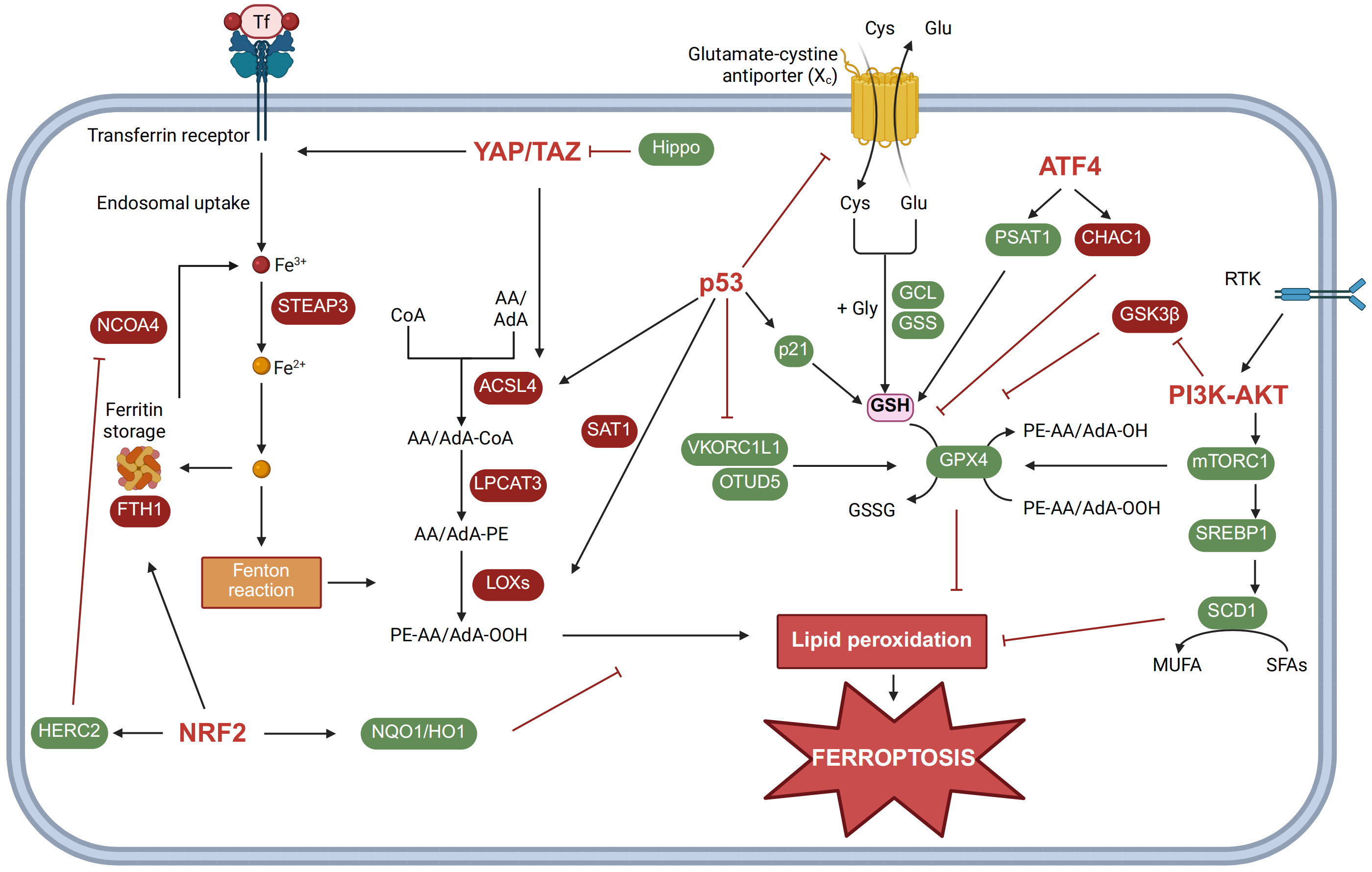
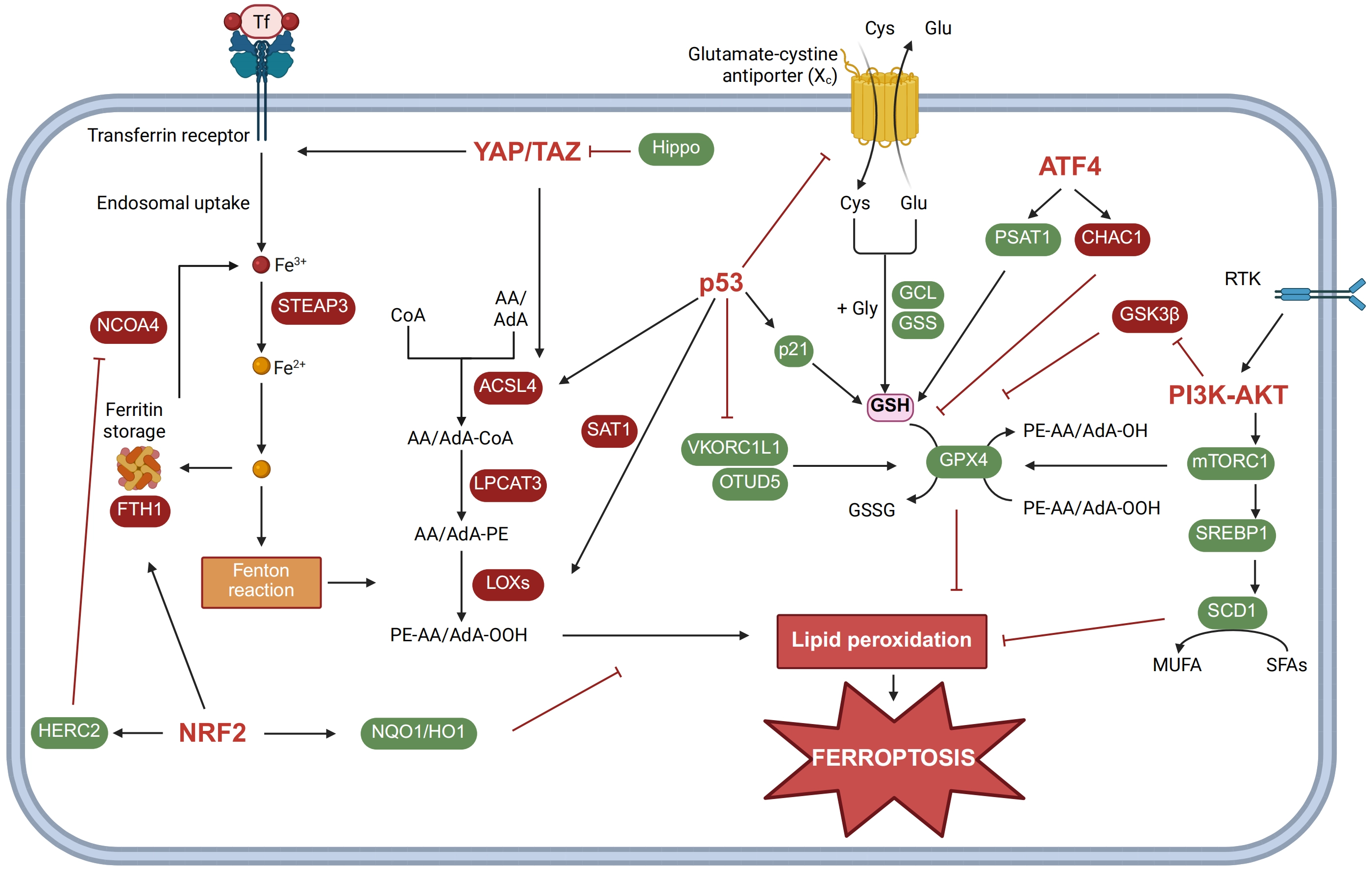
Figure 2. Signaling pathways dictate ferroptosis sensitivity. Signaling pathways shape ferroptosis sensitivity by coordinating iron metabolism, lipid remodeling, and antioxidant defense. Hippo signaling suppresses YAP/TAZ activity through phosphorylation-dependent inhibition. When released from Hippo restraint, YAP/TAZ promote TFRC-mediated iron uptake and enhance ACSL4-dependent lipid remodeling, thereby sensitizing cells to ferroptosis. NRF2 suppresses ferroptosis by inducing FTH1 to sequester iron. NRF2 also upregulates NQO1 and HO1 to limit oxidative stress and lipid peroxidation. In addition, NRF2 inhibits NCOA4-dependent ferritinophagy and further restricts iron release. p53 promotes ferroptosis by inducing SAT1 to upregulate ALOX15 expression. p53 also represses SLC7A11 and VKORC1L1, thereby weakening glutathione- and vitamin K-dependent defense systems. In contrast, the canonical p53 target p21 suppresses ferroptosis by maintaining glutathione homeostasis. ATF4 has dual roles in ferroptosis regulation. ATF4 promotes glutathione synthesis through SLC7A11 induction, but also upregulates CHAC1 to degrade glutathione. The PI3K-AKT-mTOR pathway generally protects cells from ferroptosis. PI3K-AKT signaling inhibits GSK3β, whereas mTORC1 activates the SREBP1-SCD1 axis to promote MUFA synthesis and membrane remodeling. Created in BioRender. Zhao, B. (2026) https://BioRender.com/hdoxh5d. YAP: Yes-associated protein; TAZ: transcriptional coactivator with PDZ-binding motif; TFRC: transferrin receptor; ACSL4: acyl-CoA synthetase long-chain family member 4; NRF2: nuclear factor erythroid 2-related factor 2; FTH1: ferritin heavy chain 1; NQO1: NAD(P)H:quinone oxidoreductase 1; HO1: heme oxygenase-1; NCOA4: nuclear receptor coactivator 4; SAT1: spermidine/spermine N1-acetyltransferase 1; ALOX15: arachidonate 15-lipoxygenase; SLC7A11: solute carrier family 7 member 11; VKORC1L1: vitamin K epoxide reductase complex subunit 1-like 1; ATF4: activating transcription factor 4; CHAC1: ChaC glutathione specific gamma-glutamylcyclotransferase 1; PI3K: phosphatidylinositide 3-kinase; mTOR: mechanistic target of rapamycin; GSK3β: glycogen synthase kinase-3beta; mTORC1: mechanistic target of rapamycin complex 1; SREBP1: sterol regulatory element-binding protein-1; SCD1: stearoyl-CoA desaturase 1; MUFA: monounsaturated fatty acid.
Beyond this lipid remodeling program, recent studies have uncovered a parallel mechanism, whereby PI3K-AKT signaling promotes ferroptosis resistance by enhancing mitochondrial fitness[95]. Cancer cells with constitutive PI3K activation often display elevated mitochondrial mass, membrane potential, and antioxidant capacity. Conversely, pharmacologic inhibition of the PI3K-AKT-mTORC1 axis compromises mitochondrial fitness, triggers mitophagy, and sensitizes cells to ferroptosis. Therefore, expression of oncogenic PIK3CA or activation of AKT confers ferroptosis resistance in a mitochondria-dependent manner[95].
In addition, mTORC1 impinges on ferroptosis defense at the level of protein synthesis. It was shown that mTORC1 can promote GPX4 protein synthesis through its role in translation regulation[96] (Figure 2). Consequently, mTORC1 inhibition sensitizes cells to ferroptosis, and this pro-ferroptotic effect is further amplified by the concomitant induction of autophagy. For instance, mTORC1 inhibitors promote ferroptosis in head and neck squamous cell carcinoma cells by reducing the phosphorylation of unc-51 like autophagy activating kinase 1 (ULK1) and autophagy-related protein 13 (ATG13), which enhances ferritinophagy and elevates the intracellular LIP[97].
In tumor cells harboring isocitrate dehydrogenase mutations, accumulated oncometabolite D-2-hydroxyglutarate (D-2-HG) can activate PI3K signaling. This aberrant activation leads to the inhibition of the GSK3β/β-TrCP E3 ligase complex, thereby dampening nuclear factor erythroid 2-related factor 2 (NRF2) degradation. The stabilized NRF2 then activates a series of downstream antioxidant target genes, ultimately alleviating cellular oxidative stress and protecting against ferroptosis[98].
Collectively, RTK-PI3K signaling and the downstream pathways engage multiple mechanisms to orchestrate a robust cellular ferroptosis-resistant state.
3.2 p53
TP53, encoding the p53 protein, is the most frequently mutated tumor suppressor gene in human cancers. p53 functions primarily as a transcription factor that orchestrates diverse tumor suppressive programs, including cell cycle arrest, metabolic reprogramming, senescence, and apoptosis[99,100]. Recent studies have revealed that p53 also plays a dual role in ferroptosis[100,101].
The best-characterized mechanism by which p53 sensitizes cells to ferroptosis involves the cystine–glutamate antiporter SLC7A11. It was shown that p53 directly suppresses SLC7A11 transcription, thereby limiting cystine uptake and depleting the intracellular GSH pool[102] (Figure 2). Reduced SLC7A11 expression simultaneously relieves SLC7A11-mediated inhibition of ALOX12, thus enhancing phospholipid peroxidation and driving ferroptosis[103].
Beyond the SLC7A11–GSH axis, p53 activation was shown to downregulate VKORC1L1, the reductase essential for sustaining the VKH2 pool, thereby disrupting this protective pathway[48] (Figure 2). Furthermore, p53 also targets ferroptosis defense mechanisms at the post-translational level. In gastric cancer cells, p53 transcriptionally represses the deubiquitinase OTU deubiquitinase 5 (OTUD5), leading to decreased GPX4 protein stability[104]. In parallel, p53-mediated transcriptional activation of spermidine/spermine N1-acetyltransferase 1 (SAT1) promotes ferroptosis, likely through remodeling polyamine metabolism and upregulating ALOX15 expression[105] (Figure 2).
Paradoxically, p53 activation can also confer ferroptosis resistance in certain contexts. For instance, p53 was shown to suppress ferroptosis by activating iPLA2β, an enzyme that hydrolyzes oxidized fatty acyl chains and reduces lipid peroxidation levels[106], as discussed above. Moreover, stabilization of p53 delays cysteine deprivation-induced ferroptosis by upregulating its target gene CDKN1A (encoding p21). This transcriptional response slows GSH consumption and limits the accumulation of toxic lipid ROS, thereby postponing the onset of cell death[107]. On the other hand, certain clinically observed mutant forms of p53 also confer ferroptosis resistance by activating the NRF2 pathway, leading to the upregulation of GSH-dependent peroxidases microsomal glutathione S-transferase 3 (MGST3) and PRDX6, which collaboratively mitigate lipid peroxidation and reinforce the ferroptosis-resistant state[108].
3.3 NRF2
The transcription factor NRF2 is a master regulator of antioxidant defense. Under basal conditions, the cellular level of NRF2 is tightly controlled by the E3 ubiquitin ligase kelch-like ECH-associated protein 1 (KEAP1), which facilitates degradation of NRF2 through the ubiquitin-proteasome system[109]. Oxidative stress triggers the liberation of NRF2 from KEAP1, allowing its nuclear accumulation and rapid induction of a defensive transcriptional program[110]. A central component of this program is the GSH system. NRF2 promotes GSH production by inducing the expression of biosynthetic enzymes including GCLC, GCLM, and GSS. In the meantime, NRF2 upregulates various GSH- and NADPH-dependent enzymes, such as glutathione S-transferases, peroxiredoxins, and thioredoxin reductase 1 (TXNRD1). This dual control over both the GSH production and utilization pathways amplifies the overall antioxidant capacity, constituting a crucial barrier against ferroptosis[111]. Notably, the NRF2-driven antioxidant network is also subject to context-dependent negative feedback. A recent study revealed that TXNRD1 can paradoxically stabilize KEAP1 and promote NRF2 degradation, thereby suppressing NRF2-mediated GPX4 expression and sensitizing cancer cells to ferroptosis[112].
Suppression of ferroptosis by NRF2 is further achieved through coordinated induction of additional downstream effectors. For instance, NAD(P)H quinone dehydrogenase 1 (NQO1) alleviates oxidative stress by scavenging pro-oxidant precursors through its reductase activity, whereas HMOX1 degrades pro-oxidant heme and generates the antioxidant metabolite bilirubin. Furthermore, NRF2 facilitates the expression of FTH1, thereby enhancing the cellular iron storage capacity and restricting the LIP available for propagating lipid peroxidation[60,61] (Figure 2). Consistent with this protective role, loss of NRF2 in ovarian cancer cells was found to promote ferroptosis susceptibility by disrupting the iron regulatory network. In the absence of NRF2, both F-box and leucine rich repeat protein 5 (FBXL5) and NCOA4 are stabilized, resulting in dysregulated ferritin biosynthesis and degradation that favor an increase in the LIP[113]. Meanwhile, NRF2 loss suppresses vesicle associated membrane protein 8 (VAMP8) through the mTORC1–TFEB axis, further disrupting iron homeostasis as a result of dysregulated ferritinophagy[113].
Given its central role in ferroptosis defense, targeting the KEAP1-NRF2 interaction has emerged as a promising therapeutic strategy. It was recently found that hydrogen sulfide (H2S) promotes S-sulfhydration of KEAP1, dampening its inhibitory effect on NRF2. The consequent NRF2 activation alleviates doxorubicin-induced ferroptosis in cardiomyocytes, suggesting that targeting H2S metabolism may represent a therapeutic approach for limiting cardiotoxicity[114]. In addition, the natural compound Micheliolide was shown to directly bind to the Arg483 residue of KEAP1, disrupting KEAP1-NRF2 binding and enhancing NRF2 activity to suppress ferroptosis in macrophages, thereby mitigating the development of atherosclerosis[115,116].
3.4 Activating transcription factor 4 (ATF4)
ATF4 is a stress-inducible transcription factor activated by various adverse conditions, including endoplasmic reticulum (ER) stress and nutrient deficiency. As a master regulator of the integrated stress response, ATF4 transcriptionally activates genes involved in amino acid metabolism, proteostasis, and redox homeostasis, thereby determining the balance between cell survival and death under stress conditions[117]. As a transcriptional activator of the SLC7A11 gene, ATF4 was shown to synergize with NRF2 to promote SLC7A11 expression and support ferroptosis resistance[118]. Consistently, hepatocytes lacking ATF4 are sensitized to ER stress-induced ferroptosis due to impaired SLC7A11 expression and decreased GSH biosynthesis[118]. In contrast to this pro-survival role, ATF4 can also promote ferroptosis under certain contexts. For example, ATF4 activation induces the expression of ChaC glutathione specific gamma-glutamylcyclotransferase 1 (CHAC1), an enzyme that degrades GSH and sensitizes cells to ferroptosis[119,120] (Figure 2). These opposing effects, mediated through distinct downstream targets, likely reflect the context-dependent role of ATF4 in fine-tuning cell fate according to the type and duration of stress.
Beyond GSH metabolism, ATF4 regulates ferroptosis through additional mechanisms. In glioma cells, dihydroartemisinin induces ER stress and activates ATF4, which in turn upregulates the ER chaperone heat shock protein family A member 5 (HSPA5) to enhance GPX4 levels and activity, thereby protecting against ferroptosis[121]. Furthermore, ATF4 can directly bind to the promoter of nuclear protein 1 (NUPR1) and activate its transcription under oxidative stress. Increased NUPR1 then suppresses ferroptosis by maintaining mitochondrial integrity and iron homeostasis[122-123]. By contrast, ATF4 can also promote ferroptosis through the induction of thioredoxin-interacting protein (TXNIP). TXNIP suppresses the thioredoxin antioxidant system, leading to the accumulation of ROS[124]. This ATF4-TXNIP regulatory axis has been implicated in pathological settings such as polycystic ovary syndrome and sepsis-induced cardiomyopathy[125,126]. Collectively, these findings establish ATF4 as a context-dependent modulator of ferroptosis, capable of either suppressing or promoting cell death through distinct transcriptional programs.
3.5 The Hippo pathway
The Hippo pathway integrates diverse signals, including mechanical forces, cell polarity and hormones, to regulate cellular proliferation, survival, motility and differentiation. The core of the Hippo pathway consists of the mammalian sterile 20-like kinases 1 and 2 (MST1/2), large tumor suppressor kinases 1 and 2 (LATS1/2) kinase cascade, which in turn phosphorylates YAP and TAZ to prevent their nuclear translocation[127]. In the context of ferroptosis, YAP/TAZ activation directly upregulates two key pro-ferroptotic effectors: ACSL4 and TFRC. In epithelial cells, E-cadherin-mediated cell-cell adhesion suppresses ferroptosis by activating the Merlin-Hippo signaling axis, which phosphorylates and inactivates YAP. Disruption of cell adhesion or antagonism of this pathway releases YAP, allowing its nuclear translocation to promote ferroptosis by transcriptionally upregulating ACSL4 and TFRC[128]. Similarly, in prostate cancer, the deubiquitinase CYLD was shown to potentiate ferroptosis by stabilizing YAP and enhancing the expression of these ferroptosis regulators[129].
Beyond direct transcriptional control of ACSL4 and TFRC, YAP also modulates ferroptosis through additional targets. For instance, YAP directly promotes the expression of E3 ubiquitin ligase S-phase kinase-associated protein 2 (SKP2), which governs the degradation of cell cycle regulators and has been implicated in ferroptosis resistance through multiple mechanisms[130]. Knockdown or pharmacologic inhibition of SKP2 has been reported to influence the mRNA levels of TTK protein kinase and TFRC, thereby impairing ferroptosis execution[130]. Consistent with this regulatory role, YAP has also been implicated in the control of ferritinophagy. In a model of sepsis-induced acute lung injury, YAP confers ferroptosis protection by disrupting the NCOA4–FTH1 interaction, thereby inhibiting ferritinophagy and reducing intracellular LIP available to ferroptosis[131]. Thus, YAP/TAZ signaling can either promote or inhibit ferroptosis depending on the downstream targets engaged and the cellular context.
4. Coordinated Regulation of Ferroptosis by Multiple Organelles
In addition to metabolic and signaling regulation, as described above, ferroptosis is a highly complex process and requires a coordinated effort of multiple intracellular organelles. As a result, understanding where the regulatory steps of ferroptosis occur at the subcellular level is critical in gaining mechanistic understanding of ferroptosis. Since the onset of ferroptosis is a result of lipid peroxidation, organelles enveloped by phospholipid membranes likely play a more direct role. This section will discuss the distinct roles as well as functional interactions of several key subcellular compartments in ferroptosis.
4.1 Plasma membrane
The characteristic shrinkage-associated cell death phenotype observed in ferroptosis stems from the accumulation of lipid peroxidation within the cell membrane. Concurrently, a number of proteins function to counteract and repair this membrane lipid peroxidation. The plasma membrane is primarily composed of a phospholipid bilayer. Its outer surface hosts proteins that function as transporters or ligand receptors, while the inner leaflet serves as the main site for lipid peroxidation and detoxification. This inherent asymmetry enables the cell membrane to play multiple critical roles in ferroptosis.
In response to extracellular and intracellular cues, the receptors and transporters on the plasma membrane mediate the uptake of key ferroptosis-relevant metabolites. As discussed above, TFRC-mediated endocytosis of transferrin is a major way by which cells acquire iron for ferroptosis[58]. In addition, LRP8 mediates the uptake of SELENOP, supporting selenoprotein translation and ferroptosis defense[79,132] (Figure 3). Beyond receptor-mediated endocytosis, a variety of small metabolites are exchanged across the plasma membrane through their transporters. The cystine-glutamate antiporter SLC7A11 is one of the first characterized transporters directly involved in ferroptosis regulation[133]. Furthermore, the plasma membrane and the ion transporters work together to maintain cellular osmotic balance. Calcium influx across the plasma membrane during ferroptosis can induce a swelling morphology that may contribute to the wave-like spread of ferroptosis. In this process, calcium influx recruits the endosomal sorting complex required for transport-III complex, together with charged multivesicular body protein 5 (CHMP5), CHMP6, and CHMP4B, to repair membrane damage[134,135]. Consistently, recent evidence suggested that lipid peroxidation increases plasma membrane tension and activates mechanosensitive channels including Piezo1 and transient receptor potential (TRP), thereby enhancing cation permeability to promote ferroptosis[136]. In addition to calcium, the transportation of sodium and potassium ions is also essential for maintaining cellular osmotic balance. However, whether and how cells regulate these ion fluxes to influence ferroptosis susceptibility is an intriguing question that warrants further investigation.
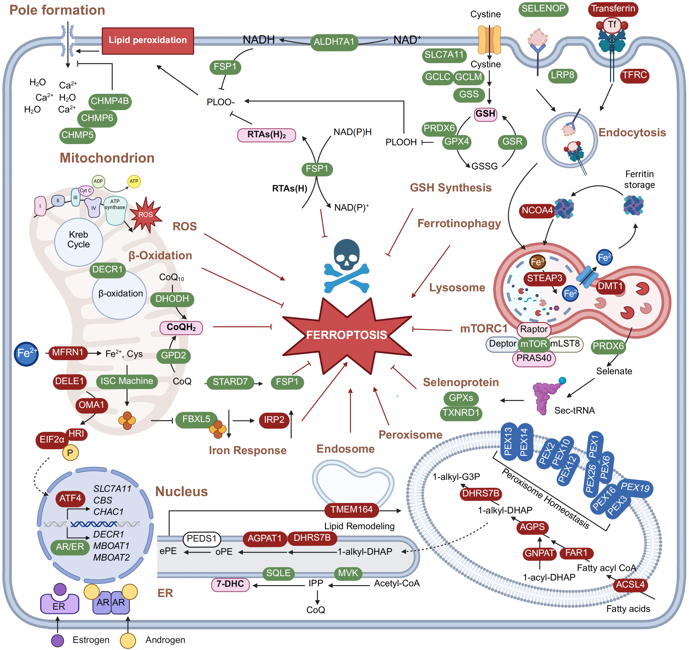
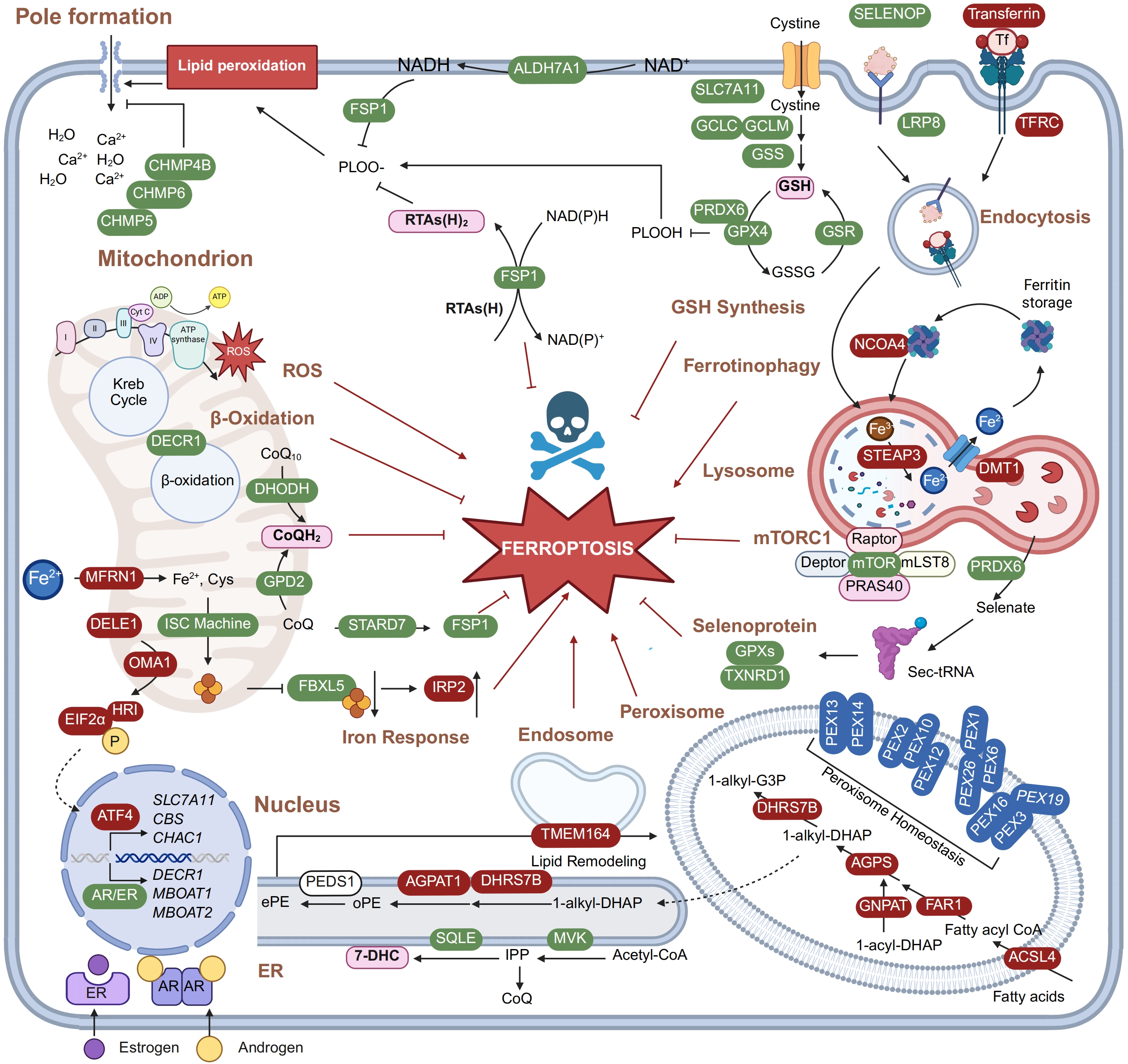
Figure 3. Coordinated regulation of ferroptosis by multiple organelles. Ferroptosis is orchestrated by spatially organized metabolic and redox processes across multiple subcellular compartments. At the plasma membrane, xCT imports cystine, LRP8 recognizes SELENOP, and TFRC internalizes transferrin. ALDH7A1 supplies NADH for FSP1-dependent ubiquinol regeneration at the plasma membrane. The plasma membrane is also the major site of lipid peroxidation-driven rupture. ESCRT-III components, including CHMP4B, CHMP5, and CHMP6, oppose membrane damage and delay ferroptotic membrane breakdown. In lysosomes, endocytosed iron and selenium are released after cargo processing. NCOA4-mediated ferritinophagy supplies iron for Fenton chemistry. Lysosomal mTORC1 coordinates nutrient signaling and ferroptosis resistance. In mitochondria, TCA cycle- and ETC-derived ROS promote ferroptosis. In contrast, DHODH and GPD2 regenerate CoQH2 to suppress lipid peroxidation. STARD7 transfers mitochondrial CoQ to other membranes and extends CoQ-dependent protection beyond mitochondria. DELE1-HRI-ATF4 signaling connects mitochondrial stress to nuclear transcriptional responses and shapes ferroptosis sensitivity. Peroxisomes initiate plasmalogen biosynthesis. The ER supports plasmalogen maturation, phospholipid remodeling, 7-DHC production, and IPP-dependent CoQ biosynthesis. The nucleus integrates transcriptional programs, including hormone-responsive pathways. The cytosol provides GSH-, GPX4-, and FSP1-dependent reducing capacity to remove lipid peroxides. Created in BioRender. Zhao, B. (2026) https://BioRender.com/hdoxh5d. LRP8: lipoprotein receptor-related protein 8; SELENOP: selenoprotein P; TFRC: transferrin receptor; ALDH7A1: aldehyde dehydrogenase 7 family member A1; NADH: reduced nicotinamide adenine dinucleotide; FSP1: ferroptosis suppressor protein 1; ESCRT-III: endosomal sorting complex required for transport-III; CHMP: charged multivesicular body protein; NCOA4: nuclear receptor coactivator 4; mTORC1: mechanistic target of rapamycin complex 1; TCA: tricarboxylic acid; ETC: electron transport chain; ROS: reactive oxygen species; DHODH: dihydroorotate dehydrogenase; GPD2: glycerol-3-phosphate dehydrogenase 2; CoQ: coenzyme Q; DELE1: DAP3-binding cell death enhancer 1; HRI: heme-regulated inhibitor kinase; ATF4: activating transcription factor 4; ER: endoplasmic reticulum; 7-DHC: 7-dehydrocholesterol; IPP: isopentenyl pyrophosphate; GSH: glutathione; GPX4: glutathione peroxidase 4.
Moreover, the plasma membrane also hosts enzymes that directly remodel phospholipid composition. For example, upregulated membrane bound glycerophospholipid O-acyltransferase 1 and 2 (MBOAT1/2) under hormone stimulation (such as ER and androgen receptor) protect cells from ferroptotic stress by promoting the Lands cycle[137]. More recently, the lipid flippase SLC47A1 was shown to inhibit ferroptosis by influencing the asymmetric distribution of PUFA-PLs and CoQ across the lipid bilayer[138].
During ferroptosis, the plasma membrane is also a primary site for lipid oxidation and reduction. Iron-dependent phospholipid peroxidation occurs within the inner leaflet and can culminate in membrane rupture that defines ferroptotic cell death[1]. This lethal process is driven in part by the interaction between PEBP1 and ALOX15, a key regulatory step that can be blocked by ferrostatin-1[55,139]. Conversely, FSP1 functions as a protective oxidoreductase anchored to the plasma membrane, where it regenerates CoQH2 from CoQ to act as a lipophilic RTA and safeguard cells against ferroptosis[44,45]. Structural analysis of the FSP1 protein revealed that the active form of FSP1 is a protein dimer, although whether dimerization is necessary for its antioxidant function needs further confirmation[140]. Moreover, regulation of FSP1 through the formation of phase-separated condensates was reported to modulate its protective role in ferroptosis[141]. Nonetheless, FSP1 expression strongly correlates with ferroptosis resistance across multiple experimental models of cancer in vitro and in vivo, and has emerged as a potential therapeutic target. Pharmacologic or genetic inhibition of FSP1 led to tumor shrinkage and could synergize with radiotherapy in cancer treatment, highlighting CoQ redox metabolism at the plasma membrane as a promising target for ferroptosis-based therapy design[142,143]. In addition, aldehyde dehydrogenase 7 family member A1 (ALDH7A1) was shown to generate reduced nicotinamide adenine dinucleotide (NADH) at the plasma membrane and facilitate FSP1-dependent ferroptosis suppression, thereby connecting aldehyde detoxification with ferroptosis control at the cell surface[144].
While FSP1 has a well-characterized subcellular location at the plasma membrane, another key ferroptosis suppressor, GPX4, lacks a clear membrane targeting sequence yet still contributes to the reduction of peroxidized lipid near the cell membrane[145]. Recently, PRDX6 has been implicated in the recruitment of GPX4 to the plasma membrane, by forming a PRDX6-GPX4 complex through disulfide linkage under ferroptotic stress[146], which may provide a mechanism underlying GPX4 localization.
In contrast to apoptosis or pyroptosis, ferroptosis can engage wave-like spreading across cells. This intercellular spreading depends critically on plasma membrane contacts, which regulate ferroptosis through two distinct mechanisms. The first is adhesion-dependent signaling: E-cadherin-mediated cell-cell contact activates the NF2–Hippo–YAP cascade, which transcriptionally programs ferroptosis resistance, whereas loss of contact sensitizes cells to ferroptotic death[128]. In contrast, the second mechanism involves direct membrane–membrane interaction: physical contact between plasma membranes is sufficient to transmit ferroptotic signals, as demonstrated by liposome reconstitution studies[147]. Therefore, the plasma membrane not only serves as the subcellular platform for ferroptotic execution but also mediates the intercellular communication that underlies its unique spatiotemporal dynamics.
4.2 Mitochondrion
Mitochondria regulate ferroptosis through multiple mechanisms and can either promote or inhibit ferroptosis in a context-dependent manner. One important aspect of this regulation involves iron homeostasis (Figure 3). Mitochondria harbor the Fe-S biogenesis pathway and contribute to cellular iron homeostasis by preventing excessive accumulation of labile iron. Genetic or pharmacologic disruption of Fe-S biogenesis activates cellular iron starvation response and sensitizes cells to ferroptosis[65,148-150]. Conversely, increased mitochondrial iron uptake from the cytosol via SLC25A37 (also known as MFRN1) was also shown to enhance ferroptosis sensitivity, although the underlying molecular mechanism remains unclear[151].
In addition to the regulation of iron homeostasis, CoQ metabolism provides an independent layer of ferroptosis defense. DHODH in the pyrimidine biosynthesis pathway and glycerol-3-phosphate dehydrogenase 2 (GPD2) of the glycerol phosphate shuttle both couple electron transport to the regeneration of CoQH2 at the inner mitochondrial membrane, thereby directly quenching phospholipid peroxyl radicals[46,152]. However, the role of DHODH in mitochondrial ferroptosis regulation remains controversial. A subsequent study reported that the ferroptosis-sensitizing effect of the DHODH inhibitor may be attributable to its off-target inhibition of FSP1 rather than to DHODH inhibition itself[153]. Therefore, whether DHODH directly contributes to ferroptosis defense in mitochondria warrants further clarification. Consistent with a role for mitochondrial CoQ metabolism in ferroptosis protection, cytosolic enzymes of the de novo pyrimidine biosynthesis pathway were found to confer ferroptosis resistance, likely by sustaining substrate flux toward the DHODH-catalyzed reaction[154]. Furthermore, CoQ synthesized within mitochondria can be exported to other compartments via STARD7, linking the mitochondrial CoQ pool to broader antioxidant systems such as the FSP1-CoQ axis[155].
On the other hand, mitochondrial catabolic metabolism has been widely implicated in promoting ferroptosis. ROS generated from the ETC are a key driving force of lipid peroxidation and ferroptosis[1,156,157]. Consistently, the entry of glutamine into the TCA cycle via glutaminolysis was shown to promote ferroptosis under various conditions[57,156]. Beyond these metabolic functions, mitochondria also initiate stress response signaling that impinges on ferroptosis regulation. For example, the DELE1-HRI-ATF4 pathway can be activated by mitochondrial stress and contribute to GSH biosynthesis through ATF4-mediated transcriptional programs. As a result, this pathway was found to govern the sensitivity of drug-tolerant persister cells to RSL3 treatment[158,159]. Furthermore, mitochondrial dynamics, including the balance between fission and fusion regulated by DRP1, are essential for maintaining mitochondrial morphology and have been shown to dictate ferroptosis sensitivity[76]. Recent evidence indicates that disruption of mitochondrial dynamics mediated by DRP1 can paradoxically attenuate ferroptosis and chemotherapy-induced toxicity through NRF2-mediated upregulation of FSP1, providing a mechanistic link between mitochondrial morphology, antioxidant defense, and ferroptosis resistance[160]. Collectively, mitochondria integrate metabolic, redox, and stress signaling inputs to determine the cellular response to ferroptotic stimuli.
4.3 ER
ER is a primary site of phospholipid biosynthesis and remodeling, and consequently a major subcellular location of lipid peroxidation. The Kennedy pathway operates primarily in the ER and generates both structural components of membranes and PUFA-containing substrates for lipid peroxidation[161,162] (Figure 3). Indeed, genes involved in PE synthesis, including selenoprotein I (SELENOI) and phosphatidylethanolamine cytidyltransferase (PCYT2), have been identified in CRISPR -based genetic screens as ferroptosis regulators[163,164]. During phospholipid biosynthesis, ACSL4 and LPCAT3 incorporate arachidonic and adrenic acid into PE, generating PUFA-PLs that are highly susceptible to oxidation[18]. This remodeling step also provides a mechanistic basis for how other acyl-CoA synthetases influence ferroptosis sensitivity by shaping the fatty acyl composition of membrane phospholipids. For instance, ACSL3, which localizes to both the ER and lipid droplets, preferentially channels MUFAs into membranes and may thereby counterbalance PUFA incorporation under certain conditions[165]. Once PUFA-PLs are produced, ER-associated redox enzymes determine whether these lipids undergo peroxidation. POR-dependent electron transfer to cytochrome P450 enzymes can fuel lipid peroxide generation, while LOX can directly oxidize PUFA-containing phospholipids[12,56]. Therefore, the ER serves as both a biosynthetic platform that supplies ferroptosis-prone lipid substrates and a redox-active compartment that amplifies their oxidation. Consistent with this compartment-centered view, chemical mapping studies have identified essential ferroptosis-associated peroxidation sites within ER-localized phospholipids[166]. In addition to phospholipid production, ER also harbors enzymes of the cholesterol biosynthesis pathway, the intermediates of which, as discussed above, are essential regulators of ferroptosis[36,38,39,40].
Beyond its biosynthetic functions, the ER also serves as a platform for intracellular signaling that instructs metabolic regulation and gene transcription relevant to ferroptosis. For example, lipid biosynthesis is dictated by a signaling cascade that activates SREBF1 (also known as SREBP1) mediated transcriptional program[91]. Moreover, nuclear factor erythroid 2-related factor 1 (NRF1) is an ER-localized transcription factor involved in redox control. Together with its cleavage enzyme DNA-damage inducible 1 homolog 2 (DDI2) and the deglycosylation enzyme N-glycanase 1 (NGLY1), NRF1 promotes ferroptosis resistance by activating downstream target genes, such as HMOX1 and proteasome subunit genes like proteasome 20S subunit alpha 1 (PSMA1) and proteasome 20S subunit beta 1 (PSMB1), which collectively enhance GPX4 protein stability[167-169]. In addition, ER stress response mediated by PRKR-like endoplasmic reticulum kinase-ATF4 signaling was also reported to modulate ferroptosis sensitivity[121,170].
4.4 Lipid droplets
Lipid droplets have emerged as central players in ferroptosis by serving as a dynamic storage for fatty acids. As discussed above, lipid droplets sequester PUFAs as TAGs to limit the pool of phospholipids accessible to oxidation, while their lipolytic degradation releases these PUFAs to promote ferroptosis[24-26]. Similarly, MUFAs are dynamically stored in and mobilized from lipid droplets to modulate membrane composition and ferroptosis sensitivity[34]. Beyond their canonical storage function, lipid droplets also serve as a platform for local antioxidant defense. For example, FSP1 localizes to the surface of lipid droplets, where it can detoxify oxidized TAGs and cholesteryl esters, thereby providing protection against lipid peroxidation[171]. Certain genetic alterations, such as loss of the tumor suppressor CDKN2A, were found to disrupt lipid droplet homeostasis and increase ferroptosis susceptibility by promoting fatty acid release[172]. Thus, by coordinating lipid storage, mobilization, and local antioxidant activity, lipid droplets serve as a key metabolic node to regulate ferroptosis.
4.5 Lysosome
Lysosomes act as a hub of metabolic signaling and nutrient processing. Lysosomal degradation of transferrin, ferritin and heme-containing proteins can liberate both imported and intracellularly stored iron into the cytosol[57,58,173]. In agreement, the lysosomotropic iron chelator salinomycin was shown to protect cells from ferroptosis[174] (Figure 3). Beyond simply releasing iron, lysosomes may also influence how iron is partitioned between organelles. Emerging evidence suggests that lysosome-mitochondria contact sites, acting through BDH2-dependent siderophore-mediated iron transfer, can determine iron partitioning between these two compartments and thereby prime mesenchymal cancer cells for ferroptosis[175].
In addition to iron metabolism, lysosomes also control ferroptosis through cysteine availability and amino acid recycling. They support the cysteine pool through two principal routes: the release of cysteine derived from the degradation of endocytosed proteins, and the autophagic recycling of intracellular proteins that provides cysteine-containing precursors. Through these activities, lysosomal nutrient processing sustains GSH production and helps maintain GPX4-dependent detoxification of lipid peroxides[176]. Interestingly, SLC7A11 localized on the lysosomes was reported to function as an unconventional proton transporter, which helps to maintain lysosomal pH and modulate ferroptosis sensitivity independently of its role in cystine uptake[177]. This lysosome-centered supply of amino acids is further coupled to nutrient-sensing pathways on the lysosomal surface. mTORC1, anchored on the lysosomal surface, senses amino acid sufficiency and couples cysteine availability to GPX4 translation and cellular lipogenesis, thus integrating amino acid status with ferroptosis sensitivity[91,96]. Consistently, lysosomal catabolism of extracellular proteins, such as albumin, also supplies amino acids to support GSH biosynthesis and to modulate mTORC1 signaling, providing an alternative ferroptosis resistance mechanism in nutrient limited regions of tumors[178].
Beyond serving as a reservoir and processing hub for ferroptosis-related metabolites, lysosomes also function as an active initiation site of lipid peroxidation. Pharmacological mobilization of lysosomal iron has been shown to catalyze local radical generation within the acidic lysosomal lumen, thereby triggering membrane lipid oxidation and ferroptosis[58]. Consistently, lysosomal lipid peroxidation can induce lysosomal membrane permeabilization, resulting in iron leakage into the cytosol and the subsequent propagation of lipid peroxidation throughout the cell[179]. Moreover, lysosomal acidity appears to be a key determinant of this process. In senescent cells, alkalinization of lysosomes traps ferrous iron within the organelle and suppresses cystine deprivation-induced lipid peroxidation, whereas restoration of lysosomal acidity re-sensitizes cells to ferroptosis[180]. Thus, lysosomal pH, iron reactivity, and membrane integrity together define a local pro-ferroptotic platform that initiates and amplifies lipid damage.
Collectively, lysosomes coordinate iron trafficking, amino acid supply, stress signaling, and local iron-driven lipid peroxidation to exert multifaceted control over ferroptosis initiation and propagation.
4.6 Peroxisome and other organelles
Peroxisomal activities are increasingly recognized as key determinants of ferroptosis, primarily through the regulation of both lipid and plasmalogen metabolism. Plasmalogen biosynthesis is initiated in the peroxisome, where the characteristic vinyl ether bond is formed and subsequent maturation and remodeling take place in the ER and endosomes. This pathway generates PUFA-containing plasmalogens, such as plasmalogen-PE, which can be incorporated into cellular membranes and serve as potent substrates for lipid peroxidation (Figure 3). Consequently, genetic inactivation of peroxisomal ether-lipid biosynthetic enzymes reduces plasmalogen-PE content and blunts ferroptosis, whereas ether-lipid-rich cells exhibit heightened ferroptosis sensitivity[21,181]. In addition to plasmalogen biosynthesis, peroxisomes host the β-oxidation of very long-chain PUFAs and generate H2O2 as a byproduct, thereby influencing both the supply of long-chain PUFA acyl-CoAs available for membrane incorporation and the overall oxidative status in the cell[6]. Thus, peroxisomes shape ferroptosis sensitivity by controlling both the membrane lipid composition and the cellular redox environment.
Finally, emerging studies have begun to reveal the role of other organelles in ferroptosis regulation. For example, monocyte to macrophage differentiation associated (MMD) in the Golgi apparatus was shown to promote the activity of the ACSL4-MBOAT7 axis, thereby increasing cellular vulnerability to ferroptotic stress[182]. In the endosomal compartment, TMEM164 was identified as an acyltransferase that produces ferroptotic C20:4 ether phospholipids, further expanding the repertoire of organelle-specific lipid remodeling events that influence ferroptosis[183]. The nucleus has so far been implicated in ferroptosis regulation mainly as the site where gene transcription occurs. Whether the nucleus or other organelles play more direct roles in ferroptotic execution remains an open question for future investigation.
5. Perspective
Studies in the past years have greatly advanced our understanding of ferroptosis. In this Review, we summarized recent progress in the field from the perspective of cellular metabolism. We discussed major intracellular metabolites, signaling pathways, and inter-organellar communications that are involved in ferroptosis regulation. Looking forward, several key questions remain. For example, how metabolic pathways coordinate to determine ferroptosis sensitivity in a cell type-specific manner is not fully understood. How interactions between cells and the extracellular microenvironment shape ferroptosis regulation also awaits further investigation. With the growing knowledge of the metabolic basis of ferroptosis under both physiological and pathological conditions, we have started to see the development of intervention strategies that exploit ferroptosis regulation in preclinical models of diseases including ischemic organ injury, neurodegeneration, and cancer. Future efforts may focus on developing highly specific metabolic modulators of ferroptosis, identifying biomarkers to predict treatment response, and designing combinatorial regimens that benefit from both ferroptosis intervention and conventional therapy. A deeper understanding of the metabolic principles that govern ferroptosis will pave the way for precision medicine approaches in pathologies involving this cell death modality.
Acknowledgements
We thank members of the Zhu laboratory for critical discussions during manuscript preparation.
Authors contribution
Zhang L, Zhao ZB, Li W: Investigation, visualization, writing-original draft, writing-review & editing.
Zhu J: Conceptualization, supervision, writing-review & editing.
Conflicts of interest
Not applicable.
Ethical approval
Not applicable.
Consent to participate
Not applicable.
Consent for publication
Not applicable.
Availability of data and materials
Not applicable.
Funding
This work was supported by National Natural Science Foundation of China (Grant Nos. 32541044 and 32270819), Zhejiang Provincial Clinical Research Center for Pediatric Diseases (ZJEK2301Z), Tsinghua University Initiative Scientific Research Program (20263080054), Tsinghua University Dushi Special Fund, and Tsinghua-Peking Center for Life Sciences (This program does not have a grant number).
Copyright
© The Author(s) 2026.
References
-
1. Dixon SJ, Lemberg KM, Lamprecht MR, Skouta R, Zaitsev EM, Gleason CE, et al. Ferroptosis: An iron-dependent form of nonapoptotic cell death. Cell. 2012;149(5):1060-1072.[DOI]
-
5. Wang W, Green M, Choi JE, Gijón M, Kennedy PD, Johnson JK, et al. CD8+ T cells regulate tumour ferroptosis during cancer immunotherapy. Nature. 2019;569(7755):270-274.[DOI]
-
6. Liang D, Minikes AM, Jiang X. Ferroptosis at the intersection of lipid metabolism and cellular signaling. Mol Cell. 2022;82(12):2215-2227.[DOI]
-
8. Conrad M, Pratt DA. The chemical basis of ferroptosis. Nat Chem Biol. 2019;15(12):1137-1147.[DOI]
-
15. Ma X, Xiao L, Liu L, Ye L, Su P, Bi E, et al. CD36-mediated ferroptosis dampens intratumoral CD8+ T cell effector function and impairs their antitumor ability. Cell Metab. 2021;33(5):1001-1012.[DOI]
-
17. Lee JY, Nam M, Son HY, Hyun K, Jang SY, Kim JW, et al. Polyunsaturated fatty acid biosynthesis pathway determines ferroptosis sensitivity in gastric cancer. Proc Natl Acad Sci U S A. 2020;117(51):32433-32442.[DOI]
-
19. Jiang X, Stockwell BR, Conrad M. Ferroptosis: Mechanisms, biology and role in disease. Nat Rev Mol Cell Biol. 2021;22(4):266-282.[DOI]
-
22. Beharier O, Tyurin VA, Goff JP, Guerrero-Santoro J, Kajiwara K, Chu T, et al. PLA2G6 guards placental trophoblasts against ferroptotic injury. Proc Natl Acad Sci U S A. 2020;117(44):27319-27328.[DOI]
-
24. Olzmann JA, Carvalho P. Dynamics and functions of lipid droplets. Nat Rev Mol Cell Biol. 2019;20(3):137-155.[DOI]
-
25. Dixon SJ, Olzmann JA. The cell biology of ferroptosis. Nat Rev Mol Cell Biol. 2024;25(6):424-442.[DOI]
-
26. Bai Y, Meng L, Han L, Jia Y, Zhao Y, Gao H, et al. Lipid storage and lipophagy regulates ferroptosis. Biochem Biophys Res Commun. 2019;508(4):997-1003.[DOI]
-
28. Nassar ZD, Mah CY, Dehairs J, Burvenich IJ, Irani S, Centenera MM, et al. Human DECR1 is an androgen-repressed survival factor that regulates PUFA oxidation to protect prostate tumor cells from ferroptosis. eLife. 2020;9:e54166.[DOI]
-
38. Freitas FP, Alborzinia H, dos Santos AF, Nepachalovich P, Pedrera L, Zilka O, et al. 7-Dehydrocholesterol is an endogenous suppressor of ferroptosis. Nature. 2024;626(7998):401-410.[DOI]
-
43. Soula M, Weber RA, Zilka O, Alwaseem H, La K, Yen F, et al. Metabolic determinants of cancer cell sensitivity to canonical ferroptosis inducers. Nat Chem Biol. 2020;16(12):1351-1360.[DOI]
-
45. Doll S, Freitas FP, Shah R, Aldrovandi M, da Silva MC, Ingold I, et al. FSP1 is a glutathione-independent ferroptosis suppressor. Nature. 2019;575(7784):693-698.[DOI]
-
46. Mao C, Liu X, Zhang Y, Lei G, Yan Y, Lee H, et al. DHODH-mediated ferroptosis defence is a targetable vulnerability in cancer. Nature. 2021;593(7860):586-590.[DOI]
-
49. Matsushita M, Freigang S, Schneider C, Conrad M, Bornkamm GW, Kopf M. T cell lipid peroxidation induces ferroptosis and prevents immunity to infection. J Exp Med. 2015;212(4):555-568.[DOI]
-
52. Tschuck J, Padmanabhan Nair V, Galhoz A, Zaratiegui C, Tai HM, Ciceri G, et al. Suppression of ferroptosis by vitamin A or radical-trapping antioxidants is essential for neuronal development. Nat Commun. 2024;15(1):7611.[DOI]
-
53. Gaschler MM, Stockwell BR. Lipid peroxidation in cell death. Biochem Biophys Res Commun. 2017;482(3):419-425.[DOI]
-
58. Cañeque T, Baron L, Müller S, Carmona A, Colombeau L, Versini A, et al. Activation of lysosomal iron triggers ferroptosis in cancer. Nature. 2025;642(8067):492-500.[DOI]
-
62. Hou W, Xie Y, Song X, Sun X, Lotze MT, Zeh III HJ, et al. Autophagy promotes ferroptosis by degradation of ferritin. Autophagy. 2016;12(8):1425-1428.[DOI]
-
63. Gao M, Monian P, Pan Q, Zhang W, Xiang J, Jiang X. Ferroptosis is an autophagic cell death process. Cell Res. 2016;26(9):1021-1032.[DOI]
-
67. Yang WS, SriRamaratnam R, Welsch ME, Shimada K, Skouta R, Viswanathan VS, et al. Regulation of ferroptotic cancer cell death by GPX4. Cell. 2014;156(1-2):317-331.[DOI]
-
70. Amelio I, Cutruzzolá F, Antonov A, Agostini M, Melino G. Serine and glycine metabolism in cancer. Trends Biochem Sci. 2014;39(4):191-198.[DOI]
-
72. Yang M, Vousden KH. Serine and one-carbon metabolism in cancer. Nat Rev Cancer. 2016;16(10):650-662.[DOI]
-
75. Suzuki S, Venkatesh D, Kanda H, Nakayama A, Hosokawa H, Lee E, et al. GLS2 is a tumor suppressor and a regulator of ferroptosis in hepatocellular carcinoma. Cancer Res. 2022;82(18):3209-3222.[DOI]
-
80. Esaki N, Nakamura T, Tanaka H, Soda K. Selenocysteine lyase, a novel enzyme that specifically acts on selenocysteine. Mammalian distribution and purification and properties of pig liver enzyme. J Biol Chem. 1982;257(8):4386-4391.[PubMed]
-
81. Tobe R, Mihara H. Delivery of selenium to selenophosphate synthetase for selenoprotein biosynthesis. Biochim Biophys Acta Gen Subj. 2018;1862(11):2433-2440.[DOI]
-
86. Labunskyy VM, Hatfield DL, Gladyshev VN. Selenoproteins: Molecular pathways and physiological roles. Physiol Rev. 2014;94(3):739-777.[DOI]
-
87. Burk RF, Hill KE. Regulation of selenium metabolism and transport. Annu Rev Nutr. 2015;35:109-134.[DOI]
-
91. Glaviano A, Foo ASC, Lam HY, Yap KCH, Jacot W, Jones RH, et al. PI3K/AKT/mTOR signaling transduction pathway and targeted therapies in cancer. Mol Cancer. 2023;22(1):138.[DOI]
-
99. Liu Y, Su Z, Tavana O, Gu W. Understanding the complexity of p53 in a new era of tumor suppression. Cancer Cell. 2024;42(6):946-967.[DOI]
-
101. Zhou Q, Meng Y, Li D, Yao L, Le J, Liu Y, et al. Ferroptosis in cancer: From molecular mechanisms to therapeutic strategies. Sig Transduct Target Ther. 2024;9:55.[DOI]
-
102. Jiang L, Kon N, Li T, Wang SJ, Su T, Hibshoosh H, et al. Ferroptosis as a p53-mediated activity during tumour suppression. Nature. 2015;520(7545):57-62.[DOI]
-
110. Rojo de la Vega M, Chapman E, Zhang DD. NRF2 and the hallmarks of cancer. Cancer Cell. 2018;34(1):21-43.[DOI]
-
111. Dodson M, Castro-Portuguez R, Zhang DD. NRF2 plays a critical role in mitigating lipid peroxidation and ferroptosis. Redox Biol. 2019;23:101107.[DOI]
-
116. Luo X, Wang Y, Zhu X, Chen Y, Xu B, Bai X, et al. Corrigendum to “MCL attenuates atherosclerosis by suppressing macrophage ferroptosis via targeting KEAP1/NRF2 interaction”[Redox Biol. 69 (2024) 102987]. Redox Biol. 2024;69:103009.[DOI]
-
122. Liu J, Song X, Kuang F, Zhang Q, Xie Y, Kang R, et al. NUPR1 is a critical repressor of ferroptosis. Nat Commun. 2021;12:647.[DOI]
-
123. Huang C, Santofimia-Castaño P, Iovanna J. NUPR1: A critical regulator of the antioxidant system. Cancers. 2021;13(15):3670.[DOI]
-
127. Ma S, Meng Z, Chen R, Guan KL. The hippo pathway: Biology and pathophysiology. Annu Rev Biochem. 2019;88:577-604.[DOI]
-
139. Anthonymuthu TS, Tyurina YY, Sun WY, Mikulska-Ruminska K, Shrivastava IH, Tyurin VA, et al. Resolving the paradox of ferroptotic cell death: Ferrostatin-1 binds to 15LOX/PEBP1 complex, suppresses generation of peroxidized ETE-PE, and protects against ferroptosis. Redox Biol. 2021;38:101744.
-
141. Nakamura T, Hipp C, Santos Dias Mourão A, Borggräfe J, Aldrovandi M, Henkelmann B, et al. Phase separation of FSP1 promotes ferroptosis. Nature. 2023;619(7969):371-377.[DOI]
-
142. Wu K, Vaughan AJ, Bossowski JP, Hao Y, Ziogou A, Kim SM, et al. Targeting FSP1 triggers ferroptosis in lung cancer. Nature. 2026;649:487-495.[DOI]
-
156. Gao M, Yi J, Zhu J, Minikes AM, Monian P, Thompson CB, et al. Role of mitochondria in ferroptosis. Mol Cell. 2019;73(2):354-363.[DOI]
-
168. Forcina GC, Pope L, Murray M, Dong W, Abu-Remaileh M, Bertozzi CR, et al. Ferroptosis regulation by the NGLY1/NFE2L1 pathway. Proc Natl Acad Sci U S A. 2022;119(11):e2118646119.[DOI]
-
173. Gryzik M, Asperti M, Denardo A, Arosio P, Poli M. NCOA4-mediated ferritinophagy promotes ferroptosis induced by erastin, but not by RSL3 in HeLa cells. Biochim Biophys Acta Mol Cell Res. 2021;1868(2):118913.[DOI]
-
177. Zhou N, Chen J, Hu M, Wen N, Cai W, Li P, et al. SLC7A11 is an unconventional H+ transporter in lysosomes. Cell. 2025;188(13):3441-3458.[DOI]
-
183. Reed A, Ware T, Li H, Bazan JF, Cravatt BF. TMEM164 is an acyltransferase that forms ferroptotic C20: 4 ether phospholipids. Nat Chem Biol. 2023;19(3):378-388.[DOI]
Copyright
© The Author(s) 2026. This is an Open Access article licensed under a Creative Commons Attribution 4.0 International License (https://creativecommons.org/licenses/by/4.0/), which permits unrestricted use, sharing, adaptation, distribution and reproduction in any medium or format, for any purpose, even commercially, as long as you give appropriate credit to the original author(s) and the source, provide a link to the Creative Commons license, and indicate if changes were made.
Publisher’s Note
Science Exploration remains a neutral stance on jurisdictional claims in published
maps
and institutional affiliations. The views expressed in this article are solely those
of
the author(s) and do not reflect the opinions of the Editors or the publisher.
Share And Cite


Science Exploration Style
Zhang L, Zhao ZB, Li W, Zhu J. Regulation of ferroptosis by inter-organellar metabolic signaling network. Ferroptosis Oxid Stress. 2026;2:202628. https://doi.org/10.70401/fos.2026.0038
Tips
Copy completed.
Submit a Manuscript
Author Instructions
Cite this Article
Article Metrics
0
View
0
Download
Cited
Article Updates

- Abstract
- Keywords
- 1. Introduction
- 2. Metabolites Are Key Players in Ferroptosis
- 3. Signaling Pathways Dictate Ferroptosis Sensitivity
- 4. Coordinated Regulation of Ferroptosis by Multiple Organelles
- 5. Perspective
- Acknowledgements
- Authors contribution
- Conflicts of interest
- Ethical approval
- Consent to participate
- Consent for publication
- Availability of data and materials
- Funding
- References
- Copyright
 Get citation
Science Exploration Style

Zhang L, Zhao ZB, Li W, Zhu J. Regulation of ferroptosis by inter-organellar metabolic signaling network. Ferroptosis Oxid Stress. 2026;2:202628. https://doi.org/10.70401/fos.2026.0038
copy
Share Link
copy