-
EXO – Beyond the Cell is a quarterly, gold open-access journal published by Science Exploration Press. The journal highlights groundbreaking discoveries on how cells engage with their environments and how these interactions shape biology and medicine. With an emphasis on spatial organization, dynamic communication, and cross-scale integration, EXO – Beyond the Cell serves as a hub for innovative research at the interface of cell biology, technology, and translational science. By fostering rigor, creativity, and accessibility, the journal seeks to accelerate insights that redefine our understanding of life beyond the boundaries of the cell. more >
Articles
Extracellular vesicles in Drosophila and mammals: Conserved mechanisms and emerging functional roles
-
Extracellular vesicles (EVs) are membrane-enclosed particles released by cells carrying proteins, lipids, metabolites and nucleic acids that can alter the behavior of recipient cells. In mammalian systems, EVs have been studied extensively as important ...
MoreExtracellular vesicles (EVs) are membrane-enclosed particles released by cells carrying proteins, lipids, metabolites and nucleic acids that can alter the behavior of recipient cells. In mammalian systems, EVs have been studied extensively as important mediators of intercellular and interorgan communication in development, tissue homeostasis, immunity, regeneration, metabolism, cancer and neurobiology. In parallel, Drosophila has emerged as a powerful in vivo model for EV research owing to its genetic tractability and the availability of well-established tools for studying interorgan communication. Work in Drosophila has shown that EVs participate in synaptic cargo transfer, selected developmental signaling contexts and neuronal homeostasis. Importantly, most of the pathways that regulate endosomal sorting, multivesicular body dynamics, membrane budding and vesicle secretion are conserved between flies and mammals. This review summarizes current understanding of EV nomenclature, biogenesis, cargo selection and biological function, with emphasis on points of convergence and divergence between mammalian and Drosophila systems. It further discusses the strengths and limitations of Drosophila as a model for mammalian EV biology and highlights how comparative approaches can sharpen mechanistic insight and translational EV studies.
Less -
Kyosuke Yanagawa, Norbert Perrimon
-
DOI: https://doi.org/10.70401/EXO.2026.0014 - June 23, 2026
Anchoring virtual cells on gene programs: From biological networks to patient digital twins
-
Predicting developmental trajectories and disease progression is a central goal of virtual biology. While conventional biological network models struggle with noise and computational complexity, black-box deep learning obscures mechanistic interpretability. ...
MorePredicting developmental trajectories and disease progression is a central goal of virtual biology. While conventional biological network models struggle with noise and computational complexity, black-box deep learning obscures mechanistic interpretability. In this Perspective, we introduce “gene programs” (GPs) as the essential intermediate layer to close this gap. Extracted from massive single-cell datasets, GPs provide relatively robust and interpretable modules that capture coherent biological functions. Supported by recent multi-modal and large-scale perturbation advances, GPs may reflect underlying cross-layer biophysical structures. We propose a framework where next-generation virtual cells encode GPs as latent variables in neural networks. By incorporating spatial multicellular contexts and multimodal GP extraction, this GP-centric framework will reduce data noise and allow direct perturbation-to-phenotype mapping, enabling high-fidelity simulations of complex tissue dynamics. Anchoring virtual models on GPs paves the way for highly personalized “Patient Digital Twins”, empowering predictive in silico clinical simulations and advancing mechanism-driven precision medicine.
Less -
Tianhao Wang, ... Jie Liao
-
DOI: https://doi.org/10.70401/EXO.2026.0013 - June 11, 2026
Democratizing the spatial view: STAMP technology from an analytical perspective
-
Single-cell RNA sequencing (scRNA-seq) has transformed the profiling of cellular heterogeneity; however, tissue dissociation for scRNA-seq eliminates three critical classes of information that often define cell state: spatial organization, morphology, ...
MoreSingle-cell RNA sequencing (scRNA-seq) has transformed the profiling of cellular heterogeneity; however, tissue dissociation for scRNA-seq eliminates three critical classes of information that often define cell state: spatial organization, morphology, and protein localization. Spatial transcriptomics partially restores this context, yet many platforms are limited by cost, throughput, or experimental complexity. Single-cell transcriptomics analysis and multimodal profiling (STAMP) tackle these limitations by immobilizing cells or nuclei on a slide, enabling high-content imaging prior to molecular readout and consequently preserving each cell’s visual characteristics alongside high-plex transcript and/or protein measurements. This perspective focuses on the downstream analytical workflow for STAMP datasets using Python, illustrating how standard single-cell methods can be applied to image-derived cell-by-feature matrices enriched with per-cell covariates. The pooled MIX sub-STAMP serves as a working example to illustrate practical steps for quality control, normalization, dimensionality reduction, clustering or label transfer, and program-level interpretation. Morpho-transcriptomic coupling is highlighted as an exploratory strategy that links inferred gene programs to image-derived morphology. Collectively, these analyses establish STAMP as a practical bridge between single-cell transcriptomics and quantitative cell phenotype, enabling interpretable state inference that can be validated by imaging-derived measurements.
Less -
Suresh Poudel, ... Douglas R. Green
-
DOI: https://doi.org/10.70401/EXO.2026.0012 - June 03, 2026
Ferroptosis spreading through propagative signals
-
Ferroptosis is a regulated mechanism of cell death caused by the uncontrolled peroxidation of cellular lipids that has an unusual ability to propagate or spread between cells. Here, we review studies identifying regulators of ferroptosis propagation to consider ...
MoreFerroptosis is a regulated mechanism of cell death caused by the uncontrolled peroxidation of cellular lipids that has an unusual ability to propagate or spread between cells. Here, we review studies identifying regulators of ferroptosis propagation to consider a working model that might explain this unusual feature. Recent findings implicating the spread of lipid peroxides and iron suggest that these main catalysts of ferroptosis may also be primary vehicles that can spread death between cells. Experiments revealing localized versus long-range effects of propagation are discussed, as well as sensitizing factors that may expand the propagative potential of death induced by Class II ferroptosis-inducing compounds. As ferroptosis propagation may underlie the loss of large groups of cells in degenerative diseases and may also have a specialized role in normal development, consideration of how ferroptosis can spread through propagative signals may be important for understanding both normal tissue dynamics and disease.
Less -
Saloni K. Hombalkar, ... Michael Overholtzer
-
DOI: https://doi.org/10.70401/EXO.2026.0011 - May 26, 2026
Computational workflows and data infrastructures for spatial omics analysis
-
Spatial omics is a broad term referring to technologies that allow for biomolecules to be observed within their native tissue context. These technologies have been used by biomedical researchers to gain a better understanding of cellular interactions, tumor ...
MoreSpatial omics is a broad term referring to technologies that allow for biomolecules to be observed within their native tissue context. These technologies have been used by biomedical researchers to gain a better understanding of cellular interactions, tumor microenvironment dynamics, and immune cell infiltration. While the basic outputs, such as spatial coordinates, segmentation masks, and transcript/protein matrices, are provided by the instrument software, the true biological insights come from several downstream, specialized analysis steps. Since spatial omics remains a relatively new field, no unified analysis pipeline has yet been established to encompass all platforms. Most workflows are adapted from single-cell RNA sequencing analysis frameworks, while incorporating additional steps that are specific to spatial data, especially for imaging-based technologies. At the same time, the diversity of platforms, data modalities, and output formats has introduced substantial challenges for data representation, interoperability, and cross-platform integration, highlighting the need for flexible, spatially aware, and user-friendly data structures made specifically for imaging-based data, not merely adapted from other methods. This review summarizes the general analytical steps following spatial omics data acquisition, commonly used data infrastructures and tools, existing gaps, and future directions in the field.
Less -
Margaret Alexander, ... Jasmine Plummer
-
DOI: https://doi.org/10.70401/EXO.2026.0010 - May 15, 2026
EXO - Beyond the Cell, a journal about how cells interact with their environment
-
Brent R. Stockwell
-
DOI: https://doi.org/10.70401/EXO.2026.0001 - January 13, 2026
Human iPSC-derived macrophages for studying intrinsic and extrinsic factors in cystic fibrosis
-
Background: Cystic fibrosis (CF) is a progressive genetic disease characterized by defective ion transport, mucus accumulation, chronic infection, and inflammation that drive airway damage and ultimately end-stage lung failure. Previous studies ...
MoreBackground: Cystic fibrosis (CF) is a progressive genetic disease characterized by defective ion transport, mucus accumulation, chronic infection, and inflammation that drive airway damage and ultimately end-stage lung failure. Previous studies show that high levels of proteolytic enzymes in the sputum of CF patients correlate with declining lung function, but the related effects on distal lung extracellular matrix (ECM) and immune responses are unclear.
Methods: To address this gap, induced pluripotent stem cell (iPSC) lines from healthy donors and CF patients were differentiated into macrophages, and stimulated with lipopolysaccharide (LPS) to compare their inflammatory responses. Bulk RNA sequencing, functional assays, and secreted protein profiling revealed key differences between healthy and CF-derived macrophages, providing insight into how these cells may contribute to inflammatory responses in CF patients. Further, human lung ECM from distal CF lung tissue was isolated, used to generate ECM biomaterials, and combined with iPSC-derived macrophages from healthy and CF donors in vitro. Macrophage phenotype was evaluated through cytokine profiling and RNA sequencing.
Results: CF macrophage inflammation was dysregulated, with elevated baseline IL-8, IL-18, and MCP-1 expression, and a blunted inflammatory response to CF ECM compared to healthy macrophages. By using CF ECM and healthy macrophages, we characterized how healthy cells may be altered in a persistent CF milieu after anticipated CFTR modulator therapy.
Conclusion: These findings reveal altered innate immune behavior in CF and demonstrate the utility of iPSC-derived macrophages for modeling extrinsic immune-ECM interactions in disease.
Less -
Daniel Naveed Tavakol, ... Gordana Vunjak-Novakovic
-
DOI: https://doi.org/10.70401/EXO.2026.0005 - April 10, 2026
Ferroptosis in BRCA-associated disorders: Extracellular vesicles as potential messengers beyond the cell
-
BRCA1 and BRCA2 deficiencies are classically defined by impaired homologous recombination–mediated DNA repair; however, their pathological consequences extend far beyond cell-autonomous genomic instability. Accumulating evidence indicates that ...
MoreBRCA1 and BRCA2 deficiencies are classically defined by impaired homologous recombination–mediated DNA repair; however, their pathological consequences extend far beyond cell-autonomous genomic instability. Accumulating evidence indicates that BRCA deficiency is accompanied by iron dysregulation and persistent lipid peroxidation, placing cells under chronic ferroptotic pressure. Studies using BRCA1/2 rat models demonstrate that ferroptosis functions as a decisive biological checkpoint with gene-specific outcomes. Under BRCA1 haploinsufficiency, iron-driven oxidative stress accelerates carcinogenesis by selecting for ferroptosis-resistant clones, whereas BRCA2 haploinsufficiency enhances ferroptotic execution, thereby preventing iron-induced cancer promotion. In contrast, reproductive tissues lacking adaptive escape capacity manifest BRCA deficiency as a direct ferroptosis-driven cellular loss, resulting in male and female infertility. Importantly, ferroptosis is not a silent, cell-confined event. Experimental evidence from asbestos-induced carcinogenesis demonstrates that macrophages undergoing ferroptosis after asbestos phagocytosis release CD63-positive, ferritin-containing extracellular vesicles (EVs) that induce oxidative stress in recipient mesothelial cells, establishing EVs as active mediators of ferroptotic stress propagation. We propose that BRCA deficiency generates a state of ferroptotic priming in which oxidized lipids, iron-related factors, and nucleic acids are disseminated via EVs, thereby shaping tissue- and organ-level pathology. From an evolutionary perspective, the persistence of pathogenic BRCA variants may reflect adaptive advantages conferred by haploinsufficiency in iron-limited, short-lived ancestral environments; under modern conditions of iron abundance and extended lifespan, this once-adaptive state becomes maladaptive, predisposing carriers to cancer and degenerative disorders beyond the cell.
Less -
Shinya Toyokuni, ... Yashiro Motooka
-
DOI: https://doi.org/10.70401/EXO.2026.0002 - February 14, 2026
Heme, copper, and a new way to kill cancer cells
-
Heme homeostasis influences mitochondrial metabolism and leukemia stem cell biology in acute myeloid leukemia. Lewis et al. uncover a surprising metabolic vulnerability in acute myeloid leukemia: suppression of heme biosynthesis primes leukemic ...
MoreHeme homeostasis influences mitochondrial metabolism and leukemia stem cell biology in acute myeloid leukemia. Lewis et al. uncover a surprising metabolic vulnerability in acute myeloid leukemia: suppression of heme biosynthesis primes leukemic cells for cuproptosis, a form of copper-dependent cell death. By linking heme depletion to mitochondrial cytochrome c oxidase (Complex IV) dysfunction, copper accumulation, and cuproptosis, the study integrates transcriptional regulation, mitochondrial metabolism, and metal homeostasis into a unified framework for selective cancer cell killing.
Less -
Xi Zhao, ... Boyi Gan
-
DOI: https://doi.org/10.70401/EXO.2026.0004 - March 19, 2026
Computational workflows and data infrastructures for spatial omics analysis
-
Spatial omics is a broad term referring to technologies that allow for biomolecules to be observed within their native tissue context. These technologies have been used by biomedical researchers to gain a better understanding of cellular interactions, tumor ...
MoreSpatial omics is a broad term referring to technologies that allow for biomolecules to be observed within their native tissue context. These technologies have been used by biomedical researchers to gain a better understanding of cellular interactions, tumor microenvironment dynamics, and immune cell infiltration. While the basic outputs, such as spatial coordinates, segmentation masks, and transcript/protein matrices, are provided by the instrument software, the true biological insights come from several downstream, specialized analysis steps. Since spatial omics remains a relatively new field, no unified analysis pipeline has yet been established to encompass all platforms. Most workflows are adapted from single-cell RNA sequencing analysis frameworks, while incorporating additional steps that are specific to spatial data, especially for imaging-based technologies. At the same time, the diversity of platforms, data modalities, and output formats has introduced substantial challenges for data representation, interoperability, and cross-platform integration, highlighting the need for flexible, spatially aware, and user-friendly data structures made specifically for imaging-based data, not merely adapted from other methods. This review summarizes the general analytical steps following spatial omics data acquisition, commonly used data infrastructures and tools, existing gaps, and future directions in the field.
Less -
Margaret Alexander, ... Jasmine Plummer
-
DOI: https://doi.org/10.70401/EXO.2026.0010 - May 15, 2026
Human iPSC-derived macrophages for studying intrinsic and extrinsic factors in cystic fibrosis
-
Background: Cystic fibrosis (CF) is a progressive genetic disease characterized by defective ion transport, mucus accumulation, chronic infection, and inflammation that drive airway damage and ultimately end-stage lung failure. Previous studies ...
MoreBackground: Cystic fibrosis (CF) is a progressive genetic disease characterized by defective ion transport, mucus accumulation, chronic infection, and inflammation that drive airway damage and ultimately end-stage lung failure. Previous studies show that high levels of proteolytic enzymes in the sputum of CF patients correlate with declining lung function, but the related effects on distal lung extracellular matrix (ECM) and immune responses are unclear.
Methods: To address this gap, induced pluripotent stem cell (iPSC) lines from healthy donors and CF patients were differentiated into macrophages, and stimulated with lipopolysaccharide (LPS) to compare their inflammatory responses. Bulk RNA sequencing, functional assays, and secreted protein profiling revealed key differences between healthy and CF-derived macrophages, providing insight into how these cells may contribute to inflammatory responses in CF patients. Further, human lung ECM from distal CF lung tissue was isolated, used to generate ECM biomaterials, and combined with iPSC-derived macrophages from healthy and CF donors in vitro. Macrophage phenotype was evaluated through cytokine profiling and RNA sequencing.
Results: CF macrophage inflammation was dysregulated, with elevated baseline IL-8, IL-18, and MCP-1 expression, and a blunted inflammatory response to CF ECM compared to healthy macrophages. By using CF ECM and healthy macrophages, we characterized how healthy cells may be altered in a persistent CF milieu after anticipated CFTR modulator therapy.
Conclusion: These findings reveal altered innate immune behavior in CF and demonstrate the utility of iPSC-derived macrophages for modeling extrinsic immune-ECM interactions in disease.
Less -
Daniel Naveed Tavakol, ... Gordana Vunjak-Novakovic
-
DOI: https://doi.org/10.70401/EXO.2026.0005 - April 10, 2026
EXO - Beyond the Cell, a journal about how cells interact with their environment
-
Brent R. Stockwell
-
DOI: https://doi.org/10.70401/EXO.2026.0001 - January 13, 2026
Nutrient-sensing and mTORC1 regulation in neuronal homeostasis: from metabolic signaling to neurodegeneration
-
Neurons rely on precise nutrient-sensing mechanisms to sustain proteostasis and stress resilience across a lifetime. Among these, mechanistic target of rapamycin complex 1 (mTORC1) functions as a central metabolic hub, integrating amino acid availability, ...
MoreNeurons rely on precise nutrient-sensing mechanisms to sustain proteostasis and stress resilience across a lifetime. Among these, mechanistic target of rapamycin complex 1 (mTORC1) functions as a central metabolic hub, integrating amino acid availability, growth factor signals, and energetic status to coordinate protein synthesis, autophagy, and neuronal survival. Neuronal mTORC1 regulation is highly specialised, reflecting unique metabolic demands, axonal compartmentalisation, and dependence on long-term homeostatic control that is not shared by non-neuronal cell types. Beyond canonical PI3K–Akt and AMP-activated protein kinase (AMPK) signaling, emerging evidence highlights metabolic intermediates, most notably leucine-derived acetyl-coenzyme A (AcCoA), as critical upstream regulators that couple nutrient flux to mTORC1 activity via EP300-mediated Raptor acetylation. Chronic dysregulation of these pathways drives persistent mTORC1 hyperactivation, progressive autophagy impairment, and accumulation of proteotoxic species, collectively contributing to neurodegeneration. In Alzheimer’s disease, aberrant mTORC1 activity is linked to tau hyperphosphorylation and amyloid-β accumulation; in Parkinson’s disease, to α-synuclein aggregation and mitophagy failure; in Huntington’s disease, to impaired clearance of mutant huntingtin; and in amyotrophic lateral sclerosis (ALS), to dysregulated proteostasis in motor neurons. This mini review synthesizes current understanding of neuronal mTORC1 regulation, with an emphasis on the AcCoA–acetylation axis as an emerging metabolic control mechanism, its disease-specific implications across major neurodegenerative conditions, and the therapeutic opportunities these insights reveal upstream of mTORC1.
Less -
Sung Min Son, ... David C. Rubinsztein
-
DOI: https://doi.org/10.70401/EXO.2026.0009 - May 15, 2026
Heme, copper, and a new way to kill cancer cells
-
Heme homeostasis influences mitochondrial metabolism and leukemia stem cell biology in acute myeloid leukemia. Lewis et al. uncover a surprising metabolic vulnerability in acute myeloid leukemia: suppression of heme biosynthesis primes leukemic ...
MoreHeme homeostasis influences mitochondrial metabolism and leukemia stem cell biology in acute myeloid leukemia. Lewis et al. uncover a surprising metabolic vulnerability in acute myeloid leukemia: suppression of heme biosynthesis primes leukemic cells for cuproptosis, a form of copper-dependent cell death. By linking heme depletion to mitochondrial cytochrome c oxidase (Complex IV) dysfunction, copper accumulation, and cuproptosis, the study integrates transcriptional regulation, mitochondrial metabolism, and metal homeostasis into a unified framework for selective cancer cell killing.
Less -
Xi Zhao, ... Boyi Gan
-
DOI: https://doi.org/10.70401/EXO.2026.0004 - March 19, 2026
EXO Chats
The Lymphatic-Immune Dialogue in Cancer: How Endothelial Cells Hold the Keys to Immunotherapy
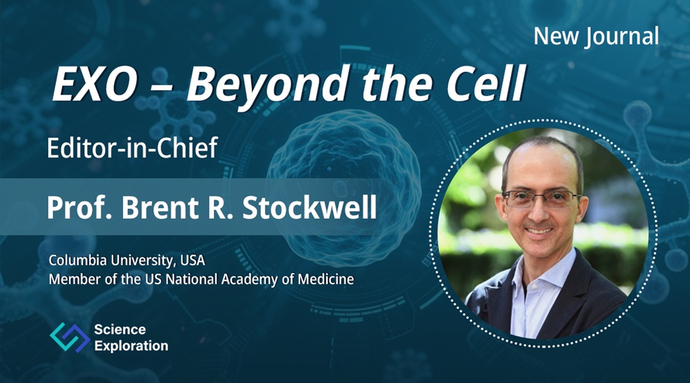
Department of Biological Sciences, Department of Chemistry, Department of Pathology and Cell Biology, Columbia University, New York, NY, USA.
Dr. Kathryn A Jacobs
Cell Death Research and Therapy Laboratory, Center for Cancer Biology, VIB, Leuven, Belgium.
Department of Cellular and Molecular Medicine, KU Leuven, Leuven, Belgium.
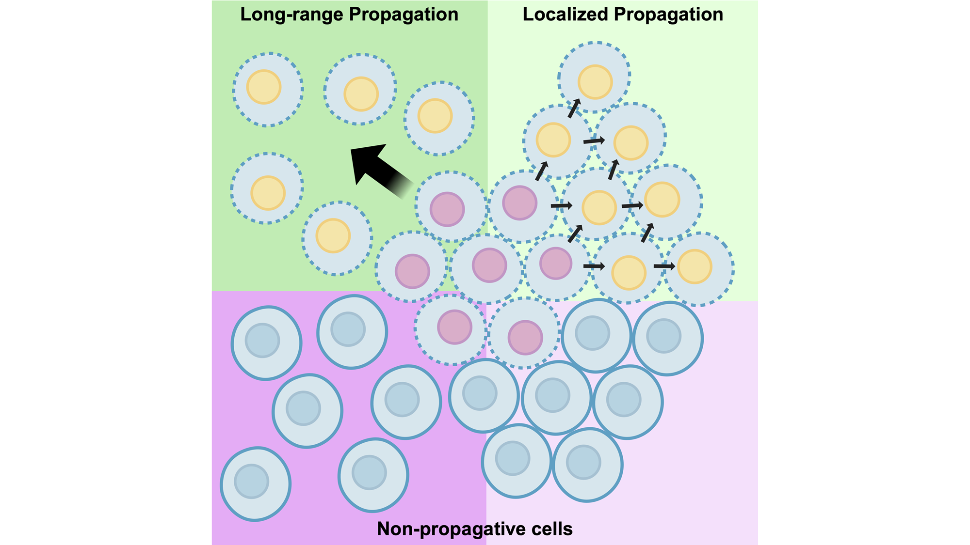
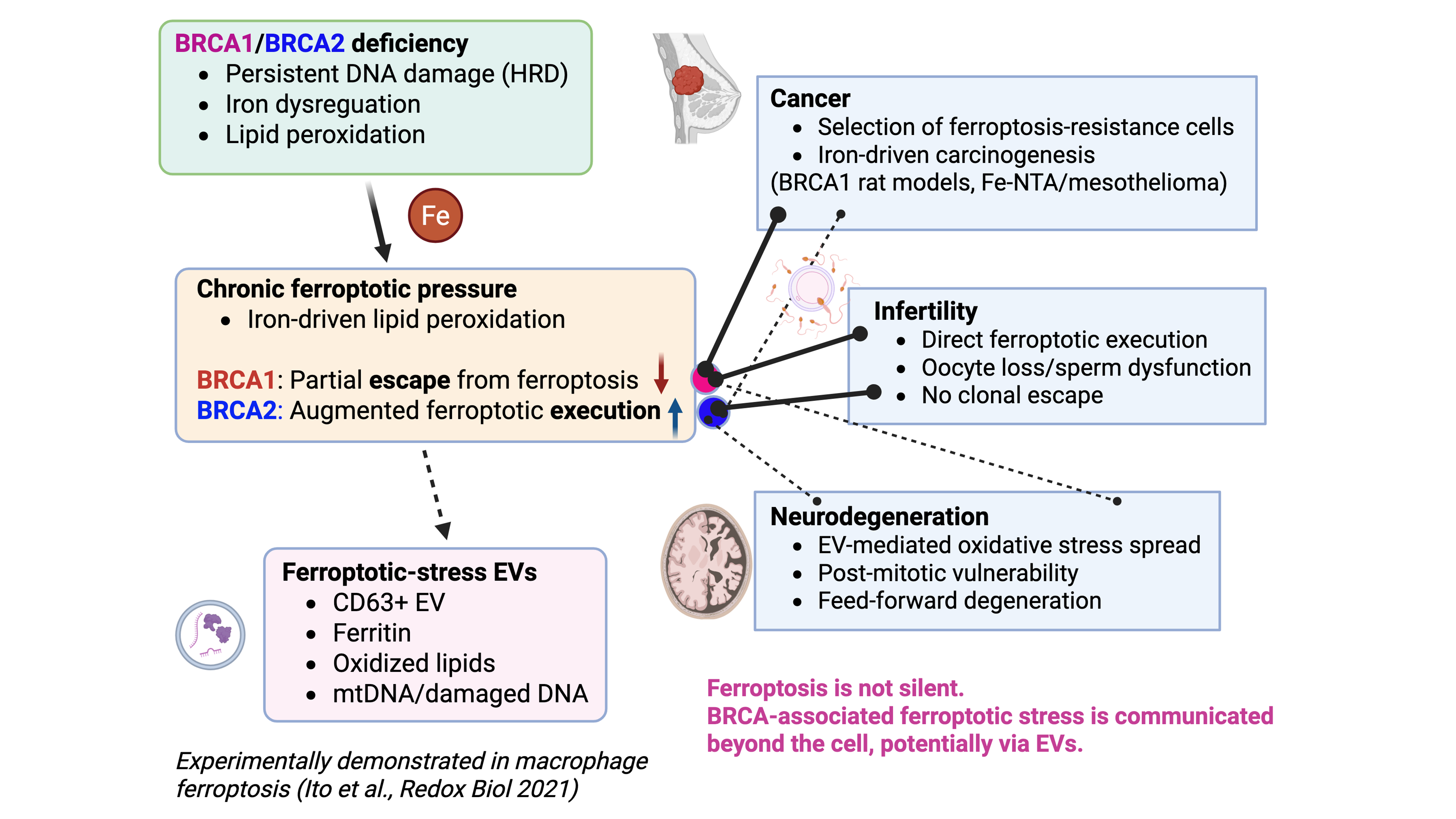
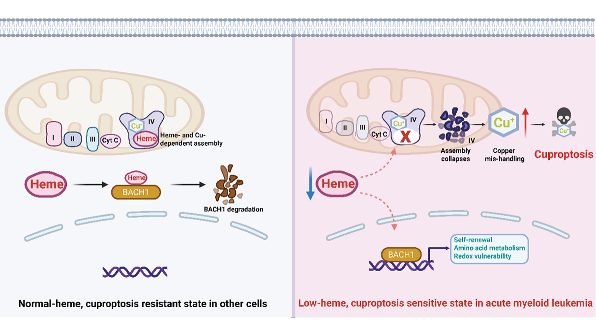
Ferroptosis and BRCA: New Mechanisms in Cancer Biology
Department of Biological Sciences, Department of Chemistry, Department of Pathology and Cell Biology, Columbia University, New York, NY, USA.
Prof. Shinya Toyokuni
Department of Pathology and Biological Responses, Nagoya University Graduate School of Medicine, Nagoya, Japan.
Dr. Yingyi Kong
Department of Pathology and Biological Responses, Nagoya University Graduate School of Medicine, Nagoya, Japan.


