Oxidative stress and inflammation: A panoply of pathways but a paucity of perspective

Aoife Costigan

Seamus J. Martin
*
*Correspondence to:
Seamus J. Martin, Molecular Cell Biology Laboratory, Department of Genetics, The Smurfit Institute, Trinity College, Dublin 2, Ireland.
E-mail: martinsj@tcd.ie
Ferroptosis Oxid Stress. 2027;3:202617. 10.70401/fos.2026.0040
Received: April 29, 2026Accepted: August 03, 2026Published: August 03, 2026
Abstract
Reactive oxygen species (ROS) are typically elevated at inflammatory sites due to the influx of large numbers of neutrophils and other myeloid cells that produce high levels of oxygen free radicals as microbicidal agents. Reactive oxygen intermediates are also generated as a byproduct of cellular metabolism, due to mitochondrial damage, exposure to toxins or radiation, and are also frequently increased during inflammatory reactions. However, although oxidative stress is often implicated as an initiator or amplifier of inflammation, particularly in the context of cancer, how reactive oxygen modulates inflammatory signalling remains poorly understood. Here we discuss the diversity of pathways and molecules implicated in promoting or suppressing inflammation in response to ROS. As we shall see, reactive oxygen has been reported to influence inflammatory outputs through direct effects on phosphatases that regulate nuclear factor kappa B (NF-κB) activation, through activation of the nuclear factor erythroid 2-related factor 2 (Nrf2) or hypoxia-inducible factor (HIF)-1 transcription factors that can directly or indirectly promote inflammatory gene expression, by provoking DNA damage leading to activation of the cyclic guanosine monophosphate (GMP)-adenosine monophosphate (AMP) synthase (cGAS)/stimulator of interferon genes (STING) pathway, or through promoting assembly of inflammasomes. Thus, there are a multitude of routes by which ROS can influence inflammatory responses. How reactive oxygen promotes inflammation in specific contexts is likely to be influenced by several factors, including the source of ROS, whether intracellular or extracellular, the responding cell type, as well as the extent of deviation from normal homeostatic setpoints. Understanding how reactive oxygen shapes inflammation has important implications for therapeutic intervention in multiple disease states.
Keywords
TNF, IL-1, inflammation, HIF-1, NF-κB, Keap1, Nrf2, reactive oxygen
1. Introduction
Reactive oxygen species (ROS) are highly reactive metabolites of oxygen that possess strong oxidizing capabilities and are generated as byproducts of cellular metabolism, particularly during mitochondrial respiration, but are also produced at high concentrations by immune cells as microbicidal agents[1]. Thus, ROS are frequently elevated at inflammatory sites due to the rapid influx of large numbers of neutrophils and other myeloid cells within minutes of the initiation of inflammation[2]. Activated neutrophils mount a robust respiratory burst via the plasma membrane-associated nicotinamide adenine dinucleotide phosphate (NADPH) oxidase complex (NOX2) that produces large amounts of ROS, which have potent microbicidal effects, but can also provoke local tissue damage[3]. At high concentrations, ROS are deleterious to cells, inflicting damage to cellular constituents and typically resulting in cell death. However, at low or physiological concentrations, ROS can function as signalling intermediates, through post-translational modification of redox-sensitive proteins including kinases, phosphatases, and transcription factors. Redox-sensitive proteins typically contain functionally significant cysteine residues that can undergo oxidation, which can alter protein activity and function[4].
Oxidative stress is frequently implicated in promoting or enhancing inflammation, but the underlying mechanism(s) are still unresolved[5,6]. Persistent reports also suggest that tumor necrosis factor (TNF) receptor stimulation can promote production of ROS via activation of the NOX1 or NOX2 complexes, depending on the cell type, as well as through production of mitochondrial ROS[7]. However, despite the association between elevated levels of reactive oxygen and inflammation, how ROS initiates or amplifies inflammation, as well as the impact of cellular antioxidant defence systems on the pro-inflammatory effects of major cytokines such as TNF and interleukin (IL)-1, are poorly understood. Here, we overview current data linking ROS-sensing pathways to inflammation. As we shall see, elevated ROS can exert effects on inflammatory outputs through activating the nuclear factor erythroid 2-related factor 2 (Nrf2) or hypoxia-inducible factor (HIF)-1 transcription factors, by provoking DNA damage leading to activation of the cyclic guanosine monophosphate (GMP)-adenosine monophosphate (AMP) synthase (cGAS)/stimulator of interferon genes (STING) pathway, through promoting assembly of NOD-like receptor pyrin domain-containing 3 (NLRP3) inflammasomes, as well as by direct modification of cysteine residues within phosphatases that influence activation of the nuclear factor kappa B (NF-κB) transcription factor, a master regulator of inflammation (Figure 1).
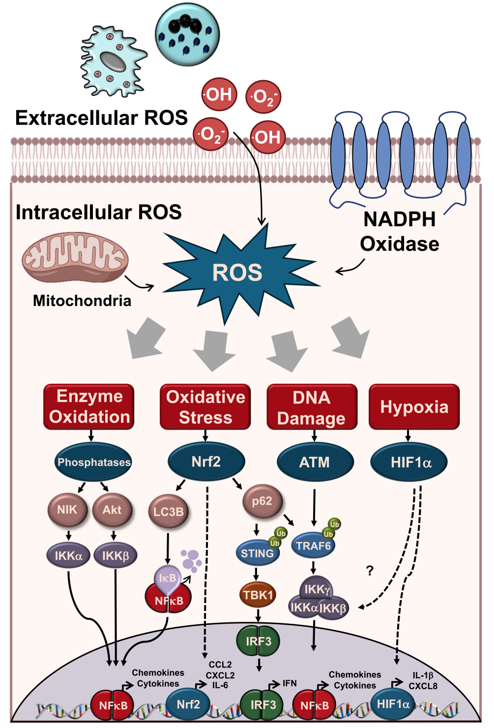
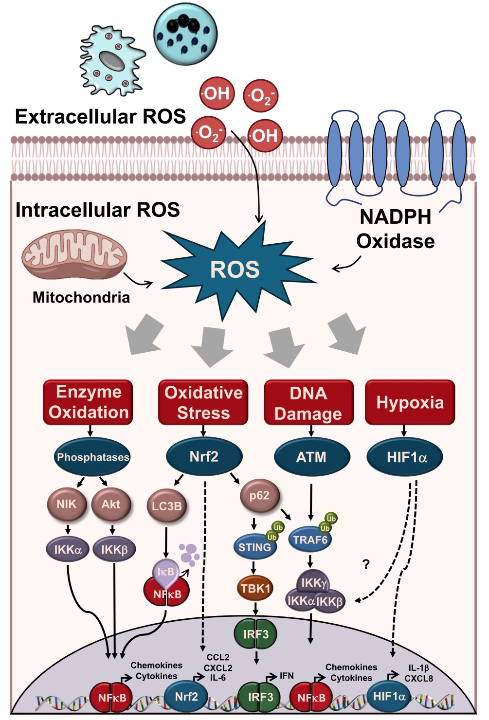
Figure 1. Diversity of pathways implicated in ROS-mediated regulation of inflammation. ROS: reactive oxygen species; ATM: ataxia-telangiectasia mutated; Nrf2: nuclear factor erythroid 2-related factor 2; HIF: hypoxia-inducible factor; NIK: NF-κB-inducing kinase; Akt: protein kinase B; STING: stimulator of interferon genes; IKK: IkappaB kinase; IL: interleukin; NF-κB: nuclear factor kappa B; IRF3: interferon-regulatory factor 3.
Generation of ROS extracellularly from immune cells (e.g. neutrophils) or intracellularly from mitochondria or NADPH oxidase activates multiple pathways that stimulate inflammation. Oxidation of redox-sensitive protein tyrosine phosphatases can impair their enzymatic activity, relieving inhibition of the downstream kinases NF-κB-inducing kinase (NIK) and protein kinase B (Akt) and facilitating NF-κB activation. The transcription factor Nrf2 is stabilized in the presence of oxidative stress and can promote inflammation directly by binding to the promoters of inflammatory genes, or indirectly via upregulation of p62 or light chain 3B (LC3B). p62 can stimulate interferon (IFN) induction by promoting STING ubiquitination or NF-κB activation through ubiquitination of tumor necrosis factor receptor-associated factor 6 (TRAF6). LC3B promotes NF-κB activation by mediating degradation of IkBa . Activation of ataxia-telangiectasia mutated (ATM) in response to ROS-induced DNA damage stimulates NF-κB activation by ubiquitination of TRAF6 and activation of NF-kappa B essential modulator (NEMO). Lastly, ROS-mediated hypoxia can stabilize HIF-1α, which can promote inflammation directly by binding to the promoters of inflammatory genes or through activation of the NF-κB signalling pathway.
2. The NF-κB Pathway as a Key Driver of Inflammation
Although several transcription factors can influence the production of inflammatory cytokines and other inflammatory mediators, as we shall discuss below, the NF-κB transcription factor is a major driver of the production of numerous inflammatory cytokines, chemokines, adhesion molecules, and other inflammatory factors[8-10]. Indeed, the most potent pro-inflammatory cytokines, including TNF and members of the extended IL-1 family, all exert their pro-inflammatory effects through activating NF-κB[11-13]. Thus, much of the following discussion will focus on interactions between ROS and NF-κB activation pathways.
The NF-κB transcription factor family includes several members, including: v-rel avian reticuloendotheliosis viral oncogene homolog A (RelA) (p65), p50, v-rel avian reticuloendotheliosis viral oncogene homolog B (RelB), p52 and cellular Rel (c-Rel)[14,15]. The two primary NF-κB transcriptional complexes, comprised of p65: p50 heterodimers (the canonical pathway) or p52: RelB heterodimers (the non-canonical pathway), are responsible for the majority of NF-κB-dependent inflammation[14]. Canonical NF-κB activation typically occurs downstream of the IkappaB kinase (IKK) complex (Figure 2), which is comprised of inhibitor of nuclear factor kappa-B kinase subunit alpha (IKKα), inhibitor of nuclear factor kappa-B kinase subunit beta (IKKβ) and their associated regulatory subunit inhibitor of nuclear factor kappa-B kinase subunit gamma (IKKγ) (NEMO). Activation of the IKK complex, which is frequently achieved through recruitment of NEMO/IKKγ to linear ubiquitin chains, or through phosphorylation of NEMO by upstream kinases, results in phosphorylation and degradation of the NF-κB inhibitor, IκB, which liberates p65: p50 heterodimers or p65 homodimers that can then translocate to the nucleus and promote inflammatory gene expression downstream[16,17]. In contrast, the non-canonical NF-κB pathway is typically activated through stabilization of the NIK (Figure 2), which can in turn activate IKKα leading to
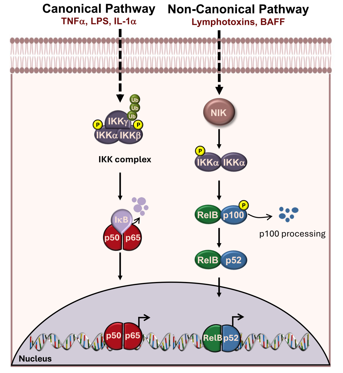
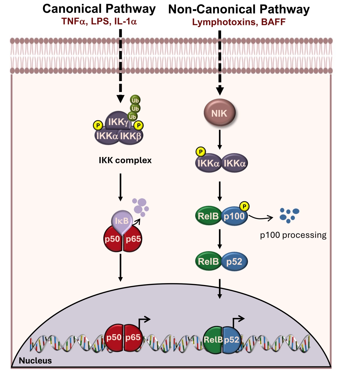
Figure 2. NF-κB activation pathways. TNF-α, LPS and IL-1α stimulate the canonical NF-κB pathway. K63 polyubiquitination of IKKγ activates the IKK complex which phosphorylates IkBa, promoting its proteasomal degradation and nuclear translocation of NF-κB p50:p65 heterodimers. Activation of the non-canonical NF-κB pathway by lymphotoxins or BAFF stabilizes NIK levels which phosphorylates IKKa. Activated IKKa phosphorylates p100, stimulating the proteasomal-mediated processing of p100 to p52. The p52 subunit forms a heterodimer with the NF-κB subunit RelB, and translocates to the nucleus to regulate expression of numerous inflammatory genes.
3. ROS-Induced Activation of NF-κB Through Direct Effects on Upstream Phosphatases
A number of reports have suggested that reactive oxygen can activate inflammatory signalling pathways through phosphorylation of NF-κB signalling proteins (Figure 1), although there is disagreement on the precise mechanism[20-23]. Some studies suggest that H2O2 promotes NF-κB activation via phosphorylation of the IKK complex, through activation of NIK or Akt[5,21,22], while others suggest that H2O2 activates NF-κB through activating a kinase that phosphorylates IkBa at tyrosine 42, rather than at the canonical phosphorylation sites serine 32 and 34[24-26]. Furthermore, ROS was also reported to promote phosphorylation of NF-κB p65 (RelA) through activating cAMP dependent protein kinase A (PKAc)[27]. Thus, it has been suggested that ROS can promote phosphorylation of multiple NF-κB regulators by several different mechanisms. One possibility, which might account for the diversity of mechanisms, is that elevated ROS inhibits multiple protein tyrosine phosphatases, which are highly sensitive to oxidation (Figure 1). Protein tyrosine phosphatases are redox-sensitive due to their active site cysteine residues, which exhibit a notably low pKa in comparison to other cysteine residues, thereby rendering the cysteine molecule vulnerable to oxidation[28,29]. For example, the phosphatase phosphatase and tensin homolog (PTEN), an inhibitor of Akt, contains a catalytic cysteine molecule that is ROS sensitive. Oxidation of PTEN impairs its phosphatase activity, thereby promoting Akt kinase activity[30] (Figure 1). Depending on the severity of ROS exposure, inhibition of multiple phosphatases may, in turn, lead to an increased activity of several distinct kinases promoting NF-κB activation and inflammation downstream (Figure 1). Aside from general phosphatase inhibition, ROS may also promote inflammation via activation of specific molecular pathways including Nrf2/Keap1, HIF, NLRP3, and cGAS-STING, as we shall see below.
4. Nrf2 as a Key Effector of ROS-Mediated Inflammatory Signalling
Previous reports have also implicated the key ROS sensor, Nrf2 (Figure 3), as a modulator of inflammation in a number of contexts[31]. On the one hand, Nrf2 has been implicated as a negative regulator of inflammation in response to lipopolysaccharide (LPS) or TNF stimulation[32-34]. However, Nrf2 has also been reported to serve as an enhancer of inflammation in the context of wound healing[35,36]. Cellular context and/or reactive oxygen concentration may dictate whether Nrf2 activation results in pro-inflammatory or
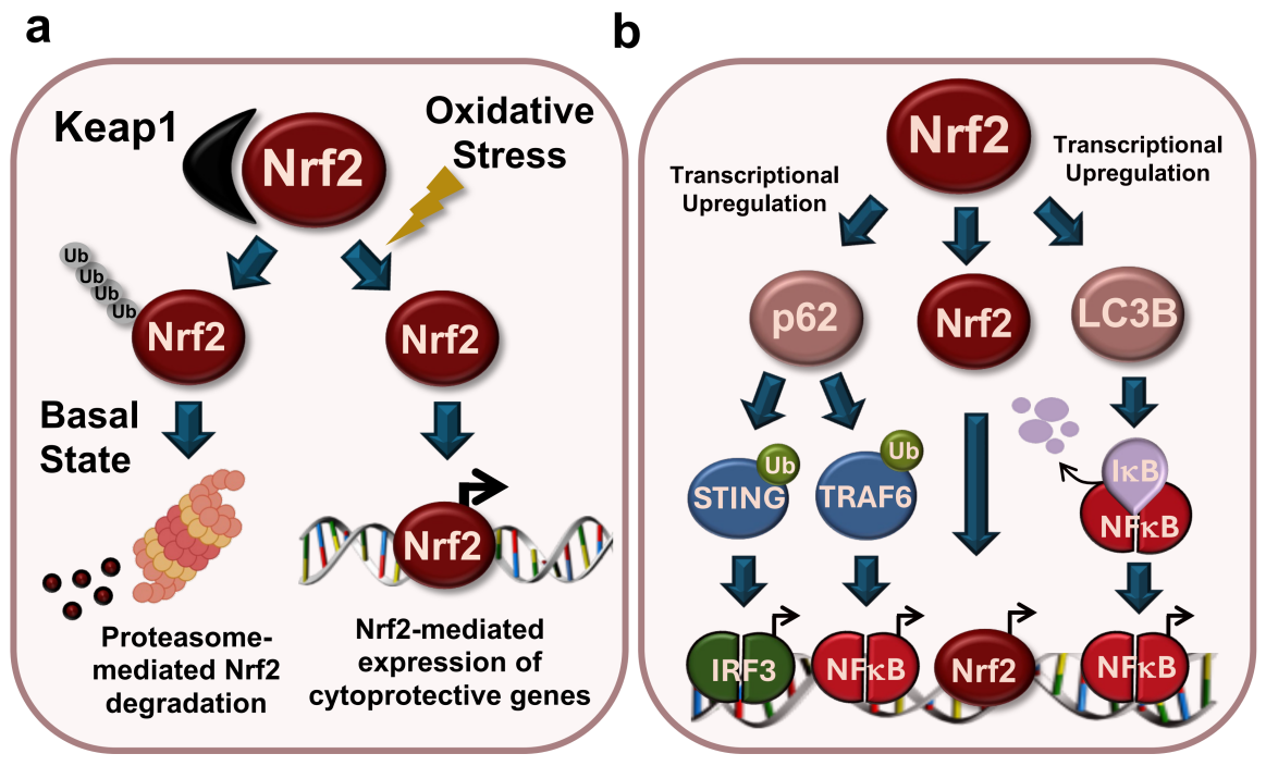
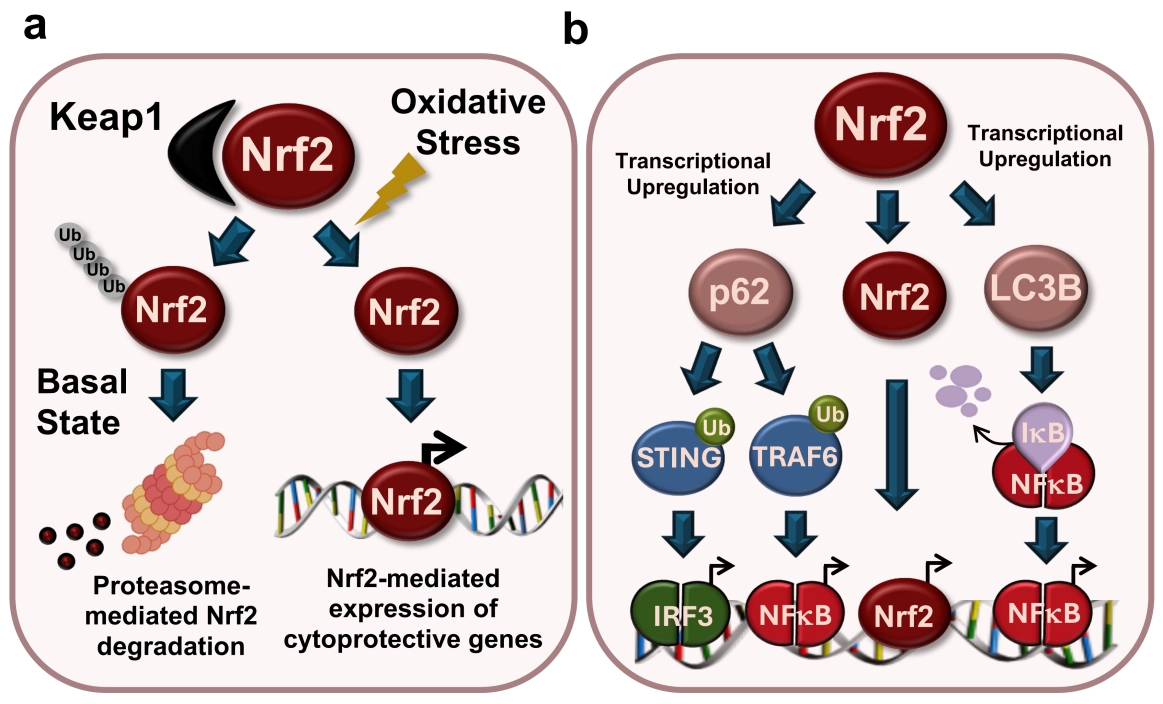
Figure 3. Nrf2 activation and inflammatory pathways. (A) Under basal conditions, the stability of Nrf2 is regulated by Keap1, an adaptor molecule for the Cullin-3 (Cul3) ubiquitin ligase complex. Keap1 constitutively promotes the ubiquitination and proteasomal degradation of Nrf2. In response to oxidative stress, oxidation of cysteine molecules in Keap1 disrupts its ability to interact with Nrf2, leading to stabilization of the latter. Once liberated from Keap1, Nrf2 translocates to the nucleus and upregulates a battery of antioxidant genes; (B) Nrf2 can stimulate inflammation directly through its effects on inflammatory gene promoters or indirectly via the upregulation of target genes. Nrf2 directly induces inflammation by binding to the promoters of inflammatory genes and initiating or enhancing their transcription. The Nrf2 target gene p62 can induce inflammation by promoting ubiquitination of TRAF6 and STING, thereby activating the NF-κB and IFN pathways, respectively. The Nrf2 target gene LC3B promotes
Nrf2/antioxidant response element (ARE) signalling plays a key role in the protection against oxidative stress and is responsible for the maintenance of homeostasis and redox balance in cells and tissues[37]. The Nrf2 transcription factor is stabilized through oxidative or electrophilic stress and its activation is typically cytoprotective due to its ability to regulate a battery of antioxidant response genes[38]. Nrf2 is regulated through the Keap1/Cullin-3 axis, where Keap1 acts as an adaptor for the Cullin-3 ubiquitin ligase complex to promote constitutive ubiquitin-dependent degradation of Nrf2 at steady state (Figure 3a)[39,40]. In response to oxidative or electrophilic stresses, Keap1 is inactivated, leading to stabilization of Nrf2 and upregulation of Nrf2-regulated genes such as HO-1 and the autophagy adaptor p62[41,42]. Previous studies have shown that induction of Nrf2 results in the suppression of apoptosis in response to multiple stress-inducing agents, including H2O2, RSL3 and proteasome inhibitors[43-45]. In addition, Nrf2 has also been implicated as a modulator of inflammation in a number of contexts[32,46-48]. However, despite the importance of Nrf2 as a sensor of oxidative stress, how this transcription factor exerts its influence on inflammatory signalling pathways is still poorly understood, with evidence supporting a pro- as well as an anti-inflammatory role for Nrf2 in the context of oxidative stress, as noted earlier[37].
4.1 Pro-inflammatory effects of the Keap1/Nrf2 pathway
While Nrf2 is primarily known for regulating genes that protect against oxidative stress, it is becoming increasingly apparent that Nrf2 can also regulate the expression of a number of genes beyond its canonical anti-oxidant targets (Figure 3b)[49]. Several studies suggest that Nrf2 can modulate the expression of pro-inflammatory cytokines and chemokines by binding to their proximal promoters[32,47,50]. Overexpression of Nrf2 has been reported to be sufficient to activate the interleukin (IL)-6 promoter, which was attenuated upon mutation of the ARE within the latter promoter[47]. Furthermore, chromatin immunoprecipitation sequencing
Other studies also argue a role for Nrf2 in driving inflammatory gene expression through direct promoter binding. Using transcription factor binding analysis combined with chromatin immunoprecipitation followed by quantitative polymerase chain reaction
In addition to promoting expression of genes encoding inflammatory cytokines directly, Nrf2 target genes have also been implicated as modulators of inflammation. Nrf2 upregulates many autophagy-related proteins as part of its anti-oxidant function[52]. Autophagy is activated as an adaptive response to increase the clearance of damaged organelles and protein aggregates that can arise during oxidative stress. p62/sequestosome 1 (SQSTM1) is an autophagy cargo protein and an established Nrf2 target gene[42]. While p62 is well known to function as a major autophagy cargo adaptor, this multifunctional protein has also been implicated as a regulator of inflammatory signalling pathways[53,54]. p62 has recently been reported to regulate type I IFN production through activation of the cytosolic DNA sensor STING, by binding to the latter and displacing NBRI, an autophagy adaptor which promotes lysosomal degradation of STING[54]. Binding of p62 to STING was reported to stimulate the ubiquitination and oligomerization of STING, leading to the activation of TBK1 and downstream transcription factor interferon-regulatory factor 3 (IRF3), subsequently stimulating the release of type I interferons (Figure 1)[54]. Several studies also propose that p62 can activate NF-κB signalling. As previously discussed, the canonical NF-κB heterodimer, p65:p50, is typically held inactive in the cytoplasm by its cognate inhibitor IkBa. Phosphorylation of IkBa by the upstream kinase complex IKK, stimulates proteasomal degradation of IkBa and translocation of NF-κB to the nucleus[14]. Several studies suggest that p62 can promote NF-κB activation by binding to and promoting ubiquitination of the E3 Ub-ligase TRAF6, resulting in activation of its E3 ligase activity to generate K63 polyubiquitin chains that recruit and activate the IKK complex, leading in turn to NF-κB activation downstream[55]. Consistent with this, activation of NF-κB in response to nerve growth factor, oncogenic Ras or in polarized Th2 cells, was blunted in p62-deficient cells[56-58]. In all cases, inhibition of NF-κB in p62-deficient cells correlated with a decrease in TRAF6 polyubiquitination, which blunted activation of the IKK complex and NF-κB downstream. Furthermore, secretion of IL-4, IL-6 and cell survival were impaired in p62-deficient cells[56,57]. In addition to the p62-dependent routes discussed above, Nrf2 has also been implicated as a regulator of the NF-κB pathway through upregulation of LC3B, an autophagy adaptor protein involved in autophagosome formation and maturation. In response to proteasome inhibition, Nrf2 was reported to activate NF-κB through upregulation of LC3B and promoting the degradation of IkBa[59]. It is evident that Nrf2 plays a crucial role in promoting inflammation in a variety of contexts. However, as we will discuss below, there is also a body of evidence to suggest that ROS-mediated Nrf2 activation functions to suppress inflammation.
4.2 Anti-inflammatory effects of the Nrf2/Keap1 pathway
A number of studies have implicated Nrf2 as a restraining influence on inflammation, possibly through regulating the expression of cellular antioxidants, thereby counteracting the effects of ROS. It has been reported that Nrf2-/- mice displayed increased cytokine production, immune cell infiltration, and mortality in response to LPS stimulation, compared to their wildtype counterparts[33,60]. Similarly, Nrf2 deficiency enhanced inflammatory signalling in monocytes following TNF-α treatment[34]. Given that ROS can promote inflammation, the anti-inflammatory effects of Nrf2 may be a direct consequence of its antioxidative activity, rather than a direct role for Nrf2 as a suppressor of inflammation. In line with this interpretation, the antioxidant N-acetyl cysteine (NAC) was reported to suppress LPS-induced inflammation and mortality in Nrf2-deficient mice, as well as TNF-driven inflammation[34,60,61]. Furthermore, upregulation of the Nrf2-inducible antioxidant genes heme oxygenase 1 (HO-1) and NADPH quinone oxidoreductase (NQO1) also reduced inflammation following LPS exposure[46]. Thus, the reported anti-inflammatory effects of Nrf2 may be a secondary outcome of its antioxidant activity, rather than a direct role for Nrf2 as a transcriptional suppressor of inflammatory genes.
However, there are reports that suggest Nrf2 may suppress inflammation independently of its antioxidant functions. One study reported that Nrf2 can downregulate the expression of IL-6 and IL-1β in macrophages through inhibiting the recruitment of RNA polymerase II to the promoter regions of the genes encoding the latter cytokines[32]. Other studies have described an antagonistic relationship between Nrf2 and the NF-κB subunit p65, further supporting an anti-inflammatory role for Nrf2[62]. Nrf2 and p65 compete for the same co-transcriptional activator cAMP response element-binding protein (CREB) binding protein (CBP/p300). Upon phosphorylation, p65 has a higher affinity for CBP/p300 and therefore sequesters CBP/p300 from Nrf2. p65 has also been reported to recruit histone deacetylase 3 (HDAC3) to promoters which impairs acetylation of the Nrf2-MafK heterodimer, thereby reducing Nrf2 mediated ARE transcription[63]. Furthermore, NF-κB p65 can inhibit Nrf2 activity by binding to Keap1 and facilitating Nrf2 ubiquitination and degradation[64]. Conversely, Nrf2 has also been described to inhibit NF-κB activation. It has been reported that
The dual and often contradictory role of Nrf2 in inflammation is well illustrated by the findings of Yamamoto and colleagues[32]. While the main conclusion of the latter study is that Nrf2 activation can blunt transcription of pro-inflammatory genes through inhibition of RNA Polymerase II recruitment, several observations from the same study indicate that Nrf2 activation can also promote inflammation[32]. These opposing functions are likely context-dependent, with the degree of oxidative stress and/or the level of Nrf2 activation in specific cell types all influencing the effect of Nrf2 on inflammatory outcomes. In many studies that describe an
5. The HIF-1 Pathway as a Modulator of Inflammation Associated with ROS Production
Oxygen homeostasis and ROS generation are inherently interlinked within the cell. Hypoxia can stimulate ROS production by dysregulating the mitochondrial electron transport chain, generating unpaired electrons that react with cellular oxygen to produce superoxide ions. ROS can also promote tissue hypoxia by impairing endothelial function, thereby limiting oxygen delivery, or by altering mitochondrial function, leading to increased oxygen consumption. Cellular oxygen homeostasis is tightly monitored by the transcription factor HIF. HIF is a heterodimeric protein composed of two subunits, a constitutively expressed HIFb subunit and an oxygen sensitive a-subunit, that can be either of the three isoforms that have been identified (HIF-1α, HIF-2α, HIF-3α)[67]. HIF-1α is ubiquitously expressed, whereas HIF-2α and HIF-3α are expressed within specific cells, such as macrophages. However, all isoforms can regulate the transcriptional response to hypoxia. Under normoxic conditions, HIF-1α stability is tightly regulated by
5.1 Direct regulation of inflammatory genes by HIF-1α and HIF-1β
Inflammatory sites are typically associated with hypoxia due to immune cell infiltration, vascular impairment, and elevated ROS production. HIF has previously been implicated as a key driver of inflammation by stimulating cytokine production, promoting immune cell function, and initiating NF-κB activation[72,73]. Thus, HIF represents a molecular link through which ROS and hypoxia can induce inflammation. Multiple studies have previously described a central role for HIF-1α in stimulating pro-inflammatory cytokine production. In response to hypoxia, the expression of vascular endothelial growth factor (VEGF), CXCL8 and IL-1β were reduced in the absence of HIF-1α[74]. Similarly, LPS-induced IL-1β release was markedly impaired in HIF-1α null macrophages compared to wild type cells[75]. Loss of HIF-1α also impaired IL-6 and IL-8 secretion in Rheumatoid Arthritis synovial fibroblasts, while overexpression of the transcription factor enhanced cytokine signalling[76]. In a mouse model of colitis, constitutive activation of HIF by VHL deletion correlated with increased inflammatory cytokine production and more severe clinical symptoms, effects that were reversed following deletion of HIF-1β[77]. At the molecular level, HIF-1α appears to directly regulate the expression of pro-inflammatory cytokines by binding to promoter elements within these inflammatory genes (Figure 1). HIF-1α regulation of IL-1β was dependent on the hypoxic response element within the proximal regions of the IL-1β promoter[78]. HIF-2α was also reported to mediate expression of inflammatory mediators in M1 macrophages in response to hypoxia, also through direct promoter binding[79].
5.2 Indirect regulation of inflammation by HIF-1α through activation of NF-κB and induction of glycolysis
Aside from directly regulating the expression of pro-inflammatory cytokines, HIF-1α may also mediate inflammation indirectly, through activation of NF-κB[72] (Figure 1). Although it is well established that NF-κB can positively modulate HIF expression, the role of HIF in the regulation of NF-κB remains largely obscure[72,80]. Scortegagna and colleagues illustrated that constitutive HIF-1α expression in keratinocytes enhanced cytokine secretion in response to an inflammatory challenge, which could be reversed upon co-expression of the IkBa super-repressor[81]. Constitutive expression of HIF-1α also correlated with increased phosphorylation of IkBa, IKKβ, and p65 suggesting that HIF-1α may promote inflammation via NF-κB activation[81]. Furthermore, in response to hypoxia, NF-κB activation was observed following suppression of PHD hydroxylation activity and enhanced phosphorylation of IKKβ[82]. Thus, there are some indications that the HIF pathway may positively regulate NF-κB activity. However, further investigation is required to fully characterise the relationship between these two pathways.
HIF-1α has frequently been reported to be an important regulator of immune cell function[73,83], possibly via its effects on NF-κB activation. Decreased levels of IKKβ, IkBa and p65 were observed in HIF-1α-deficient neutrophils following hypoxic stress[84]. This reduction in NF-κB signalling correlated with an increase in apoptosis in HIF-1α-null neutrophils compared to their wild-type counterparts[84]. HIF-1α can also modulate immune cell function by promoting a metabolic shift from oxidative phosphorylation to glycolysis during immune cell activation. Because oxygen deprivation is a common feature of inflamed tissues, activation of glycolysis is an essential adaptive mechanism to ensure proper immune cell function at hypoxic sites. HIF-1α has been reported to regulate glycolysis in multiple immune cell types through direct transcriptional upregulation of key glycolytic genes including glucose transporters (e.g. GLUT-1) and glycolytic enzymes (e.g. pyruvate kinase M2 (PKM2))[73]. In macrophages, HIF-1α deficiency has been reported to reduce cellular adenosine triphosphate (ATP) levels, which correlated with impaired mobility and phagocytosis in response to bacterial infection[85]. In dendritic cells, loss of HIF-1α impaired LPS-induced GLUT-1 expression, which correlated with a decrease in dendritic cell maturation and T cell activation[86]. Thus, HIF-1α can modulate inflammation through a variety of mechanisms and represents a key mechanistic link between ROS and inflammation.
6. ROS-Mediated Activation of Inflammasomes
Inflammasomes are complexes between Nod-like receptor (NLR) proteins and inflammatory caspases (predominantly caspase-1) that form in response to homeostatic disturbances associated with stress and infection[87]. Assembly of inflammasomes leads to recruitment and activation of Caspase-1, which can directly promote inflammation through proteolytic processing of the potent inflammatory cytokines IL-1β and IL-18, which are then liberated through assembly of Gasdermin D pores within plasma membranes as a consequence of Caspase-1-mediated cleavage of the latter protein[88,89]. A very well studied NLR complex, the NLRP3 inflammasome, is assembled in response to LPS in combination with bacterial virulence factors and is a key driver of inflammation in numerous contexts. However, for full activation of the NLRP3 inflammasome and liberation of IL-1β, a second signal in the form of a membrane damaging bacterial toxin (which can also be mimicked by a variety of noxious agents) is typically required. This second signal appears to permit the efflux of K+ ions from the cytosol, which permits full assembly of the inflammasome, caspase-1 activation and processing of IL-1β and IL-18 downstream. Several studies have suggested that ROS can serve as a second signal for inflammasome assembly[90-92]. Inhibition of ROS by the antioxidants NAC or (2R, 4R)-4-aminopyrrolidine-2,4-dicarboxylate (APDC) impaired NLRP3 mediated IL-1β release and caspase-1 activation following asbestos exposure[91]. Loss of mitochondrial ROS, through silencing of voltage-dependent anion channels or inhibition of mitophagy, impaired inflammasome activation in response to various stimuli including LPS and monosodium urate (MSU) crystals [90].
Mechanistically, how ROS promotes inflammasome activation is unclear, but cysteine oxidation has been suggested as a potential mechanism. It has been reported that ROS induces phosphorylation of related kinase 7 (NEK7), a key component of the NLRP3 inflammasome, promoting NEK7 binding to the leucine-rich repeat (LRR) domain of NLRP3 and subsequent inflammasome
7. DNA damage-Initiated Routes to Inflammation
Reactive oxygen can also result in genotoxic damage, either through direct modification of DNA, or through depletion of nucleotide pools in the cytoplasm as a consequence of oxidation and this can lead to DNA damage due to misincorporation of nucleotides during DNA replication[101]. DNA damage can, in turn, promote inflammation in a number of ways. First, activation of the DNA damage sensing kinase ATM has been directly implicated in activating the NF-κB-activating kinase IKKg (NEMO) through direct phosphorylation of the latter within the nucleus, leading to its transport into the cytoplasm and activation of the IKK complex downstream[102-104]. DNA damage-induced activation of ATM has also been reported to lead to formation of an ATM-TRAF6 signalling complex that can promote NF-κB activation[105-107]. The latter complex has also been implicated in NF-κB-dependent upregulation of TNF, which can induce feed-forward inflammatory signalling by promoting further NF-κB activation downstream[107]. ATM has also been implicated in mediating inflammation through promoting the formation of an NF-κB-activating complex containing p53‐induced death‐domain (PIDD) and receptor‐interacting protein kinase 1 (RIPK1), called the PIDDosome, in response to DNA damaging agents such as etoposide, as well as centrosome duplication-associated with polyploidy[108,109].
Second, extensive DNA damage can trigger entry of cells into a non-replicating state, called premature replicative senescence[110]. Senescence is a radical departure from normal cellular function, as these cells typically exit the cell cycle, greatly increase in size, activate DNA sensing kinases, and secrete a range of pro-inflammatory and proliferative factors, collectively called the
Campisi and colleagues have published a series of studies suggesting that IL-1α is an important driver of senescence-associated inflammation in response to DNA damage, oncogene activation or chromatin perturbation[111,117,118]. Although the upstream initiators of IL-1α expression in this context remain obscure, the DNA damage-sensing kinase, ATM, was again implicated[118]. ROS-mediated DNA damage has also been reported to trigger the SASP through promoting the release of genomic DNA into the cytoplasm as cytoplasmic chromatin fragments. The latter event leads to activation of the cGAS/STING pathway, leading to inflammatory cytokine production through activation of STING, which is well established to promote NF-κB- and IRF-dependent inflammatory pathways downstream[119-122]. Cytoplasmic DNA fragments are a well-established feature of senescence and accumulate in the cytoplasm following the loss of nuclear envelope integrity, driven by lamin B1 degradation. Activation of the autophagy-lysosome pathway has been reported to mediate lamin B1 degradation in senescent cells[123,124]. Autophagy-related 7 (ATG7) deficiency has been reported to attenuate lamin B1 loss in the context of oxidative stress-induced senescence, as well as delay the onset of senescence in response to oncogene activation[123]. Given that reactive oxygen is a potent activator of autophagy[125], ROS-mediated autophagy activation may be an important regulator of senescence and the SASP through degradation of lamin B1, leading to the escape of chromatin into the cytoplasmic compartment leading to cGAS/STING-initiated inflammation.
As previously mentioned, the SASP is a double-edged sword. While SASP release has been reported to facilitate immune-mediated clearance of pre-malignant cells, thereby contributing to tumour suppression[119], chronic SASP production has been implicated in tumour progression and the pathogenesis of a range of age-related diseases[126,127]. ‘Inflammaging’ was coined by Franceschi and colleagues to describe the low grade chronic inflammation that typically develops with age and can exacerbate disease[128]. Accumulation of senescent cells in many tissues, including the immune system, is suspected to be a central driver of
8. Therapeutic Implications of ROS-induced Inflammation
It is well established that ROS and oxidative stress play a central role in a wide range of pathologies. ROS can act as the primary cause of pathology, as in paraquat poisoning or radiation-induced lung damage, or act as a secondary amplifier in conditions such as cancer and inflammatory diseases[136,137]. Thus, identifying the key ROS sensing pathways that promote inflammation has important therapeutic implications. The use of antioxidants to treat various conditions has yielded little clinical success to date due to the diverse sources of intracellular ROS and the robustness of endogenous antioxidant defence systems, which minimize the overall impact of exogenous ROS scavengers[137,138]. Activation of endogenous antioxidant defence systems, such as the Nrf2 pathway, may be a more fruitful approach to tackling ROS and its downstream consequences[139]. For example, the Nrf2 activator dimethyl fumarate (DMF) has shown promising results in relapsing-remitting multiple sclerosis (MS)[140-142]. Conversely, in situations where Nrf2 activation is associated with poor prognosis, such as in cancer[143,144], Nrf2 inhibitors may have therapeutic benefits by reducing inflammation and disease progression. NLRP3 represents another attractive target for the suppression of ROS-driven inflammation. To date, no Food and Drug Administration (FDA)-approved NLRP3 inhibitors are available. Although initially promising results were seen with MCC950, a small molecular NLRP3 inhibitor which traps the latter in an inactive state[145], this compound was ultimately discontinued due to liver toxicity[146]. While the optimization of compounds with greater specificity towards NLRP3 is underway, targeting ROS-mediated activation of NLRP3 may represent an alternative strategy[98]. Other approaches to ameliorate the downstream effects of ROS involve the identification of ‘senolytics’ or ‘senomorphics’, compounds that can eliminate or modify the behaviour of senescent cells, thereby suppressing their pro-inflammatory effects[114,147,148].
9. Concluding Remarks
Reactive oxygen has been implicated as an important modulator of inflammation for many years. Despite this, the molecular mechanisms of ROS-driven inflammation have remained relatively obscure. Here, we have discussed how ROS may influence inflammatory outputs by a diversity of mechanisms, including activation of the major ROS-sensing transcription factor Nrf2, as well as the key oxygen sensor HIF-1. ROS can also shape inflammatory responses through initiating DNA damage, leading to activation of the cGAS/STING pathway, by influencing assembly of NLRP3 inflammasomes, as well as via direct effects on phosphatases that influence activation of NF-κB (Figure 4). The specific route to ROS-mediated inflammation that is employed in a particular context is likely to depend on several factors, including the source of ROS, whether intracellular or extracellular, the responding cell type (whether a professional immunocyte, such as a macrophage, or an epithelial cell) and its natural ROS-buffering systems, as well as the degree of ROS elevation relative to normal setpoints in the cell type or tissue in question. Further work is clearly required to clarify the precise nature of the ROS-sensing pathways that serve to initiate or escalate inflammation in specific disease contexts, such as cancer, or in the context of age-related inflammation (inflammaging) where excessive ROS production and/or reduced antioxidant capacity are suspected to be contributing factors. Clarifying the major pathways linking ROS sensing to inflammatory outputs would undoubtedly have very significant benefits for the treatment of multiple conditions where ROS is a major factor in driving inflammation.
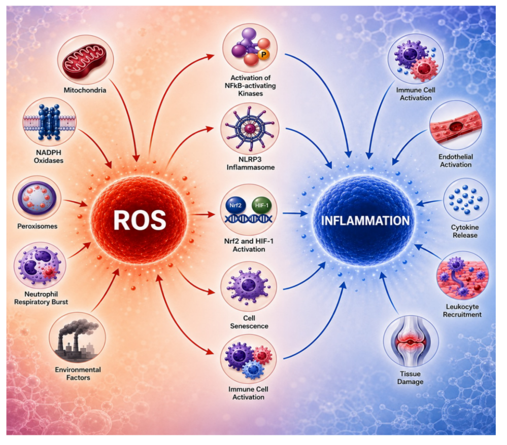
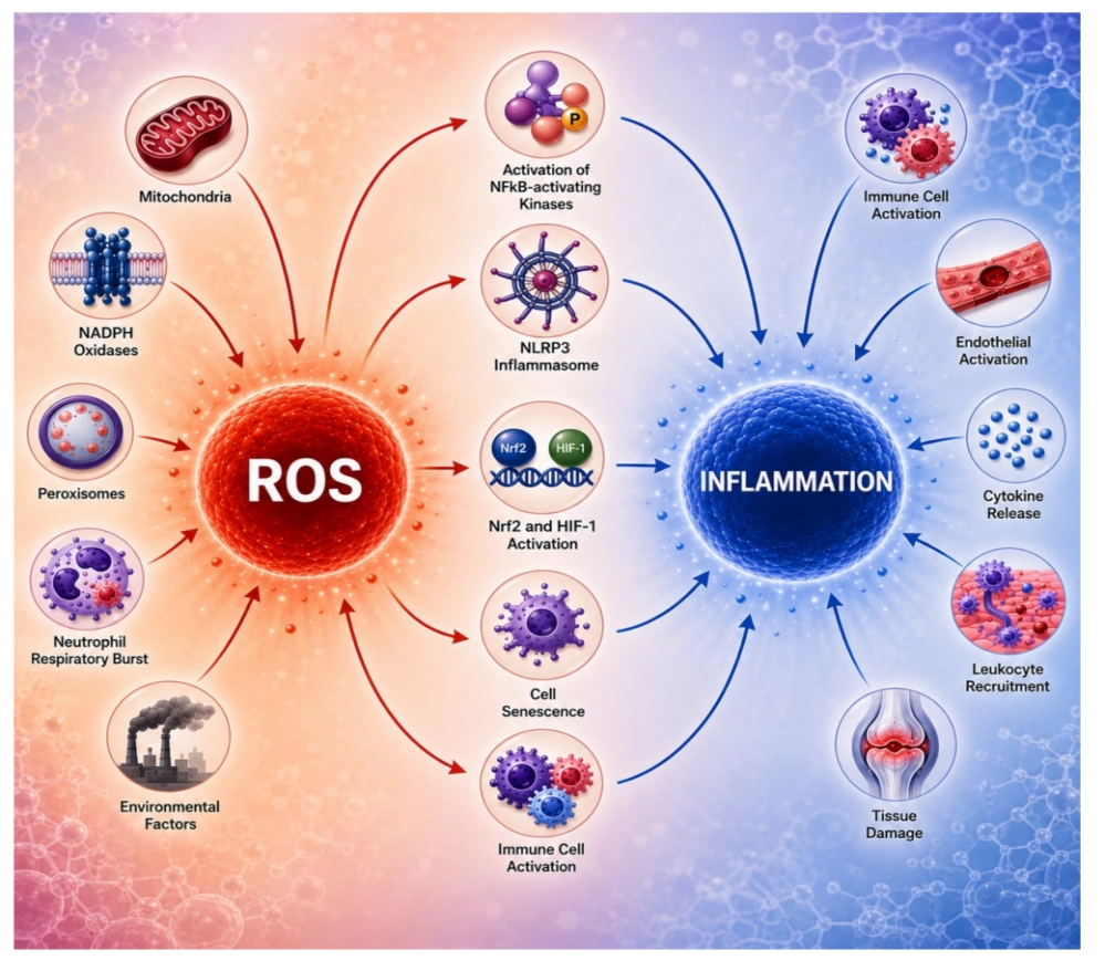
Figure 4. Summary of pathways linking ROS generation to inflammatory outputs. See main text for further details. ROS: reactive oxygen species; Nrf2: nuclear factor erythroid 2-related factor 2; HIF: hypoxia-inducible factor; NADPH: nicotinamide adenine dinucleotide phosphate; NLRP3: NOD-like receptor pyrin domain-containing 3.
Acknowledgments
During manuscript preparation, OpenAI GPT-5.5 was used to assist in generating the schematic illustration in Figure 4. The authors reviewed, edited, and verified the final figure and take full responsibility for its content.
Authors contribution
Costigan A, Martin SJ: Conceptualization, methodology, writing-original draft.
Conflicts of interest
Seamus J. Martin is an Editorial Board Member of Ferroptosis and Oxidative Stress. The other author declares no conflicts of interest.
Ethical approval
Not applicable.
Consent to participate
Not applicable.
Consent for publication
Not applicable.
Availability of data and materials
Not applicable.
Funding
The Martin laboratory is supported by grants from the Irish Research Council Laureate Award Programme (Grant No. IRCLA/2019/133) and the European Research Council Advanced Grant Programme (DESTRESS, Grant No. 101020534).
Copyright
© The Author(s) 2026.
References
-
1. Sies H, Berndt C, Jones DP. Oxidative stress. Annu Rev Biochem. 2017;86:715-748.[DOI]
-
2. Mittal M, Siddiqui MR, Tran K, Reddy SP, Malik AB. Reactive oxygen species in inflammation and tissue injury. Antioxid Redox Signal. 2014;20(7):1126-1167.[DOI]
-
4. Parvez S, Long MJC, Poganik JR, Aye Y. Redox signaling by reactive electrophiles and oxidants. Chem Rev. 2018;118(18):8798-8888.[DOI]
-
5. Morgan MJ, Liu ZG. Crosstalk of reactive oxygen species and NF-κB signaling. Cell Res. 2011;21(1):103-115.[DOI]
-
6. Yu Y, Liu S, Yang L, Song P, Liu Z, Liu X, et al. Roles of reactive oxygen species in inflammation and cancer. MedComm. 2024;5(4):e519.[DOI]
-
10. Qian Z, Lenardo MJ, Baltimore D. 30 years of NF-κB: A blossoming of relevance to human pathobiology. Cell. 2017;168(1):37-57.[DOI]
-
14. Oeckinghaus A, Ghosh S. The NF-κB family of transcription factors and its regulation. Cold Spring Harb Perspect Biol. 2009;1(4):a000034.[DOI]
-
15. Baeuerle PA, Baltimore D. NF-κB: Ten years after. Cell. 1996;87(1):13-20.[DOI]
-
19. Sun SC. The non-canonical NF-κB pathway in immunity and inflammation. Nat Rev Immunol. 2017;17(9):545-558.[DOI]
-
25. Schoonbroodt S, Ferreira V, Best-Belpomme M, Boelaert JR, Legrand-Poels S, Korner M, et al. Crucial role of the amino-terminal tyrosine residue 42 and the carboxyl-terminal PEST domain of I kappa B alpha in NF-kappa B activation by an oxidative stress. J Immunol. 2000;164(8):4292-4300.
-
26. Takada Y, Mukhopadhyay A, Kundu GC, Mahabeleshwar GH, Singh S, Aggarwal BB. Hydrogen peroxide activates NF-kappa B through tyrosine phosphorylation of I kappa B alpha and serine phosphorylation of p65: Evidence for the involvement of I kappa B alpha kinase and Syk protein-tyrosine kinase. J Biol Chem. 2003;278(26):24233-24241.
-
30. Lee SR, Yang KS, Kwon J, Lee C, Jeong W, Rhee SG. Reversible inactivation of the tumor suppressor PTEN by H2O2. J Biol Chem. 2002;277(23):20336-20342.[DOI]
-
31. Saha S, Buttari B, Panieri E, Profumo E, Saso L. An overview of Nrf2 signaling pathway and its role in inflammation. Molecules. 2020;25(22):5474.[DOI]
-
39. Itoh K, Wakabayashi N, Katoh Y, Ishii T, Igarashi K, Engel JD, et al. Keap1 represses nuclear activation of antioxidant responsive elements by Nrf2 through binding to the amino-terminal Neh2 domain. Genes Dev. 1999;13(1):76-86.[DOI]
-
40. Kobayashi A, Kang MI, Okawa H, Ohtsuji M, Zenke Y, Chiba T, et al. Oxidative stress sensor Keap1 functions as an adaptor for Cul3-based E3 ligase to regulate proteasomal degradation of Nrf2. Mol Cell Biol. 2004;24(16):7130-7139.[DOI]
-
41. Dinkova-Kostova AT, Holtzclaw WD, Cole RN, Itoh K, Wakabayashi N, Katoh Y, et al. Direct evidence that sulfhydryl groups of Keap1 are the sensors regulating induction of phase 2 enzymes that protect against carcinogens and oxidants. Proc Natl Acad Sci U S A. 2002;99(18):11908-11913.[DOI]
-
48. Zhang X, Chen X, Song H, Chen HZ, Rovin B. Activation of the Nrf2/antioxidant response pathway increases IL-8 expression. Eur J Immunol. 2005;35(11):3258-3267.[DOI]
-
52. Pajares M, Jiménez-Moreno N, García-Yagüe ÁJ, Escoll M, de Ceballos ML, Van Leuven F, et al. Transcription factor NFE2L2/NRF2 is a regulator of macroautophagy genes. Autophagy. 2016;12(10):1902-1916.[DOI]
-
53. Sánchez-Martín P, Saito T, Komatsu M. p62/SQSTM1: ‘Jack of all trades’ in health and cancer. FEBS J. 2019;286(1):8-23.[DOI]
-
57. Duran A, Linares JF, Galvez AS, Wikenheiser K, Flores JM, Diaz-Meco MT, et al. The signaling adaptor p62 is an important NF-κB mediator in tumorigenesis. Cancer Cell. 2008;13(4):343-354.[DOI]
-
58. Wooten MW, Geetha T, Seibenhener ML, Babu JR, Diaz-Meco MT, Moscat J. The p62 scaffold regulates nerve growth factor-induced NF-κB activation by influencing TRAF6 polyubiquitination. J Biol Chem. 2005;280(42):35625-35629.[DOI]
-
60. Kong X, Thimmulappa R, Craciun F, Harvey C, Singh A, Kombairaju P, et al. Enhancing Nrf2 pathway by disruption of Keap1 in myeloid leukocytes protects against sepsis. Am J Respir Crit Care Med. 2011;184(8):928-938.[DOI]
-
63. Liu GH, Qu J, Shen X. NF-κB/p65 antagonizes Nrf2-ARE pathway by depriving CBP from Nrf2 and facilitating recruitment of HDAC3 to MafK. Biochim Biophys Acta Mol Cell Res. 2008;1783(5):713-727.[DOI]
-
64. Yu M, Li H, Liu Q, Liu F, Tang L, Li C, et al. Nuclear factor p65 interacts with Keap1 to repress the Nrf2-ARE pathway. Cell Signal. 2011;23(5):883-892.[DOI]
-
69. Maxwell PH, Wiesener MS, Chang GW, Clifford SC, Vaux EC, Cockman ME, et al. The tumour suppressor protein VHL targets hypoxia-inducible factors for oxygen-dependent proteolysis. Nature. 1999;399(6733):271-275.[DOI]
-
71. Lee G, Won HS, Lee YM, Choi JW, Oh TI, Jang JH, et al. Oxidative dimerization of PHD2 is responsible for its inactivation and contributes to metabolic reprogramming via HIF-1α activation. Sci Rep. 2016;6:18928.[DOI]
-
72. D’Ignazio L, Bandarra D, Rocha S. NF-κB and HIF crosstalk in immune responses. FEBS J. 2016;283(3):413-424.[DOI]
-
73. McGettrick AF, O’Neill LAJ. The role of HIF in immunity and inflammation. Cell Metab. 2020;32(4):524-536.[DOI]
-
87. Lamkanfi M, Dixit VM. Mechanisms and functions of inflammasomes. Cell. 2014;157(5):1013-1022.[DOI]
-
90. Zhou R, Yazdi AS, Menu P, Tschopp J. A role for mitochondria in NLRP3 inflammasome activation. Nature. 2011;469(7329):221-225.[DOI]
-
91. Dostert C, Pétrilli V, Van Bruggen R, Steele C, Mossman BT, Tschopp J. Innate immune activation through Nalp3 inflammasome sensing of asbestos and silica. Science. 2008;320(5876):674-677.[DOI]
-
92. Tschopp J, Schroder K. NLRP3 inflammasome activation: The convergence of multiple signalling pathways on ROS production? Nat Rev Immunol. 2010;10(3):210-215.[DOI]
-
93. Shi H, Wang Y, Li X, Zhan X, Tang M, Fina M, et al. NLRP3 activation and mitosis are mutually exclusive events coordinated by NEK7, a new inflammasome component. Nat Immunol. 2016;17(3):250-258.[DOI]
-
94. Groß CJ, Mishra R, Schneider KS, Médard G, Wettmarshausen J, Dittlein DC, et al. K+ efflux-independent NLRP3 inflammasome activation by small molecules targeting mitochondria. Immunity. 2016;45(4):761-773.[DOI]
-
95. Lackner A, Leonidas L, Macapagal A, Lee H, McNulty R. How interactions between oxidized DNA and the NLRP3 inflammasome fuel inflammatory disease. Trends Biochem Sci. 2025;50(10):931-944.[DOI]
-
97. Shimada K, Crother TR, Karlin J, Dagvadorj J, Chiba N, Chen S, et al. Oxidized mitochondrial DNA activates the NLRP3 inflammasome during apoptosis. Immunity. 2012;36(3):401-414.[DOI]
-
98. Cabral A, Cabral JE, Wang A, Zhang Y, Liang H, Nikbakht D, et al. Differential binding of NLRP3 to non-oxidized and Ox-mtDNA mediates NLRP3 inflammasome activation. Commun Biol. 2023;6:578.[DOI]
-
101. Cooke MS, Evans MD, Dizdaroglu M, Lunec J. Oxidative DNA damage: Mechanisms, mutation, and disease. FASEB J. 2003;17(10):1195-1214.[DOI]
-
108. Janssens S, Tinel A, Lippens S, Tschopp J. PIDD mediates NF-κB activation in response to DNA damage. Cell. 2005;123(6):1079-1092.[DOI]
-
111. Coppé JP, Desprez PY, Krtolica A, Campisi J. The senescence-associated secretory phenotype: The dark side of tumor suppression. Annu Rev Pathol. 2010;5:99-118.[DOI]
-
113. Wang B, Kohli J, Demaria M. Senescent cells in cancer therapy: Friends or foes? Trends Cancer. 2020;6(10):838-857.[DOI]
-
114. Wang L, Lankhorst L, Bernards R. Exploiting senescence for the treatment of cancer. Nat Rev Cancer. 2022;22(6):340-355.[DOI]
-
119. Dou Z, Ghosh K, Vizioli MG, Zhu J, Sen P, Wangensteen KJ, et al. Cytoplasmic chromatin triggers inflammation in senescence and cancer. Nature. 2017;550(7676):402-406.[DOI]
-
122. Yang H, Wang H, Ren J, Chen Q, Chen ZJ. cGAS is essential for cellular senescence. Proc Natl Acad Sci U S A. 2017;114(23):E4612-E4620.[DOI]
-
123. Dou Z, Xu C, Donahue G, Shimi T, Pan JA, Zhu J, et al. Autophagy mediates degradation of nuclear lamina. Nature. 2015;527(7576):105-109.[DOI]
-
124. Ivanov A, Pawlikowski J, Manoharan I, van Tuyn J, Nelson DM, Rai TS, et al. Lysosome-mediated processing of chromatin in senescence. J Cell Biol. 2013;202(1):129-143.[DOI]
-
126. Faget DV, Ren Q, Stewart SA. Unmasking senescence: Context-dependent effects of SASP in cancer. Nat Rev Cancer. 2019;19(8):439-453.[DOI]
-
128. Franceschi C, Bonafè M, Valensin S, Olivieri F, De Luca M, Ottaviani E, et al. Inflamm-aging: An evolutionary perspective on immunosenescence. Ann N Y Acad Sci. 2000;908(1):244-254.[DOI]
-
131. Baker DJ, Childs BG, Durik M, Wijers ME, Sieben CJ, Jian Z, et al. Naturally occurring p16Ink4a-positive cells shorten healthy lifespan. Nature. 2016;530(7589):184-189.[DOI]
-
133. Sayed N, Huang Y, Nguyen K, Krejciova-Rajaniemi Z, Grawe AP, Gao T, et al. An inflammatory aging clock (iAge) based on deep learning tracks multimorbidity, immunosenescence, frailty and cardiovascular aging. Nat Aging. 2021;1(7):598-615.[DOI]
-
136. Reddy VP. Oxidative stress in health and disease. Biomedicines. 2023;11(11):2925.[DOI]
-
137. Forman HJ, Zhang H. Targeting oxidative stress in disease: Promise and limitations of antioxidant therapy. Nat Rev Drug Discov. 2021;20(9):689-709.[DOI]
-
138. Meng J, Lv Z, Zhang Y, Wang Y, Qiao X, Sun C, et al. Precision redox: The key for antioxidant pharmacology. Antioxid Redox Signal. 2021;34(14):1069-1082.[DOI]
-
139. Robledinos-Antón N, Fernández-Ginés R, Manda G, Cuadrado A. Activators and inhibitors of NRF2: A review of their potential for clinical development. Oxid Med Cell Longev. 2019;2019:9372182.[DOI]
-
140. Fox RJ, Miller DH, Phillips JT, Hutchinson M, Havrdova E, Kita M, et al. Placebo-controlled phase 3 study of oral BG-12 or glatiramer in multiple sclerosis. N Engl J Med. 2012;367(12):1087-1097.[DOI]
-
141. Gold R, Kappos L, Arnold DL, Bar-Or A, Giovannoni G, Selmaj K, et al. Placebo-controlled phase 3 study of oral BG-12 for relapsing multiple sclerosis. N Engl J Med. 2012;367(12):1098-1107.[DOI]
-
143. Rojo de la Vega M, Chapman E, Zhang DD. NRF2 and the hallmarks of cancer. Cancer Cell. 2018;34(1):21-43.[DOI]
-
145. Coll RC, Robertson AAB, Chae JJ, Higgins SC, Muñoz-Planillo R, Inserra MC, et al. A small-molecule inhibitor of the NLRP3 inflammasome for the treatment of inflammatory diseases. Nat Med. 2015;21(3):248-255.[DOI]
-
146. Mangan MSJ, Olhava EJ, Roush WR, Seidel HM, Glick GD, Latz E. Targeting the NLRP3 inflammasome in inflammatory diseases. Nat Rev Drug Discov. 2018;17(8):588-606.[DOI]
-
148. Lei Z, Pitcher LE, Prahalad V, Niedernhofer LJ, Robbins PD. Targeting cellular senescence with senotherapeutics: Senolytics and senomorphics. FEBS J. 2023;290(5):1362-1383.[DOI]
Copyright
© The Author(s) 2026. This is an Open Access article licensed under a Creative Commons Attribution 4.0 International License (https://creativecommons.org/licenses/by/4.0/), which permits unrestricted use, sharing, adaptation, distribution and reproduction in any medium or format, for any purpose, even commercially, as long as you give appropriate credit to the original author(s) and the source, provide a link to the Creative Commons license, and indicate if changes were made.
Publisher’s Note
Science Exploration remains a neutral stance on jurisdictional claims in published
maps
and institutional affiliations. The views expressed in this article are solely those
of
the author(s) and do not reflect the opinions of the Editors or the publisher.
Share And Cite


Science Exploration Style
Costigan A, Martin SJ. Oxidative stress and inflammation: A panoply of pathways but a paucity of perspective. Ferroptosis Oxid Stress. 2027;3:202617. https://doi.org/10.70401/fos.2026.0040
Tips
Copy completed.
Submit a Manuscript
Author Instructions
Cite this Article
Article Metrics
0
View
0
Download
Cited
Article Updates

- Abstract
- Keywords
- 1. Introduction
- 2. The NF-κB Pathway as a Key Driver of Inflammation
- 3. ROS-Induced Activation of NF-κB Through Direct Effects on Upstream Phosphatases
- 4. Nrf2 as a Key Effector of ROS-Mediated Inflammatory Signalling
- 5. The HIF-1 Pathway as a Modulator of Inflammation Associated with ROS Production
- 6. ROS-Mediated Activation of Inflammasomes
- 7. DNA damage-Initiated Routes to Inflammation
- 8. Therapeutic Implications of ROS-induced Inflammation
- 9. Concluding Remarks
- Acknowledgments
- Authors contribution
- Conflicts of interest
- Ethical approval
- Consent to participate
- Consent for publication
- Availability of data and materials
- Funding
- References
- Copyright
 Get citation
Science Exploration Style

Costigan A, Martin SJ. Oxidative stress and inflammation: A panoply of pathways but a paucity of perspective. Ferroptosis Oxid Stress. 2027;3:202617. https://doi.org/10.70401/fos.2026.0040
copy
Share Link
copy