Dietary modulation of ferroptosis sensitivity in glioblastoma and other treatment-resistant cancers

Olivia E. Gilbert
*

,
Sebastian John
Ali D. Fahim
Dominique M.O. Higgins
*Correspondence to:
Olivia E. Gilbert, Department of Neurosurgery, University of North Carolina, Chapel Hill, NC 27599, USA.
E-mail: olivia.gilbert@unchealth.unc.edu
Ferroptosis Oxid Stress. 2027;3:202631. 10.70401/fos.2026.0042
Received: July 01, 2025Accepted: August 31, 2026Published: August 31, 2026
Abstract
Ferroptosis sensitivity in cancer cells is directly governed by cellular metabolism and nutritional inputs, including amino acid availability, lipid composition, and redox homeostasis. This suggests that dietary interventions might systemically shift the metabolic landscape of tumors toward ferroptotic susceptibility. Specifically, dietary restriction of methionine and cysteine, iron supplementation, selenium or lipid modulation, ketogenic diets, and fasting-mimicking regimens each target distinct aspects of the ferroptosis regulatory network and offer pharmacologic parallels that converge on complementary mechanisms. In glioblastoma (GBM), preclinical studies demonstrate that restriction of dietary methionine and cysteine sensitizes gliomas to ferroptosis and synergizes with glutathione peroxidase 4 (GPX4) inhibitors, while radiation itself triggers ferroptotic cell death. Emerging data extend these principles across multiple tumor types, including pancreatic, gastric, esophageal, and colorectal cancers, as well as leukemia, suggesting broad applicability. Here, we synthesize the rapidly growing literature that links these nutritional interventions to ferroptosis sensitization in cancer, with particular focus on GBM as a paradigmatic treatment-resistant malignancy.
Keywords
Dietary, ketogenic, glioblastoma, lipid, amino acid, methionine, cysteine, cysteine and methionine deprivation, ferroptosis, iron, selenium
1. Introduction
Glioblastoma (GBM) is the most malignant form of primary brain tumor in adults and presents a devastating example of therapeutic resistance. Despite standard of care treatment with surgery plus adjuvant chemotherapy and radiation, GBM inevitably recurs with fatal consequences. Finding new therapeutic strategies that can effectively and preferentially target tumor cells would represent a major advance in the treatment paradigm of GBM and other treatment-resistant malignancies.
Emerging evidence suggests that ferroptosis might be exploited as such, as many aggressive cancers including GBM depend heavily on antioxidant defenses that, when compromised, render them vulnerable to lipid peroxidation. Ferroptosis is a regulated form of cell death that results from lethal lipid peroxidation and is distinct from other types of cell death including apoptosis, necrosis, and necroptosis[1,2]. The central regulatory axis, System Xc--glutathione (GSH)-glutathione peroxidase 4 (GPX4), imports cystine for GSH biosynthesis, which in turn enables GPX4 to reduce toxic lipid hydroperoxides to non-toxic alcohols. Ferroptosis sensitivity is thus tightly linked to amino acid metabolism and redox homeostasis, processes significantly influenced by diet and nutritional factors. Cysteine and methionine supply the substrates for GSH synthesis, while polyunsaturated fatty acids (PUFAs) incorporated into membrane phospholipids provide the oxidizable substrate for lethal lipid peroxidation. Energy-sensing kinases further integrate nutrient status with the lipid peroxidation threshold. This metabolic dependency means that ferroptosis sensitivity is not fixed: it can be dynamically modulated by altering the nutritional environment of the cell[3,4].
Cancer cells undergo extensive metabolic reprogramming that simultaneously sustains proliferation and reshapes ferroptosis vulnerability. Many tumors upregulate System Xc- and GPX4 to evade ferroptosis, yet therapy-resistant and mesenchymal-state cancer cells paradoxically incorporate high levels of PUFAs into their membranes, creating an intrinsic susceptibility to lipid peroxidation[2,5]. Beyond direct tumor cell killing, ferroptosis is now recognized as a key modulator of the immune landscape in glial tumors, shaping immune cell composition and function within the glioma milieu and revealing synergistic potential when ferroptosis-inducing agents are combined with immunotherapies such as immune checkpoint blockade[6]. Combining nutritional-based sensitization with ferroptosis-inducing drugs may therefore offer a novel therapeutic approach to targeting these resistant tumor cells. Dietary restriction of methionine in preclinical and clinical studies has been well characterized. Although methionine is an essential amino acid, its restriction paradoxically results in improved lifespan and metabolic health in preclinical models, effects now understood to intersect directly with ferroptosis regulation[5,7].
This review synthesizes the rapidly growing evidence linking dietary and metabolic interventions to ferroptosis induction in cancer (Table 1), with particular emphasis on GBM as a paradigmatic treatment-resistant malignancy uniquely suited to this approach given its dependence on cystine import, sensitivity to methionine deprivation, and established clinical interest in metabolic therapies.
Table 1. Comparative summary of dietary ferroptosis-sensitizing strategies with corresponding references cited.
| Strategy | Core mechanism | Tumor types with data | Research stage | GBM-specific evidence | Ferroptosis directly demonstrated | References |
| Methionine restriction | Reduced trans-sulfuration flux to cysteine and GSH; SAM-dependent redox and epigenetic effects | Glioma, gastric, esophageal, melanoma, lung | Preclinical + phase II | Xenograft growth suppression; impaired oncogenic kinase signaling; glioma included in phase II trial | Ferroptosis-validated (gastric, esophageal, lung); in GBM only in combination with cysteine deprivation | [11-14,25-36,79-80,85,87-88] |
| Cysteine depletion/cyst(e)inase | Direct disabling of System Xc-–GSH–GPX4 axis | Glioma, pancreatic, renal (HLRCC) | Preclinical; enzyme in translational development | CMD diet synergizes with RSL3 and prolongs survival in syngeneic orthotopic murine glioma | Ferroptosis-validated, including orthotopic GBM | [13,37-42] |
| Iron supplementation | Expanded labile Fe²⁺ pool driving Fenton-mediated lipid peroxidation | Colorectal, hepatic, glioma | Preclinical; dietary iron largely indirect | Dysregulated TfR1/TfR2 and ferritin trafficking; TFR2 promotes ferroptosis and temozolomide sensitivity | Ferroptosis-validated for cellular iron manipulation; dietary iron modulation is mechanistic rationale only, and labile iron pool dynamics do not universally predict ferroptosis | [15-24] |
| Selenium restriction | Limits selenocysteine incorporation required for GPX4 activity | Broad; colorectal, glioma | Preclinical, largely cell-based | Selenoprotein P knockdown lowers GPX4 and enhances RSL3-induced ferroptosis | Ferroptosis-validated in GBM cells; dietary selenium restriction is mechanistic rationale only | [43,48,81] |
| Ketogenic diet | Lipid remodeling, altered redox balance, NADPH depletion | Glioma, leukemia, colorectal liver metastasis | Preclinical + early-phase clinical | Reduced proliferation and stemness in glioma stem-like cells attributed to increased ROS | Ferroptosis-validated in non-CNS models; ROS/metabolic endpoints only in GBM | [49-53,77,82-84,86] |
| PUFA supplementation | Expands oxidizable membrane phospholipid pool, notably di-PUFA species | Colorectal, pancreatic, melanoma | Preclinical | pH-selective peroxidation rationale applies to the acidic GBM microenvironment; direct GBM data lacking | Ferroptosis-validated in non-CNS models; mechanistic rationale only in GBM | [52,54-63] |
| MUFA restriction/SCD inhibition | Prevents displacement of oxidizable PUFAs from membrane phospholipids | Glioma, melanoma | Preclinical; phase 1 SCD1 inhibitor | KB-1460A1.5 promotes ferroptosis via mTOR/SREBP-1/SCD1 in vitro and in subcutaneous glioma | Ferroptosis-validated in glioma; not yet in orthotopic models | [64-68] |
| Fasting-mimicking diet/caloric restriction | Reprograms energy-sensing pathways; differential stress sensitization | Colorectal, glioma | Preclinical + feasibility/case series | Genotype-dependent tumor suppression; sensitization of glioma models; feasible during reirradiation | Ferroptosis-validated in colorectal; ROS/metabolic endpoints only in GBM | [69-74,78,86] |
| Radiotherapy (combination) | Radiation-induced ACSL4 upregulation and lipid peroxidation cross the ferroptotic threshold; adaptive SLC7A11/GPX4 induction is the axis dietary strategies target | Broad; glioma, lung, colorectal, nasopharyngeal | Preclinical + established clinical standard; diet–RT combinations in phase 1 and randomized trials | Synergy in patient-derived glioma models; ketogenic diet more than additive with whole-brain RT intracranially; fasting sensitizes intracranial glioma to RT and TMZ; selenite radiosensitizes glioma cells; multiple phase 1 diet + chemoradiation trials completed | Ferroptosis-validated for radiation itself; diet–RT combination studies used survival and growth endpoints, so the ferroptotic contribution to that synergy is inferred rather than directly measured | [75-88] |
GBM: glioblastoma; GSH: glutathione; SAM: S-adenosylmethionine; GPX4: glutathione peroxidase 4; CMD: cysteine and methionine deprivation; RSL3: RAS-selective lethal small molecule 3; TfR1/TfR2: transferrin receptor 1/2; NADPH: nicotinamide adenine dinucleotide phosphate; ROS: reactive oxygen species; CNS: central nervous system; PUFA: polyunsaturated fatty acid; MUFA: monounsaturated fatty acid; SCD1: stearoyl-CoA desaturase-1; mTOR: mechanistic target of rapamycin; SREBP-1: sterol regulatory element-binding protein 1; ACSL4: acyl-CoA synthetase long-chain family member 4; SLC7A11: solute carrier family 7 member 11; HLRCC: hereditary leiomyomatosis and renal cell carcinoma; RT: radiotherapy; TMZ: temozolomide.
2. GBM Metabolic Landscape and Ferroptosis Susceptibility
GBM cells exhibit several metabolic features that render them susceptible to ferroptosis induction. Preclinical studies have demonstrated that direct pharmacological induction of ferroptosis is effective against glioma cells. RAS-selective lethal small molecule 3 (RSL3), a GPX4 inhibitor, triggers autophagic cell death in glioma cells by causing glycolysis dysfunction, highlighting GPX4 as a viable therapeutic target in this tumor type[8]. Similarly, dihydrotanshinone I has been shown to inhibit human glioma cell proliferation through activation of ferroptosis[9]. Importantly, Banu, et al. recently demonstrated that quiescent astrocyte-like glioma cell states exhibit a specific metabolic vulnerability to GPX4-dependent ferroptosis driven by increased mitochondrial lipid peroxidation and depleted reduced glutathione, revealing that ferroptosis can target treatment-resistant GBM subpopulations[10].
GBM cells also display a pronounced dependence on methionine metabolism, which creates an additional axis of vulnerability. Long-term depletion of plasma methionine significantly suppressed tumor growth and improved survival in human brain tumor xenograft models, and methionine deprivation has been shown to compromise GBM proliferation by disrupting oncogenic kinase signaling[11,12]. Building on these observations, Upadhyayula and Higgins, et al. demonstrated that cysteine and methionine deprivation (CMD) synergizes with RSL3 to increase ferroptotic cell death and lipid peroxidation in both murine and human glioma cell lines and in ex vivo organotypic slice cultures[13]. Specifically, a cysteine-depleted, methionine-restricted diet improved therapeutic response to RSL3 and prolonged survival in their syngeneic orthotopic murine glioma model. Notably, a Phase II clinical trial evaluating a methionine-free diet in combination with cystemustine in patients with melanoma and glioma demonstrated the feasibility of dietary methionine restriction in a clinical setting, though efficacy results were modest and highlighted the need for optimized combinatorial approaches[14].
Together, these findings establish GBM as a tumor type with intrinsic metabolic vulnerabilities that can be targeted through ferroptosis induction, particularly when combined with dietary strategies that deplete key antioxidant precursors.
3. Iron Intake and Iron Metabolism
Iron is the namesake element of ferroptosis, and dietary iron intake represents an additional nutritional lever for modulating ferroptosis sensitivity. Cancer cells exhibit an enhanced demand for iron relative to normal cells, upregulating transferrin receptor 1 (TfR1) expression and downregulating the iron exporter ferroportin to maintain elevated intracellular iron pools that support rapid proliferation[15]. This “iron addiction” paradoxically renders tumor cells vulnerable to ferroptosis, as excess labile Fe2+ catalyzes the Fenton reaction to generate hydroxyl radicals that drive lethal lipid peroxidation[16,17]. In preclinical models, high-iron diets increased oxidative damage and lowered hepatocyte glutathione concentrations, and iron supplementation potentiated ferroptotic cell death in colorectal cancer cells[18,19]. Heme iron, the form most efficiently absorbed from red meat, has been shown to promote lipid peroxidation through both Fenton chemistry and catalytic generation of reactive aldehydes[20,21].
In GBM specifically, iron metabolism is dysregulated, with elevated TfR1 and transferrin receptor 2 (TfR2) expression contributing to iron accumulation; overexpression of TfR2 has been shown to promote ferroptosis and enhance temozolomide sensitivity in glioma cells[22-24]. However, the relationship between dietary iron and tumor ferroptosis is nuanced. Individuals with intact hepcidin-ferroportin regulatory axes tightly control systemic iron levels, and whether dietary iron modulation alone can meaningfully shift intratumoral iron availability remains to be established[17,19]. Careful investigation into the timing, dosing, and combination of iron-based strategies with other ferroptosis-sensitizing dietary interventions will be needed to harness this axis safely and effectively.
4. Methionine Restriction and Ferroptosis
The link between methionine restriction (MR) and ferroptosis is rooted in the trans-sulfuration pathway, which converts methionine to cysteine (the rate-limiting precursor for GSH synthesis). The trans-sulfuration pathway occupies a central position in cancer biology, serving dual roles in both cancer prevention and promotion depending on context[25]. Increased trans-sulfuration activity has been shown to mediate the longevity and health benefits of dietary restriction in Drosophila models, establishing an evolutionarily conserved connection between sulfur amino acid metabolism and stress resistance[26]. When methionine is restricted, flux through this pathway is reduced, leading to measurable depletion of tissue glutathione and cysteine levels, as demonstrated in methionine-restricted rats[27]. This GSH depletion disables the GPX4-dependent antioxidant defense system, thereby lowering the threshold for ferroptotic cell death.
Recent evidence has highlighted that methionine restriction represents a cancer-specific metabolic vulnerability. Miyashi et al. demonstrated that tumor cells are selectively sensitive to methionine deprivation, whereas normal cells can compensate through alternative metabolic pathways, highlighting the therapeutic window afforded by MR[28]. Interestingly, Xia et al. reported that methionine-S-adenosylmethionine (SAM) metabolism-dependent ubiquinone synthesis was associated with lipid peroxide accumulation during cystine deprivation, with SAM supplementation associated with greater ferroptotic cell death and inhibition of SAM production associated with protection[29]. Methionine metabolism also exerts broad epigenetic effects on gene expression in cancer cells, as methionine restriction alters histone methylation patterns and transcriptional programs through changes in SAM availability[30]. Furthermore, methionine metabolism is linked with phospholipid and glutamine metabolism to drive ferroptosis, suggesting that MR disrupts multiple interconnected pathways simultaneously rather than acting through a single linear pathway[31].
The ferroptosis-promoting effects of MR have been validated across multiple cancer types. MR has also been shown to promote ferroptosis in gastric cancer through transcriptional regulation of ferroptosis defense genes[32]. In esophageal squamous cell carcinoma, SLC43A2 and nuclear factor kappa-light-chain-enhancer of activated B cells (NF-κB) signaling form a feedback loop that regulates methionine/cystine restriction-induced ferroptosis. This suggests that tumor cells activate compensatory signaling pathways to resist MR-induced ferroptotic stress[33].
From a translational perspective, the dosing schedule of MR may be as important as the degree of restriction. Xue et al. showed that intermittent dietary methionine deprivation facilitates tumoral ferroptosis and synergizes with immune checkpoint blockade, achieving enhanced antitumor efficacy while potentially improving tolerability compared to continuous restriction[34]. In humans, a double-blind randomized controlled pilot study of dietary methionine and cysteine restriction in women with obesity demonstrated measurable changes in plasma biomarkers, such as elevated serum fibroblast growth factor 21 (FGF21) and alterations in adipose tissue gene expression, supporting that clinically meaningful metabolic shifts can be achieved through dietary manipulation alone[35]. Beyond methionine and cysteine specifically, broader amino acid restriction has been shown to sensitize lung cancer cells to ferroptosis through general control nonderepressible 2 (GCN2)-dependent activation of the integrated stress response[36].
Together, these findings establish MR as a multifaceted strategy for ferroptosis sensitization with emerging clinical evidence supporting its feasibility in humans.
5. Cysteine Depletion and Cyst(e)inase Enzyme Therapy
While methionine restriction reduces cysteine availability indirectly through the trans-sulfuration pathway, direct cysteine depletion represents a more immediate strategy for disabling the GSH-GPX4 antioxidant axis and triggering ferroptosis. Amino acids serve as critical regulators of ferroptosis sensitivity through multiple mechanisms, including GSH biosynthesis, redox homeostasis, and integrated stress response signaling[37]. Cysteine occupies a uniquely central position among these, as it is the rate-limiting substrate for GSH synthesis and thus the most direct metabolic gatekeeper of ferroptosis resistance.
Badgley et al. provided landmark evidence that cysteine depletion induces ferroptosis in pancreatic tumors in mice. The genetic deletion of the cystine transporter SLC7A11 triggered ferroptotic cell death and suppressed tumor growth in vivo[38]. As a pharmacologic strategy, Cramer et al. developed cyst(e)inase, an engineered enzyme that systemically depletes both cysteine and cystine from plasma, and showed that this approach increases reactive oxygen species (ROS) and suppresses tumor growth across multiple cancer models[39]. Further, Kerimoglu et al. demonstrated that cyst(e)inase combined with rapamycin induces ferroptosis in both in vitro and in vivo models of hereditary leiomyomatosis and renal cell cancer, suggesting that mechanistic target of rapamycin (mTOR) inhibition amplifies the ferroptotic effects of cysteine depletion by blocking compensatory survival pathways[40].
Beyond purified enzyme approaches, innovative bioengineering strategies have emerged to achieve localized cysteine depletion within the tumor microenvironment. Qiao et al. developed engineered bacteria that manipulate cysteine metabolism to boost ferroptosis-based therapy in pancreatic ductal adenocarcinoma, offering a strategy that concentrates cysteine depletion at the tumor site while potentially minimizing systemic toxicity[41]. These approaches sit within a broader landscape of pharmacologic amino acid depletion strategies for cancer therapy, which have been comprehensively reviewed by Wilder et al. and include enzymatic degradation, transport inhibition, and dietary restriction as complementary modalities[42].
6. Selenium and Selenoprotein Metabolism
Selenium is the essential micronutrient required for biosynthesis of GPX4, the principal regulator of ferroptosis. GPX4 contains a selenocysteine residue at its catalytic site that confers resistance to irreversible overoxidation; cells expressing a cysteine variant of GPX4 display markedly heightened sensitivity to peroxide-induced ferroptosis[43]. Cancer cells use this dependency by upregulating selenium uptake and selenoprotein synthesis to sustain GPX4 production and resist ferroptosis[44,45]. In GBM specifically, selenoprotein P (SeP) is expressed at significantly higher levels compared to healthy brain tissue and functions as an autocrine/paracrine selenium storage system that preserves GPX4 levels. Knockdown of SeP in GBM cells decreased GPX4 protein levels and enhanced RSL3-induced ferroptotic cell death, an effect that was rescued by exogenous selenite supplementation[46]. This suggests that disrupting selenium availability to tumor cells could sensitize them to ferroptosis. Notably, selenium exhibits dose-dependent and context-dependent effects: while physiologic selenium supports GPX4-mediated ferroptosis resistance, high-dose sodium selenite has been shown to paradoxically induce ferroptosis in colorectal cancer cells by directly targeting the nuclear factor erythroid 2-related factor 2 (Nrf2)/GPX4 axis[47]. From a dietary perspective, selenium restriction represents a potential strategy to limit the substrate available for selenoprotein synthesis and thereby lower the ferroptosis threshold in tumors, though this must be balanced against the essential role of selenium in normal physiology, including neuronal survival[48].
7. Ketogenic Diets and Ferroptosis
The ketogenic diet (KD), characterized by high fat and very low carbohydrate intake, has attracted considerable interest as an adjunctive metabolic therapy in oncology, particularly for glioblastoma. By restricting glucose availability and forcing reliance on fatty acid oxidation and ketone body metabolism, KD fundamentally alters the cellular lipid landscape and redox equilibrium. These metabolic shifts have direct implications for ferroptosis sensitivity.
The clearest connection between KD and ferroptosis involves lipid remodeling and nicotinamide adenine dinucleotide phosphate (NADPH)-dependent redox pathways. Within tumor cells, the high-fat environment of a KD increases lipid peroxidation and saturates the glutathione system, leading to ferroptotic cell death. However, the systemic consequences of this redox imbalance can be detrimental to the host. In IL-6-producing cancers, KD-induced NADPH depletion impairs corticosterone biosynthesis, accelerating cachexia onset and shortening survival despite delayed tumor growth[49]. This uncoupling of antitumor efficacy from host toxicity underscores a critical translational challenge: KD may effectively promote tumor ferroptosis while simultaneously compromising the host’s metabolic resilience. Notably, glucocorticoid supplementation can rescue the cachexia phenotype while preserving the antitumor effects, suggesting that pharmacologic co-interventions may be necessary to safely exploit KD-induced ferroptosis in the clinical setting.
Adding further complexity, the relationship between ketogenesis and ferroptosis is not uniformly tumor-suppressive. Han et al. recently demonstrated that a ketogenesis-ferroptosis axis can paradoxically maintain leukemic stem cell survival and promote leukemia progression[50]. In this context, ketone body metabolism supports rather than undermines cancer cell viability, revealing that the ferroptotic consequences of ketogenesis are highly dependent on tumor type and the metabolic wiring of the specific malignancy. This finding serves as an important counterpoint to the prevailing narrative that KD universally sensitizes tumors to ferroptosis.
The immunological consequences of KD-induced ferroptosis are also intricate. Cai et al. showed that KD impairs natural killer (NK) cell cytotoxic function in colorectal cancer liver metastases by inducing ferroptosis in NK cells themselves through suppression of the p62-Keap1-Nrf2 pathway[51]. This finding reveals a potential immunological cost of KD: while the diet may promote ferroptosis in tumor cells, it can simultaneously trigger ferroptotic death in critical immune effector cells, potentially undermining antitumor immunity. The balance between tumor-directed and immune-directed ferroptosis may ultimately determine the net therapeutic benefit of KD in any given clinical scenario.
At the subcellular level, lipid droplet (LD) dynamics represent a key metabolic switch governing ferroptosis sensitivity under KD conditions. Hanano and Yousfan described how LD biogenesis functions as a metabolic buffer that regulates ferroptosis sensitivity in cancer cells[52]. Lipid droplets sequester PUFAs within neutral lipid stores, diverting them away from membrane phospholipids where they would otherwise serve as substrates for iron-dependent lipid peroxidation. Under KD conditions, the increased flux of dietary fatty acids through the cell creates a dynamic tension between LD-mediated PUFA sequestration and membrane PUFA incorporation. When LD biogenesis is overwhelmed or pharmacologically inhibited, excess PUFAs are redirected into membrane phospholipids, dramatically increasing ferroptosis susceptibility. This mechanism has particular relevance for GBM, where the high lipid content of the brain microenvironment and the metabolic reprogramming characteristic of glioma cells may create unique conditions for LD-mediated ferroptosis regulation.
For GBM specifically, where clinical trials of KD are actively underway and early-phase data suggest safety and feasibility, understanding these mechanisms is essential for optimizing dietary protocols and identifying rational combination strategies that maximize tumor-directed ferroptosis while protecting the host and preserving antitumor immunity. Ji et al. demonstrated that ketogenic conditions reduce proliferation and attenuate stemness markers in patient-derived glioma stem-like cells through increased ROS production, which is the final driver of ferroptotic cell death[53].
Collectively, these findings paint a nuanced picture of KD as a ferroptosis modulator. The diet can promote tumor ferroptosis through lipid peroxidation and GSH depletion, but this benefit must be weighed against the potential risks of host cachexia, paradoxical tumor support in certain malignancies, and collateral ferroptotic damage to immune cells.
8. PUFA Supplementation and Lipid Remodeling
A more targeted approach to ferroptosis sensitization involves direct supplementation with PUFAs. Long-chain omega-3 and omega-6 PUFAs contain bis-allylic hydrogen atoms highly susceptible to iron-catalyzed peroxidation, making them the primary substrates for the lipid peroxidation that executes ferroptosis. Increasing membrane PUFA content through dietary supplementation expands the pool of oxidizable lipids without requiring the systemic metabolic shifts of a full ketogenic diet.
Dierge et al. demonstrated that peroxidation of both omega-3 and omega-6 PUFAs in the acidic tumor microenvironment leads to ferroptosis-mediated anticancer effects, with the acidic pH characteristic of solid tumors selectively promoting PUFA peroxidation in cancer cells while sparing those in physiologic pH environments[54]. This pH-dependent selectivity is relevant for GBM, where high glycolytic flux and poor vascularization create a characteristically acidic microenvironment. Subsequent work confirmed these findings across tumor types: docosahexaenoic acid (DHA) induces ferroptosis in colorectal cancer patient-derived organoids and drug-tolerant cells, and PUFA supplementation triggers ferroptosis and suppresses pancreatic cancer growth[55,56]. Notably, the ability of DHA to overcome drug tolerance suggests that PUFA supplementation targets a metabolic vulnerability that persists even in treatment-resistant cells.
Importantly, ferroptosis sensitivity is not determined by total cellular PUFA content alone, but by the specific phospholipid species into which dietary fatty acids are incorporated. Qiu et al. demonstrated that phosphatidylcholines bearing two polyunsaturated acyl tails (PC-PUFA2s), a low-abundance species comprising only a small fraction of the membrane lipidome, accumulate following free fatty acid or phospholipid treatment and correlate closely with cancer cell sensitivity to ferroptosis. These diacyl PUFA species interact with the mitochondrial electron transport chain to generate ROS that initiate lipid peroxidation, and mitochondria-targeted antioxidants are sufficient to protect cells from PC-PUFA2 driven death[57]. Consistent with this, di-PUFA phosphatidylethanolamines are preferred substrates of 15-lipoxygenase and are peroxidized early during ferroptosis execution[58]. This has direct implications for dietary PUFA supplementation in GBM, as therapeutic outcome may depend less on the magnitude of PUFA delivery than on the efficiency with which supplemented fatty acids are channeled into di-PUFA phospholipid pools, suggesting that PC-PUFA2 abundance could potentially serve as a pharmacodynamic biomarker of dietary response.
The efficacy of this strategy is modulated by LD dynamics. As discussed above, LD biogenesis buffers ferroptosis sensitivity by sequestering PUFAs in neutral lipid stores away from membrane phospholipids[52]. When exogenous PUFA loading exceeds LD storage capacity, excess PUFAs are incorporated into membranes, dramatically increasing ferroptotic vulnerability. This suggests that pharmacologic inhibition of LD formation could synergize with PUFA supplementation, and that LD content may serve as a biomarker for predicting sensitivity to this approach. From a translational standpoint, PUFA supplementation is additive rather than restrictive, potentially better tolerated in cancer patients at risk for cachexia, and could be readily integrated into existing treatment protocols as a low-cost adjunctive strategy.
The fate of dietary fatty acids is governed by the enzymatic machinery of phospholipid remodeling. Doll et al. established that acyl-CoA synthetase long-chain family member 4 (ACSL4) might dictate ferroptosis sensitivity by shaping cellular lipid composition, with ACSL4 preferentially activating arachidonic and adrenic acid for esterification and its loss conferring marked ferroptosis resistance[59]. Pharmacologic ACSL4 inhibition with thiazolidinediones has reproduced this protection. ACSL4 and lysophosphatidylcholine acyltransferase 3 (LPCAT3) together direct PUFAs into the membrane phospholipid pool where peroxidation executes ferroptosis[60]. A parallel peroxisomal route produces polyunsaturated ether phospholipids, and clustered regularly interspaced short palindromic repeats (CRISPR) screens have demonstrated carcinoma cells evading ferroptosis in vivo through downregulation of PUFA-ether phospholipids[61]. Because of these pathways, expression of ACSL4, LPCAT3, and peroxisomal biogenesis genes may predict which tumors respond to dietary lipid manipulation.
Dietary fats can directly reprogram anti-tumor immunity by altering dendritic cell metabolism and survival. Omega-3 PUFAs enhance dendritic cell redox homeostasis, preserving antigen presentation and strengthening T cell–mediated anti-tumor immunity, making them well-suited to synergize with immunotherapies such as vaccines and immune checkpoint inhibitors. At high levels, omega-6 PUFAs are instead capable of inducing oxidative stress in dendritic cells, also generating lipid peroxidation and ROS[62]. In the future, this might be leveraged therapeutically in combination with radiation therapy, which itself relies on oxidative damage to eliminate tumor cells.
Dietary modulation through high-fiber, microbiome-supportive nutrition also promotes short-chain fatty acid production, which can help to shift the gut microbiota toward enhancing ferroptosis sensitivity and improving antitumor immune responses[63].
The balance between monounsaturated fatty acids (MUFAs) and polyunsaturated fatty acids in membrane phospholipids is a critical determinant of ferroptosis sensitivity. MUFAs promote a ferroptosis-resistant cell state by displacing oxidizable PUFAs from membrane phospholipids, effectively diluting the pool of substrates available for lethal lipid peroxidation[64]. The rate-limiting enzyme in MUFA biosynthesis, stearoyl-CoA desaturase (SCD), has emerged as a key therapeutic target in this context. In glioma, the long non-coding RNA KB-1460A1.5 was shown to promote ferroptosis by suppressing the mTOR/SREBP-1/SCD1 axis, reducing cellular MUFA levels and increasing ferroptosis sensitivity both in vitro and in a subcutaneous glioma model; this effect was rescued by exogenous MUFA supplementation[65]. A brain-enriched isoform, SCD5, has also been identified as a critical driver of GBM stem cell maintenance, with its silencing impairing DNA repair and triggering cell death, further stressing the dependence of GBM on MUFA synthesis pathways[66]. Importantly, Oatman et al. demonstrated that combining pharmacologic SCD1 inhibition with a low-oleic acid diet produced robust tumor suppression in melanoma models through reduced MUFA incorporation and increased membrane saturation, and that this drug-diet combination further enhanced the efficacy of anti-PD1 immunotherapy[67]. A Phase 1 clinical trial of the SCD1 inhibitor MTI-301 in solid tumors has recently been initiated, representing the first clinical-stage evaluation of this target[68]. These findings suggest that simultaneously restricting MUFA synthesis while enriching membrane PUFAs may represent a particularly potent strategy that is directly applicable to GBM given its demonstrated dependence on SCD-mediated lipid remodeling.
9. Fasting-Mimicking Diets and Caloric Restriction
Fasting and fasting-mimicking diets (FMDs) represent another dietary strategy that operates through broad metabolic reprogramming rather than restriction of a single nutrient. FMDs reduce circulating glucose, insulin, and insulin-like growth factor 1, while simultaneously increasing ROS in cancer cells through differential stress sensitization. Whereas normal cells enter a stress-resistant mode, oncogene-driven tumor cells cannot adapt to nutrient deprivation[69]. In GBM, this metabolic vulnerability is particularly pronounced given the tumor’s heavy reliance on glycolysis and its limited metabolic flexibility. Case studies in patients with glioblastoma suggest that intermittent fasting can suppress tumor growth and extend survival, and preclinical work has demonstrated that fasting-mimicking conditions sensitize glioma cell models to therapeutic agents by disrupting their glucose-dependent oncogenic phenotype[69,70]. Notably, recent evidence indicates that the efficacy of intermittent fasting in GBM may be genotype-dependent, with significant tumor suppression observed in Tp53-driven but not CDKN2a-driven glioma models, mediated through gut microbiota alterations in methionine sulfoxide metabolism and downstream RNA modification[71].
The direct link between fasting and ferroptosis was established by Liu et al., who demonstrated that fasting or FMD causes colorectal cancer cells to enter a slow-cycling, quiescent state that is resistant to conventional chemotherapy but highly susceptible to ferroptosis induction[72]. mRNA sequencing revealed ferroptosis as the pathway most influenced by fasting, and combining FMD with ferroptosis inducers eradicated these drug-tolerant persister cells by boosting autophagy-dependent lipid peroxidation. This finding is directly relevant to GBM, where treatment-resistant quiescent cell populations are a major driver of inevitable recurrence. Caloric restriction more broadly has been shown to be safe and feasible in glioma patients, with clinical trials combining ketogenic diets and intermittent fasting during reirradiation demonstrating tolerability and metabolic activity, though short-duration protocols have not yet improved progression-free survival[73,74]. The integration of FMD with ferroptosis-inducing strategies may address this limitation by adding a mechanistically distinct cell death pathway to the metabolic stress imposed by caloric restriction.
10. Radiation-Ferroptosis Synergy
Radiation therapy is a cornerstone of GBM treatment, and literature shows that ionizing radiation itself functions as a ferroptosis inducer. This provides a rationale for combining dietary ferroptosis-sensitization strategies with standard radiotherapy. Ye et al. demonstrated that radiation induces lipid peroxidation in cancer cells and that this radiation-induced lipid peroxidation triggers ferroptosis[75]. Notably, the study showed that ferroptosis inducers, including GPX4 inhibitors and system Xc- inhibitors, synergize with radiation to amplify cell death, while ferroptosis inhibitors such as ferrostatin-1 and liproxstatin-1 partially rescue radiation-induced cell death, confirming that ferroptosis constitutes a mechanistically distinct component of radiation cytotoxicity.
This finding has direct implications for GBM, where radiation is administered as part of the standard Stupp protocol following surgical resection[76]. If dietary interventions such as methionine/cysteine restriction deplete GSH and disable GPX4-dependent antioxidant defenses prior to or during radiotherapy, the radiation-induced lipid peroxidation that would otherwise be buffered by intact antioxidant systems may instead propagate unchecked, tipping cells past the ferroptotic threshold. The same logic applies to PUFA supplementation, which increases the pool of oxidizable membrane lipids available as substrates for radiation-generated free radicals, and to ketogenic diets, which alter lipid composition and redox balance. In essence, each of the dietary strategies discussed in the preceding sections has the potential to amplify the ferroptotic component of radiation-induced cell death, converting a sublethal radiation dose into a lethal one through metabolic priming.
Several of these dietary strategies have already been tested in combination with radiation and/or temozolomide, and the results extend beyond mechanistic rationale. In preclinical GBM models, Abdelwahab et al. showed that a ketogenic diet combined with whole-brain radiation produced more-than-additive tumor suppression, with tumor signal falling below detection in 9 of 11 irradiated animals and no recurrence for over 200 days after return to standard diet[77]. Safdie et al. demonstrated that 48-hour fasting sensitized both subcutaneous and intracranial GL26 glioma to radiotherapy and temozolomide, extending survival without sensitizing normal glia[78]. For methionine restriction, Kokkinakis et al. showed that total methionine depletion tripled the efficacy of temozolomide against brain tumor xenografts, with downregulation of MGMT identified as a contributing mechanism, and Miousse et al. demonstrated dose-dependent radiosensitization by a methionine-deficient diet in vivo[79,80]. Selenite has been shown to radiosensitize C6 glioma cells in vitro, reducing the surviving fraction at 2 Gy from 0.72 to 0.46[81].
In human patients, Amaral et al. completed a phase 1 trial of a ketogenic diet combined with standard Stupp protocol chemoradiation in 17 newly diagnosed GBM patients, demonstrating safety, universal adherence to ketosis, and a median overall survival of 29.4 months[82]. Both Champ et al. and Porper et al. reported tolerability of ketogenic diets during concurrent chemoradiotherapy[83,84]. A case report described marked tumor shrinkage and 19-month stable disease in a patient with high-grade glioma treated with oral recombinant methioninase and a low-methionine diet alongside temozolomide and radiation[85]. However, the randomized ERGO2 trial of a calorically restricted ketogenic diet with intermittent fasting added to reirradiation achieved metabolic ketosis but did not improve progression-free survival, although patients reaching below-median glucose had significantly longer survival, suggesting that the depth and duration of metabolic perturbation, rather than the diet itself, may be the operative variable[86]. Importantly, none of these combination studies employed ferroptosis-specific endpoints. The ferroptotic contribution to the observed synergy is inferred from the parallel mechanistic literature rather than confirmed within the same experiments, and this gap represents a clear priority for future trial design.
11. Translational Challenges and Future Directions
Several translational barriers must be addressed before these dietary modification approaches can be integrated into clinical practice. The most direct clinical precedent comes from the phase II trial conducted by Thivat et al., in which a methionine-free diet was combined with cystemustine in patients with melanoma and glioma[14]. While the study demonstrated that dietary methionine elimination is feasible in cancer patients and can achieve measurable reductions in plasma methionine levels, the trial was limited by small sample size and the lack of ferroptosis-specific endpoints, which were not yet recognized at the time of study design.
The obstacles this trial exposed remain the core bottlenecks today. Methionine-restricted diets can be poorly palatable and difficult to sustain. A subsequent phase 1 trial combining methionine restriction with definitive radiotherapy closed early for slow accrual and reached a mean on-treatment plasma methionine of 18.8 μM against a prespecified target of 13 μM[87]. Plasma methionine reductions of this magnitude fall short of the concentrations that meaningfully compromise glutathione synthesis in preclinical models, which might support why a modest efficacy is observed to date. In addition, this argues for possible enzymatic depletion, such as methioninase or cyst(e)inase, as an adjunct or replacement for diet alone. Compounding this, methionine dependence is heterogeneous across tumors, so patient selection is crucial[88]. Sustained amino acid or caloric restrictions carry a real nutritional risk in a population who is already vulnerable to cachexia. As mentioned, there are no standard ferroptosis biomarkers for clinical use, so prospective validation is needed to confirm pharmacodynamic endpoints[89]. Complementing this, the randomized controlled pilot study by Olsen et al. confirmed that dietary methionine and cysteine restriction produces measurable metabolic shifts in humans, including elevated serum FGF21 and altered adipose tissue gene expression, establishing biomarker frameworks that could be incorporated into future GBM-specific trials[35].
Another central challenge for clinical translation is that dietary strategies promoting ferroptosis in tumor cells can simultaneously induce ferroptosis in immune effector cells. As mentioned above, Cai et al. demonstrated that ketogenic diets impair NK cell cytotoxic function by triggering ferroptosis through suppression of the p62-Keap1-Nrf2 pathway[51]. This is particularly concerning in GBM, where antitumor immune responses are already compromised by the suppressive tumor microenvironment. Intermittent dietary restriction may help balance antitumor efficacy against collateral immune cell death[34].
Enzymatic approaches such as cyst(e)inase, which systemically depletes cysteine and cystine, can be combined with pharmacologic agents to induce ferroptosis in treatment-resistant tumors[40]. Engineered bacterial systems that locally manipulate cysteine metabolism within the tumor microenvironment represent another innovative strategy to achieve tumor-selective ferroptosis without systemic nutrient depletion[41]. For GBM specifically, the blood-brain barrier poses an additional challenge. While dietary interventions and systemic enzymatic depletion can alter plasma amino acid levels, the degree to which these changes translate into the central nervous system (CNS) compartment requires further investigation. Fasting-mimicking diets and caloric restriction protocols that are feasible in cancer patients offer additional dietary modalities that warrant evaluation in GBM-specific clinical trials[73,74].
Future clinical trial design for dietary ferroptosis sensitization in GBM should consider incorporating several key elements: ferroptosis-specific biomarkers as pharmacodynamic endpoints, imaging correlates of metabolic response, intermittent dosing schedules that balance tumor ferroptosis sensitization with immune and nutritional preservation, dietician-supervised adherence monitoring, and thoughtful combination with standard Stupp protocol radiochemotherapy. The dietary strategies reviewed here have established strong preclinical evidence for ferroptosis sensitization in GBM and now demand rigorous clinical investigation to determine whether they can meaningfully improve outcomes in this devastating disease.
12. Conclusion
The evidence reviewed here establishes that dietary and metabolic interventions, including methionine and cysteine restriction, iron supplementation, selenium or lipid modulation, ketogenic diets, and fasting-mimicking regimens, can sensitize cancer cells and GBM cells in particular, to ferroptosis (Figure 1). This occurs through convergent disruption of antioxidant defenses, enrichment of oxidizable membrane lipids, and reprogramming of cellular energy metabolism. These dietary strategies synergize with established ferroptosis inducers and radiation therapy, which triggers ferroptotic cell death of its own accord. While preclinical data are compelling and early clinical studies confirm the feasibility of dietary amino acid restriction in cancer patients, noteworthy challenges remain, including the potential for collateral ferroptosis in immune effector cells, uncertainty regarding CNS bioavailability of dietary metabolic effects across the blood-brain barrier, and the need for ferroptosis-specific biomarkers to guide clinical trial design. Addressing these challenges through well-designed GBM-specific clinical trials that integrate dietary ferroptosis sensitization with standard radiation plus chemotherapy represents a promising and actionable frontier in the treatment of this devastating disease.
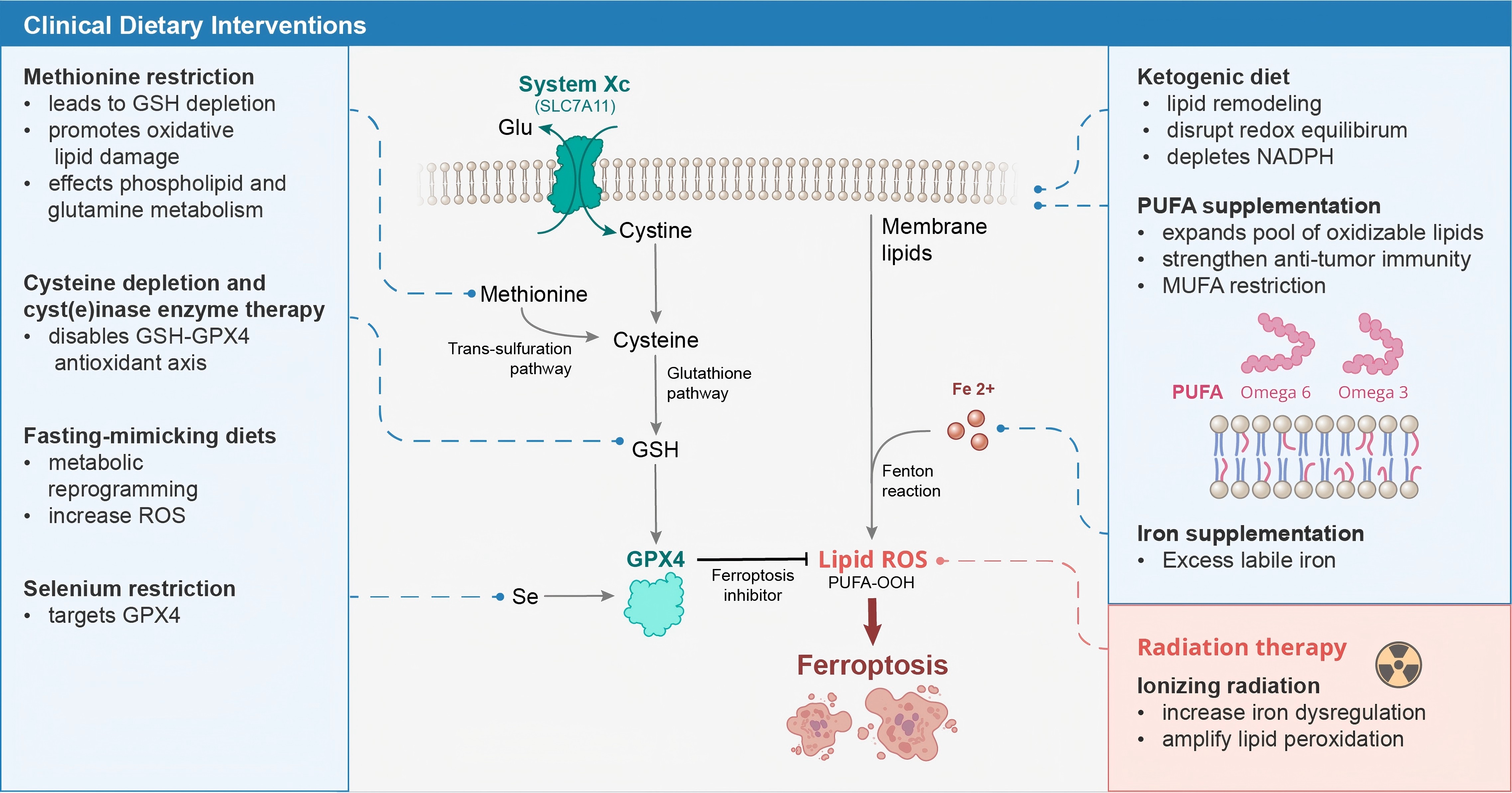
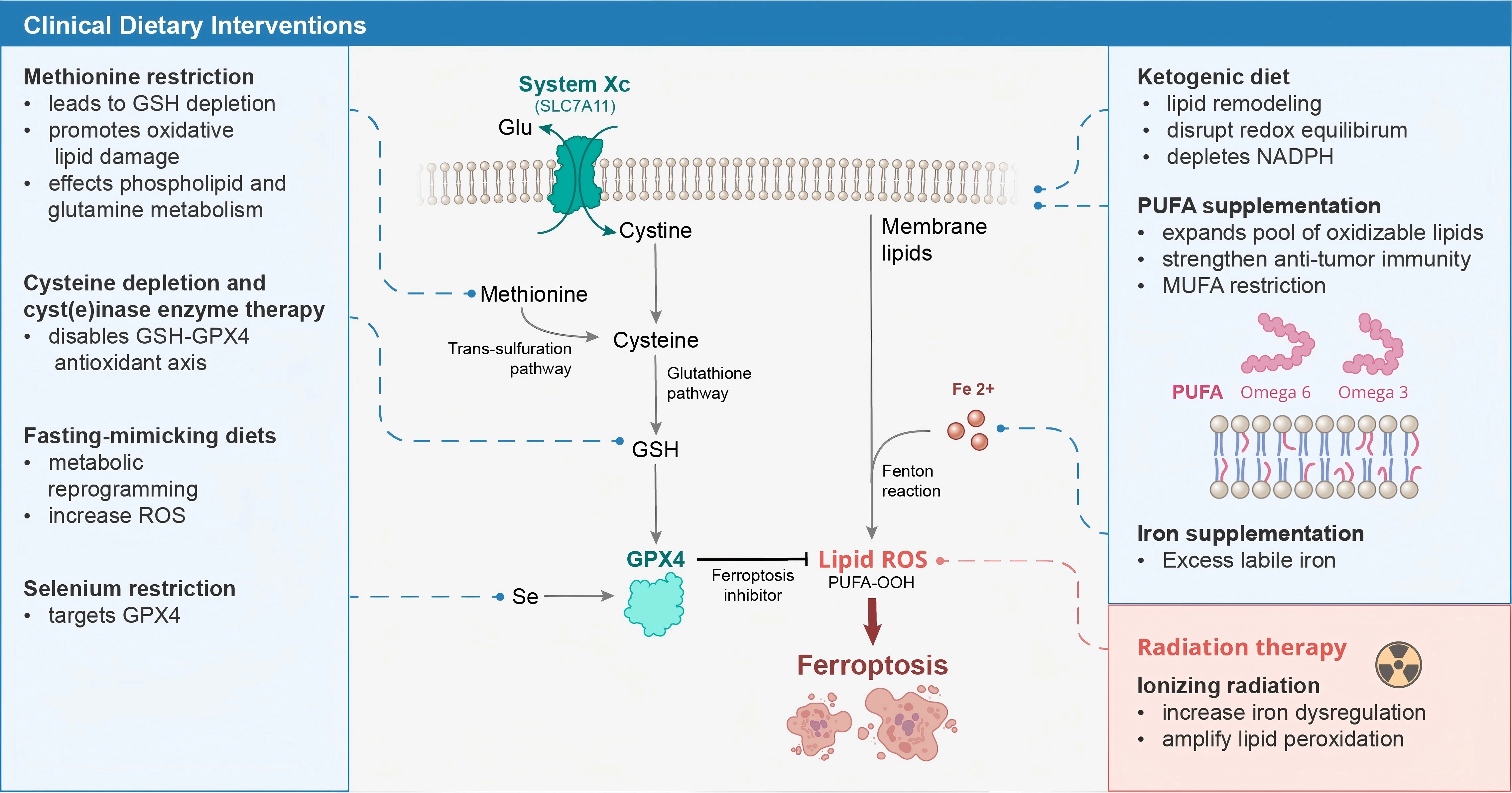
Figure 1. Overview of dietary interventions that converge on ferroptosis sensitization in cancer cells by targeting antioxidant defenses, membrane lipid composition, and redox homeostasis (Fe2+: ferrous iron; Glu, glutamate; GPX4: glutathione peroxidase 4; GSH: glutathione; MUFA: monounsaturated fatty acid; NADPH: nicotinamide adenine dinucleotide phosphate; PUFA: polyunsaturated fatty acid; PUFA-OOH: polyunsaturated fatty acid hydroperoxide; ROS: reactive oxygen species; Se: selenium; SLC7A11: solute carrier family 7 member 11 (System Xc- light chain).
Acknowledgements
The authors would like to acknowledge Xian Boles, MFA, for illustrating the schematic in Figure 1.
Authors contribution
Gilbert OE, John S: Writing-original draft, writing-review & editing, conceptualization, investigation.
Fahim AD: Writing-review & editing.
Higgins DMO: Conceptualization, supervision, writing-review & editing.
Conflicts of interest
The authors declare no conflicts of interest.
Ethical approval
Not applicable.
Consent to participate
Not applicable.
Consent for publication
Not applicable.
Availability of data and materials
Not applicable.
Funding
None.
Copyright
© The Author(s) 2026.
References
-
1. Yang WS, Stockwell BR. Ferroptosis: Death by lipid peroxidation. Trends Cell Biol. 2016;26(3):165-176.[DOI]
-
5. Tschuck J, Skafar V, Friedmann Angeli JP, Hadian K. The metabolic code of ferroptosis: Nutritional regulators of cell death. Trends Biochem Sci. 2025;50(8):663-676.[DOI]
-
6. Argenziano MG, Neelakantan TV, Sperring C, Adeuyan O, Higgins DMO, Bruce JN, et al. Emerging roles of ferroptosis in modulating the immune landscape of glial tumours. Nat Cell Biol. 2026;28(7):1364-1378.[DOI]
-
10. Banu MA, Dovas A, Argenziano MG, Zhao W, Sperring CP, Cuervo Grajal H, et al. A cell state-specific metabolic vulnerability to GPX4-dependent ferroptosis in glioblastoma. EMBO J. 2024;43(20):4492-4521.[DOI]
-
11. Kokkinakis DM, Schold SC Jr, Hori H, Nobori T. Effect of long-term depletion of plasma methionine on the growth and survival of human brain tumor xenografts in athymic mice. Nutr Cancer. 1997;29(3):195-204.[DOI]
-
14. Thivat E, Farges MC, Bacin F, D’Incan M, Mouret-Reynier MA, Cellarier E, et al. Phase II trial of the association of a methionine-free diet with cystemustine therapy in melanoma and glioma. Anticancer Res. 2009;29(12):5235-5240.[PubMed]
-
16. Sajeev A, Manickasamy MK, Vishwa R, Alqahtani MS, Abbas M, Sethi G, et al. Iron homeostasis and ferroptosis: A converging axis in cancer therapy. Biochem Pharmacol. 2026;249:117769.[DOI]
-
17. Toyokuni S, Kong Y, Maeda Y, Lyu Q, Ohara Y, Sato K, et al. Ferroptosis and cancer: When iron turns against tumors. Cell Mol Life Sci. 2025;83(1):16.[DOI]
-
18. Ponnusamy V, Randall DR, Lee ZH, Das NK, Zhao L, Buscher K, et al. Labile iron pool dynamics do not drive ferroptosis in colorectal cancer cells. J Biol Chem. 2026;302(5):111357.[DOI]
-
20. Gamage SMK, Lee KTW, Dissabandara DLO, Lam AK, Gopalan V. Dual role of heme iron in cancer; promotor of carcinogenesis and an inducer of tumour suppression. Exp Mol Pathol. 2021;120:104642.[DOI]
-
23. Tufano E, Connor JR. Ferritin in motion: How systemic iron balance and tumor trafficking shape glioblastoma. Biochim Biophys Acta BBA Rev Cancer. 2026;1881(3):189573.[DOI]
-
25. Rosado JO, Salvador M, Bonatto D. Importance of the trans-sulfuration pathway in cancer prevention and promotion. Mol Cell Biochem. 2007;301(1):1-12.[DOI]
-
27. Richie JP, Komninou D, Leutzinger Y, Kleinman W, Orentreich N, Malloy V, et al. Tissue glutathione and cysteine levels in methionine-restricted rats. Nutrition. 2004;20(9):800-805.[DOI]
-
29. Xia C, Peng P, Zhang W, Xing X, Jin X, Du J, et al. Methionine-SAM metabolism-dependent ubiquinone synthesis is crucial for ROS accumulation in ferroptosis induction. Nat Commun. 2024;15:8971.[DOI]
-
30. Dai Z, Mentch SJ, Gao X, Nichenametla SN, Locasale JW. Methionine metabolism influences genomic architecture and gene expression through H3K4me3 peak width. Nat Commun. 2018;9:1955.[DOI]
-
31. Kim JW, Jang SY, Roh YJ, Ju Y, Kang JB, Park BS, et al. Methionine metabolism is linked with phospholipid and glutamine metabolism to drive ferroptosis. Cell Rep. 2026;45(4):117157.[DOI]
-
32. Xin L, Fan LJ, Liu C, Lu H, Zou YH, Xu HS, et al. MR promotes ferroptosis in gastric cancer by regulating FANCD2 expression mediated by M6A modification. Appl Biochem Biotechnol. 2025;197(10):6805-6819.[DOI]
-
35. Olsen T, Øvrebø B, Haj-Yasein N, Lee S, Svendsen K, Hjorth M, et al. Effects of dietary methionine and cysteine restriction on plasma biomarkers, serum fibroblast growth factor 21, and adipose tissue gene expression in women with overweight or obesity: A double-blind randomized controlled pilot study. J Transl Med. 2020;18(1):122.
-
37. Yao H, Jiang W, Liao X, Wang D, Zhu H. Regulatory mechanisms of amino acids in ferroptosis. Life Sci. 2024;351:122803.[DOI]
-
41. Qiao C, Wang L, Huang C, Jia Q, Bao W, Guo P, et al. Engineered bacteria manipulate cysteine metabolism to boost ferroptosis-based pancreatic ductal adenocarcinoma therapy. Adv Mater. 2025;37(6):2412982.[DOI]
-
42. Wilder CS, Zhao C, DiGiovanni J. Pharmacologic approaches to amino acid depletion for cancer therapy. Mol Carcinog. 2022;61(2):127-152.[DOI]
-
45. Lee J, Roh JL. Selenium and selenoproteins: Key regulators of ferroptosis and therapeutic targets in cancer. J Mol Med. 2025;103(8):899-911.[DOI]
-
47. Zhao M, Xu M, Zhang W, Tang M, Ji S, Shi J. Selenium induces ferroptosis in colorectal cancer cells via direct interaction with Nrf2 and Gpx4. Biol Trace Elem Res. 2026;204(5):3338-3349.[DOI]
-
55. di Blasio L, Vara-Messler M, Peracino B, Masti E, Cepas-López V, Trusolino L, et al. Omega-3 fatty acid DHA induces ferroptosis in colorectal cancer patient-derived organoids and drug-tolerant cells. Cell Death Dis. 2026;17:464.[DOI]
-
56. Suda A, Umaru BA, Yamamoto Y, Shima H, Saiki Y, Pan Y, et al. Polyunsaturated fatty acids-induced ferroptosis suppresses pancreatic cancer growth. Sci Rep. 2024;14:4409.[DOI]
-
58. Samovich SN, Mikulska-Ruminska K, Dar HH, Tyurina YY, Tyurin VA, Souryavong AB, et al. Strikingly high activity of 15-lipoxygenase towards di-polyunsaturated arachidonoyl/adrenoyl-phosphatidylethanolamines generates peroxidation signals of ferroptotic cell death. Angew Chem Int Ed. 2024;63(9):e202314710.
-
66. Mnatsakanyan H, Sammarco A, Hewett A, Awwad R, Roumieh E, Pechdimaljian C, et al. SCD1 and SCD5 modulate PARP-dependent DNA repair via fatty acid desaturation in glioblastoma. Cell Rep. 2026;45(4):117223.[DOI]
-
68. Gleba JJ, Copland JA, Tun HW. Targeting the lipid desaturation network in cancer: From metabolic plasticity to precision therapeutics. J Exp Clin Cancer Res. 2026;45(1):187.[DOI]
-
70. Roughley K, Khochaiche A, Landstra A, Valceski M, Hollis C, Lerch M, et al. A fasting-mimicking environment enhances procaspase-activating compound 1 in 2D and 3D glioma cell models. Cell Cycle. 2026;25(1):1-12.[DOI]
-
71. Lin Y, Li S, Xu X, Hu X, Li Y, Liang S, et al. Intermittent fasting inhibits Tp53-driven glioma through gut microbiota-mediated methionine-M6A regulation. Nat Commun. 2026;17:1804.[DOI]
-
78. Safdie F, Brandhorst S, Wei M, Wang W, Lee C, Hwang S, et al. Fasting enhances the response of glioma to chemo- and radiotherapy. PLoS One. 2012;7(9):e44603.[DOI]
-
79. Kokkinakis DM, Hoffman RM, Frenkel EP, Wick JB, Han Q, Xu M, et al. Synergy between methionine stress and chemotherapy in the treatment of brain tumor xenografts in athymic mice. Cancer Res. 2001;61(10):4017-4023.[PubMed]
-
81. Schueller P, Puettmann S, Micke O, Senner V, Schaefer U, Willich N. Selenium influences the radiation sensitivity of C6 rat glioma cells. Anticancer Res. 2004;24(5A):2913-2917.[PubMed]
-
82. Amaral LJ, Gresham G, Kim S, Tighiouart M, Nelson TA, Welborn A, et al. A phase 1 safety and feasibility trial of a ketogenic diet plus standard of care for patients with recently diagnosed glioblastoma. Sci Rep. 2025;15:21064.[DOI]
-
84. Porper K, Shpatz Y, Plotkin L, Pechthold RG, Talianski A, Champ CE, et al. A Phase I clinical trial of dose-escalated metabolic therapy combined with concomitant radiation therapy in high-grade glioma. J Neurooncol. 2021;153(3):487-496.[DOI]
-
85. Sato M, Han Q, Mizuta K, Mori R, Kang BM, Morinaga S, et al. Extensive shrinkage and long-term stable disease in a teenage female patient with high-grade glioma treated with temozolomide and radiation in combination with oral recombinant methioninase and a low-methionine diet. In Vivo. 2024;38(3):1459-1464.
-
86. Voss M, Wagner M, von Mettenheim N, Harter PN, Wenger KJ, Franz K, et al. ERGO2: A prospective, randomized trial of calorie-restricted ketogenic diet and fasting in addition to reirradiation for malignant glioma. Int J Radiat Oncol. 2020;108(4):987-995.[DOI]
-
87. Mattes MD, Koturbash I, Leung CN, Sijin W, Jacobson GM. A phase I trial of a methionine restricted diet with concurrent radiation therapy. Nutr Cancer. 2024;76(6):463-468.[DOI]
-
89. Taki AG, Shareef A, Arora V, Oweis R, Jyothi SR, Singh U, et al. Ferroptosis in cancer: Integrating multi-omics, AI-driven biomarkers, and translational challenges. Clin Chim Acta. 2026;591:121131.[DOI]
Copyright
© The Author(s) 2027. This is an Open Access article licensed under a Creative Commons Attribution 4.0 International License (https://creativecommons.org/licenses/by/4.0/), which permits unrestricted use, sharing, adaptation, distribution and reproduction in any medium or format, for any purpose, even commercially, as long as you give appropriate credit to the original author(s) and the source, provide a link to the Creative Commons license, and indicate if changes were made.
Publisher’s Note
Science Exploration remains a neutral stance on jurisdictional claims in published
maps
and institutional affiliations. The views expressed in this article are solely those
of
the author(s) and do not reflect the opinions of the Editors or the publisher.
Share And Cite


Science Exploration Style
Gilbert OE, John S, Fahim AD, Higgins DM. Dietary modulation of ferroptosis sensitivity in glioblastoma and other treatment-resistant cancers. Ferroptosis Oxid Stress. 2027;3:202631. https://doi.org/10.70401/fos.2026.0042
Tips
Copy completed.
Submit a Manuscript
Author Instructions
Cite this Article
Article Metrics
0
View
0
Download
Cited
Article Updates

- Abstract
- Keywords
- 1. Introduction
- 2. GBM Metabolic Landscape and Ferroptosis Susceptibility
- 3. Iron Intake and Iron Metabolism
- 4. Methionine Restriction and Ferroptosis
- 5. Cysteine Depletion and Cyst(e)inase Enzyme Therapy
- 6. Selenium and Selenoprotein Metabolism
- 7. Ketogenic Diets and Ferroptosis
- 8. PUFA Supplementation and Lipid Remodeling
- 9. Fasting-Mimicking Diets and Caloric Restriction
- 10. Radiation-Ferroptosis Synergy
- 11. Translational Challenges and Future Directions
- 12. Conclusion
- Acknowledgements
- Authors contribution
- Conflicts of interest
- Ethical approval
- Consent to participate
- Consent for publication
- Availability of data and materials
- Funding
- References
- Copyright
 Get citation
Science Exploration Style

Gilbert OE, John S, Fahim AD, Higgins DM. Dietary modulation of ferroptosis sensitivity in glioblastoma and other treatment-resistant cancers. Ferroptosis Oxid Stress. 2027;3:202631. https://doi.org/10.70401/fos.2026.0042
copy
Share Link
copy