Tumor but not tumor cells: Ferroptosis in non-tumor cells within the tumor

Liang Gong
1,#

,
Zhicheng Gong
2,#
,
Kang Wang
3
,
Qiang Zhang
4
,
Kaisa Cui
1,2,*
*Correspondence to:
Kaisa Cui, College of Synthetic Biology Industry, Hunan University of Arts and Science, Changde 415000, Hunan, China; Wuxi Cancer Institute, Affiliated Hospital of Jiangnan University, Wuxi 214062, Jiangsu, China.
E-mail: ksxg@foxmail.com
Ferroptosis Oxid Stress. 2027;3:202619. 10.70401/fos.2026.0044
Received: May 06, 2026Accepted: September 15, 2026Published: September 18, 2026
Abstract
Ferroptosis is an iron-dependent form of regulated cell death that has attracted considerable attention as a promising therapeutic strategy because of its ability to eliminate tumor cells. However, tumors are multicellular ecosystems in which malignant cells coexist with diverse stromal and immune cell populations that critically shape tumor progression, anti-tumor immunity, and therapeutic responses. Emerging evidence indicates that ferroptosis exerts fundamentally distinct biological effects across these non-malignant cell populations, extending far beyond direct tumor cell killing. In this review, we propose a cell type-centered conceptual framework in which the consequences of ferroptosis are determined primarily by cellular context rather than by ferroptosis itself. While ferroptosis resistance enables certain cell types, such as fibroblasts and macrophages, within the tumor microenvironment (TME) to maintain tumor-promoting functions, ferroptosis compromises the persistence and effector activity of cytotoxic lymphocytes, including CD8+ T cells and natural killer cells. Conversely, ferroptosis in neutrophils and polymorphonuclear myeloid-derived suppressor cells may generate oxidized lipid mediators that paradoxically reinforce immune suppression despite reducing cell abundance. Therefore, in this review, we integrate recent advances in understanding ferroptosis across major nonmalignant cell populations within the TME, focusing on its effects on cell fate, functional plasticity, and anti-tumor immunity. We further discuss how these insights may guide the development of precision ferroptosis-based therapies that selectively target tumor-promoting cell states while preserving protective immune responses.
Keywords
Ferroptosis, tumor, fibroblasts, macrophages, neutrophils, T cells, NK cells, cancer therapy
1. Introduction
Ferroptosis is an iron-dependent form of regulated cell death driven by the accumulation of toxic lipid peroxides; it was originally identified through studies on cancer cells more than a decade ago[1]. Because many malignant cells exhibit heightened susceptibility to ferroptosis, the pharmacological induction of ferroptosis has emerged as a promising strategy for cancer therapy[1-6]. Consequently, early studies largely viewed ferroptosis as a tumor-suppressive process, and substantial efforts have focused on understanding its molecular regulation and therapeutic exploitation.
This tumor cell-centric view has evolved considerably with the growing appreciation that tumors function as complex multicellular ecosystems rather than collections of malignant cells alone[7-10]. The tumor microenvironment (TME) comprises diverse immune and stromal populations that actively regulate tumor progression, metastasis, immune surveillance, and therapeutic responses through extensive interactions with cancer cells[7-10]. Our previous review demonstrates that these nonmalignant cells also engage in ferroptosis-associated pathways in tumor cells[4]. Importantly, the biological consequences of ferroptosis differ markedly among cell types. Although ferroptosis in tumor cells is generally considered beneficial for cancer treatment, ferroptosis in nontumoral cells may either enhance or impair anti-tumor immunity depending on the cellular context. Thus, the consequences of ferroptosis cannot be understood simply by asking whether ferroptosis occurs, but rather which cells undergo ferroptosis, how ferroptosis sensitivity is regulated, and how ferroptotic cells or sublethal ferroptosis stress reshapes the surrounding microenvironment.
The core molecular mechanisms regulating ferroptosis have been extensively reviewed elsewhere[2], including our previous review[4]. Briefly, ferroptosis results from excessive lipid peroxidation driven by iron-dependent and enzymatic oxidative reactions[1,11] (Figure 1). Cellular susceptibility is primarily restrained by antioxidant defense systems centered on the cysteine-glutathione (GSH)-glutathione peroxidase 4 (GPX4) axis[12], ferroptosis suppressor protein 1 (FSP1)[13,14], and dihydroorotate dehydrogenase (DHODH)[15] (Figure 1). Meanwhile, additional regulatory pathways continue to emerge (Figure 1). Our team recently showed that lactylated lysine-specific demethylase 1 (LSD1) promotes recurrent melanoma cell survival by blocking transferrin receptor protein 1 (TFRC)-mediated ferroptosis, while a c-Myc-DHODH feedback loop enhances pyrimidine biosynthesis to suppress ferroptosis[16,17]. The AMP-activated protein kinase (AMPK)-regulated pyrimidinosome coordinates pyrimidine biosynthesis and confers ferroptosis resistance to cancer cells[18]. Extracellular signal-regulated kinase 2 (ERK2) as a creatine sensor stabilizes FSP1 to facilitate ferroptosis resistance[19]. Two back-to-back studies demonstrate that vitamin B2 (riboflavin) is essential for FSP1 stability, thereby inhibiting ferroptosis in tumor cells[20,21]. Peroxiredoxin 6 (PRDX6) promotes ferroptosis resistance by regulating both GPX4 expression and subcellular localization[22,23]. Another study reveals that OSBPL8 recruits GPX1 to the endoplasmic reticulum (ER), where GPX1 directly reduces oxidized phosphatidic acid (PA), thereby preventing ferroptosis[24]. In contrast to several recent reviews including our previous review that summarized these molecular mechanisms and discussed ferroptosis in the TME from a largely tumor cell-oriented perspective, this review focuses specifically on the cell type-specific biological consequences of ferroptosis in non-malignant stromal and immune populations and how these distinct ferroptosis states collectively shape tumor progression and anti-tumor immunity.
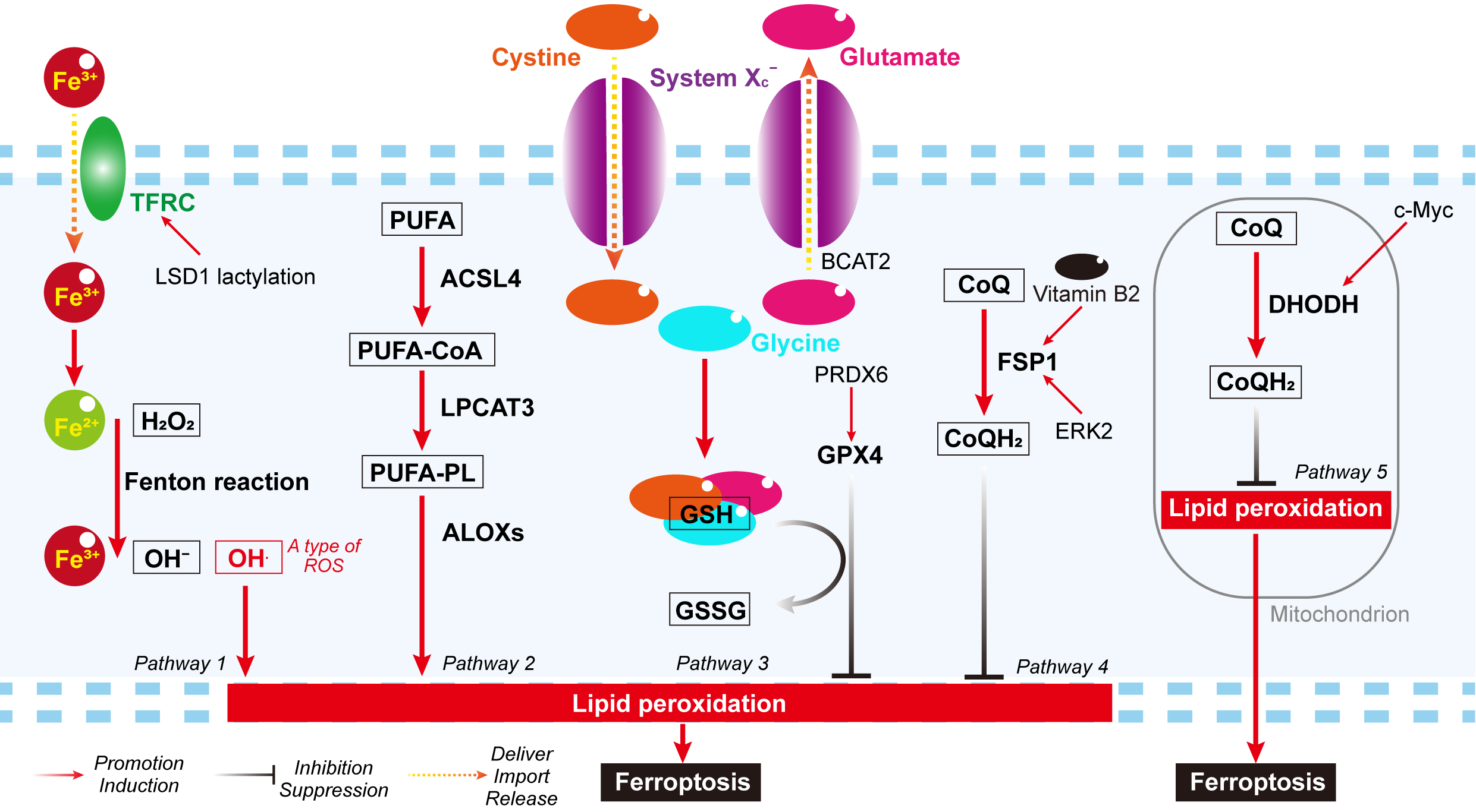
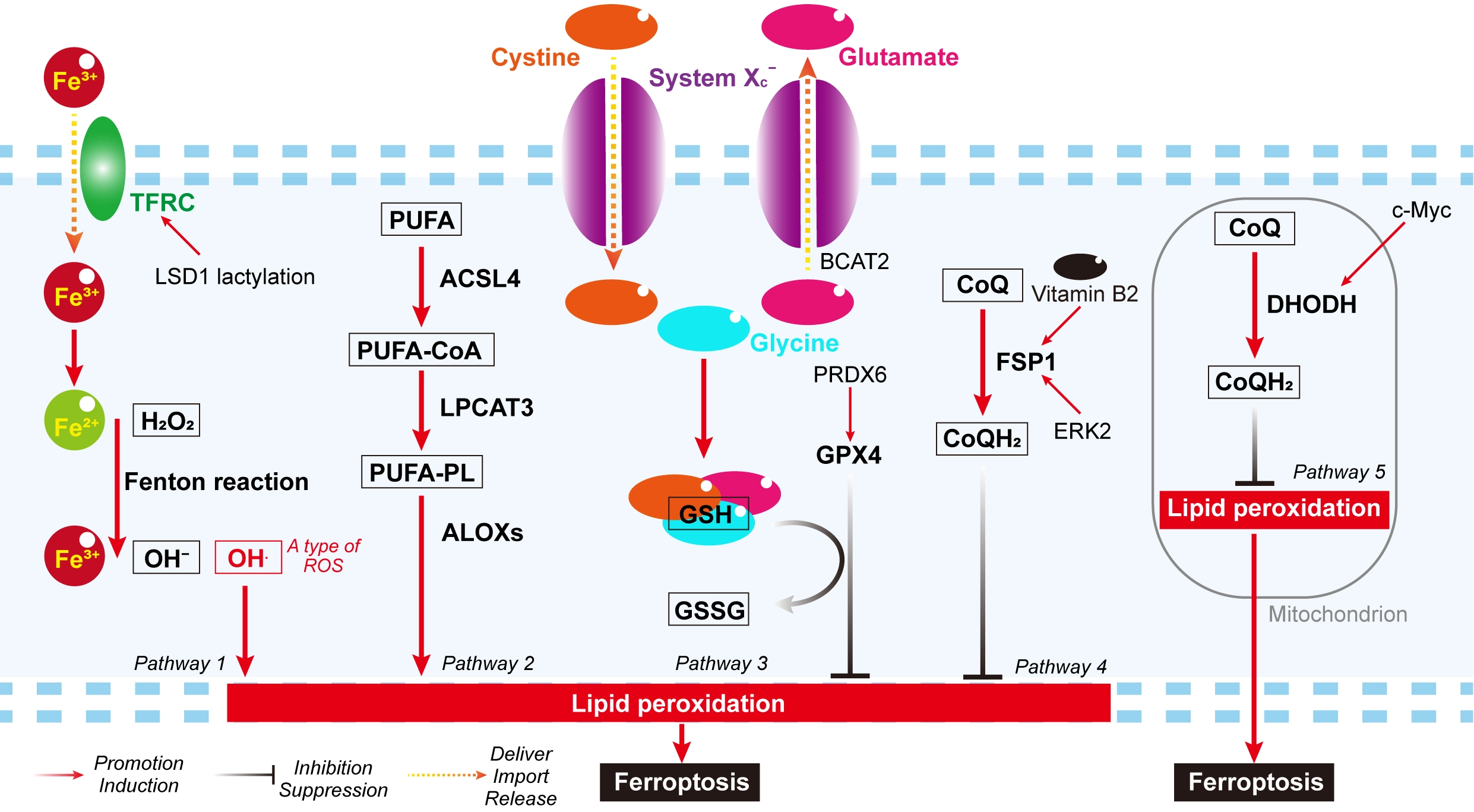
Figure 1. Major pathway regulating ferroptosis. Primary mechanisms driving ferroptosis: Pathway 1 shows the non-enzymatic pathway Fenton reaction; Pathway 2 shows the enzymatic pathway of PUFA to PUFA-PL regulated by the ACSL4/LPCAT3/ALOXs axis. Major mechanisms against ferroptosis: Pathway 3 shows the cysteine/GSH/GPX4 mechanism; Pathway 4 shows the FSP1 mechanism; Pathway 5 shows the DHODH mechanism. Additionally, recent studies have demonstrated that PRDX6 regulates GPX4, Vitamin B2 and ERK2 regulate FSP1, lactylated LSD1 regulates TFRC, and c-Myc regulates DHODH. OH: hydroxyl radical; ROS: reactive oxygen species; PUFA: polyunsaturated fatty acid; PL: phospholipids; ACSL4: acyl-CoA synthetase long chain family member 4; LPCAT3: lysophosphatidylcholine acyltransferase 3; ALOXs: arachidonate lipoxygenases; GSH: glutathione; GSSG: Oxidized Glutathione; GPX4: glutathione peroxidase 4; PRDX6: peroxiredoxin 6; ERK2: extracellular signal-regulated kinase 2; FSP1: ferroptosis suppressor protein 1; LSD1: lysine-specific demethylase 1; TFRC: transferrin receptor protein 1; DHODH: dihydroorotate dehydrogenase; System Xc-: Cystine-glutamate antiporter; BCAT2: branched-chain amino acid aminotransferase 2; CoQ: ubiquinone-10 (also known as coenzyme Q 10); CoQH2: ubiquinol.
Recent advances in single-cell transcriptomics, spatial omics, and functional immunology have fundamentally changed our understanding of the TME and revealed extensive heterogeneity among immune and stromal cell populations. Similar to other biological processes, such as ribosome biogenesis and metabolic reprogramming[8,25], ferroptosis is increasingly recognized as a context-dependent process whose consequences extend beyond cell death. In many cases, activation of ferroptosis-associated pathways that can be defined as sublethal ferroptosis stress regulate cell plasticity, functional reprogramming, and intercellular communication in addition to determining cell survival. These findings suggest that ferroptosis should be viewed as a broader regulator of TME organization and immune remodeling rather than solely as a mechanism for eliminating tumor cells.
In this review, we summarize the current knowledge concerning ferroptosis in nontumoral cell populations, including cancer-associated fibroblasts (CAFs), tumor-associated macrophages (TAMs), neutrophil/myeloid-derived suppressor cells (MDSCs), CD8+ T cells, regulatory T (Treg) cells, and natural killer (NK) cells. Rather than cataloging individual studies, we discuss each cell type using a common conceptual framework: (i) whether ferroptosis is generally tumor-suppressive or tumor-promoting; (ii) the molecular determinants of ferroptosis sensitivity and resistance; (iii) the consequences of ferroptosis for anti-tumor immunity; (iv) the major unanswered questions and therapeutic opportunities. We propose that understanding the cell type-specific functions of ferroptosis will provide a conceptual basis for developing precision ferroptosis-based therapies that remodel the TME while preserving protective anti-tumor immunity.
2. Adaptation to Ferroptosis Stress in CAFs
CAFs are highly heterogeneous stromal cells that shape almost every aspect of the TME, including extracellular matrix remodeling, metabolic adaptation, immune regulation, and therapeutic responses[26]. Although several CAF subtypes are constantly exposed to oxidative stress, lipid peroxidation, and altered iron metabolism within tumors, accumulating evidence suggests that they rarely undergo ferroptosis[27,28]. Instead, they appear to engage in ferroptosis-associated pathways while simultaneously activating compensatory mechanisms that preserve cell survival. Thus, rather than simply classifying CAFs as ferroptosis-sensitive or ferroptosis-resistant, an emerging concept is that CAFs frequently exist in a ferroptosis-adapted state, in which oxidative and iron-dependent stress actively shapes CAF phenotypes without necessarily committing them to cell death.
Several studies support this concept. Single-cell transcriptomic analyses identified a hypoxia-driven CUB domain containing protein 1 (CDCP1)+ ferritin light chain (FTL)+ CAF population in pancreatic ductal adenocarcinoma that exhibits enhanced glycolytic metabolism together with increased resistance to ferroptosis (Figure 2)[27]. Similarly, FerroCAFs, an iron-enriched CAF subset identified in prostate cancer, accumulate intracellular iron, generate large amounts of reactive oxygen species (ROS), and undergo extensive lipid peroxidation[28]; however, these cells remain viable through the activation of antioxidant defenses (Figure 2). Importantly, these cells actively promote tumor progression by establishing an immunosuppressive microenvironment through iron-dependent epigenetic remodeling[28]. Together, these observations suggest that adaptation to ferroptotic stress is not merely a survival mechanism but may also endow CAFs with functions that support tumor growth and immune evasion.
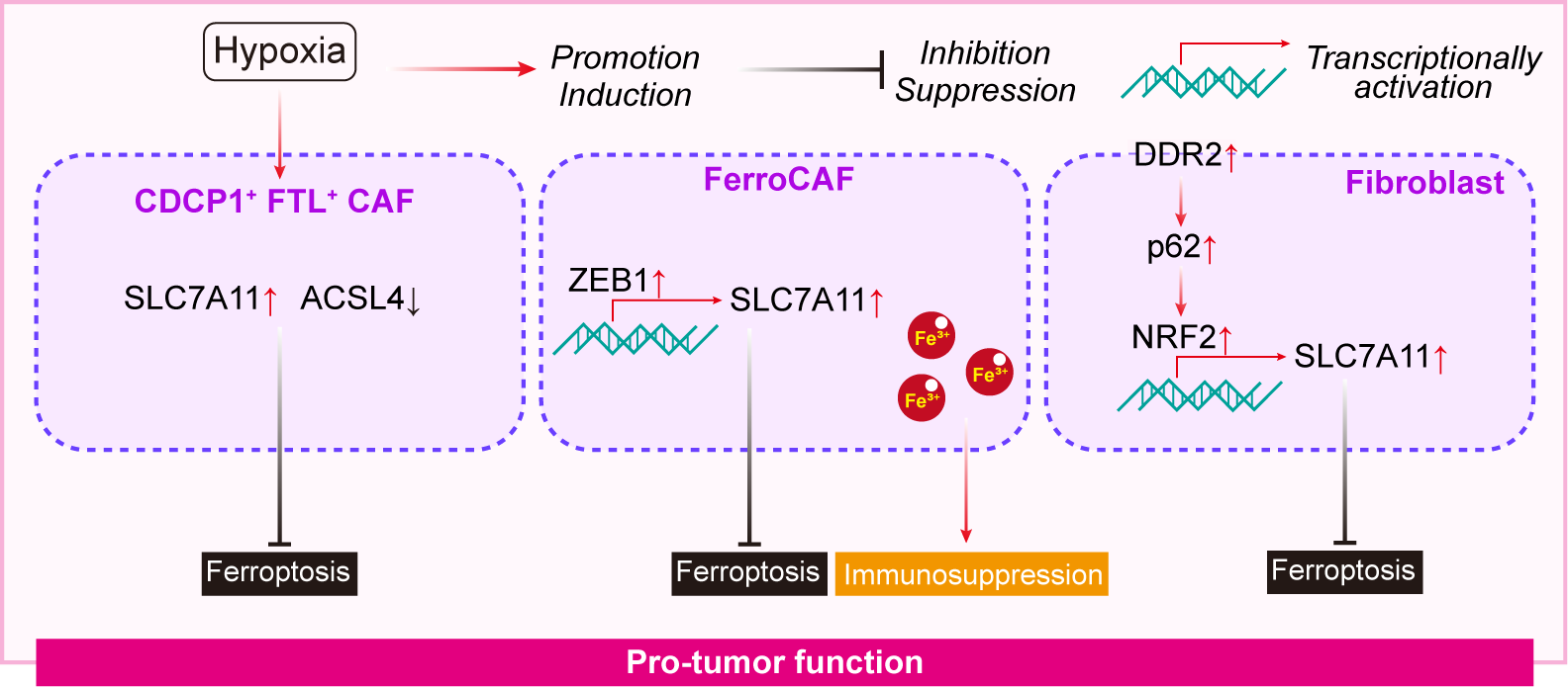
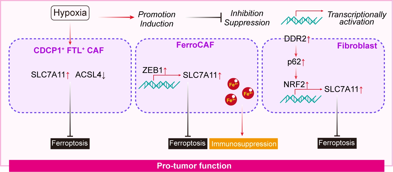
Figure 2. Ferroptosis in CAFs within the tumor. FerroCAFs and other ferroptosis-resistant CAF subpopulations generally display tumor-promoting properties within the TME. TME: tumor microenvironment; CAFs: cancer-associated fibroblasts; CDCP1: CUB domain containing protein 1; FTL: ferritin light chain; SLC7A11: solute carrier family 7 member 11; ACSL4: acyl-CoA synthetase long chain family member 4; ZEB1: zinc finger E-box binding homeobox 1; DDR2: discoidin domain receptor tyrosine kinase 2; NRF2: nuclear factor erythroid 2-related factor 2.
It is beginning to emerge how CAFs survive this persistent ferroptotic stress. Current evidence converges on antioxidant defense pathways as central determinants of CAF survival. In FerroCAFs, the transcription factor zinc finger E-box binding homeobox 1 (ZEB1) upregulates the cystine-glutamate antiporter (System Xc-) components solute carrier family 3 member 2 (SLC3A2) and solute carrier family 7 member 11 (SLC7A11, also named xCT), thereby maintaining cystine uptake and preventing ferroptosis (Figure 2)[29]. Consistent with this model, pharmacological inhibition of System Xc- using erastin or sulfasalazine restored ferroptosis sensitivity in FerroCAFs[29]. Similarly, discoidin domain receptor tyrosine kinase 2 (DDR2) protects CAFs by activating the xCT-GSH-GPX4 antioxidant axis through noncanonical sequestosome 1 (p62)-dependent nuclear factor erythroid 2-related factor 2 (NRF2) signaling while simultaneously regulating intracellular iron metabolism (Figure 2)[30]. These findings collectively suggest that CAFs actively rewire redox homeostasis to tolerate an otherwise lethal ferroptotic microenvironment.
The consequences of this ferroptosis-adapted state are likely to extend beyond the survival of CAFs. Ferroptosis-resistant CAFs appear to sustain tumor-promoting functions despite continuous oxidative stress, raising the possibility that ferroptosis adaptation contributes to immune suppression, metabolic support of cancer cells, and therapeutic resistance[28-30]. However, direct evidence linking CAF ferroptosis to anti-tumor immunity remains limited. Whether ferroptotic CAFs release immunomodulatory mediators or whether preventing ferroptosis in CAFs alters immune cell recruitment and activation remains largely unexplored. Defining these interactions is essential for understanding how ferroptosis shapes stromal–immune cell crosstalk within the TME.
Notably, CAFs are functionally diverse. While FerroCAFs and other ferroptosis-resistant subpopulations generally display tumor-promoting properties, several CAF subsets, including Meflin+ and α-smooth muscle actin-positive (αSMA+) CAFs, have been reported to restrain pancreatic tumor progression[31,32]. Therefore, the indiscriminate induction of ferroptosis in all CAFs is unlikely to represent an optimal therapeutic strategy. Instead, selectively targeting ferroptosis-adapted, tumor-promoting CAF subsets while preserving or reprogramming tumor-restraining CAFs may offer greater therapeutic benefit.
Several key questions remain unanswered. First, it is unclear whether ferroptosis adaptation represents a universal feature of CAFs or is restricted to specific CAF states generated by hypoxia, iron overload, or other environmental cues. Second, the mechanisms by which ferroptosis-adapted CAFs regulate anti-tumor immunity require further investigation. Finally, identifying subtype-specific ferroptosis vulnerabilities may enable the rational combination of ferroptosis-targeting therapies with immunotherapy and other stromal-directed strategies.
3. Ferroptosis-Regulated Plasticity in TAMs
TAMs are among the most abundant immune cell populations within the TME and orchestrate tumor progression, metastasis, immune suppression, and therapeutic responses through dynamic interactions with tumor and stromal cells[33-35]. As professional phagocytes responsible for iron recycling and inflammatory regulation, macrophages are intrinsically coupled to iron metabolism and redox homeostasis, making them particularly responsive to ferroptosis-related stress[36,37]. Emerging evidence suggests that the role of ferroptosis extends beyond determining macrophage survival. Instead, sublethal ferroptosis stress appears to function as a regulator of macrophage plasticity, linking iron metabolism and lipid peroxidation to macrophage polarization, metabolic adaptation, and immunoregulatory activity rather than triggering cell death. Thus, ferroptosis should not be viewed merely as a mechanism for eliminating TAMs; sublethal ferroptosis-associated stress may also shape macrophage states within the TME.
Current evidence generally supports a tumor-suppressive role for ferroptosis in immunosuppressive TAM subpopulations. Multiple studies have reported that promoting ferroptosis preferentially disrupts M2-like macrophages, thereby reducing immune suppression and inhibiting tumor progression. For example, xCT expression is positively correlated with M2-like polarization in hepatocellular carcinoma (HCC), whereas xCT deficiency induces macrophage ferroptosis, suppresses TAM infiltration, and inhibits tumor growth through the GPX4/ribonucleotide reductase regulatory subunit M2 (RRM2) pathway (Figure 3)[38]. Similarly, inhibition of apolipoprotein C1 (APOC1) induces sublethal ferroptosis-associated stress, thereby increasing intracellular ROS levels and reprogramming M2-like TAMs toward a more inflammatory phenotype (Figure 3)[39]. In addition, several natural compounds have been suggested to induce ferroptosis or sublethal ferroptosis stress to regulate polarization in TAMs (Figure 3)[40-42]. For example, Macelignan induces ferroptosis in TAMs, thereby disrupting the signal transducer and activator of transcription 6 (STAT6)/peroxisome proliferator-activated receptor gamma (PPAR-γ)/Krüppel-like factor 4 (KLF4) axis and limiting M2 polarization[40]. Cucurbitacin B triggers ferroptosis in M2 macrophages, thereby increasing the release of malondialdehyde (MDA), ROS, and lipid peroxidation (LPO) and facilitating M1 polarization[41]. Dauricine induces sublethal ferroptosis-associated stress to suppress M2 polarization in TAMs[42]. Collectively, these studies suggest that resistance to ferroptosis stress enables immunosuppressive TAMs to persist within tumors and sustain a permissive microenvironment for cancer progression.
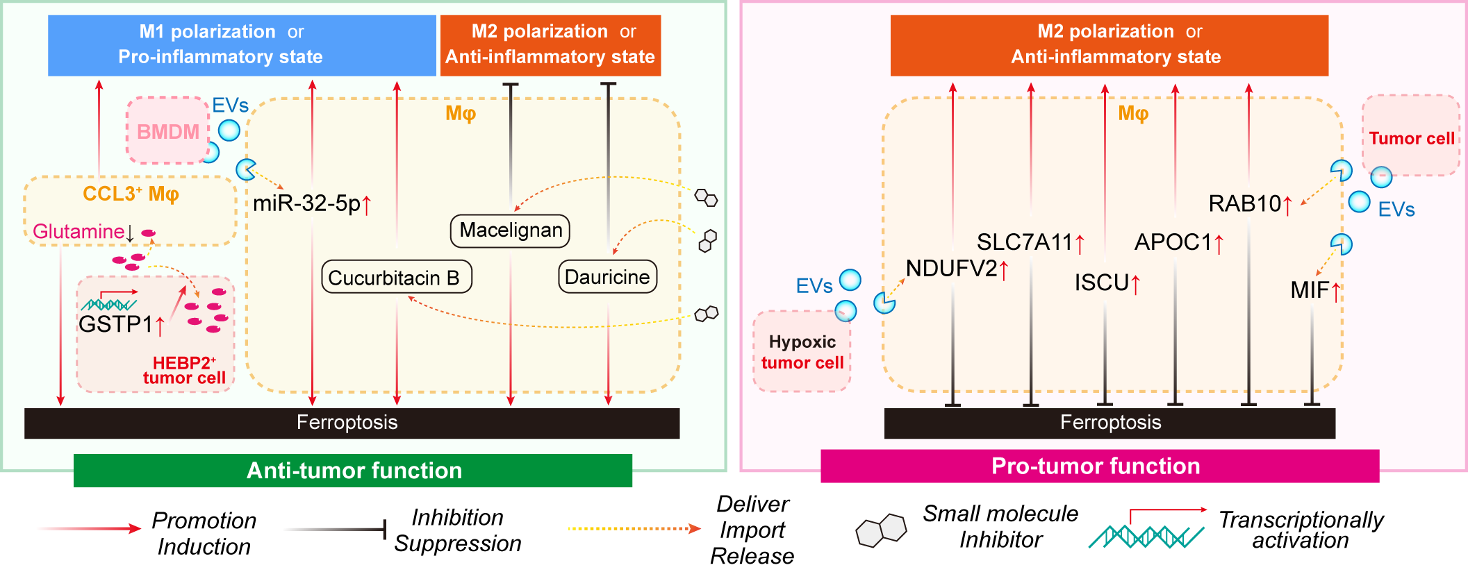
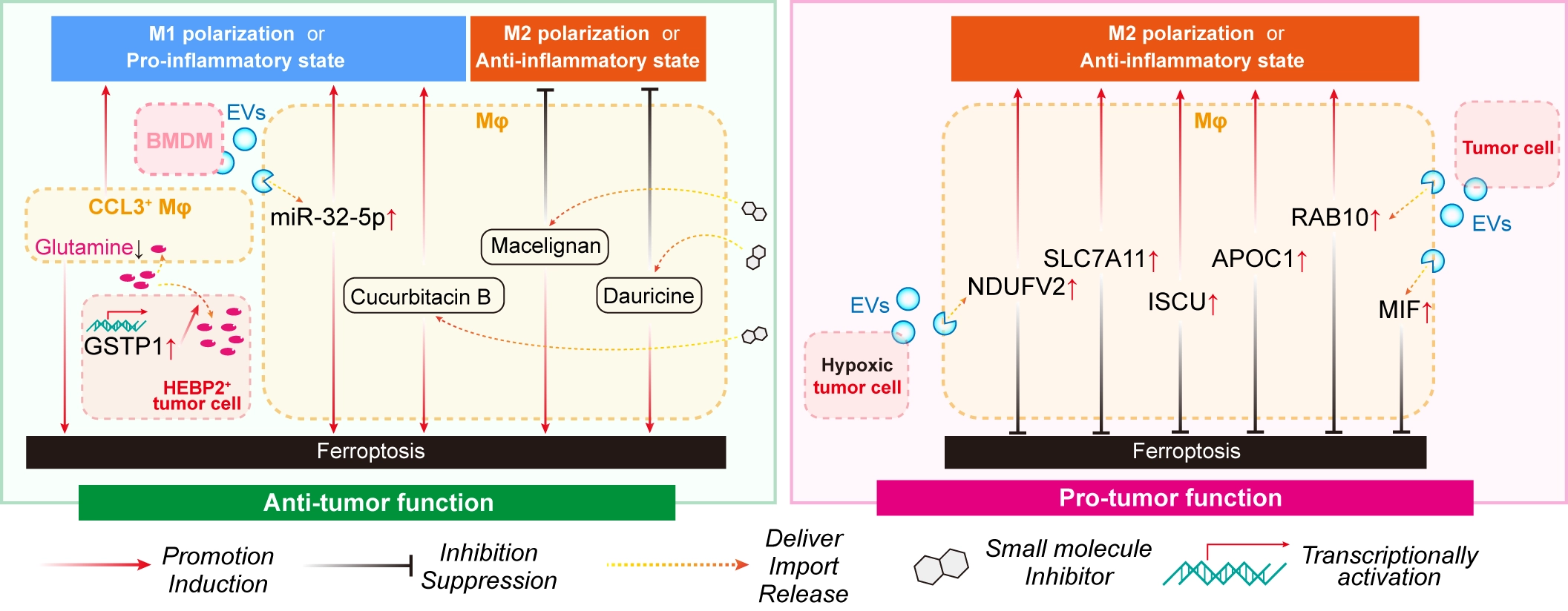
Figure 3. Ferroptosis in macrophages within the tumor. Ferroptosis resistance generally plays a pro-tumor role in immunosuppressive macrophage subpopulations within the TME. The TME can regulate macrophage ferroptosis through intercellular communication. TME: tumor microenvironment; Mφ: macrophage; APOC1: apolipoprotein C1; ISCU: iron-sulfur cluster assembly enzyme; EVs: extracellular vesicles; RAB10: Ras-related in brain 10; NDUFV2: NADH: ubiquinone oxidoreductase core subunit V2; MIF: macrophage migration inhibitory factor; SLC7A11: solute carrier family 7 member 11; BMDM: bone marrow-derived macrophages; CCL3: C-C motif chemokine ligand 3; HEBP2: heme binding protein 2; GSTP1: glutathione S-transferase pi 1.
However, the relationship between ferroptosis and macrophage function is unlikely to conform to the traditional M1/M2 paradigm. In glioblastoma, for example, elevated expression of the ferroptosis-related protein FTL promotes iron accumulation and ROS generation and is associated with macrophage ferroptosis, M2 polarization, and poor patient survival[43]. Rather than contradicting previous findings, these observations suggest that ferroptosis-associated stress may regulate macrophage phenotypes in a context-dependent manner that differs across tumor types and macrophage states. Therefore, the biological outcome of ferroptosis likely depends not only on whether macrophages undergo ferroptosis but also on how ferroptotic signaling reshapes their transcriptional and metabolic programs.
Mechanistically, ferroptosis sensitivity in TAMs is determined by the balance between oxidative stress and antioxidant defense. Central regulators include the xCT-GSH-GPX4 axis, iron metabolism, and NRF2 signaling[44]. Early studies demonstrated that iron regulates SLC7A11 expression through the ROS-NRF2 pathway, establishing a direct link between iron homeostasis and ferroptosis resistance in macrophages[45]. More recently, iron-sulfur cluster assembly enzyme (ISCU) was shown to suppress associated stress, including ROS, lipid ROS, and Fe2+ levels, by promoting cytoplasmic translocation of p53, thereby relieving p53-mediated repression of xCT and facilitating M2-like polarization in esophageal squamous cell carcinoma (Figure 3)[46]. Likewise, hemangioma-derived stem cells enhance NRF2 nuclear translocation and GPX4 expression in macrophages, increasing ferroptosis resistance and promoting infantile hemangioma progression[47]. Together, these studies identify antioxidant defense pathways as central determinants of macrophage adaptation to ferroptotic stress.
The TME also actively regulates macrophage ferroptosis through intercellular communication. Tumor-derived extracellular vesicles (EVs), such as exosomes, have emerged as important mediators of this process. Exosomes carrying Ras-related in brain 10 (RAB10), NADH: ubiquinone oxidoreductase core subunit V2 (NDUFV2)-associated signals or macrophage migration inhibitory factor (MIF) suppress macrophage ferroptosis by increasing their antioxidant capacity or preserving mitochondrial metabolism, thereby promoting M2 polarization and tumor progression (Figure 3)[48-50]. Conversely, monocyte-derived exosomal microRNA (miR)-32-5p induces ferroptosis and inflammatory activation in TAMs through activation of the cyclic GMP-AMP synthase (cGAS)-stimulator of interferon genes (STING) pathway, resulting in enhanced anti-tumor responses in HCC (Figure 3)[51]. These findings indicate that tumor cells actively manipulate ferroptosis sensitivity in macrophages to remodel the immune landscape in their favor.
The immunological consequences of ferroptosis in macrophages are therefore likely to depend on the macrophage subpopulations that undergo ferroptosis stress. In many settings, the induction of ferroptosis in immunosuppressive TAMs is accompanied by enhanced inflammatory responses and improved anti-tumor immunity, suggesting that selectively targeting ferroptosis-resistant TAMs may augment immunotherapy. However, recent single-cell RNA sequencing studies have revealed macrophage states that extend well beyond the conventional M1/M2 classification. For example, a C-C motif chemokine ligand 3 (CCL3)+ macrophage subset that correlated with increased immune checkpoint blockade responses was identified in triple-negative breast cancer and this macrophage subset exhibited an enhanced dependence on glutamine metabolism to resist ferroptosis (Figure 3)[52]. Tumor-derived heme binding protein 2 (HEBP2) promotes ferroptosis in these macrophages through metabolic reprogramming, thereby weakening anti-tumor immunity (Figure 3)[52]. These findings suggest that indiscriminate induction of ferroptosis could inadvertently eliminate macrophage populations that support effective immune responses. Therefore, therapeutic strategies should focus on selectively targeting ferroptosis-adapted, tumor-promoting macrophage populations while preserving or enhancing macrophage subsets that contribute to immune surveillance.
Despite substantial progress, several important questions remain unanswered. First, it is unclear whether ferroptosis-regulated macrophage plasticity represents a universal feature across cancer types or is restricted to specific TAM states generated by distinct metabolic and inflammatory cues. Second, the molecular mechanisms through which ferroptosis signaling shapes macrophage-lymphocyte crosstalk and responses to immunotherapy remain poorly understood. Finally, the integration of single-cell, spatial transcriptomic, and metabolic profiling approaches is essential for identifying subtype-specific ferroptosis vulnerabilities and developing ferroptosis-based strategies that remodel, rather than indiscriminately deplete, the macrophage compartment.
4. Ferroptosis as a Driver of Neutrophil/MDSC Functional Plasticity
Neutrophils are the most abundant circulating leukocytes and are among the earliest immune cells recruited into tumors[53-56]. Within the TME, neutrophils acquire remarkable phenotypic and functional diversity, ranging from anti-tumor effector cells to highly immunosuppressive subpopulations traditionally referred to as polymorphonuclear myeloid-derived suppressor cells (PMN-MDSCs)[56,57]. However, accumulating single-cell studies have indicated that PMN-MDSCs likely represent pathological activation states rather than a distinct neutrophil lineage, highlighting the continuum of neutrophil plasticity in cancer[58,59]. Throughout this section, we therefore discuss both tumor-associated neutrophils (TANs) and PMN-MDSCs while preserving the terminology used in the original studies.
Among immune cell populations within the TME, neutrophils exhibit an unusually high susceptibility to ferroptosis. Unlike macrophages or fibroblasts, which frequently adapt to ferroptosis stress through robust antioxidant programs, TANs readily undergo ferroptosis because of their elevated ROS levels, large amounts of polyunsaturated phospholipids, and altered lipid metabolism[57]. This intrinsic susceptibility suggests that ferroptosis is not simply a mechanism of neutrophil elimination but also a regulator of neutrophil-mediated immune suppression.
Current evidence indicates that the consequences of ferroptosis in neutrophils are paradoxical. On the one hand, ferroptosis reduces the abundance of immunosuppressive neutrophils and PMN-MDSCs. On the other hand, ferroptotic neutrophils actively suppress adaptive immunity through the release of oxidized lipids and other immunomodulatory mediators (Figure 4). Kim et al. reported that compared with neutrophils residing in the bone marrow or spleen, tumor-infiltrating pathologically activated neutrophils (PMNs) are markedly more susceptible to ferroptosis[57]. Ferroptotic PMNs released oxidized phospholipids that impaired T-cell function, whereas pharmacological or genetic inhibition of ferroptosis abolished PMN-mediated immunosuppression and enhanced immune checkpoint blockade (ICB) efficacy (Figure 4)[57]. Similarly, tumor-derived caveolin-1 induced endoplasmic reticulum stress and mitochondrial dysfunction in MDSCs, promoting ferroptosis and the subsequent release of oxidized phosphatidylcholine, which inhibited T-cell activity (Figure 4)[60]. Together, these findings suggest that ferroptosis can paradoxically amplify the immunosuppressive function of neutrophils through the bioactive lipid mediators generated during ferroptosis.
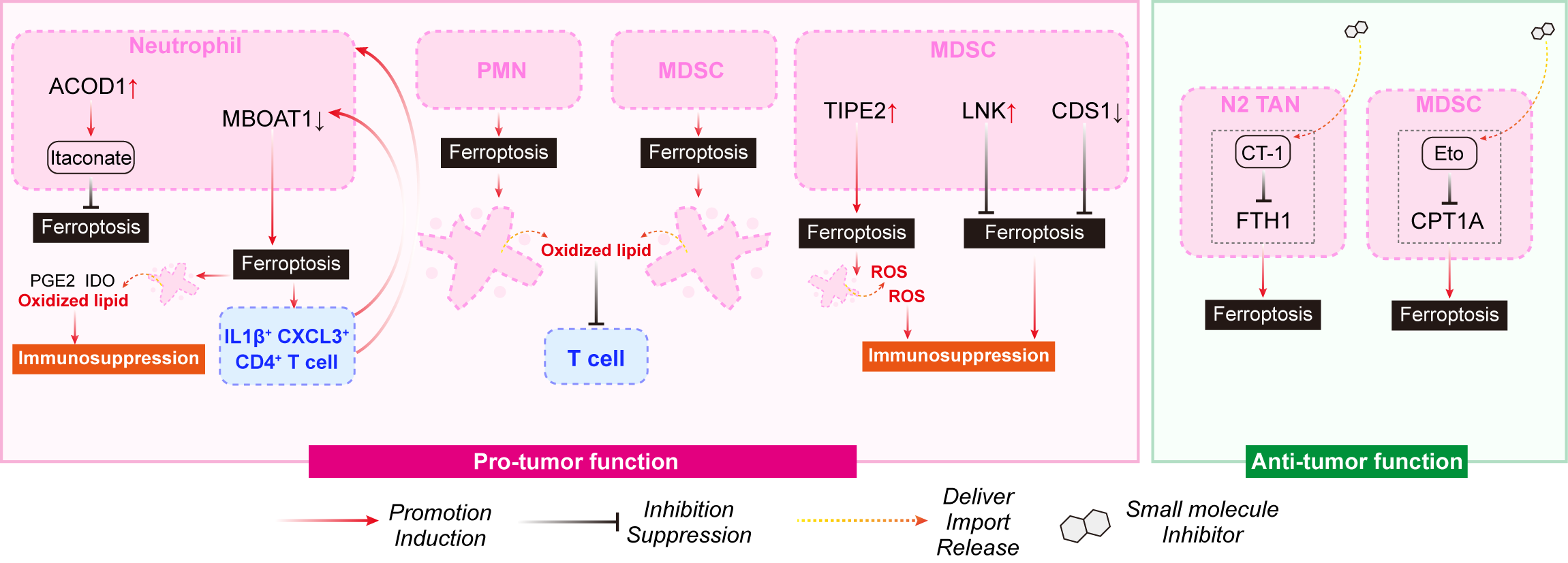
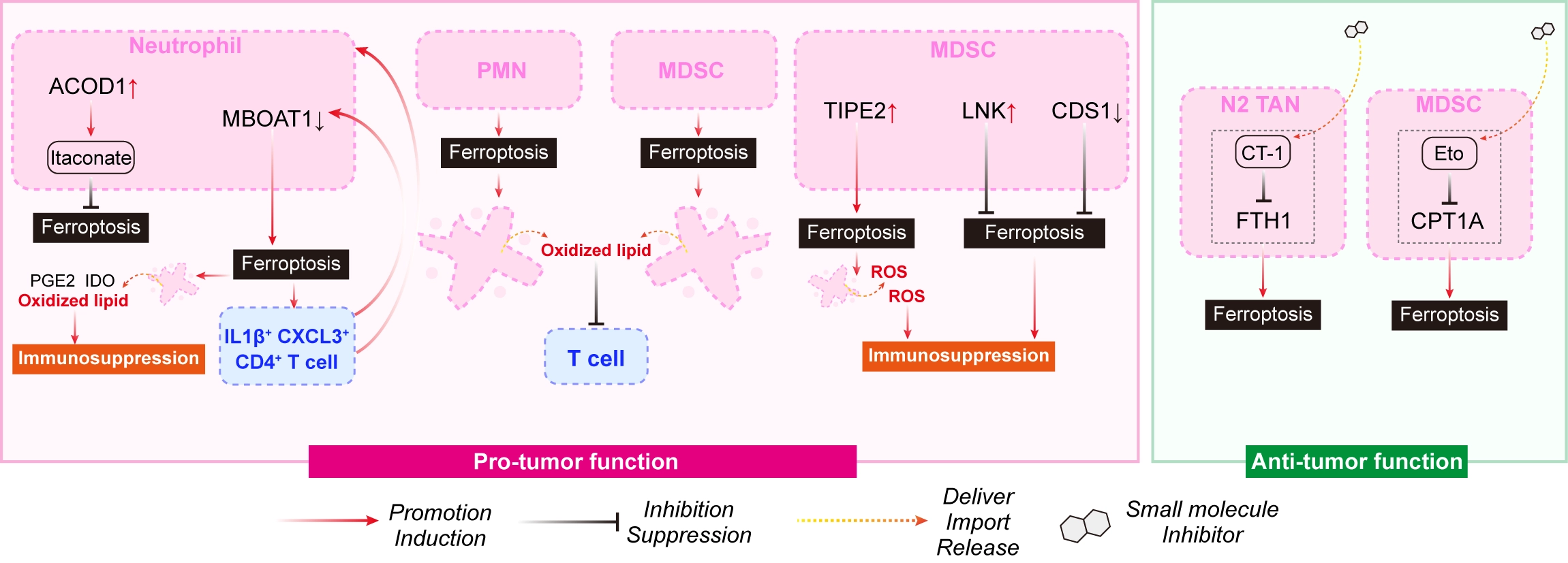
Figure 4. Ferroptosis in neutrophil/MDSC within the tumor. Neutrophils/MDSC generally exhibit an unusually high susceptibility to ferroptosis among immune cell populations within the TME. The consequences of neutrophils/MDSC ferroptosis are paradoxical. TME: tumor microenvironment; MDSC: myeloid-derived suppressor cell; PMN: pathologically activated neutrophil; IDO: indoleamine 2,3-dioxygenase; PGE2: prostaglandin E2; IL1β: interleukin 1β; CXCL3: C-X-C motif chemokine ligand 3; ACOD1: Aconitate decarboxylase 1; TIPE2: TNF alpha induced protein 8 like 2; LNK: lymphocyte adaptor protein, also known as SH2B3; ROS: reactive oxygen species; TAN: tumor-associated neutrophils; MBOAT1: membrane bound glycerophospholipid O-acyltransferase 1; CPT1A: carnitine palmitoyltransferase 1A; CDS1: CDP-diacylglycerol synthase 1; Eto: etomoxir sodium salt; FTH1: ferritin heavy chain 1.
The relationship between ferroptosis and the functional state of neutrophils is also highly context-dependent. Chemotherapy-resistant breast tumors contain neutrophils with elevated lipid peroxidation and a unique phospholipid composition that renders them particularly vulnerable to ferroptosis[61]. Rather than limiting immune suppression, ferroptotic neutrophils promote an immunosuppressive microenvironment through the production of prostaglandin E2 (PGE2), indoleamine 2,3-dioxygenase (IDO) and oxidized phospholipids (Figure 4)[61]. Intriguingly, an interleukin 1β (IL1β)+ C-X-C motif chemokine ligand 3 (CXCL3)+ CD4+ T-cell subset enhanced neutrophil ferroptosis through IL1β-interleukin 1 receptor type 1 (IL1R1)-nuclear factor kappa B (NF-κB) signaling while simultaneously replenishing neutrophil recruitment, thereby establishing a positive feedback loop that reinforced immune suppression (Figure 4)[61]. Similarly, compared with anti-tumor N1 neutrophils, N2-like TANs appear to be more susceptible to ferroptosis, and pharmacological induction of ferroptosis by the cryptotanshinone derivative CT-1 selectively eliminated N2 neutrophils through nuclear receptor coactivator 4 (NCOA4)-mediated ferritinophagy by targeting ferritin heavy chain 1 (FTH1) (Figure 4)[62,63]. These observations suggest that the susceptibility to ferroptosis differs substantially among different neutrophil states and may indicate an exploitable vulnerability of tumor-promoting neutrophils.
Multiple intrinsic and extrinsic mechanisms determine the sensitivity of neutrophils to ferroptosis. Antioxidant pathways centered on NRF2 remain major regulators of neutrophil survival. Aconitate decarboxylase 1 (ACOD1)-derived itaconate activates NRF2 signaling and suppresses ferroptosis in tumor neutrophils, whereas ACOD1 deficiency increases ferroptosis and ICB responses in metastatic breast cancer (Figure 4)[64-66]. Conversely, several pathways actively promote ferroptosis. Loss of lymphocyte adaptor protein (LNK, also known as SH2B3) activates the Fms-related tyrosine kinase 3 (FLT3)-STAT1-interferon regulatory factor 1 (IRF1)-arachidonate 12-lipoxygenase, 12S type (ALOX12) axis, resulting in excessive lipid peroxidation and ferroptosis of MDSCs[67]. Tumor necrosis factor (TNF) alpha-induced protein 8-like 2 (TIPE2) regulates susceptibility to ferroptosis by remodeling the phospholipid composition, whereas inhibition of carnitine palmitoyltransferase 1A (CPT1A) or activation of CDP-diacylglycerol synthase 1 (CDS1) disrupts the SLC7A11-GPX4 antioxidant axis and sensitizes MDSCs to ferroptosis (Figure 4)[68-70]. These studies collectively indicate that lipid metabolism and antioxidant defense cooperate to determine whether neutrophils survive or succumb to ferroptosis stress.
Emerging evidence also suggests that ferroptosis in neutrophils is influenced by systemic factors beyond the tumor itself. A recently identified resistin-like gamma (Retnlg)+ lipocalin 2 (Lcn2)+ senescence-like neutrophil population preferentially accumulates in the male bladder cancer microenvironment and exhibits potent immunosuppressive activity[71]. Interestingly, the female-enriched gut bacterium Alistipes shahii produces the metabolite lurasidone, which targets the iron-binding protein LCN2 and selectively induces ferroptosis in these senescent neutrophils, thereby suppressing tumor growth[71]. These findings highlight an unexpected microbiota-neutrophil-ferroptosis axis that may contribute to sex-specific differences in anti-tumor immunity.
Collectively, the current evidence suggests that ferroptosis in neutrophils differs fundamentally from ferroptosis in other stromal and immune cell populations. Rather than simply eliminating tumor-promoting neutrophils, ferroptosis can simultaneously generate immunosuppressive lipid mediators that dampen T-cell responses. Therefore, therapeutic strategies should not focus solely on increasing ferroptosis in neutrophils but instead consider how ferroptotic neutrophils influence the surrounding immune landscape. Future studies should determine which neutrophil subpopulations benefit from ferroptosis induction, identify the immunosuppressive mediators released during ferroptosis, and establish whether the selective targeting of ferroptosis-prone neutrophil populations can enhance immunotherapy efficacy without exacerbating lipid-mediated immune dysfunction.
5. Ferroptosis as a Determinant of CD8+ T-cell Fitness
CD8+ T cells are the principal cytotoxic lymphocytes responsible for recognizing and eliminating malignant cells and constitute a central effector population for ICB and adoptive cell therapy (ACT)[72]. However, durable anti-tumor immunity requires not only T-cell activation but also sustained metabolic fitness within the TME[73,74]. The TME is characterized by oxidative stress, lipid accumulation, nutrient competition, and iron dysregulation, all of which converge on ferroptosis pathways. Similar to the abovementioned cell types within the TME, emerging evidence suggests that ferroptosis is not simply a mechanism of CD8+ T-cell death but also a critical regulator of T-cell persistence, functional exhaustion, and responsiveness to immunotherapy.
Unlike tumor cells, activated CD8+ T cells appear to be intrinsically vulnerable to ferroptosis. A high-throughput metabolic vulnerability screen first revealed that activated CD8+ T cells are considerably more sensitive to GPX4 inhibition than tumor cells, and that tumor-infiltrating CD8+ T cells accumulate substantial lipid peroxidation within the TME (Figure 5)[75]. Importantly, enhancing ferroptosis resistance through GPX4 or FSP1 overexpression preserved T-cell survival without compromising cytotoxic function, whereas inhibition of acyl-CoA synthetase long chain family member 4 (ACSL4) reduced ferroptosis but simultaneously impaired anti-tumor activity (Figure 5), suggesting that the lipid remodeling required for T-cell effector function also creates an inherent susceptibility to ferroptosis[75]. Consistent with these findings, GPX4-mediated antioxidant defense cooperates with GSH metabolism and adenosine A2A receptor signaling to maintain CD8+ T-cell expansion, survival, and anti-tumor immunity[76]. Together, these observations support the concept that ferroptosis represents a metabolic vulnerability of activated CD8+ T cells rather than merely a consequence of T-cell dysfunction.
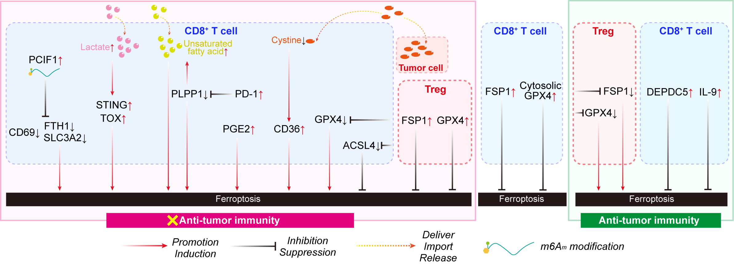
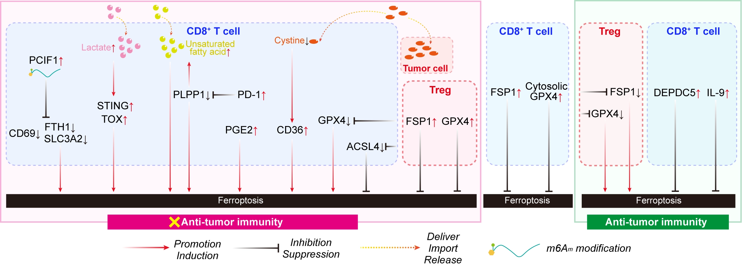
Figure 5. Ferroptosis in T cells within the tumor. Activated CD8+ T cells generally appear to be intrinsically vulnerable to ferroptosis, which in turn contributes to T cell exhaustion within the TME, regulated by intrinsic metabolic, transcriptional programs, epigenetic and posttranscriptional mechanisms. Ferroptosis is generally detrimental to Treg-mediated immune suppression and therefore favors anti-tumor immunity. TME: tumor microenvironment; Treg: regulatory T; ACSL4: acyl-CoA synthetase long chain family member 4; GPX4: glutathione peroxidase 4; IL-9: interleukin 9; STING: stimulator of interferon genes; TOX: thymocyte selection associated high mobility group box; PCIF1: phosphorylated CTD-interacting factor 1; FTH1: ferritin heavy chain 1; SLC3A2: solute carrier family 3 member 2; PLPP1: phospholipid phosphatase 1; PD-1: programmed death-1; PGE2: prostaglandin E2; FSP1: ferroptosis suppressor protein 1; DEPDC5: DEP domain containing 5, GATOR1 subcomplex subunit; m6Am: N6 2′-O-dimethyladenosine.
Accumulating evidence further indicates that ferroptosis contributes directly to T-cell exhaustion within tumors. CD36, a major scavenger receptor responsible for lipid uptake, promotes the accumulation of oxidized lipids, iron, and ROS in tumor-infiltrating CD8+ T cells, thereby driving ferroptosis and reducing the production of cytotoxic cytokines (Figure 5)[77,78]. Likewise, metabolic competition between tumor cells and T cells for cystine limits glutathione synthesis, resulting in oxidative stress, CD36-dependent lipid accumulation, ferroptosis, and terminal T-cell exhaustion (Figure 5)[77,78]. Restricting cystine uptake by tumor cells alleviates these metabolic constraints, reduces ferroptosis, and restores CD8+ T-cell function (Figure 5)[79]. These findings identify ferroptosis as a key mechanism linking metabolic competition within the TME to the loss of T-cell effector activity.
The susceptibility of CD8+ T cells to ferroptosis is determined by multiple intrinsic metabolic and transcriptional programs. Several studies have identified pathways that enhance ferroptosis resistance while preserving anti-tumor function. Compared with conventional Tc1 cells, Tc9 cells display lower levels of lipid peroxidation and greater resistance to ferroptosis through IL-9-mediated activation of STAT3 and fatty acid oxidation, contributing to their superior persistence and therapeutic efficacy following adoptive transfer (Figure 5)[80,81]. Conversely, PGE2, an immunosuppressive metabolite enriched within tumors, promotes lipid peroxidation and ferroptosis by disrupting the IL-2-mammalian target of rapamycin (mTOR)-peroxisome proliferator-activated receptor γ coactivator 1α (PGC1α) metabolic axis in tumor-infiltrating lymphocytes (Figure 5)[82]. Similarly, lactate accumulation activates the STING-thymocyte selection-associated high mobility group box (TOX) pathway, suppresses heme oxygenase-1 (HO-1) expression, and promotes iron accumulation and mitochondrial oxidative stress, thereby sensitizing CD8+ T cells to ferroptosis (Figure 5)[83]. Collectively, these studies suggest that metabolic adaptation rather than T-cell receptor signaling alone determines whether activated T cells maintain effector function or undergo ferroptosis-related dysfunction.
Beyond metabolic regulation, recent studies have identified epigenetic and posttranscriptional mechanisms that govern ferroptosis sensitivity in CD8+ T cells. Loss of the RNA N6 2′-O-dimethyladenosine (m6Am) methyltransferase phosphorylated CTD-interacting factor 1 (PCIF1) decreases intracellular iron levels, enhances cystine uptake and glutathione synthesis, and protects CD8+ T cells from ferroptosis through altered expression of ferroptosis-associated genes such as FTH1 and SLC3A2 (Figure 5)[84]. Similarly, phospholipid phosphatase 1 (PLPP1) preserves phospholipid homeostasis and prevents unsaturated fatty acid-induced ferroptosis, whereas programmed death-1 (PD-1) signaling suppresses PLPP1 expression through the protein kinase B (Akt)-GATA binding protein 1 (GATA1) pathway, thereby coupling immune checkpoint signaling to ferroptotic dysfunction (Figure 5)[85]. In addition, DEP domain containing 5, GATOR1 subcomplex subunit (DEPDC5) deficiency activates mTOR complex 1 (mTORC1)-activating transcription factor 4 (ATF4)-dependent xanthine oxidase signaling, increasing ROS generation and lipid peroxidation without directly regulating canonical ferroptosis genes (Figure 5)[86]. These observations indicate that ferroptosis sensitivity is integrated with broader transcriptional and metabolic programs that govern T-cell differentiation and activation.
Extrinsic factors also substantially influence CD8+ T-cell ferroptosis. Dietary lipid composition alters phospholipid metabolism independently of the gut microbiota and profoundly affects ferroptosis susceptibility in tumor-infiltrating CD8+ T cells[87]. Compared with mice receiving standard laboratory diets, mice receiving purified diets presented lower lipid ROS levels, enhanced resistance to ferroptosis, increased production of interferon γ (IFNγ) and TNF, and improved tumor control[87]. Likewise, treatment with ferrostatin-1 selectively increased the accumulation of tumor-infiltrating CD8+ T cells and improved anti-tumor immunity under ferroptosis-promoting dietary conditions[87]. In addition, pathological systemic environments can further sensitize T cells to ferroptosis. In sickle cell disease, SWI/SNF related BAF chromatin remodeling complex subunit B1 (SMARCB1)-dependent chromatin remodeling suppresses SLC7A11 and hydrogen sulfide biosynthesis, thereby rendering CD8+ T cells highly susceptible to ferroptosis despite concomitant ferroptosis resistance in renal medullary carcinoma cells[88]. These findings emphasize that systemic metabolism and disease states can profoundly reshape T-cell ferroptosis independently of tumor-intrinsic mechanisms.
Collectively, the current evidence consistently supports a tumor-promoting role for CD8+ T-cell ferroptosis. Unlike macrophages or neutrophils, where the consequences of ferroptosis are often context-dependent, ferroptosis in CD8+ T cells almost invariably impairs anti-tumor immunity by limiting T-cell survival, promoting exhaustion, and reducing cytotoxic function. Consequently, enhancing ferroptosis resistance in therapeutic T cells represents an attractive strategy for increasing the efficacy of ICB and ACT. However, important questions remain unresolved. It is still unclear whether ferroptosis sensitivity differs among distinct exhausted, stem-like, or tissue-resident CD8+ T-cell subsets identified by single-cell analyses. Moreover, because lipid metabolism simultaneously supports T-cell effector function and predisposes cells to ferroptosis, future studies should determine how to selectively enhance ferroptosis resistance without compromising the metabolic programs required for durable anti-tumor immunity.
6. Selective Ferroptosis Protection Sustains Treg Cell Function
Tregs constitute a major immunosuppressive population within the TME and represent one of the principal barriers to effective cancer immunotherapy[89]. By suppressing effector T-cell activation through cell-cell interactions and the secretion of immunoregulatory cytokines, Tregs establish an immune-tolerant environment that facilitates tumor progression[90,91]. Emerging evidence suggests that maintaining resistance to ferroptosis stress is an essential component of Treg biology, as it enables these cells to preserve immune homeostasis and sustain immunosuppressive activity within the oxidatively stressed TME.
Current evidence indicates that ferroptosis is generally detrimental to Treg-mediated immune suppression and therefore favors anti-tumor immunity. GPX4 serves as a central antioxidant defense that protects Tregs from lipid peroxidation and ferroptosis while maintaining their suppressive function[90]. Notably, the Treg-specific deletion of GPX4 disrupts immune regulation despite having little effect on Treg survival under steady-state conditions, suggesting that GPX4 primarily preserves Treg function rather than simply preventing cell death (Figure 5)[90]. However, GPX4 is also indispensable for the survival and anti-tumor activity of effector CD8+ T cells (Figure 5)[75,76]. Consequently, systemic inhibition of GPX4 is expected to impair both immunosuppressive and cytotoxic T-cell populations, limiting its therapeutic value.
Recent work has identified FSP1 as a more selective regulator of Treg function. Unlike GPX4, FSP1 expression is induced following T-cell receptor activation, indicating that distinct ferroptosis-defense programs are engaged during T-cell activation[91]. Importantly, genetic ablation of FSP1 in T cells selectively impaired the immunosuppressive function of tumor-infiltrating Tregs without compromising effector T-cell-mediated anti-tumor immunity or causing systemic autoimmune toxicity (Figure 5)[91]. These findings suggest that Tregs rely on a unique ferroptosis-defensive program that can be therapeutically targeted independently of the GPX4 pathway.
Collectively, the current evidence suggests that ferroptosis resistance is critical for maintaining the immunosuppressive activity of Tregs within tumors. More importantly, these studies report that not all ferroptosis-related defense mechanisms are functionally equivalent. While GPX4 broadly supports T-cell survival, FSP1 appears to preferentially sustain Treg-mediated immune suppression, identifying FSP1 as a promising target for selectively destabilizing intratumoral Tregs while preserving protective anti-tumor immunity. Nevertheless, the molecular basis for this selective dependence remains poorly understood, and future studies should determine whether additional ferroptosis-regulatory pathways similarly distinguish immunosuppressive Tregs from effector T cells.
7. TME–Driven Ferroptosis Restrains NK Cell Immunity
NK cells are innate cytotoxic lymphocytes that eliminate transformed or virus-infected cells independently of antigen recognition and constitute important components of anti-tumor immunity[92]. The increasing clinical success of NK cell-based immunotherapies has highlighted the importance of maintaining NK cell fitness within the TME[93,94]. Emerging evidence suggests that ferroptosis represents a major mechanism through which the metabolically hostile TME compromises NK cell survival and cytotoxic function. Rather than being driven primarily by intrinsic defects, NK cell ferroptosis appears to be orchestrated by multiple tumor-derived metabolic and stromal signals that converge on oxidative stress, lipid peroxidation, and iron dysregulation.
Current evidence consistently supports a tumor-promoting role for NK cell ferroptosis. Tumor-derived metabolic products directly impair NK cell function by inducing ferroptosis stress. For example, gastric cancer cells utilize IDO to generate L-kynurenine, which induces ferroptosis in NK cells independently of aryl hydrocarbon receptor (AHR) signaling (Figure 6)[95,96]. Notably, GPX4 overexpression protects NK cells from L-kynurenine-induced ferroptosis, highlighting the importance of antioxidant defense in preserving NK cell function[96]. Similarly, dietary metabolic alterations can reshape NK cell ferroptosis. Although ketogenic diets have been reported to promote ferroptosis in tumor cells[97], recent work has revealed that phosphatidylethanolamine metabolites generated under ketogenic conditions impair mitochondrial function, reduce GPX4 expression, increase lipid peroxidation, and ultimately induce ferroptosis in NK cells, thereby promoting colorectal cancer liver metastasis (Figure 6)[98]. Together, these findings indicate that tumor- and diet-derived metabolites can simultaneously exert opposing effects on tumor cells and anti-tumor immune cells through ferroptosis.
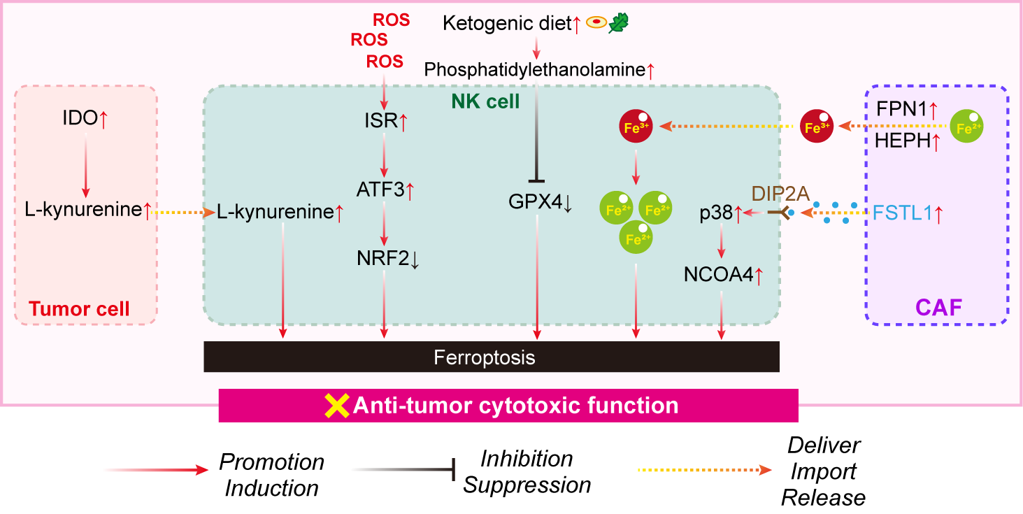
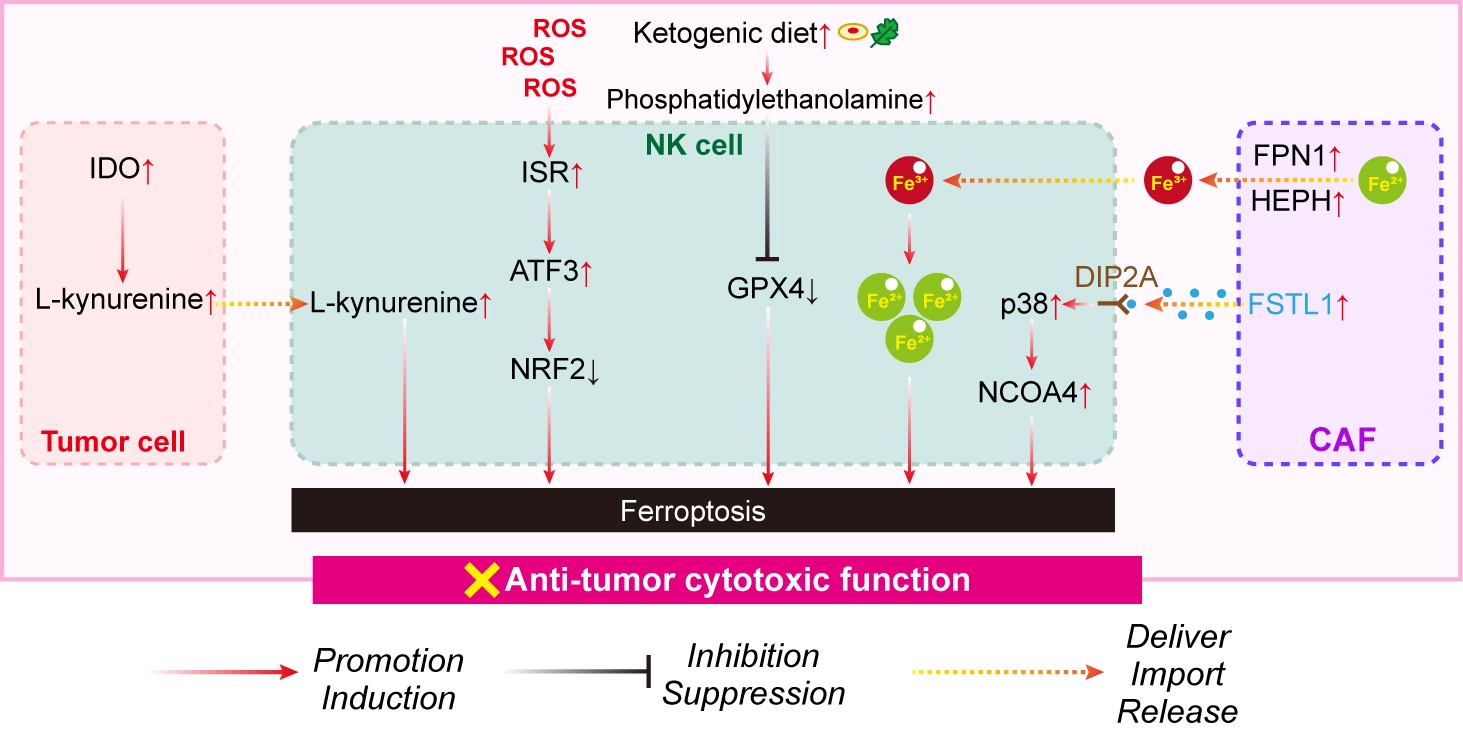
Figure 6. Ferroptosis in NK cells within the tumor. NK cell ferroptosis generally plays tumor-promoting roles within the TME. NK: natural killer; TME: tumor microenvironment; IDO: indoleamine 2,3-dioxygenase; GPX4: glutathione peroxidase 4; FPN1: ferroportin 1; HEPH: hephaestin; FSTL1: follistatin-like protein 1; ATF3: activating transcription factor 3; ISR: integrated stress response; ROS: reactive oxygen species; DIP2A: DIP2 acetate--CoA ligase A; NCOA4: nuclear receptor coactivator 4; NRF2: nuclear factor erythroid 2-related factor 2; CAF: cancer-associated fibroblast.
The TME also actively regulates NK cell ferroptosis through microbial and stromal interactions. Recent studies identified the intratumoral bacterium B. parabrevis as a positive regulator of NK cell function in HCC through the suppression of ferroptosis[99]. Mechanistically, B. parabrevis promotes acetylation of RAR related orphan receptor C (RORC), leading to NEDD4 like E3 ubiquitin protein ligase (NEDD4L)-dependent ubiquitination of iron transporters SLC39A14, SLC39A8 and six-transmembrane epithelial antigen of the prostate 3 (STEAP3), thereby limiting intracellular iron accumulation and ferroptosis-associated stress[99]. In contrast, CAFs promote NK cell ferroptosis through complementary mechanisms. CAFs increase iron availability by upregulating ferroportin 1 (FPN1) and hephaestin (HEPH), thereby expanding the labile iron pool in NK cells (Figure 6)[100]. In parallel, CAF-derived follistatin-like protein 1 (FSTL1) activates DIP2A-p38 signaling to induce NCOA4-mediated ferritinophagy, further sensitizing NK cells to ferroptosis[100]. These observations illustrate how distinct components of the TME cooperate to regulate NK cell iron metabolism and ferroptosis.
Cell-intrinsic stress responses also influence the susceptibility of NK cells to ferroptosis. High levels of ROS within the TME activate the integrated stress response (ISR), resulting in the induction of activating transcription factor 3 (ATF3) and the suppression of NRF2-mediated antioxidant signaling, thereby promoting ferroptosis (Figure 6)[101]. These findings link environmental oxidative stress to impaired antioxidant capacity and highlight the interplay between stress adaptation and ferroptosis in NK cells.
Collectively, the current evidence indicates that ferroptosis is a major mechanism limiting NK cell-mediated anti-tumor immunity. Unlike tumor cells, which often acquire robust ferroptosis resistance, NK cells remain highly susceptible to the ferroptosis stress generated by metabolic competition, stromal interactions, oxidative stress, and microbial metabolites within the TME. Consequently, protecting NK cells from ferroptosis may represent an attractive strategy for enhancing the efficacies of NK cell-based immunotherapies. However, several important questions remain unresolved. It remains unclear whether distinct NK cell subsets differ in terms of ferroptosis sensitivity, how ferroptosis contributes to NK cell dysfunction during ICB or adoptive NK cell therapy, and whether selectively enhancing ferroptosis resistance in therapeutic NK cells can improve clinical efficacy without reducing ferroptosis-mediated tumor cell death.
8. Conclusion and Perspective
Although ferroptosis was originally characterized as a mechanism of tumor cell death, accumulating evidence demonstrates that sublethal ferroptosis associated stress is equally important in shaping the biology of non-tumoral cells within the TME. Rather than acting solely as a cell death program, ferroptosis-associated stress is tightly involved in controlling the cell identity, functional plasticity, metabolic adaptation, and intercellular communication across diverse stromal and immune cell populations. Throughout this review, we propose a conceptual framework in which the biological consequences of ferroptosis are determined primarily by cellular context rather than by ferroptosis itself. As highlighted throughout this review, the consequences of ferroptosis are highly cell-type-specific. Whereas ferroptosis resistance enables tumor-promoting CAFs and immunosuppressive macrophages to maintain their protumor functions, ferroptosis impairs the persistence and effector activity of cytotoxic lymphocytes such as CD8+ T cells and NK cells. Moreover, in neutrophils and PMN-MDSCs, ferroptosis generates oxidized lipid mediators that may paradoxically reinforce immune suppression despite reducing cell numbers. Together, these observations indicate that ferroptosis cannot simply be classified as either tumor-suppressive or tumor-promoting. Instead, its biological consequences are determined by the cellular context in which it occurs.
This conceptual shift has important therapeutic implications. Current ferroptosis-based strategies largely aim to maximize ferroptosis in tumor cells. However, indiscriminate induction or inhibition of ferroptosis or its related stress may simultaneously alter stromal and immune cell populations in ways that either enhance or compromise anti-tumor immunity. Future therapeutic approaches should therefore move beyond globally targeting ferroptosis and instead selectively modulate ferroptosis in specific cellular compartments of the TME. Recent studies identifying cell-type-specific regulators, such as FSP1, in regulatory T cells, suggest that selective ferroptosis intervention can be achieved.
Several important questions remain unresolved. First, the molecular determinants governing ferroptosis sensitivity across distinct stromal and immune cell states remain incompletely understood, particularly given the extensive heterogeneity revealed by single-cell and spatial transcriptomic technologies. Second, whether the biological effects of ferroptosis primarily reflect the depletion of specific cell populations or functional reprogramming of surviving cells, as increasingly observed in macrophages and other immune populations, remains unclear. Third, the mechanisms by which ferroptotic nontumoral cells communicate with neighboring immune and stromal cells through oxidized lipids, cytokines, EVs, and other mediators require further investigation. Finally, defining quantitative ferroptosis vulnerabilities across individual cell populations is essential for designing therapies that maximize tumor cell killing while preserving or even enhancing protective anti-tumor immunity.
Overall, the emerging view is that ferroptosis is not merely a mechanism for eliminating cells within tumors but also a fundamental regulator of TME organization and immune function. A deeper understanding of cell-type-specific ferroptosis programs will facilitate the development of precision ferroptosis-based therapies that simultaneously target malignant cells, remodel the TME, and improve responses to immunotherapy.
Acknowledgements
The authors declare that GPT-5.6 was used solely for first-round language polishing during the manuscript preparation process. Then, the authors reviewed and further sent this manuscript for the second-round language polishing by American Journal Experts English language editing service. All research content, including review design, reference analysis, interpretations and figures is original and was not generated using AI tools. The authors reviewed, revised, and approved the final manuscript and take full responsibility for its content.
Authors contribution
Gong L, Gong Z: Data curation, writing-original draft, writing review & editing.
Wang K, Zhang Q: Data curation, writing review & editing.
Cui K: Supervision, conceptualization, data curation, funding acquisition, writing-original draft, writing review & editing.
Conflicts of interest
The authors declare no conflicts of interest.
Ethical approval
Not applicable.
Consent to participate
Not applicable.
Consent for publication
Not applicable.
Availability of data and materials
Not applicable.
Funding
The project was supported by the grant from the National Natural Science Foundation of China (Grant No. 82472671) and the Doctoral Research Initiation Project of Hunan University of Arts and Science (Grant No. 25BSQD72).
Copyright
© The Author(s) 2026.
References
-
4. Cui K, Wang K, Huang Z. Ferroptosis and the tumor microenvironment. J Exp Clin Cancer Res. 2024;43(1):315.[DOI]
-
6. Jain C, Shah YM. Iron: Regulation, redox homeostasis, and ferroptosis in cancer. Ferroptosis Oxid Stress. 2026;2(3):202526.[DOI]
-
8. Cui K, Liu B, Gong L, Wan Q, Tang H, Gong Z, et al. Targeting ribosomes reprograms the tumour microenvironment and augments cancer immunotherapy. Br J Cancer. 2025;133(6):756-770.[DOI]
-
16. Li A, Gong Z, Long Y, Li Y, Liu C, Lu X, et al. Lactylation of LSD1 is an acquired epigenetic vulnerability of BRAFi/MEKi-resistant melanoma. Dev Cell. 2025;60(14):1974-1990.e11.[DOI]
-
17. Zhang Q, Cui K, Kong Y, Yu J, Luo Z, Yang X, et al. Targeting both the enzymatic and non-enzymatic functions of DHODH as a therapeutic vulnerability in c-Myc-driven cancer. Cell Rep. 2025;44(3):115327.[DOI]
-
19. Zhou X, Wang G, Wu Y, Wu M, Zhai X, Tian C, et al. SLC6A8-mediated creatine uptake suppresses ERK2-FSP1 signaling and induces ferroptosis in colorectal cancer. Cell Rep. 2025;44(9):116139.[DOI]
-
22. Hu Y, Li Z, Li M, Wu X, Zhang S, Tang M, et al. Targeting PRDX6-dependent localization and function of GPX4 enhances ferroptosis-mediated tumor suppression. Mol Cell. 2025;85(24):4602-4620.e9.[DOI]
-
23. Ito J, Nakamura T, Toyama T, Chen D, Berndt C, Poschmann G, et al. PRDX6 dictates ferroptosis sensitivity by directing cellular selenium utilization. Mol Cell. 2024;84(23):4629-4644.e9.[DOI]
-
26. Chen Y, McAndrews KM, Kalluri R. Clinical and therapeutic relevance of cancer-associated fibroblasts. Nat Rev Clin Oncol. 2021;18(12):792-804.[DOI]
-
27. Tao M, Liu W, Chen J, Liu R, Zou J, Yu B, et al. Transcriptome landscape of cancer-associated fibroblasts in human PDAC. Adv Sci. 2025;12(20):2415196.[DOI]
-
29. Zhang Y, Zhao H, Xu P, Liu K, Du G, Chen X, et al. The Zeb1/system Xc- axis safeguards iron-loaded cancer-associated fibroblasts against ferroptosis and promotes immunosuppression in prostate cancer. Cancer Lett. 2026;639:218243.[DOI]
-
33. Nogales-Pons M, Munárriz-Paños M, Aceña-Gonzalo T, Casanova-Acebes M. Myeloid diversity in tumors: Shaped by genes, location, and time. J Exp Med. 2026;223(4):e20252267.[DOI]
-
36. Jin R, Neufeld L, McGaha TL. Linking macrophage metabolism to function in the tumor microenvironment. Nat Cancer. 2025;6(2):239-252.[DOI]
-
39. Hao X, Zheng Z, Liu H, Zhang Y, Kang J, Kong X, et al. Inhibition of APOC1 promotes the transformation of M2 into M1 macrophages via the ferroptosis pathway and enhances anti-PD1 immunotherapy in hepatocellular carcinoma based on single-cell RNA sequencing. Redox Biol. 2022;56:102463.
-
40. Sun D, Liu X, Mao C, Zhao F, Fan W, Li C, et al. Macelignan targets SLC7A11 to induce ferroptosis and modulate macrophage polarization via STAT6/PPAR-γ/KLF4 signaling. Bioorg Chem. 2026;169:109422.[DOI]
-
41. Cheng C, Chai J, Zhang H, Wang Y, Zhang L, Yang M, et al. Cucurbitacin B suppresses malignant progression of oral leukoplakia via ferroptosis-induced macrophage polarization. Int Immunopharmacol. 2025;165:115485.[DOI]
-
45. Wang H, An P, Xie E, Wu Q, Fang X, Gao H, et al. Characterization of ferroptosis in murine models of hemochromatosis. Hepatology. 2017;66(2):449-465.[DOI]
-
48. Li R, Liu Y, Pang B, Liu X, Zhu C, Tang G, et al. RAB10 mediates metabolic regulation of ferroptosis in breast Cancer and synergistically remodels the tumour immune microenvironment via macrophage M2 polarization. Int Immunopharmacol. 2026;172:116201.[DOI]
-
52. Xiao Y, Xu Y, Wang H, Yang F, Ding XH, Fu T, et al. HEBP2-governed glutamine competition between tumor and macrophages dictates immunotherapy efficacy in triple-negative breast cancer. Cell Metab. 2025;37(10):2030-2047.e7.[DOI]
-
54. Gabrilovich DI, Bronte V, Chen SH, Colombo MP, Ochoa A, Ostrand-Rosenberg S, et al. The terminology issue for myeloid-derived suppressor cells. Cancer Res. 2007;67(1):425.[DOI]
-
55. Hedrick CC, Malanchi I. Neutrophils in cancer: Heterogeneous and multifaceted. Nat Rev Immunol. 2022;22(3):173-187.[DOI]
-
56. Quail DF, Amulic B, Aziz M, Barnes BJ, Eruslanov E, Fridlender ZG, et al. Neutrophil phenotypes and functions in cancer: A consensus statement. J Exp Med. 2022;219(6):e20220011.[DOI]
-
57. Kim R, Hashimoto A, Markosyan N, Tyurin VA, Tyurina YY, Kar G, et al. Ferroptosis of tumour neutrophils causes immune suppression in cancer. Nature. 2022;612(7939):338-346.[DOI]
-
58. Veglia F, Hashimoto A, Dweep H, Sanseviero E, De Leo A, Tcyganov E, et al. Analysis of classical neutrophils and polymorphonuclear myeloid-derived suppressor cells in cancer patients and tumor-bearing mice. J Exp Med. 2021;218(4):e20201803.[DOI]
-
60. Wang Y, Zhang X, Yang W, Liu X, Xie S, Zhang L, et al. Caveolin-1 drives ferroptosis in MDSCs via PKA-DRP1-mediated ER-mitochondria crosstalk to shape breast cancer immunosuppression. Free Radic Biol Med. 2026;242:601-613.[DOI]
-
62. Liu Y, Sun Q, Guo J, Yan L, Yan Y, Gong Y, et al. Dual ferroptosis induction in N2-TANs and TNBC cells via FTH1 targeting: A therapeutic strategy for triple-negative breast cancer. Cell Rep Med. 2025;6(1):101915.[DOI]
-
63. Mancias JD, Wang X, Gygi SP, Harper JW, Kimmelman AC. Quantitative proteomics identifies NCOA4 as the cargo receptor mediating ferritinophagy. Nature. 2014;509(7498):105-109.[DOI]
-
67. Zhou J, Yin H, Pan J, Yin R, Wei X, Shen M, et al. Lnk deficiency attenuates the immunosuppressive capacity of MDSCs via ferroptosis to suppress tumor development. Cell Death Dis. 2025;16(1):610.[DOI]
-
69. Mohamady Farouk Abdalsalam N, Liang Z, Kashaf Tariq H, Ibrahim A, Li R, Wan X, et al. Etomoxir sodium salt promotes imidazole ketone erastin-induced myeloid-derived suppressor cell ferroptosis and enhances cancer therapy. Biology. 2024;13(11):949.[DOI]
-
70. Li C, Liu W, Zhang Y, Yu S, Li D. CDS1 deficiency promotes colorectal cancer progression by suppressing CDP-DAG-induced ferroptosis in MDSCs. Cell Signal. 2026;137:112196.[DOI]
-
71. Zhu Q, Zhang G, Cao M, Huang H, He D, Zang Z, et al. Microbiota-shaped neutrophil senescence regulates sexual dimorphism in bladder cancer. Nat Immunol. 2025;26(5):722-736.[DOI]
-
73. Philip M, Schietinger A. CD8+ T cell differentiation and dysfunction in cancer. Nat Rev Immunol. 2022;22(4):209-223.[DOI]
-
74. Sun Q, Dong C. Regulators of CD8+ T cell exhaustion. Nat Rev Immunol. 2026;26(2):129-151.[DOI]
-
83. Zhu Q, Zhang JB, Nie CP, Liu XF, Zhan LP, Li M, et al. STING synergizes with TOX suppressing HO-1 expression to trigger ferroptosis in tumor-infiltrating CD8+ T cell and immunotherapy resistance. Nat Commun. 2026;17(1):2543.[DOI]
-
84. Xiang B, Zhang M, Li K, Zhang Z, Liu Y, Gao M, et al. The epitranscriptional factor PCIF1 orchestrates CD8+ T cell ferroptosis and activation to control antitumor immunity. Nat Immunol. 2025;26(2):252-264.[DOI]
-
89. Imianowski CJ, Chen Q, Workman CJ, Vignali DAA. Regulatory T cells in the tumour microenvironment. Nat Rev Cancer. 2025;25(9):703-722.[DOI]
-
94. Kochs T, Nath P, Chen A, Romee R, Maia A. Reprogramming antitumor immunity: NK cell strategies to navigate the immunosuppressive tumor microenvironment. Adv Sci. 2026;13(35):e75326.[DOI]
-
97. Ferrer M, Mourikis N, Davidson EE, Kleeman SO, Zaccaria M, Habel J, et al. Ketogenic diet promotes tumor ferroptosis but induces relative corticosterone deficiency that accelerates cachexia. Cell Metab. 2023;35(7):1147-1162.e7.[DOI]
-
99. Pan B, Zhang X, Ye D, Yao Y, Zhang Z, Luo Y, et al. Intratumoral Brevibacillus parabrevis enhances antitumor immunity by inhibiting NK cell ferroptosis in hepatocellular carcinoma. Cell Death Dis. 2025;16(1):407.[DOI]
Copyright
© The Author(s) 2027. This is an Open Access article licensed under a Creative Commons Attribution 4.0 International License (https://creativecommons.org/licenses/by/4.0/), which permits unrestricted use, sharing, adaptation, distribution and reproduction in any medium or format, for any purpose, even commercially, as long as you give appropriate credit to the original author(s) and the source, provide a link to the Creative Commons license, and indicate if changes were made.
Publisher’s Note
Science Exploration remains a neutral stance on jurisdictional claims in published
maps
and institutional affiliations. The views expressed in this article are solely those
of
the author(s) and do not reflect the opinions of the Editors or the publisher.
Share And Cite


Science Exploration Style
Gong L, Gong Z, Wang K, Zhang Q, Cui K. Tumor but not tumor cells: Ferroptosis in non-tumor cells within the tumor. Ferroptosis Oxid Stress. 2027;3:202619. https://doi.org/10.70401/fos.2026.0044
Tips
Copy completed.
Submit a Manuscript
Author Instructions
Cite this Article
Article Metrics
0
View
0
Download
Cited
Article Updates

- Abstract
- Keywords
- 1. Introduction
- 2. Adaptation to Ferroptosis Stress in CAFs
- 3. Ferroptosis-Regulated Plasticity in TAMs
- 4. Ferroptosis as a Driver of Neutrophil/MDSC Functional Plasticity
- 5. Ferroptosis as a Determinant of CD8+ T-cell Fitness
- 6. Selective Ferroptosis Protection Sustains Treg Cell Function
- 7. TME–Driven Ferroptosis Restrains NK Cell Immunity
- 8. Conclusion and Perspective
- Acknowledgements
- Authors contribution
- Conflicts of interest
- Ethical approval
- Consent to participate
- Consent for publication
- Availability of data and materials
- Funding
- References
- Copyright
 Get citation
Science Exploration Style

Gong L, Gong Z, Wang K, Zhang Q, Cui K. Tumor but not tumor cells: Ferroptosis in non-tumor cells within the tumor. Ferroptosis Oxid Stress. 2027;3:202619. https://doi.org/10.70401/fos.2026.0044
copy
Share Link
copy