Microglia statuses in Alzheimer’s disease and lessons from anti-tumor microglia

*Correspondence to:
Praveen Agrawal, Department of Molecular Pharmacology, Montefiore Einstein Comprehensive Cancer Center (MECCC), Cancer Dormancy Institute (CDI), Albert Einstein College of Medicine, Bronx, NY 10461, USA.
E-mail: praveen.agrawal@einsteinmed.edu
Geromedicine. 2026;2:202613. 10.70401/Geromedicine.2026.0033
Received: March 04, 2026Accepted: July 27, 2026Published: July 27, 2026
Abstract
Alzheimer’s disease (AD) and related dementias are closely associated with alterations in microglial states. These microglial changes occur in response to amyloid-β (Aβ) accumulation and aberrant tau phosphorylation and can either protect against or exacerbate AD progression, depending on factors such as disease stage, genetic background, and environmental influences. This review focuses on recent advances in understanding the protective functions and features of microglia during the early stages of AD, while highlighting the outstanding questions regarding how these protective states deteriorate and become dysfunctional as the disease progresses. Interestingly, epidemiological studies have suggested an inverse relationship between AD and cancer incidence, and in this context, we provide a comparative analysis of microglial phenotypes in AD and cancer to speculate on how insights from anti-tumor microglia may inspire new strategies for reprogramming microglia to combat AD and related dementias.
Keywords
Alzheimer’s disease, dementia, microglia, cancer, neuroimmune, therapeutics
1. Introduction
Microglia are highly dynamic and multifunctional glial cells that play essential roles in maintaining brain homeostasis. Under physiological conditions, they contribute to synaptic plasticity, clearance of cellular debris, and responses to injury, thus helping preserve brain health. Microglia continuously survey the brain environment, adapting morphology and activity in response to local cues, and participate in immune defense and tissue remodeling.
In Alzheimer’s disease (AD) research, the roles of microglia are complex, involving both protective and detrimental actions that can change as the disease progresses[1-5]. Historically, microglia were viewed primarily as drivers of neuroinflammation that contribute to neuronal dysfunction in AD. More recent research has increasingly appreciated that microglia exert important neuroprotective functions, especially in the early stages of AD. Protective microglial activities include clearing amyloid-β (Aβ) aggregates, limiting plaque formation, and releasing trophic factors and anti-inflammatory cytokines. These functions suggest that microglia can work to restrict the spread of pathology and maintain neuronal integrity when properly regulated. Advances in transcriptomic technologies, including single-cell RNA sequencing, have revealed extensive heterogeneity within the microglial population in AD. Distinct microglial subtypes have been identified, with gene expression signatures linked to beneficial functions, including enhanced phagocytic activity, metabolic resilience, and immunoregulation that counteract neurodegeneration. These findings provide a foundation for developing microglia-based strategies for AD intervention.
Despite this progress, knowledge gained from AD-focused microglial research is still limited. We predict that additional insight can be gained from other research fields involving microglia. For example, cancer biology may provide useful microglia-related information for addressing AD, especially given that epidemiological studies have reported an inverse association between AD and cancer, while admitting that this association is only a conceptual starting point and may involve multiple non-microglial mechanisms. Although the biological basis of this relationship remains incompletely understood and cannot be attributed to microglia specifically, evidence indicates that microglia participate in protective responses in both neurodegeneration and cancer. Therefore, a comparison of microglial programs across these distinct disease contexts may reveal shared immune-regulatory principles and generate new hypotheses for therapeutic intervention. Thus, lessons from anti-tumor microglial and macrophage functions may offer new ideas for microglial-targeted therapies in AD. This review focuses on recent decadal understanding of the roles of microglia in AD and then presents a comparative analysis of microglia in cancer research, which might suggest new directions for strengthening microglia-focused strategies to combat AD and related dementias.
2. Diverse Conditions of Microglia Against AD
Microglia are the principal phagocytes in the central nervous system. Early in AD, microglia efficiently internalize and degrade Aβ aggregates, a process mediated through a variety of surface receptors, including triggering receptor expressed on myeloid cells 2 (TREM2), complement receptors, lipid-binding protein CD36, and scavenger receptors[6-13]. These pathways allow microglia to clear toxic Aβ species and limit their accumulation, thereby reducing the risk of synaptic and neuronal damage. Impairments in these mechanisms, whether due to aging-related decline, genetic variants, or chronic inflammatory conditions, can significantly reduce Aβ clearance and accelerate disease progression. Experimental depletion of microglia in animal models leads to an increase in Aβ plaques and pathologies such as synaptic loss, while young microglia can help restore amyloid plaque clearance by aged microglia[14-16]. This experimental evidence provides compelling support for the function of microglia through their contribution to amyloid regulation in counteracting AD.
Beyond phagocytosis, microglia can physically limit Aβ plaques[17], modify plaque compaction[14], and constitute a barrier to the spreading of neurotoxic Aβ species[18]. This structural containment mitigates the spread of pathology and decreases neuronal exposure to toxic Aβ oligomers. Microglia are also key regulators of synaptic modeling and remodeling, which are important in the context of AD[19-23]. Thus, microglia can work to maintain synaptic homeostasis, support neural networks, and limit synaptic loss associated with AD progression.
In addition to their direct protective functions, microglia contribute to neuronal survival by releasing multiple neurotrophic factors that can enhance neuronal vitality and resilience. Among these protective molecules, brain-derived neurotrophic factor (BDNF) and glial-derived neurotrophic factor (GDNF) are notable, as they support synaptic maintenance, promote neuronal growth, and help regulate neural circuits[24,25]. In parallel with their neurotrophic roles, microglia can adopt anti-inflammatory, reparative, and immunoregulatory states that help restrain excessive inflammation and support homeostatic balance within the central nervous system. These functional states are shaped by disease stage, local cytokine signals, metabolic status, and interactions with neurons and other glial cells[26-31]. In AD, such microglial responses may counteract chronic neuroinflammation, reduce neuronal stress, and limit tissue damage. Through the combined actions of neurotrophic support and anti-inflammatory modulation, microglia serve as critical regulators of neuronal health and play a central role in shaping the brain’s adaptive response to AD-associated challenges.
3. Key Molecular Features of Microglia Against AD
Disease-associated microglia belong to a specialized subfamily of microglial cells that were initially identified in rodent models of AD[32,33]. These microglia undergo a characteristic activation process, including downregulation of homeostatic regulatory genes, which are critical for maintaining homeostatic microglia, and upregulation of genes related to phagocytosis, lipid metabolism, and plaque interaction, which reflect functional adaptation to the disease environment. Functionally, disease-associated microglia are typically present near Aβ plaques, which agrees with a role in limiting plaque toxicity and thus a protective role in AD.
TREM2 has emerged as a major regulator of protective microglial activation. TREM2 activation supports the survival, metabolic fitness, and phagocytic activity of these cells[34-36]. Genetic studies provide strong evidence for its importance, as individuals carrying TREM2 variants have a higher risk of developing AD[37-39]. Similarly, the phospholipase C gamma 2 (PLCγ2)-proline 522 to arginine (P522R) variant is associated with a reduced risk of AD[40]. This may lead to enhanced microglial activation for improving Aβ clearance. In addition to supporting receptor and signaling levels, transcription factors such as PU.1 can also reflect a protective role in microglial function. Loss-of-function mutations in PU.1 can impair the protective capabilities of microglia, suggesting the importance of transcriptional control in maintaining neuroprotective states of microglia[41-43].
Recent studies have further expanded the spectrum of microglial subtypes. Interferons (IFNs) such as IFN-γ are known to be proinflammatory and neurodegenerative[44], but recently a subtype of microglia containing IFN-γ signaling appears to participate in immune modulation and neuroprotection[45]. Microglia with a lipid burden are known to contribute to AD, and conversely, microglia with reduced lipid load have been suggested to enhance Aβ phagocytosis to protect against AD[46]. Finally, in addition to the disease-associated microglia discussed above, there may be other, still insufficiently characterized transitional microglial states that act as intermediates between homeostatic and fully activated disease-associated phenotypes. Although details remain to be explored, these cells likely participate in initiating clearance mechanisms, metabolic adaptation, and immune regulation that ultimately contribute to the microglial protective response. Together, the diverse microglial programs, ranging from highly coordinated phagocytic activity to nuanced metabolic adaptation and finely tuned immune regulation, underscore the complexity and therapeutic potential of microglial protection in AD.
4. Functional Loss and Changes of Microglia Contributing to AD
Microglial senescence is a key factor contributing to the vulnerability of the brain in AD[47-51]. As demonstrated in the body of this literature, microglia often appear senescent during late-stage AD, exhibiting morphological changes such as fragmented processes, cytoplasmic abnormalities, and irregular cell bodies. These dystrophic microglia are strongly correlated with tau pathology and neuronal loss, suggesting that the decline of protective microglial functions is particularly detrimental during the late stages of the disease. The loss of microglial surveillance, phagocytosis, and trophic support may accelerate neurodegenerative processes, highlighting the importance of maintaining healthy microglial populations to counteract disease progression.
Chronic inflammatory activation represents another mechanism by which microglial dysfunction contributes to AD[26,27,29,52]. Overall, prolonged exposure to pathological stimuli, including Aβ aggregates, tau pathology, or systemic inflammation, can drive microglia into a chronic over- activation state. These overactivated microglia overproduce pro-inflammatory cytokines and reactive oxygen species, promoting a neurotoxic environment. While acute microglial activation can be protective, chronic activation reduces their phagocytic efficiency and diminishes support for the brain. Over time, this inflammatory condition chronically contributes to sustained neuroinflammation and accelerates neuronal injury.
Aging, a key risk factor for AD, also plays a central role in microglial dysfunction. With the advent of aging, microglia exhibit impaired metabolic capacity and dysfunctions[53,54]. As appreciated in this literature, aging-associated microglial changes include impairments in lipid metabolism and mitochondrial function, which reduce their overall cellular health. Metabolically compromised microglia are less capable of clearing Aβ and supporting synaptic plasticity. These age-related declines not only limit the neuroprotective functions of microglia but also increase the susceptibility of the brain to AD pathology. Together, microglial senescence, chronic inflammatory activation, and age-related metabolic impairment converge to undermine supportive microglial functions, highlighting the importance of strategies to restore microglial health to slow or prevent the progression of AD.
While describing details of detrimental microglia is not a focus in this mini-review, Table 1 summarizes the progressive shift of microglia from homeostatic and neuroprotective functions in the early stages of AD to a dysfunctional, pro-inflammatory state in advanced disease, contributing to impaired amyloid clearance, synaptic dysfunction, and neuronal loss. Outstanding questions remain regarding how these states deteriorate and become dysfunctional as the disease advances, and addressing these questions should warrant future research.
Table 1. Transition of microglia from protective to dysfunctional states during AD progression.
| AD stage | Microglial phenotype | Major functions and dysfunctions | Key molecular features | AD pathology | References |
| Homeostatic brain (pre-pathology) | Homeostatic microglia | Immune surveillance, synaptic maintenance, debris clearance, neuronal support | P2RY12, TMEM119, CX3CR1, Sall1, PU.1 | Maintenance of CNS homeostasis | [19-23,41-43] |
| Early AD (initial Aβ accumulation) | Protective activated microglia | Aβ phagocytosis, plaque restriction, damaged synapse removal, neurotrophic support | TREM2, CD36, complement receptors, scavenger receptors, BDNF, GDNF | Reduced amyloid burden and neuronal protection | [6-18,24,25] |
| Early-to-intermediate AD | DAM with protective functions | Enhanced plaque-associated responses, lipid metabolism, metabolic adaptation, Aβ containment | TREM2, APOE, PLCγ2, TYROBP, CST7, LPL | Slower plaque expansion and reduced neurotoxicity | [32-40] |
| Intermediate AD | Transitional microglial states | Mixed protective and inflammatory responses; declining phagocytic efficiency | Reduced homeostatic genes, altered IFN signaling, emerging metabolic stress | Progressive amyloid accumulation and synaptic dysfunction | [45,46] |
| Late AD | Senescent/dystrophic microglia | Impaired surveillance, reduced trophic support, defective Aβ clearance | Mitochondrial dysfunction, lipid-droplet accumulation, reduced TREM2 activity, senescence-associated pathways | Increased plaque burden, tau pathology, neuronal loss | [47-51,53,54] |
| Advanced AD | Chronically activated neurotoxic microglia | Excessive cytokine and ROS production, chronic inflammation | TNF-α, IL-1β, IL-6, NF-κB signaling, chronic inflammatory pathways | Sustained neuroinflammation and cognitive decline | [26,27,29,52] |
| End-stage AD | Dysfunctional/senescent microglia | Failure of immune surveillance and tissue repair | Senescence programs, lysosomal dysfunction, metabolic collapse | Accelerated neurodegeneration and severe dementia | [47-51,53,54] |
AD: Alzheimer’s disease; TREM2: triggering receptor expressed on myeloid cells 2; Aβ: amyloid-β; CNS: central nervous system; DAM: disease-associated microglia; BDNF: brain-derived neurotrophic factor; GDNF: glial-derived neurotrophic factor; APOE: apolipoprotein E; PLCγ2: phospholipase C gamma 2; IFN: interferon; ROS: reactive oxygen species; TNF-α: tumor necrosis factor-alpha; P2RY12: purinergic receptor P2Y12; TMEM119: transmembrane protein 119; CX3CR1: C-X3-C motif chemokine receptor 1; Sall1: spalt-like transcription factor 1; CD36: cluster of differentiation 36; TYROBP: TYRO protein tyrosine kinase-binding protein; CST7: cystatin F; LPL: lipoprotein lipase; IL-1β: interleukin-1 beta; IL-6: interleukin-6; NF-κB: nuclear factor kappa B.
5. The Inverse Relationship between AD and Cancers
AD and cancers can be viewed as opposite extremes of dysregulated cellular homeostasis, with uncontrolled proliferation in cancer contrasting with progressive neuronal loss in AD. Of interest, multiple studies have demonstrated a strong inverse relationship between cancers and AD[55-59]. The summary below focuses specifically on evidence showing that cancer survivors exhibit a lower incidence of AD, rather than on how AD may influence cancer risk.
Neuropathology analyses support this inverse relationship, showing reduced AD hallmark pathology in individuals with cancer histories[60,61]. Population-based studies have suggested that cancer survivors have a significantly lower risk of developing AD, while AD patients have reduced cancer incidence[55,62,63]. Autopsy studies reveal that individuals with documented cancer histories exhibit lower amyloid plaque and tau tangle burden compared with cancer-free controls[64,65]. Although relatively few studies have focused specifically on primary brain tumors, recent analyses have indicated that individuals who died from AD had a significantly lower prevalence of glioma and glioblastoma compared with non-AD controls[66]. These observations raise fundamental questions regarding how biological processes diverge to produce neurodegeneration in some individuals and tumorigenesis in others.
There are several important limitations and considerations when interpreting this epidemiological evidence summarized above. One major factor is related to survival bias. Cancer patients often die at earlier ages and may not live long enough to develop AD. Nevertheless, analyses with competing-risk modeling still confirmed the inverse relationship between cancers and AD[55,67]. Another potential confounding factor is the underdiagnosis of cancer in individuals with cognitive impairment. This could lead to detection bias, but as discussed above, autopsy data still demonstrated reduced levels of pathology in AD patients with a history of cancer. Finally, therapeutic methods such as chemotherapy, radiation, or immunotherapy can affect the long-term risk of AD, potentially also confounding epidemiological observations.
Despite these limitations, the inverse epidemiological association between AD and cancer provides a useful conceptual starting point for comparing biological pathways that may operate differently in neurodegeneration and tumor immunity. This association should not be interpreted as evidence that microglia directly mediate the relationship between AD and cancer. It is no doubt that multiple mechanisms are involved, which can relate to differences in cell-cycle regulation, DNA repair, metabolism, aging-associated pathways, immune surveillance, and genetic susceptibility. Within this broader framework, microglia represent one candidate cellular system for investigation because they integrate immune regulation, tissue homeostasis, metabolic sensing, and environmental responses within the central nervous system (CNS). Therefore, in this review, we use the AD-cancer inverse association as a hypothesis-generating framework to compare microglial programs across these disease contexts, rather than as evidence for a direct causal link.
6. Microglia Conditions in Brain Tumors and Metastasis
Before discussing this topic further, it is important to distinguish resident microglia from infiltrating monocyte-derived macrophages. In brain tumors and metastases, these populations often coexist and are frequently analyzed together as tumor-associated macrophages (TAMs). Although both cell types share many functional characteristics, including phagocytosis, cytokine production, and immune regulation, these cell types differ in developmental origin, transcriptional programs, and responses to local environmental cues. Consequently, findings derived from TAM populations do not necessarily reflect microglial biology specifically. Throughout the following review, we therefore distinguish studies that directly investigate resident microglia from those examining broader TAM populations whenever possible.
Microglia are the primary innate immune cells in the central nervous system and thus play important roles in affecting tumor growth in the brain. Microglial activation states are shaped by factors such as tumor type, genetics, peripheral immune infiltration, and environmental signals. In cancer research, it has been well recognized that there are tumor-associated microglia and macrophages, collectively termed TAMs. These immune cells are often described as a factor in promoting tumor progression. Traditionally, TAMs have been associated with tumor-supportive functions ranging from immune suppression, angiogenesis, and extracellular matrix remodeling to tumor cell survival[68-70]. However, microglia also possess the roles in aberrant cell recognition, tumor cell phagocytosis, peripheral immune cell recruitment, and inflammatory cascade activation. For instance, using glioma as an example, despite glioma cells often evading phagocytosis by upregulating “don’t eat me” signals such as CD47[71], microglia can directly phagocytose glioma cells, particularly during the early phase of tumor initiation[72-74]. Microglia can upregulate major histocompatibility complex class I (MHC-I) and major histocompatibility complex class II (MHC-II) and activate T cells, thereby initiating adaptive anti-tumor responses[75]. TAM secretion of C-X-C motif chemokine ligand 9/10 (CXCL9/10) and C-C motif chemokine ligand 2 (CCL2) can recruit cytotoxic T cells and inflammatory monocytes into the tumor microenvironment[76]. TAMs have been shown to amplify responses to checkpoint inhibitors by promoting T-cell infiltration in tumor models, including glioblastoma[77,78]. In certain contexts, microglia produce pro-inflammatory and cytotoxic mediators to inhibit tumor cell proliferation[79]. IFN pathway activation in microglia is particularly important for targeting brain tumor suppression[80]. Therapeutically, STING activation, colony-stimulating factor 1 receptor (CSF1R) modulation, and checkpoint blockade, which target TAM and related immune cells, represent promising strategies for combating glioma and glioblastoma[81-85], which importantly engage the anti-tumor functions of microglia.
TAM is also significant for brain metastasis, which involves multiple stages, including extravasation, colonization, survival, and outgrowth. Microglia rapidly respond to intravascular cancer cell arrest and can attack metastatic cells through phagocytosis or reactive oxygen species production[86,87]. Experimental depletion of microglia increases metastatic colonization, highlighting a protective role of these cells[86]. Microglia help maintain the blood-brain barrier[88,89], which could help prevent extravasation of circulating tumor cells. As the blood-brain barrier is a key gateway for brain metastasis, the integration of this endothelial structure in the brain can be modulated by microglia, including a positive role through microglia-derived PDGF[89]. Also, the roles of microglia-derived cytokines are multifaceted[90,91], and it seems that microglia can release certain cytokines to inhibit metastatic cell survival and help recruit other immune cells for anti-tumor immunity[89]. Finally, microglia-astrocyte communication, as well as reactive astrocytes along with microglia, has been suggested to act as a barrier to early metastatic seeding[90,92,93]. Taken together, these observations highlight the need for studies investigating the role of the microglia-astrocyte communication in protecting against brain metastasis.
7. Microglia in AD Versus Cancers from Molecular Signaling Perspective
Microglia, the resident immune cells of the central nervous system, play pivotal roles in host defense, neurodegeneration, and tumor biology, positioning them as key candidates in mediating the inverse relationship between cancer and AD. However, research on microglia in these two classes of diseases is typically developed in a separate manner. Here, we aim to analyze the shared versus divergent signaling/transcriptional aspects of microglia in AD vs. cancer, as also outlined in Table 2.
Table 2. Shared and distinct features of protective microglia in AD and cancer.
| Feature | Microglia in AD | Microglia in cancer | Shared or distinct | Potential regulators | References |
| Early immune surveillance | Detect Aβ, tau-associated injury, stressed neurons, and damaged synapses | Detect transformed, invading, or metastatic tumor cells | Shared | Pattern-recognition receptors, TREM2, complement receptors, cytokine sensing | [6-13,68-70,86,87] |
| Phagocytosis | Clear Aβ aggregates, cellular debris, and damaged synapses | Engulf tumor cells and tumor debris, especially during early tumor initiation | Shared, but target differs | TREM2, CD36, scavenger receptors, complement receptors, CD47–SIRPα axis | [6-16,19-23,71-74] |
| Containment/barrier function | Compact and physically restrict Aβ plaques, limiting toxic Aβ spread | Help restrict tumor cell colonization and support barriers against early metastatic seeding | Shared concept, disease-specific target | TREM2-dependent plaque compaction, BBB regulation, microglia–astrocyte communication, PDGF signaling | [14,17,18,88-93] |
| Inflammatory signaling | Acute, controlled activation may support clearance; chronic activation promotes neurotoxicity | Pro-inflammatory activation can support anti-tumor immunity and tumor cell killing | Shared pathway, divergent outcome depending on context | TNF-α, IL-1β, IL-6, IFN pathways, NF-κB, STING | [27,29,44,45,81-85,121,122] |
| Type I/II interferon signaling | May support selected protective microglial states, but chronic IFN signaling can worsen neuroinflammation | Promotes anti-tumor microglial activation, cytotoxicity, and immune recruitment | Shared pathway with context-dependent effects | IFN-γ, type I IFNs, cGAS–STING, IRF/STAT signaling | [44,45,79,80,121,122] |
| Metabolic and lipid handling | TREM2–APOE axis and lipid metabolism shape DAM activation; lipid burden impairs Aβ clearance | Lipid and metabolic programs influence TAM polarization and immune activity | Shared regulatory theme, different pathological pressure | TREM2, APOE, PLCγ2, lipid-droplet pathways, mitochondrial metabolism | [32-40,53,54,123-126] |
| Neurotrophic and tissue-supportive effects | Release BDNF, GDNF, and anti-inflammatory cytokines to support neurons and synapses | Tissue-supportive programs may be tumor-promoting if they suppress immunity or support tumor survival | Divergent | BDNF, GDNF, IL-4, IL-10, TGF-β | [24-31,68-70,119,120] |
| Adaptive immune crosstalk | Limited antigen presentation and immune coordination may influence neuroinflammation | MHC-I/II expression, T-cell recruitment, and checkpoint responses support anti-tumor immunity | More prominent in cancer | MHC-I, MHC-II, CXCL9/10, CCL2, PD-1/PD-L1 | [75-78,127,128] |
| Checkpoint regulation | Potential therapeutic relevance in AD remains speculative | CD47–SIRPα and PD-1/PD-L1 signaling suppress microglial anti-tumor activity; blockade can restore phagocytosis or inflammatory competence | More established in cancer | CD47, SIRPα, PD-1, PD-L1, STAT3 | [71,127,128] |
| Disease-stage dependency | Early microglia are protective; late microglia become senescent, inflammatory, and poorly phagocytic | Early microglia may attack tumor cells; later TAMs often become immunosuppressive and tumor-supportive | Shared temporal transition | Chronic cytokine exposure, metabolic stress, tumor-derived suppressive cues, senescence pathways | [47-51,53,54,68-70] |
| Aβ signaling | Aβ triggers protective clearance and plaque containment early, but chronic exposure contributes to dysfunction | Tumor-secreted Aβ can suppress microglial and astrocytic anti-tumor responses in melanoma brain metastasis | Distinct, context-dependent | Aβ, IL-4/IL-10 signaling, astrocyte–microglia interaction | [117-120] |
| Therapeutic implication | Aim to restore phagocytosis, metabolic fitness, and balanced inflammatory signaling | Aim to enhance anti-tumor activation and overcome immune suppression | Shared principle: reprogram rather than globally activate/suppress | TREM2 agonism, CSF1R modulation, IL-33/IL-4/TGF-β, STING agonists, checkpoint blockade, IRF/STAT pathways | [35,81-85,95-97,100-107] |
AD: Alzheimer’s disease; Aβ: amyloid-beta; TREM2: triggering receptor expressed on myeloid cells 2; CD36: cluster of differentiation 36; TNF-α: tumor necrosis factor-alpha; IL-1β: interleukin-1 beta; IL-6: interleukin-6; IFN: interferon; NF-κB: nuclear factor kappa B; STING: stimulator of interferon genes; cGAS: cyclic guanosine monophosphate-adenosine monophosphate synthase; IRF: interferon regulatory factor; STAT: signal transducer and activator of transcription; APOE: apolipoprotein E; PLCγ2: phospholipase C gamma 2; DAM: disease-associated microglia; TAMs, tumor-associated macrophages; BDNF: brain-derived neurotrophic factor; GDNF: glial cell line-derived neurotrophic factor; IL-4: interleukin-4; IL-10: interleukin-10; TGF-β: transforming growth factor-beta; MHC-I/II: major histocompatibility complex class I/II; PD-1: programmed cell death protein 1; PD-L1: programmed death-ligand 1; CXCL9/10: C-X-C motif chemokine ligand 9/10; STAT3: signal transducer and activator of transcription 3; CSF1R: colony-stimulating factor 1 receptor; IL-33: interleukin-33; CCL2: C-C motif chemokine ligand 2; SIRPα: signal regulatory protein alpha; BBB: blood-brain barrier; PDGF: platelet-derived growth factor.
Although microglia can become detrimental during the late stages of either disease, they may exert protective functions early in disease development; when these early responses are effective, they may contribute to reduced AD risk or improved cancer survival. Notably, these early protective microglial functions exhibit both shared and disease-specific features in AD and cancer. Microglia exhibit several early-stage protective functions that are common to both AD and cancer. They continuously patrol the central nervous system, rapidly detecting stressed neurons in AD or tumor cells in cancer, enabling early containment of pathology. Early-stage microglia also perform phagocytosis, clearing extracellular Aβ and dystrophic synapses in AD, and engulfing tumor cells or cell debris in cancer. Additionally, microglia secrete controlled levels of cytokines such as type I interferons, tumor necrosis factor-alpha (TNF-α), and interleukin-12 (IL-12), which help maintain homeostasis and promote early protective immune responses. Microglia also support the blood-brain barrier through interactions with endothelial cells and pericytes, which may reduce vascular stress in AD and, theoretically, limit extravasation of circulating tumor cells. Finally, microglia coordinate with peripheral immune cells, recruiting monocytes, natural killer (NK) cells, or T cells, and performing limited antigen presentation, further enhancing early protective immunity.
In AD versus cancers, microglia exhibit various disease-specific protective functions. For AD conditions, microglia are critical for Aβ clearance, expressing receptors such as TREM2 and CD36 and secreting enzymes that degrade Aβ. Microglia also influence tau pathology by removing tau-containing synapses and secreting cytokines that limit tau hyperphosphorylation. Lipid-sensing and metabolic pathways, particularly the TREM2-apolipoprotein E (APOE) axis, allow microglia to maintain cellular homeostasis and efficiently clear debris. Additionally, microglia help preserve functional synapses to maintain neural networks, a mechanism that is highly relevant to AD pathology. For cancers, microglia also perform protective functions in early-stage cancer. They can directly kill tumor cells through phagocytosis, reactive oxygen species, and production of nitric oxide and cytotoxic cytokines. Pattern-recognition receptor pathways, including TLRs and STING, can activate microglia into an anti-tumor phenotype, a mechanism largely absent in AD. Microglia additionally recruit and activate NK and cytotoxic T cells through chemokine secretion, enhancing anti-tumor immunity. Finally, microglia may remodel the extracellular matrix and perivascular structures to restrict early tumor invasion, a cancer-specific function.
Overall, early protective microglial functions encompass both shared and disease-specific mechanisms. Shared features include rapid surveillance, phagocytosis, controlled inflammatory signaling, blood-brain barrier support, and immune crosstalk. Disease-specific mechanisms are tailored to the pathology, for example, Aβ and tau regulation and synaptic maintenance in AD, versus cytotoxicity, NK/T-cell recruitment, and extracellular matrix (ECM) remodeling in cancer. Understanding these common and distinct microglial roles may provide insight into the inverse relationship between cancer and AD and guide potential therapeutic strategies targeting microglial functions.
An important limitation when comparing AD and cancer is that microglia represent the dominant innate immune population in AD, whereas cancer studies frequently investigate mixed populations of resident microglia and infiltrating macrophages. Consequently, some anti-tumor functions discussed in the cancer literature may reflect contributions from both cell types rather than microglia alone. This distinction should be considered when extrapolating lessons from cancer-associated myeloid cells to microglial biology in AD.
8. Microglia in AD Versus Cancers from Metabolic Perspective
In addition to transcriptional and signaling differences, microglial metabolism differs substantially between AD and cancer, which is also outlined in Table 2. Metabolic state is increasingly recognized as a major determinant of microglial function because phagocytosis, cytokine production, antigen presentation, and tissue remodeling are energetically demanding processes. In AD, microglia progressively develop impairments in lipid metabolism and mitochondrial function, leading to reduced energetic fitness and defective clearance of pathological substrates. Disease-associated microglia frequently accumulate intracellular lipid droplets and exhibit dysregulation of the TREM2-APOE pathway, which contributes to impaired Aβ phagocytosis and chronic inflammatory activation. Aging further exacerbates these defects by reducing mitochondrial respiration and metabolic flexibility[32,94]. In contrast, anti-tumor microglia often undergo metabolic activation that supports immune surveillance, phagocytosis, antigen presentation, and inflammatory responses. Activation of interferon pathways, STING signaling, and inflammatory programs is associated with increased glycolytic and biosynthetic activity required for anti-tumor functions[95,96]. Thus, whereas AD-associated microglia are frequently characterized by metabolic exhaustion and lipid dysregulation, anti-tumor microglia generally display metabolic states that support immune activation.
Taken together, microglia in AD and cancer share core immune programs, including surveillance, phagocytosis, inflammatory signaling, barrier support, and metabolic remodeling. However, the biological consequences of these programs differ substantially by disease context. In AD, protective microglia are primarily directed toward the clearance of Aβ/tau pathology and the preservation of neuronal integrity, whereas in cancer, functional microglia are directed toward tumor-cell recognition, cytotoxicity, and recruitment of adaptive immunity. These differences suggest that therapeutic strategies should not simply activate or suppress microglia, but should instead recalibrate disease-specific microglial states.
9. AD Intervention by Targeting Microglia and Lessons from Anti-Tumor Microglia
Recent advances in AD therapeutics have highlighted the central role of microglia in modulating disease progression[8,97-99]. One promising strategy involves activating TREM2 to enhance disease-associated microglial features[34]. Although some clinical trials report increased microglial activation, functional outcomes have been mixed, suggesting that simply boosting activation is insufficient. Instead, more complex modulation of microglial states is likely required. For example, partial rather than complete inhibition of CSF1R can reduce inflammatory microglia while preserving or enhancing the supporting function against neurodegenerative disorders[100,101]. These findings underscore the need to achieve a balanced modulation of microglia rather than broad suppression.
Cytokine-based approaches further reveal the potential of guided microglial reprogramming. A study demonstrated that administration of IL-33 to an AD mouse model reverses synaptic and memory impairment; reduces Aβ levels, while the mechanism involves Aβ phagocytosis and shifting of microglia toward an “anti-inflammatory” phenotype[102]. Such studies suggest that targeting both transcriptional programs and metabolic pathways may effectively enhance microglia-mediated neuroprotection. In an AD mouse model, introduction of IL-4 led to improved cognitive function in association with decreased Aβ and tau pathology[103,104]. In an AD model, administration of transforming growth factor beta 1 (TGF-β1) significantly ameliorated neurodegeneration and neuroinflammation and thus provided neuroprotective effects[105].
Focusing on intrinsic regulators of microglial identity, such as PU.1, also represents compelling intervention points. In a recent study, it was demonstrated that a subpopulation of microglia in an amyloid mouse model displays low PU.1 expression that co-localizes with amyloid plaques, and that lowering PU.1 expression in microglia reduces amyloid disease pathology in mice and is linked to enhanced neuroprotective microglial function[41]. In another study, it was reported that reduced PU.1 expression suppressed pro-inflammatory gene expression, activated antioxidant and lipid metabolism pathways, and decreased phagocytic uptake compared to overexpression[106]. Also recently, it was shown that in an Aβ-rich environment, PU.1 directly regulates the expression of TREM2, a receptor crucial for the microglial response to amyloid[107]. Together, these findings highlight PU.1 as a central transcriptional regulator whose modulation may enable microglial reprogramming in AD.
Clinical cancer immunotherapy provides a useful framework for considering how microglia might be therapeutically reprogrammed in AD. Immune checkpoint blockade, including programmed cell death protein 1 (PD-1)/programmed death-ligand 1 (PD-L1) inhibition, has transformed cancer therapy by restoring anti-tumor immune activity. Evidence suggests that PD-1/PD-L1 modulation in AD is mixed and context-dependent, for example, PD-1 blockade seems beneficial in AD mouse models[108], and astrocytic PD-L1 stimulation of microglial PD-1 suppresses neuroinflammation and AD pathology [109]. Similarly, blockade of the CD47-SIRPα “don’t eat me” pathway enhances macrophage and microglial phagocytosis of tumor cells, raising the possibility that analogous strategies could be explored to improve clearance of Aβ, tau-associated debris, or damaged synapses while question remains regarding whether CD47-SIRPα blockade may not necessarily be beneficial in AD and could potentially increase synaptic loss [110,111]. CSF1R inhibitors, which are used or investigated in tumor-associated macrophage modulation, may also be relevant to AD, although complete microglial depletion may be harmful; therefore, it could be hypothesized that partial or transient CSF1R modulation may be more valuable for preserving protective microglial functions[112,113]. STING agonists are being developed in oncology to enhance innate immune activation, but in AD, sustained STING activation may worsen microglial dysfunction and neuroinflammation, raising the possibility that pathway tuning rather than simple activation may be required[114,115]. Cytokine-based immunomodulation, including approaches related to IL-33, IL-4, and TGF-β, may also help shift microglia toward phagocytic, reparative, or anti-inflammatory states[116]. Finally, myeloid-activating approaches such as TREM2 agonism are currently under test for addressing AD; however, recent clinical results suggest that target engagement alone may not be sufficient, highlighting the need for combination or stage-specific strategies. Together, these examples suggest that lessons from tumor immunotherapy may guide AD treatment by promoting beneficial microglial reprogramming while avoiding excessive inflammation or immune suppression. Therefore, tumor immunotherapy should not be viewed as a direct form of AD treatment, but as a source of mechanistic principles for microglial reprogramming, including phagocytosis, checkpoint release, metabolic support, and controlled innate immune activation.
The therapeutic landscape of microglia-directed interventions in AD may be broadened by integrating insights from anti-tumor microglia biology. Epidemiological data indicate that cancer survivors have a lower risk of developing AD, raising the possibility that immune cells, and particularly microglia exposed to a tumor-associated environment may acquire functional states that confer protection against neurodegeneration. Therefore, it can be hypothesized thatanti-tumor immune strategies might help offer valuable clues for reprogramming microglia toward neuroprotective phenotypes in AD. The following discussion outlines several speculative concepts intended to stimulate this direction.
In tumor settings, STING agonists activate type I interferon pathways, enhancing microglial cytotoxicity and promoting T-cell recruitment. However, microglial STING signaling has received little attention in the context of AD. Partial CSF1R inhibition has been suggested to benefit AD, and cancer studies show that partial CSF1R blockade can shift microglia and macrophages toward a more inflammatory, anti-tumor state with specific molecular features that may also be relevant to AD. Checkpoint blockade of the CD47-SIRPα axis enhances microglial phagocytosis of tumor cells, while PD-1/PD-L1 inhibition may synergize with microglial activation to promote anti-tumor microglial programs. Epigenetic and transcription factor-based interventions, such as targeting interferon regulatory factor (IRF) or signal transducer and activator of transcription (STAT) pathways, have been shown to reprogram microglia toward tumor-rejecting states, suggesting that similar approaches could modulate microglia in AD. Collectively, while mixed microglia/macrophage populations in cancer studies may not directly translate to AD microglia, it can stimulate a conceptual framework through which certain anti-tumor strategies could be considered for designing microglia-centered therapeutics for AD. Overall, parallels between anti-tumor and anti-AD microglial functions suggest that microglia occupy a shared spectrum of immune states that can be strategically reprogrammed. Leveraging principles from anti-tumor microglia biology may therefore offer a novel direction for developing therapies that strengthen microglial resilience, restore immune homeostasis, and counteract neurodegeneration in AD.
9.1 Challenges and opportunities: Translating tumor immunotherapy logic to AD
Although cancer immunotherapy provides useful conceptual lessons for microglial reprogramming, direct translation to AD requires caution. In cancer, immune activation is often therapeutically desirable because the goal is to eliminate malignant cells. In AD, however, excessive immune activation can worsen neuroinflammation, synaptic dysfunction, and neuronal injury. Therefore, microglia-targeted AD therapies should aim to restore balanced functions, including Aβ and tau clearance, plaque containment, metabolic fitness, trophic support, and immune homeostasis, rather than broadly activate microglia.
Several key challenges must be considered. First, microglial responses are highly stage-dependent, as early activation may be protective, whereas late chronic activation may be detrimental. Second, brains affected by AD are affected by aging, vascular dysfunction, metabolic stress, and long-term inflammatory exposure, all of which may limit the ability of microglia to respond beneficially. Third, pathways borrowed from cancer immunotherapy, such as PD-1/PD-L1 blockade, CD47-SIRPα blockade, CSF1R targeting, and STING activation, may have different or even opposite effects in neurodegeneration. Fourth, systemic immunotherapy may not sufficiently reach the brain or may trigger peripheral immune toxicity. Therefore, brain targeted delivery, dose tuning, transient treatment windows, and biomarker-guided patient selection will be essential. Future studies are needed to define which microglial states are protective at specific AD stages, identify biomarkers that distinguish protective activation from harmful inflammation, and develop strategies that enhance phagocytosis and metabolic resilience without inducing sustained cytokine toxicity. Combination approaches may eventually be required, for example, pairing phagocytosis-enhancing strategies with metabolic support or anti-inflammatory control. Overall, the most important lesson from tumor immunotherapy is not simply to activate microglia, but to precisely reprogram them toward durable, context-appropriate protective states.
9.2 Challenges and opportunities: Context-dependent cues in AD versus cancer
The biological consequences of conserved immune and metabolic pathways in the brain are profoundly shaped by disease context. As emphasized throughout this review, microglia do not exist in intrinsically protective or pathogenic states; instead, their functions emerge from the integration of local microenvironmental cues, disease stage, and cumulative exposure to inflammatory and metabolic stressors. In AD, this context dependence is exemplified by the well-established dual roles of microglia, which initially support neuronal health through Aβ clearance, synaptic maintenance, and trophic signaling, but progressively transition toward dysfunctional states characterized by impaired phagocytosis, chronic inflammation, and neurotoxicity as the disease advances[117]. This transition reflects the progressive failure of early protective programs under sustained pathological pressure, aging, and genetic susceptibility.
Aβ provides a particularly instructive example of how identical molecular signals can drive opposing immune outcomes depending on context. In early AD, Aβ accumulation triggers protective microglial responses that promote plaque containment and clearance, thereby transiently limiting neurodegeneration. In contrast, within tumor-conditioned brain microenvironments, most notably in melanoma brain metastasis, soluble Aβ adopts an immunosuppressive role[118]. Metastatic tumor cells actively secrete Aβ to dampen innate immune surveillance, reprogramming surrounding astrocytes toward anti-inflammatory, pro-metastatic phenotypes while suppressing microglial phagocytic activity. This tumor-driven repurposing of Aβ highlights how endogenous neuroimmune pathways that are initially protective in AD can be co-opted to facilitate immune evasion and metastatic outgrowth in cancer.
Of note, these immunosuppressive microglial states appear to be reversible. In vivo studies demonstrate that disruption of anti-inflammatory signaling pathways, such as IL-10 blockade, restores microglial phagocytic competence and inflammatory responsiveness, leading to enhanced clearance of amyloid pathology[119,120]. These findings can trigger a hypothesis that immune conditioning, particularly in cancer patients exposed to immunomodulatory or immunotherapeutic interventions, may recalibrate microglial activation thresholds toward heightened immunosurveillance rather than tolerance. Such recalibration provides a plausible biological framework for epidemiological observations linking cancer survival with reduced AD incidence and raises the possibility that tumor-associated immune training may confer durable protection against neurodegeneration. Context-dependent immune wiring is further illustrated by the cyclic guanosine monophosphate-adenosine monophosphate synthase (cGAS)–STING pathway, a central regulator of innate immune activation. In cancer, STING signaling promotes anti-tumor immunity by enhancing type I interferon responses, microglial cytotoxicity, and immune cell recruitment[121]. In contrast, emerging evidence in neurodegenerative settings indicates that sustained or dysregulated STING activation in microglia contributes to chronic neuroinflammation and cognitive decline, whereas tightly regulated STING signaling appears necessary for maintaining CNS immune integrity[122]. These observations reinforce a recurring theme of this review: innate immune pathways must be precisely tuned, as insufficient activation permits tumor immune escape, whereas chronic or excessive activation accelerates neurodegeneration.
APOE provides a compelling example of disease-specific immune wiring. In Alzheimer’s disease, particularly among ε4 allele carriers, APOE is a major genetic risk factor that shapes microglial phenotype by impairing phagocytic capacity, disrupting lipid metabolism, and promoting pro-inflammatory states, thereby contributing to defective Aβ clearance and sustained neuroinflammation[123,124]. In a recent meta-analysis study, it further suggested an inverse relationship between AD and cancer and also raised the possibility that APOE gene might be involved[125]. This relationship seems particularly evident in melanoma, where APOE4 carriers show improved responses to PD-1 immune checkpoint blockade and heightened anti-tumor immunity in this type of cancer [126]. Together, these findings indicate that APOE functions as a context-dependent immune modulator, enforcing immune restraint in neurodegeneration while promoting immune activation in cancer. This context dependence extends to immune checkpoint signaling within the brain tumor microenvironment. In glioma and metastatic models, tumor-derived PD-L1 engages PD-1 on resident microglia, driving STAT3-dependent polarization toward anti-inflammatory, tumor-supportive states, whereas PD-1/PD-L1 blockade reverses this phenotype, restores inflammatory competence, and suppresses tumor growth[127,128].
10. Summary
To summarize, as schematized in Figure 1, functional microglia play an important role in protecting against AD, especially during early conditions. Many outstanding questions still remain regarding how these protective states of microglia deteriorate and become dysfunctional as the disease progresses. Through a comparative analysis of microglia in AD and cancer, there seems to exist a working model in which shared immune circuits, including Aβ signaling, STING activation, APOE-dependent lipid metabolism, and immune checkpoints, are differentially wired across neurodegeneration and cancer. This conceptual framework can suggest that therapeutic opportunities for AD may arise from recalibrating microglial activation thresholds to preserve protective innate immunity while preventing chronic suppression or excessive activation. This approach could help restore durable neuroprotection, thereby slowing the development of brain pathology and disease progression in AD and related neurodegenerative disorders.
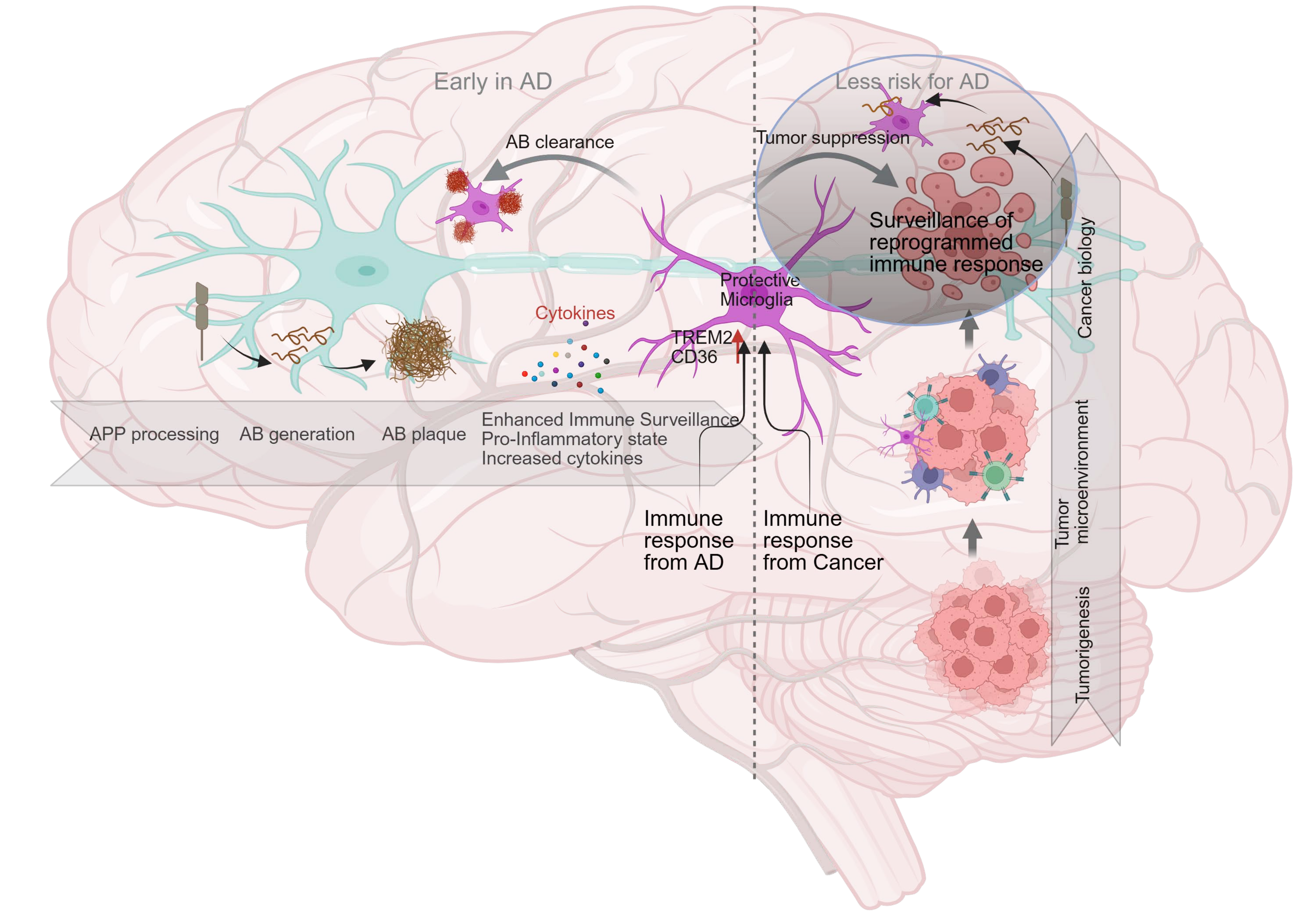
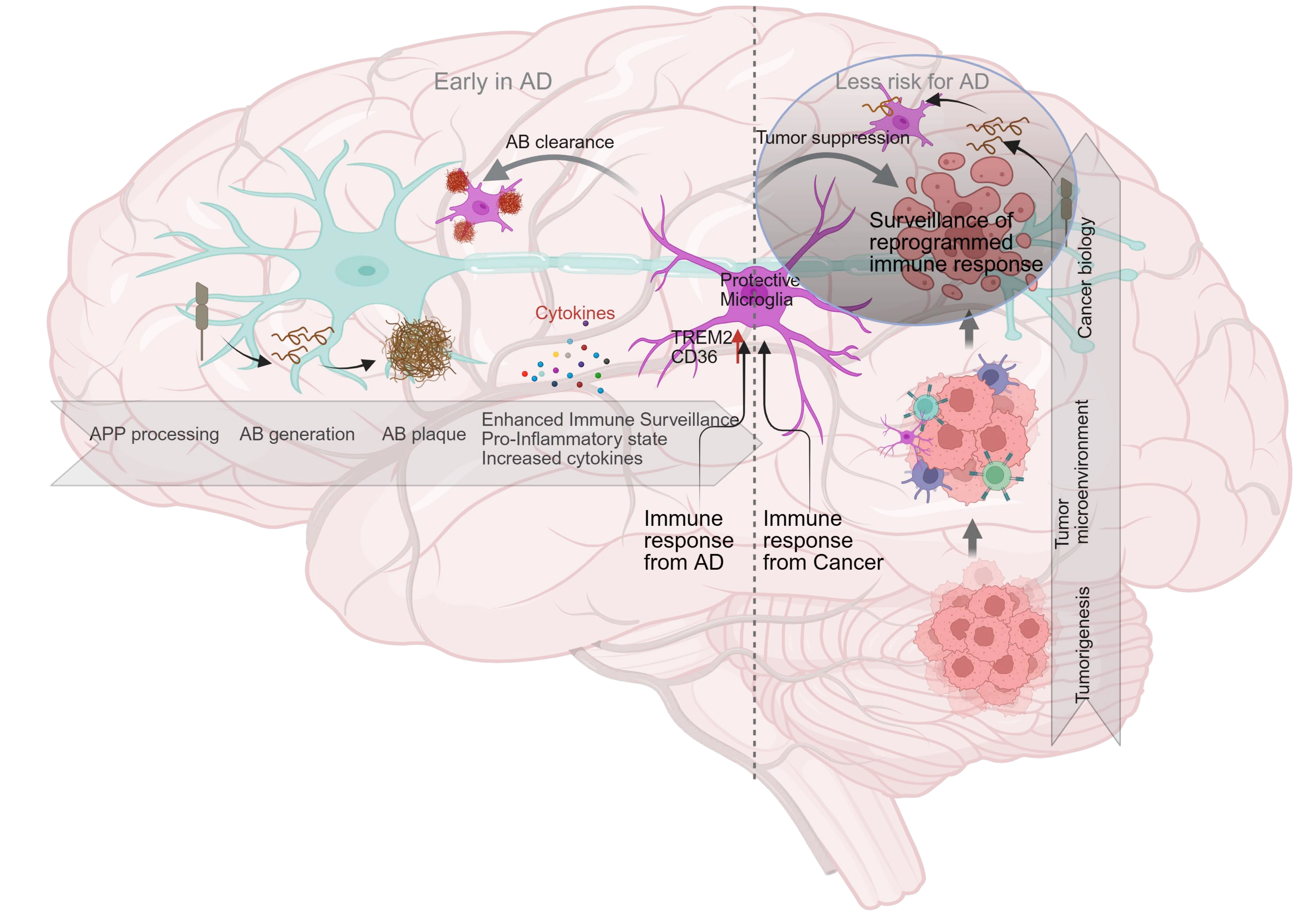
Figure 1. Microglia statuses in AD and lessons from anti-tumor microglial features. This diagram illustrates the divergent states of microglia in AD and the relationship with anti-tumor microglia through both shared and complementary mechanisms. Insights into the protective functions of anti-tumor microglia may also inform the development of novel therapeutic strategies for combating neurodegenerative disorders such as AD. It also highlights the need for future studies to elucidate the underlying molecular pathways and to develop comparative medicine approaches by targeting microglia. Created in BioRender. AD: Alzheimer’s disease; TREM2: triggering receptor expressed on myeloid cells 2; Aβ: amyloid-β; APP: amyloid precursor protein.
Acknowledgements
The authors acknowledge BioRender (www.BioRender.com) for providing scientific illustration tools.
Authors contribution
Cai JY, Vadlamudi Y: Conceptualization, writing-original draft preparation, writing-review & editing.
Agrawal P: Conceptualization, writing-review & editing.
All authors have read and agreed to the published version of the manuscript.
Conflicts of interest
Not applicable.
Ethical approval
Not applicable.
Consent to participate
Not applicable.
Consent for publication
Not applicable.
Availability of data and materials
Not applicable.
Funding
None.
Copyright
© The Author(s) 2026.
References
-
1. Ayyubova G. Dysfunctional microglia and tau pathology in Alzheimer’s disease. Rev Neurosci. 2023;34(4):443-458.[DOI]
-
2. Süß P, Schlachetzki JCM. Microglia in Alzheimer’s disease. Curr Alzheimer Res. 2020;17(1):29-43.[DOI]
-
3. Gao R, Wu H, Li Q, Zhao J. Roles of microglia in AD pathology. Curr Alzheimer Res. 2023;19(13):854-869.[DOI]
-
4. Thériault P, ElAli A, Rivest S. The dynamics of monocytes and microglia in Alzheimer’s disease. Alzheimers Res Ther. 2015;7(1):41.[DOI]
-
5. Streit WJ, Khoshbouei H, Bechmann I. Dystrophic microglia in late-onset Alzheimer’s disease. Glia. 2020;68(4):845-854.[DOI]
-
6. Merighi S, Nigro M, Travagli A, Gessi S. Microglia and Alzheimer’s disease. Int J Mol Sci. 2022;23(21):12990.[DOI]
-
7. Yeh FL, Hansen DV, Sheng M. TREM2, microglia, and neurodegenerative diseases. Trends Mol Med. 2017;23(6):512-533.[DOI]
-
8. AmeliMojarad M, AmeliMojarad M. The neuroinflammatory role of microglia in Alzheimer’s disease and their associated therapeutic targets. CNS Neurosci Ther. 2024;30(7):e14856.[DOI]
-
13. Wilkinson K, El Khoury J. Microglial scavenger receptors and their roles in the pathogenesis of Alzheimer’s disease. Int J Alzheimers Dis. 2012;2012:489456.[DOI]
-
17. Zhao R, Hu W, Tsai J, Li W, Gan WB. Microglia limit the expansion of β-amyloid plaques in a mouse model of Alzheimer’s disease. Mol Neurodegener. 2017;12(1):47.[DOI]
-
19. Miao J, Ma H, Yang Y, Liao Y, Lin C, Zheng J, et al. Microglia in Alzheimer’s disease: Pathogenesis, mechanisms, and therapeutic potentials. Front Aging Neurosci. 2023;15:1201982.[DOI]
-
27. Wang C, Zong S, Cui X, Wang X, Wu S, Wang L, et al. The effects of microglia-associated neuroinflammation on Alzheimer’s disease. Front Immunol. 2023;14:1117172.[DOI]
-
28. Chen Y, Yu Y. Tau and neuroinflammation in Alzheimer’s disease: Interplay mechanisms and clinical translation. J Neuroinflammation. 2023;20(1):165.[DOI]
-
29. Tang Y, Le W. Differential roles of M1 and M2 microglia in neurodegenerative diseases. Mol Neurobiol. 2016;53(2):1181-1194.[DOI]
-
37. Korvatska O, Leverenz JB, Jayadev S, McMillan P, Kurtz I, Guo X, et al. R47H variant of TREM2 associated with Alzheimer disease in a large late-onset family: Clinical, genetic, and neuropathological study. JAMA Neurol. 2015;72(8):920.[DOI]
-
39. Dijkstra JIR, Vermunt L, Venkatraghavan V, Ozhegov G, Coomans EM, Ossenkoppele R, et al. TREM2 risk variants and associated endophenotypes in Alzheimer’s disease. Alzheimers Res Ther. 2025;17(1):57.[DOI]
-
43. Casali BT, Reed-Geaghan EG. Microglial function and regulation during development, homeostasis and Alzheimer’s disease. Cells. 2021;10(4):957.[DOI]
-
45. Yin Z, Herron S, Silveira S, Kleemann K, Gauthier C, Mallah D, et al. Identification of a protective microglial state mediated by miR-155 and interferon-γ signaling in a mouse model of Alzheimer’s disease. Nat Neurosci. 2023;26(7):1196-1207.[DOI]
-
46. Wu X, Miller JA, Lee BTK, Wang Y, Ruedl C. Reducing microglial lipid load enhances β amyloid phagocytosis in an Alzheimer’s disease mouse model. Sci Adv. 2025;11(6):eadq6038.[DOI]
-
52. Chen X, Holtzman DM. Emerging roles of innate and adaptive immunity in Alzheimer’s disease. Immunity. 2022;55(12):2236-2254.[DOI]
-
57. Musicco M, Adorni F, Di Santo S, Prinelli F, Pettenati C, Caltagirone C, et al. Inverse occurrence of cancer and Alzheimer disease: A population-based incidence study. Neurology. 2013;81(4):322-328.[DOI]
-
59. Ganguli M. Cancer and dementia: It’s complicated. Alzheimer Dis Assoc Disord. 2015;29(2):177-182.[DOI]
-
60. Yarchoan M, James BD, Shah RC, Arvanitakis Z, Wilson RS, Schneider J, et al. Association of cancer history with Alzheimer’s disease dementia and neuropathology. J Alzheimers Dis. 2017;56(2):699-706.[DOI]
-
61. Aung KZ, Zin SS, Wu X, Myint ZW, Karanth S, Estus S, et al. Disentangling the inverse relationship between cancer and Alzheimer’s or Parkinson’s disease: A systematic review on Mendelian randomization studies. Neurobiol Dis. 2025;217:107190.[DOI]
-
62. Jeong SM, Jung W, Cho H, Choi HL, Jeon KH, Nam KW, et al. Alzheimer disease in breast cancer survivors. JAMA Netw Open. 2025;8(6):e2516468.[DOI]
-
68. Basak U, Sarkar T, Mukherjee S, Chakraborty S, Dutta A, Dutta S, et al. Tumor-associated macrophages: An effective player of the tumor microenvironment. Front Immunol. 2023;14:1295257.[DOI]
-
72. Sarkar S, Poon CC, Mirzaei R, Rawji KS, Hader W, Bose P, et al. Microglia induces Gas1 expression in human brain tumor-initiating cells to reduce tumorigenecity. Sci Rep. 2018;8(1):15286.[DOI]
-
73. Tao JC, Yu D, Shao W, Zhou DR, Wang Y, Hou SQ, et al. Interactions between microglia and glioma in tumor microenvironment. Front Oncol. 2023;13:1236268.[DOI]
-
74. Hambardzumyan D, Gutmann DH, Kettenmann H. The role of microglia and macrophages in glioma maintenance and progression. Nat Neurosci. 2016;19(1):20-27.[DOI]
-
75. Yi MH, Lee J, Moon S, So E, Bang G, Moon KS, et al. Divergent crosstalk between microglia and T cells in brain cancers: Implications for novel therapeutic strategies. Biomedicines. 2025;13(1):216.[DOI]
-
76. Ozga AJ, Chow MT, Luster AD. Chemokines and the immune response to cancer. Immunity. 2021;54(5):859-874.[DOI]
-
77. Tu S, Lin X, Qiu J, Zhou J, Wang H, Hu S, et al. Crosstalk between tumor-associated microglia/macrophages and CD8-positive T cells plays a key role in glioblastoma. Front Immunol. 2021;12:650105.[DOI]
-
78. Habibi MA, Nejati N, Najafi MB, Khodadadiyan A, Dashti M, Lorestani P, et al. Enhancing T cell infiltration in glioblastoma: A review article on challenges and therapeutic strategies. Cancer Treat Res Commun. 2025;45:100999.[DOI]
-
83. Chen J, Wu Q, Berglund AE, MacAulay RJ, Mulé JJ, Etame AB, et al. Tumor-associated macrophages in glioblastoma: Mechanisms of tumor progression and therapeutic strategies. Cells. 2025;14(18):1458.[DOI]
-
84. Przystal JM, Becker H, Canjuga D, Tsiami F, Anderle N, Keller AL, et al. Targeting CSF1R alone or in combination with PD1 in experimental glioma. Cancers. 2021;13(10):2400.[DOI]
-
88. Mayer MG, Fischer T. Microglia at the blood brain barrier in health and disease. Front Cell Neurosci. 2024;18:1360195.[DOI]
-
90. Feng Y, Hu X, Zhang Y, Wang Y. The role of microglia in brain metastases: Mechanisms and strategies. Aging Dis. 2024;15(1):169.[DOI]
-
91. Dong W, Sheng J, Cui JZM, Zhao H, Wong STC. Systems immunology insights into brain metastasis. Trends Immunol. 2024;45(11):903-916.[DOI]
-
92. Linnerbauer M, Wheeler MA, Quintana FJ. Astrocyte crosstalk in CNS inflammation. Neuron. 2020;108(4):608-622.[DOI]
-
93. Matejuk A, Ransohoff RM. Crosstalk between astrocytes and microglia: An overview. Front Immunol. 2020;11:1416.[DOI]
-
94. Marschallinger J, Iram T, Zardeneta M, Lee SE, Lehallier B, Haney MS, et al. Lipid-droplet-accumulating microglia represent a dysfunctional and proinflammatory state in the aging brain. Nat Neurosci. 2020;23(2):194-208.[DOI]
-
97. Dhapola R, Hota SS, Sarma P, Bhattacharyya A, Medhi B, Reddy DH, et al. Recent advances in molecular pathways and therapeutic implications targeting neuroinflammation for Alzheimer’s disease. Inflammopharmacology. 2021;29(6):1669-1681.[DOI]
-
98. Li Q, Wu Y, Chen J, Xuan A, Wang X. Microglia and immunotherapy in Alzheimer’s disease. Acta Neurol Scand. 2022;145(3):273-278.[DOI]
-
106. Pimenova AA, Herbinet M, Gupta I, Machlovi SI, Bowles KR, Marcora E, et al. Alzheimer’s-associated PU.1 expression levels regulate microglial inflammatory response. Neurobiol Dis. 2021;148:105217.[DOI]
-
107. Wei Z, Pan X, Cui X, Zhang J, Dai X, Zeng Y, et al. PU.1 dictates β-amyloid-induced TREM2 expression upregulation in microglia in a transgenic model of Alzheimer’s disease. Front Aging Neurosci. 2025;17:1537388.[DOI]
-
110. Weiskopf K. Cancer immunotherapy targeting the CD47/SIRPα axis. Eur J Cancer. 2017;76:100-109.[DOI]
-
117. He C, Chen B, Yang H, Zhou X. The dual role of microglia in Alzheimer’s disease: From immune regulation to pathological progression. Front Aging Neurosci. 2025;17:1554398.[DOI]
-
123. Riddell DR, Zhou H, Atchison K, Warwick HK, Atkinson PJ, Jefferson J, et al. Impact of apolipoprotein E (ApoE) polymorphism on brain ApoE levels. J Neurosci. 2008;28(45):11445-11453.[DOI]
-
127. Caffarel MM, Braza MS. Microglia and metastases to the central nervous system: Victim, ravager, or something else? J Exp Clin Cancer Res. 2022;41(1):327.[DOI]
Copyright
© The Author(s) 2026. This is an Open Access article licensed under a Creative Commons Attribution 4.0 International License (https://creativecommons.org/licenses/by/4.0/), which permits unrestricted use, sharing, adaptation, distribution and reproduction in any medium or format, for any purpose, even commercially, as long as you give appropriate credit to the original author(s) and the source, provide a link to the Creative Commons license, and indicate if changes were made.
Publisher’s Note
Science Exploration remains a neutral stance on jurisdictional claims in published
maps
and institutional affiliations. The views expressed in this article are solely those
of
the author(s) and do not reflect the opinions of the Editors or the publisher.
Share And Cite


Science Exploration Style
Cai JY, Vadlamudi Y, Agrawal P. Microglia statuses in Alzheimer’s disease and lessons from anti-tumor microglia. Geromedicine. 2026;2:202613. https://doi.org/10.70401/Geromedicine.2026.0033
Tips
Copy completed.
Submit a Manuscript
Author Instructions
Cite this Article
Article Metrics
0
View
0
Download
Cited
Article Updates

- Abstract
- Keywords
- 1. Introduction
- 2. Diverse Conditions of Microglia Against AD
- 3. Key Molecular Features of Microglia Against AD
- 4. Functional Loss and Changes of Microglia Contributing to AD
- 5. The Inverse Relationship between AD and Cancers
- 6. Microglia Conditions in Brain Tumors and Metastasis
- 7. Microglia in AD Versus Cancers from Molecular Signaling Perspective
- 8. Microglia in AD Versus Cancers from Metabolic Perspective
- 9. AD Intervention by Targeting Microglia and Lessons from Anti-Tumor Microglia
- 10. Summary
- Acknowledgements
- Authors contribution
- Conflicts of interest
- Ethical approval
- Consent to participate
- Consent for publication
- Availability of data and materials
- Funding
- References
- Copyright
 Get citation
Science Exploration Style

Cai JY, Vadlamudi Y, Agrawal P. Microglia statuses in Alzheimer’s disease and lessons from anti-tumor microglia. Geromedicine. 2026;2:202613. https://doi.org/10.70401/Geromedicine.2026.0033
copy
Share Link
copy