Senescent cell heterogeneity: The impact of targeting senescent endothelial cells in obesity

Masayoshi Suda
1,2,*

,
Tamar Tchkonia
1,2
,
James L. Kirkland
1,2,*
*Correspondence to:
James L. Kirkland, Center for Advanced Gerotherapeutics, Cedars-Sinai Health Sciences University, West Hollywood, CA 90048, USA; Division of Endocrinology, Diabetes, and Metabolism, Cedars-Sinai Health Sciences University, Los Angeles, CA 90048, USA.
E-mail: James.Kirkland@csmc.edu
Masayoshi Suda, Center for Advanced Gerotherapeutics, Cedars-Sinai Health Sciences University, West Hollywood, CA 90048, USA; Division of Endocrinology, Diabetes, and Metabolism, Cedars-Sinai Health Sciences University, Los Angeles, CA 90048, USA. E-mail: Masayoshi.Suda@csmc.edu
Masayoshi Suda, Center for Advanced Gerotherapeutics, Cedars-Sinai Health Sciences University, West Hollywood, CA 90048, USA; Division of Endocrinology, Diabetes, and Metabolism, Cedars-Sinai Health Sciences University, Los Angeles, CA 90048, USA. E-mail: Masayoshi.Suda@csmc.edu
Geromedicine. 2026;2:202610. 10.70401/Geromedicine.2026.0036
Received: February 18, 2026Accepted: September 01, 2026Published: September 02, 2026
Abstract
Cellular senescence is increasingly recognized as a key contributor to many age-related disorders and diseases, and senotherapeutics, therapies that target senescent cells (SCs), have indicated benefits in multiple preclinical models, including models of obesity, metabolic syndrome/diabetes, and their complications. Importantly, SCs are not a uniform population. They differ by originating cell type, stimuli by which senescence was induced, time since induction of senescence, and the tissue and microenvironment in which senescent cells are located, including the composition and magnitude of the senescence-associated secretory phenotype (SASP). Recent studies have indicated that senescent cell populations comprise cells that release inflammatory SASP factors and less inflammatory senescent cells that release growth factors. Senolytics, agents that eliminate senescent cells, preferentially remove the more inflammatory senescent cells while sparing the less inflammatory senescent cells that arise during processes such as acute wound healing. In another study, senescent cell heterogeneity was investigated in adipose tissue impacted by obesity, with a focus on senescence marker expression and cell-type specificity. p16+ senescent endothelial cells were a key source of SASP factors within adipose tissue, but p21+ senescent preadipocytes and immune cells also contributed substantially to the inflammatory milieu. Identifying and selectively targeting the more pathogenic, pro-inflammatory senescent cell populations may be facilitated by single-cell-level analyses to elucidate the extent of senescent cell heterogeneity and markers of different senescent cell subpopulations. Appreciating the diversity of senescent cells may be critical, since refining senotherapies based on specific SC subtypes may lead to the development of individualized and personalized therapies, potentially improving upon existing broader-spectrum strategies.
Keywords
Cellular senescence, senescent cell heterogeneity, endothelial cells, p16Ink4a, p21Cip1, senolytics, SASP factors, senotherapeutics
1. Introduction
Aging is a major risk factor for a wide range of inflammatory conditions, including disorders entailing adipose dysfunction, the geriatric syndromes, and multiple other disorders and diseases across the lifespan, with accumulating evidence indicating that cellular senescence is a fundamental biological process driving these outcomes[1-3]. Senescent cells (SCs) arise in response to diverse stressors, including DNA damage, metabolic stress, oxidative injury, lysosomal stress, and oncogenic signaling, and are characterized by essentially permanent cell-cycle arrest accompanied by a senescence-associated secretory phenotype (SASP)[1,4]. SCs cease proliferation and contribute to the loss of resilience and regenerative capacity in multiple organs and tissues. Moreover, through the secretion of pro-inflammatory cytokines, chemokines, growth factors, proteases, coding and non-coding nucleotides, bradykines, prostanoids, and other bioactive molecules, SCs can disrupt tissue homeostasis, promote fibrosis and chronic inflammation, impair tissue function, and contribute to the development of multiple pathologies[5].
Given that cellular senescence is interlinked with other hallmarks of aging, senotherapies that target SCs have emerged as a potential therapeutic strategy for a number of diseases as a root-cause therapeutic approach[6,7]. Several approaches have been explored to mitigate the detrimental effects of SCs, including interventions that suppress senescence, modulate the SASP (senomorphics), partial “reprogramming” of SCs, or selectively eliminating SCs (senolytics) (Figure 1)[8,9]. Interventions that suppress senescence aim to prevent cells from entering a senescent state by reducing cellular stress, attenuating DNA damage responses, or inhibiting upstream pathways that trigger cell-cycle arrest. Approaches such as telomere extension or modulation of p53 activity have demonstrated partial efficacy in delaying the onset of senescence[10-13]. Senomorphics are agents that reduce the production of SASP factors, the major mediators of the deleterious effects of senescent cells, and several candidates are currently being evaluated in planned clinical trials[14]. Partial reprogramming has emerged as a new approach, in which transient induction of rejuvenation-associated transcriptional programs can reverse aspects of the senescent phenotype[15,16]. In addition to these approaches, senolytics have attracted considerable attention[3,17-19]. The conceptual foundation for senolytic strategies originated from observations that interventions such as caloric restriction[20], which can extend healthspan and lifespan in mice[21] and monkeys[22], are associated with a reduced burden of SCs[23]. These findings in 2004 suggested that reducing SC burden may improve tissue function and counteract pathology. This initially prompted work to develop senolytic drugs[6] and subsequently prompted the generation of genetically modified mice, such as the p16-INK-ATTAC model, which was based on the ATTAC construct published in 2005[24] and in which elimination of cells with high p16Ink4a expression improved tissue function in progeroid mice[17]. The first published senolytics, dasatinib and quercetin (D+Q), improved function in naturally chronologically aged mice[23]. D+Q and other senolytics published subsequently, such as fisetin[25,26], navitoclax[18,27], and others[28,29], have also been tested in many preclinical models, indicating their ability to alleviate multiple disorders, diseases, and age-related phenotypes across tissues, including diabetes[30-39], liver diseases[40-44], idiopathic pulmonary fibrosis[45,46], osteoporosis[47-53], endometrial and uterine dysfunction[54,55], cancers[56-59], cardiovascular diseases[60-66], and neuropsychiatric disorders such as anxiety and dementia[3,23,42,67-70], Down syndrome[71-73], and geriatric syndromes including frailty[3,17,23,25,26,46], cognitive dysfunction[42,68], and resilience to infection[74-76]. Moreover, emerging clinical trial evidence indicates that clearance of SCs may have therapeutic potential to delay, prevent, or treat conditions associated with cellular senescence[46,49,77-79].
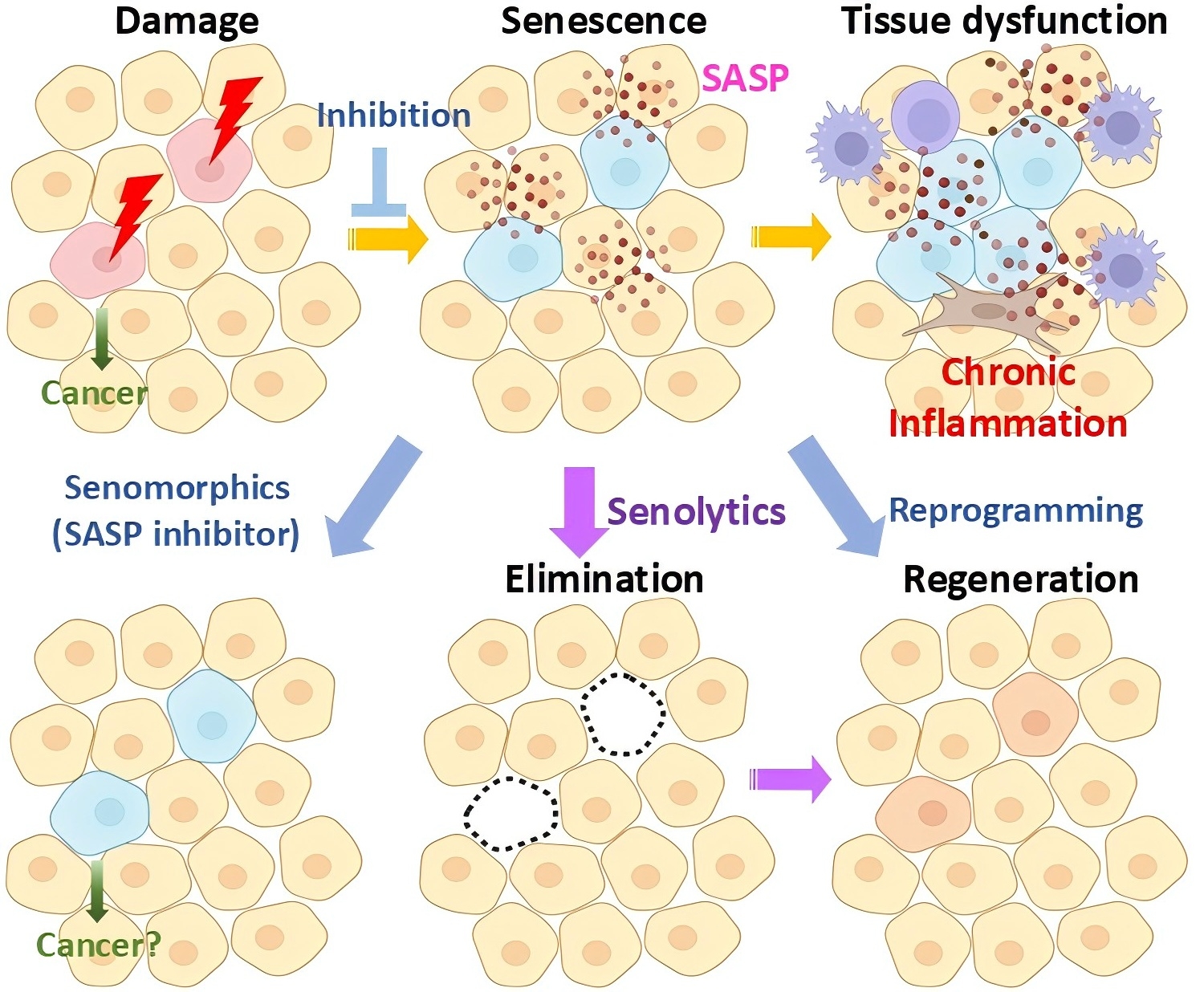
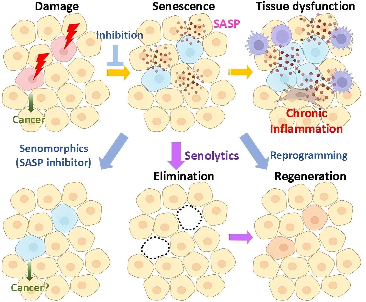
Figure 1. Therapeutic strategies for targeting senescent cells. SASP: senescence-associated secretory phenotype.
Cellular damage induces senescence, leading to stable cell-cycle arrest and the acquisition of a SASP, which can promote chronic inflammation, tissue dysfunction, and disease progression. Multiple therapeutic strategies have been proposed to target SCs. Senomorphics attenuate the deleterious effects of SCs by suppressing SASP factor production without eliminating the SCs themselves; examples include metformin, rapamycin, and the Janus kinase (JAK) inhibitor ruxolitinib. Senolytics selectively eliminate apoptosis-resistant, tissue-damaging SCs by targeting senescence-associated survival pathways, allowing intermittent “hit-and-run” dosing. Another approach involves Reprogramming, in which partial epigenetic reprogramming using Yamanaka factors (Oct4, Sox2, Klf4, and c-Myc (OSKM)) may reverse the senescent phenotype and restore proliferative capacity.
Importantly, SCs cells are not a uniform cell type. Such features of SCs, such as senescence markers[80] and SASP composition can differ depending on the cell type they originated from[81,82], the stimuli through which senescence was induced, anatomic site and their microenvironment[51,83], and how long they have been senescent (Figure 2)[84-86]. In particular, recent advances in single-cell analyses further demonstrate that senescent phenotypes differ even in chronological aging and age-related diseases such as obesity[31]. In this review, we summarize recent findings on the heterogeneity of SCs across cell types and physiological contexts and provide insights that may help overcome two major barriers in aging research: the lack of universal senescence markers and the limited efficacy of current senotherapies. The issue is that there is currently no universal marker that can identify SCs specifically and with high sensitivity. Although most senescence markers were originally established based on fundamental mechanisms of the biology of aging, many of them were derived from in vitro studies, and accumulating evidence now shows that they are not fully reliable in vivo. Recent studies suggest that different tissues may require distinct sets of senescence markers, reflecting the context-dependent nature of senescence[87,88]. This trend may, in fact, stem from the intrinsic heterogeneity of SCs. Another challenge is that some SCs, generally 30 to 70%, can exhibit resistance to currently available senolytics[23]. This implies that refining senolytic strategies based on specific senescent cell subtypes may point toward novel therapeutic opportunities, improve efficacy, and eventually lead to personalized therapies[17]. Understanding the contributions of distinct SC populations to inflammation and tissue dysfunction could help in developing targeted interventions and translating them into therapies for age-related conditions.
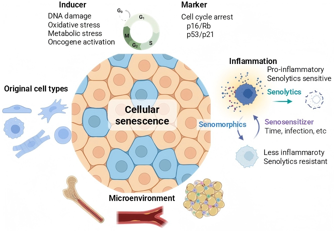
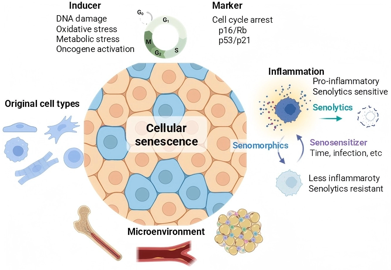
Figure 2. Heterogeneity of senescent cells. Created in BioRender. Kirkland, JL. (2026). https://BioRender.com/bhw56dv.
Cellular senescence is not a uniform biological state but instead comprises heterogeneous cell populations. SCs differ depending on senescence-inducing stimuli (e.g., DNA damage, oxidative stress, metabolic stress, oncogene activation), cellular origin, tissue and subcellular localization, microenvironmental cues, inflammatory state, and the expression of senescence-associated markers such as p16 and p21. Owing to this intrinsic heterogeneity, no single marker can universally define SCs across tissues or disease contexts. Recent advances in single-cell and spatial profiling technologies have enabled the resolution of distinct senescent subpopulations. This complexity has important implications for both the detection and therapeutic targeting of SCs, as the efficacy of senescence-targeting interventions, including senolytic strategies, may depend on the specific senescent cell populations being targeted.
2. Heterogeneity of Senescent Cells
Dasatinib preferentially targets senescent human preadipocytes, whereas quercetin targets senescent human endothelial cells, prompting their use as a combination[23,82]. Fisetin preferentially eliminates endothelial cells[25,26], further supporting the concept that senolytic sensitivity varies across SC types. Beyond cell type, recent evidence indicates that the inflammatory state of SCs influences susceptibility to senolytic interventions[82]. Even under controlled in vitro conditions, SCs exhibit substantial heterogeneity. For example, following senescence-inducing stimuli such as irradiation, senolytic treatment fails to eliminate all SCs: approximately 30-70% of SCs remain[23]. A comparative analysis of total SC populations vs. those SCs that persist after senolytic treatment (senolytic-resistant cells) revealed that the senolytic-resistant SCs had lower levels of pro-inflammatory/pro-apoptotic SASP factors such as CXCL1, CXCL5, and CXCL8 and mainly released growth factors, including platelet-derived growth factor AA (PDGF-AA)[82]. Importantly, experimentally inducing inflammation in the population of senolytic-resistant SCs using agents such as Toll-like receptor 3 (TLR3) agonists, termed senosensitizers, increased SC susceptibility to elimination by the senolytics that act by transiently disabling the SC anti-apoptotic pathways (SCAPs) that SCs use to defend themselves against pro-apoptotic factors in their SASP[14]. This suggests a causal link between the inflammatory state of SCs and their responsiveness to senolytics. Together, these findings indicate that SCs are heterogeneous with respect to their inflammatory/pro-apoptotic state, providing a mechanistic explanation for differential senolytic sensitivity and emphasizing the importance of defining functionally distinct SC subpopulations.
In addition to cellular origin and inflammatory state, SCs differ in the pathways that drive senescence and the markers SCs express[80]. The p16-INK-ATTAC mouse model was developed to allow selective elimination of p16Ink4a-positive SCs[17] based on indications that p16Ink4a is a senescence marker correlated with age-related tissue dysfunction[20,89]. Additional SC clearance models have also been generated that target other cell cycle arrest-associated molecules, including p19Arf and p21Cip1[90,91]. In mice, p19Arf is produced as an alternative transcript from the Cdkn2a locus and plays a role in regulating senescence[92]. p21Cip1 is a downstream effector of the p53 pathway that is involved in senescence induction[91]. Subsequently, SC-targeting models such as p19-diphtheria toxin receptor (DTR)[90] and p21-Cre/+; luciferase (LUC)/diphtheria toxin A (DTA) (PLD) mice[91] were developed. Clearance of SCs from these models led to attenuation of age-related phenotypes, indicating that SCs are causally involved in aging-associated dysfunction across diverse pathological contexts. Importantly, single-cell analyses using the p21-Cre/+; LUC/DTA model revealed that in adipose tissue from obese mice, p21-positive SCs accumulated and that p21-positive and p16-positive SCs were largely distinct populations[91].
In vitro studies further support this distinction, indicating that p21 expression transiently increases during the acute phase following senescence-inducing stress, subsequently declines, and then rises again during the chronic phase of senescence, whereas p16 expression predominantly increases during chronic senescence[93,94]. In the context of diet-induced metabolic stress, high-fat diet (HFD) feeding did not lead to a detectable increase in p16+ cells in p16 reporter mice, likely reflecting the relatively modest induction of p16 compared to that which occurs with aging or genotoxic stimuli as well as limited sensitivity in this non-amplifying reporter system[95]. Consistent with this, the metabolic benefits of SC elimination were relatively modest when targeting p16-positive SCs[32] compared to targeting p21[31,47,91], although it should be noted that these models are not directly comparable. Additionally, studies of radiation-induced osteoporosis indicated that selective clearance of p21-positive, but not p16-positive, SCs attenuated bone loss[47]. Based on these observations, p21-positive SCs have been considered more representative of early-phase, premature, or stress-induced senescence, such as that observed in obesity, while p16-positive SCs are thought to be more specific to chronological aging. Although more than 60 years have passed since cellular senescence was first described by Hayflick and Moorhead[96] and senolytic agents have already progressed to clinical trials[97], understanding of SC heterogeneity remains limited. This knowledge gap underscores the need for systematic, high-resolution approaches to define SC subtypes across tissues, disease states, and senescence-inducing stimuli. This knowledge gap is partly attributable to technical limitations of single-cell transcriptomic analyses, since Cdkn2a (p16) expression is often low and difficult to reliably detect by single-cell RNA sequencing[98]. These challenges provide a rationale for use of complementary technologies, such as RNA-in situ hybridization (ISH) and single-cell mass cytometry by time-of-flight (CyTOF)[53] to interrogate SC diversity in vivo at the protein level.
To address the emerging concept that SCs are not a uniform population, recent studies have applied single-cell proteomics approaches to define SC states in aged adipose tissue[26], the obese brain[67], and the aged skeletal and bone marrow microenvironments[53,99]. SCs were identified by cell cycle arrest (high p16 or p21 expression) in the absence of expression of proliferative markers (Ki-67 negativity). Within the stromal vascular fraction of gonadal white adipose tissue, both p16+ Ki67- and p21+ Ki-67- SC populations were increased. Across multiple cell types, p16+/p21+ double-positive cells-although representing a relatively small fraction of the total SC population-exhibited the highest levels of SASP expression[81], consistent with previous reports[91]. In hematopoietic cells (Cd45+) and preadipocytes, which constitute the majority of Pdgfrα+ cells in adipose tissue, single p21+ cells displayed relatively higher SASP expression than their single p16+ counterparts. In contrast, within the endothelial cell population, p16+ cells produced higher levels of SASP factors than p21+ cells, indicating a cell type-dependent pattern of senescent phenotypes (Figure 3). In the context of obesity, the population of p21+ SCs may be enriched in preadipocytes and immune cells; hence, targeting the p53/p21 pathway may exert a larger impact on adipose inflammation and systemic metabolism than targeting the p16 pathway[31,47,91]. In contrast, within the endothelial cell population, p16+ Ki-67- cells exhibited canonical hallmarks of senescence, including increased DNA damage markers such as γH2AX and robust expression of inflammatory SASP factors, including tumor necrosis factor-α (TNFα) and interleukin-6 (IL-6), whereas p21+ Ki-67- endothelial cells had minimal overlap with populations characterized by elevated γH2AX or high inflammatory SASP expression. Although p16 expression is technically challenging to evaluate using single-cell RNA sequencing and reliable antibodies for immunostaining in mouse tissues remain limited, these findings suggest that p16-based assessment may be particularly relevant for identifying functionally senescent endothelial cells in vivo. Accordingly, reliance solely on p21 or other senescence markers may underestimate the contribution of senescent endothelial cells to tissue inflammation and pathology. Given the heterogeneity of SCs, no single marker is sufficient to define cellular senescence. Although markers of cell cycle arrest are among the most useful indicators for identifying SCs, and p16, p19, or p21 reporter mice serve as valuable tools for tracing SCs in vivo, it remains essential to determine whether these cells exhibit additional defining features of senescence, such as persistent DNA damage (e.g., senescence-associated distension of satellites (SADS), telomere-associated foci (TAF), γH2AX) and production of SASP factors. The presence of these features implies the capacity to drive chronic inflammation and contribute to aging-associated pathologies. In this context, it is also important to assess auxiliary pathways linked to senescence, including lysosomal stress (as indicated by senescence-associated β-galactosidase (SA-β-gal) activity), elevated reactive oxygen species (ROS) levels, mitochondrial dysfunction, and activation of SCAPs. While cellular heterogeneity means that not all SCs uniformly express every marker, assaying only single parameters risks obscuring, rather than clarifying, the complexity of SC states. The Facilities for Geroscience Analysis (FGA), a laboratory core within the NIH-funded Translational Geroscience Network, provides standardized geroscience assays and analytical infrastructure[100]. Through this framework, aging-associated molecular features are comprehensively profiled in an unbiased manner, rather than relying on subjective or a priori-selected analyses of senescence-related molecules[46,49,77-79,101,102]. The Cellular Senescence Network (SenNet) Program, also supported by the National Institutes of Health (NIH), is generating data to comprehensively identify and characterize the differences among SCs across the organs, various states of human health, and the lifespan[88].
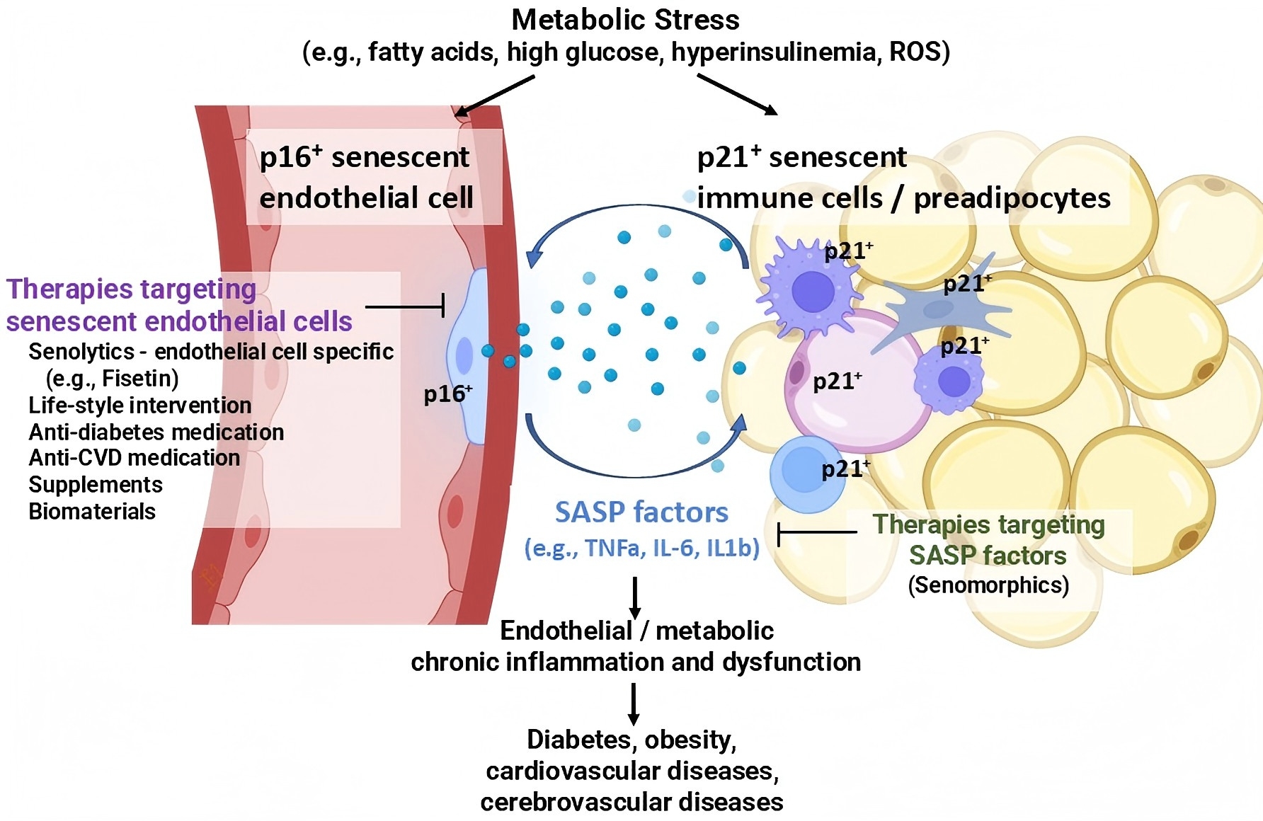
Figure 3. Heterogeneous contributions of senescent cell subpopulations to inflammation in obese adipose tissue. Created in BioRender. SASP: senescence-associated secretory phenotype; TNFα: tumor necrosis factor-α; IL-6: interleukin-6; ROS: reactive oxygen species.
In obese adipose tissue, p16-positive senescent endothelial cells are a major source of SASP factors, despite comprising a relatively small fraction of total cells. In parallel, p21-positive senescent preadipocytes and immune cells also contribute substantially to the inflammatory milieu[31,91]. p16/p21 double-positive senescent cells constitute only a minor subset of the overall senescent cell population yet have high expression of SASP factors[31,81,91]. These points highlight the heterogeneity of senescent cell populations and their distinct inflammatory contributions within adipose tissue[31,81,91]. SASP derived from senescent endothelial cells induces inflammation and secondary senescence in neighboring cells[81], while senescent cells of other lineages can, in turn, promote inflammatory activation in endothelial cells[31,91], potentially creating a vicious cycle that propagates chronic inflammation. For this reason, selectively targeting senescent endothelial cells may have therapeutic benefits not only for diabetes and obesity but also for other age-related conditions such as cardiovascular diseases, although future studies are needed to determine whether these effects extend beyond obesity, given the extensive involvement of the endothelium in regulating multiple organ systems throughout the body[81].
3. Senescent Endothelial Cells and Their Effects
Endothelial cells represent a significant cell population in the human body, comprising roughly 1-1.5% of total body mass and approximately 1013 cells. Lining the vasculature, they form a network that systemically regulates vascular function, tissue homeostasis[10,103-107], and inter-organ communication[108]. Endothelial cells are continuously exposed to circulating metabolic and inflammatory stimuli, as well as hemodynamic stresses, many of which promote senescence[106,109,110]. Across various disorders, distinct stressors induce endothelial oxidative stress, including diabetes (hyperglycemia and hyperinsulinemia)[111-113], dyslipidemia (metabolites such as free fatty acids and oxidized low-density lipoprotein (LDL))[114], and hypertension (angiotensin II)[115]. These factors increase ROS production in endothelial cells, leading to DNA damage and activation of senescence pathways, including p53/p21 and p16[4,107,116]. Senescent endothelial cells have impaired function, with reduced endothelial nitric oxide synthase (eNOS) activity and nitric oxide bioavailability[13,111,117-119]. In addition, senescent endothelial cells produce SASP factors and have upregulated adhesion molecules such as intercellular adhesion molecules (ICAM-1) that promote inflammatory cell recruitment and vascular inflammation. These changes contribute to vascular stiffening, atherosclerosis, and thrombosis[107,109]. Additionally, endothelial senescence is implicated not only in macrovascular conditions like cardiovascular and cerebrovascular diseases but also in systemic disorders through chronic inflammation and impaired microvascular homeostasis in a paracrine manner[104].
4. Targeting Senescent Endothelial Cells
A growing body of evidence indicates that endothelial senescence is a modifiable process and may be a therapeutic target. Indeed, several classes of drugs already used in clinical practice for cardiovascular and metabolic diseases have been shown to improve endothelial function and, in some contexts, attenuate endothelial cell senescence. These observations suggest that targeting senescent endothelial cells could have therapeutic benefit. Exercise[120], nitric oxide (NO) donors[111,121,122], renin-angiotensin system inhibitors, including angiotensin-converting enzyme (ACE) inhibitors and angiotensin II receptor blockers (ARBs)[123], as well as commonly used antidiabetic agents such as sodium-glucose cotransporter 2 (SGLT2) inhibitors[124], dipeptidyl peptidase-4 (DPP-4) inhibitors[125-127], and glucagon-like peptide 1 (GLP-1) receptor agonists[128,129], and statins[9,130,131] have all been reported to exert vascular-protective effects associated with improved endothelial cell function and homeostasis. In addition to conventional vascular-protective drugs, a number of senomorphic agents, including metformin[132,133], rapamycin, a mammalian target of rapamycin (mTOR) inhibitor, and JAK1/2 inhibition with ruxolitinib have been shown to modulate endothelial senescence[9,134]. Although these agents are not specifically designed to target endothelial senescence, accumulating evidence suggests that they can modulate pathways relevant to endothelial aging, supporting the concept that endothelial senescence represents a therapeutically tractable target. Indeed, several studies have reported that senolytic interventions alleviate endothelial senescence-associated dysfunction. For example, D+Q has been shown to enhance endothelial reparative functions[135], including cell migration and tube formation, and to improve endothelium-dependent vascular relaxation in vivo[23,61,136]. Most of these studies were conducted at the cellular level or relied on systemic administration of senolytics, in which effects on endothelial cells cannot be clearly distinguished from those on other cell types.
Recently, an inducible vascular senescent endothelial cell-specific depletion model, Tie2-Cre; p16-LOX-ATTAC mice was generated[81]. A loxP-flanked STOP cassette was introduced upstream of the ATTAC transgene in p16-INK-ATTAC mice, yielding the p16-LOX-ATTAC mouse model[51]. By crossing p16-LOX-ATTAC mice with Tie2-Cre mice, conditional elimination of p16-expressing senescent endothelial cells in this model was made feasible. Despite targeting a relatively small endothelial subpopulation, clearance of senescent endothelial cells resulted in reduced fat mass and lipid accumulation in gonadal white adipose tissue and improved glucose tolerance, which were associated with improvements in endothelial function such as upregulation of angiogenic factors and eNOS. Notably, AP20187 administration reduced the p16+ Ki-67- TNFα+, p16+ Ki67- IL-6+, and p16+ Ki-67- IL-1β+ cell fractions, suggesting that metabolic improvement associated with reduced chronic endothelial inflammation was sufficient to confer measurable therapeutic benefits (Figure 3). This raises the possibility that senescent endothelial cells play a particularly important role in obesity-related pathology, consistent with evidence linking endothelial dysfunction to metabolic disease[81] and that SCs can be categorized into pro-inflammatory and reparative subtypes[82]. While the effects of senolytics are often attributed to reducing overall SC burden to below a critical pathological threshold[97], this may be particularly relevant for endothelial senescent cells, which line the vasculature throughout the body and influence the function of multiple organs. An additional mechanism may involve the preferential elimination of disease-relevant, inflammatory SC subsets. Supporting this notion, SCs rendered non-inflammatory through inhibition of JAK or nuclear factor κB (NF-κB) signaling fail to induce secondary inflammatory responses in neighboring cells in vitro[81]. Also, it has been found that p21-Cre; RelA mice, in which NF-κB related inflammation in p21-positive SCs can be reduced, had decreased metabolic dysfunction[31,91]. In addition, it appears that senomorphics can alleviate age-related diseases.
Fisetin is a natural flavanol that we previously found to exhibit senolytic activity against endothelial SCs[25,136]. Fisetin reduced p16-positive cells, SA-β-gal-positive endothelial cells, and SASP factors in human fat tissue explants, with improvements in glucose tolerance and attenuation of inflammation in adipose tissue of obese mice[81]. D+Q and fisetin have been used in clinical trials of senolytics[97,137], with D+Q having broader senolytic activity across SC types than fisetin[25,26], but both approaches have been associated with preliminary changes in circulating senescence-associated and inflammatory biomarkers in humans[77,138]. The therapeutic value of selective versus broad senolytics likely depends on the underlying disease context. It will be important to define the heterogeneity of SCs across different pathological conditions in future studies. Moreover, although targeting pro-inflammatory SCs is paramount, not only senolytics but also senomorphics or other senotherapies may confer therapeutic benefit despite operating through fundamentally distinct mechanisms[9]. Notably, senolytics eliminate SCs, and because it takes time for SCs to re-accumulate, they can be administered intermittently, which may help reduce adverse effects. The approaches to suppress cellular senescence, including caloric restriction and various supplements are primarily preventive strategies and do not address SCs that have already been established, thereby requiring initiation at a relatively young age. In contrast, senolytics and cellular reprogramming may offer the advantage of being effective even when initiated later in life. Importantly, whereas many other interventions aim to modulate SCs or suppress the effects of SCs, senolytics eliminate these cells altogether, thereby also reducing the risk that SCs may later undergo malignant transformation (Figure 1).
5. Future Directions
Although p16-positive senescent endothelial cells have been identified as an inflammatory subpopulation that promotes chronic inflammation by secreting SASP factors and contributes to impaired glucose tolerance in obesity, several key questions remain unresolved. SCs are shaped by their inducing stimuli and tissue microenvironment. Hence, it is unclear whether the inflammatory phenotype of p16-positive endothelial cells observed in obesity is also conserved in aging. In vitro studies have indicated that conditioned media derived from senescent human umbilical vein endothelial cells (HUVECs) induced either by irradiation or by metabolic stressors, such as palmitic acid or high glucose, elicit inflammatory responses in non-senescent preadipocytes and HUVECs[81]. Prolonged exposure to these conditioned media subsequently promotes secondary senescence in recipient cells[81]. Although the underlying mechanisms appear to be shared across these distinct senescence-inducing stimuli, future in vivo studies will be essential to elucidate how these paracrine effects and pathogenic roles of senescent endothelial cells operate within complex tissue microenvironments.
Another important next step will be to determine whether the pro-inflammatory phenotype of p16-positive endothelial cells observed in adipose tissue is also present in other organs or disease settings beyond obesity. Although elimination of p16 positive senescent endothelial cells was not associated with overt large-vessel dysfunction or cardiac impairment in the obesity model, increases in eNOS expression and angiogenic factors were detected in the heart, suggesting that senescent endothelial cells may exert effects beyond adipose tissue and potentially influence broader cardiovascular physiology.
In addition, whether senescent endothelial cells can propagate inflammation to distant tissues such as the brain is a critical area for future investigation. Because neurons are non-regenerative, the use of senolytics for the central nervous system requires particular caution. An indirect or systemic mechanism of action of senolytics may be possible since several studies have reported cognitive benefits of senolytics despite their limited blood-brain barrier permeability[42,67,68,78,139]. Alternatively, improving vascular senescence, such as the blood-brain barrier, and reducing neuroinflammation might be considered beneficial for cognitive function[140,141].
Beyond endothelial cells, a major future challenge is to delineate the heterogeneity of SCs and specific SASP factors across multiple lineages, including adipocytes, immune cells, fibroblasts, and other vascular cell types[142]. Conditional deletion of Ercc1, a crucial DNA repair protein, in hematopoietic cells using Vav-iCre induced immunosenescence characterized by increased p16Ink4a/p21Cip1 expression and broad SASP factors, including monocyte chemoattractant protein 1 (MCP-1), TNF, IL-1β, IL-6, growth differentiation factor 15 (GDF-15), osteopontin, and β2-microglobulin[143]. This phenotype is associated with senescence and damage in multiple solid organs. In contrast, local senolysis in DMP1-Cre; p16-LOX-ATTAC mice improved selected skeletal outcomes and reduced the SASP transcriptomes such as IL-6, matrix metalloproteinase (MMPs), plasminogen activator inhibitor-1 (PAI-1), granulocyte-macrophage colony-stimulating factor (GM-CSF), TNFα, transforming growth factor-β (TGFβ), and glycoprotein nonmetastatic melanoma protein B (GPNMB), but only partially recapitulated the broader benefits of systemic senolytics[51]. It remains unclear whether the differing outcomes observed in these two studies reflect the systemic nature of immune cells versus the more localized role of osteocytes, differences in the overall burden of senescent cells, or variations in the experimental models themselves. Nevertheless, defining senescent cell populations and their lineage-specific SASP profiles for each cell type will be an important challenge for future research.
In studies of aged osteocytes, lineage-specific SASP profiles have been proposed, with factors such as IL-6, MMPs, PAI-1, sclerostin (SOST), fibroblast growth factor 23 (FGF23), and dickkopf-1 (DKK1) being associated with osteocyte senescence[51]. Because senescent cells can induce inflammatory responses in neighboring cells, it may be challenging to rigorously identify truly cell-type-specific SASP factors in vivo, particularly when relying on circulating biomarkers. Nevertheless, defining disease-specific senescence markers and establishing bona fide cell-specific SASP signatures remain important challenges for future research utilizing multiple single-cell analysis, cell-type-specific mouse models, and blood SASP factor analysis.
Moreover, it remains unclear whether senescence programs in terminally differentiated cells, such as cardiomyocytes, or in progenitor populations resemble those in proliferating cells and whether such SCs respond similarly to senolytic interventions. Importantly, physiological functions of heterogeneous SC populations remain largely undefined. In many cardiovascular diseases, senescence may not be strictly pathogenic. In pulmonary hypertension, elimination of SCs might contribute to disease progression[144], suggesting that SCs may promote protective vascular remodeling. In atherosclerosis, SCs may be of both inflammatory and fibrotic subtypes. The latter could contribute to plaque stabilization by increasing fibrous cap thickness. Similarly, in fibrotic conditions such as heart failure with preserved ejection fraction (HFpEF), pro-fibrotic SCs may represent an adaptive response that protects the heart from mechanical stress and prevents rupture, while excessive or pro-inflammatory SCs could exacerbate disease progression. Thus, while senolytic strategies hold promise, elucidating SC heterogeneity and identifying pathogenic, pro-inflammatory subpopulations could enable more selective targeting, thereby maximizing therapeutic benefit while preserving potentially adaptive senescent responses.
Future studies should also clarify how different classes of senotherapies,including dietary and exercise regimens, several supplements, cardiovascular and antidiabetic medications, senolytics, senomorphics, and biomaterial-based approaches,affect senescent endothelial cells in vivo. In particular, distinguishing whether these interventions exert direct effects on endothelial cells or act systemically through metabolic, inflammatory, or neurohumoral pathways, and determining whether their actions are endothelium-specific or broadly distributed across multiple tissues, will be essential for understanding their true therapeutic potential in vascular aging.
To further resolve the heterogeneity of SCs, future studies should incorporate emerging technologies capable of defining pathogenic senescent subpopulations in vivo. In addition to single-cell proteomics, spatial transcriptomics offers high-resolution mapping of SCs within their native tissue context, enabling the characterization of their interactions with neighboring cell types and their anatomical distribution across microenvironments. Moreover, selective clearance models such as the p16-Lox-ATTAC system provide a powerful in vivo tool to directly assess the functional contribution of SCs to tissue dysfunction and disease phenotypes.
6. Conclusions
SCs contribute to age-related diseases by inducing chronic inflammation through the secretion of SASP factors. Importantly, SCs are heterogeneous, and selectively targeting pro-inflammatory SC populations may be important to achieve therapeutic potential. Among the diverse cellular populations within tissues, endothelial cells represent a numerically modest compartment yet are ubiquitously distributed across organs and continuously exposed to a wide range of metabolic, inflammatory, and hemodynamic stimuli. Consequently, senescent endothelial cells may function as early responders and potent amplifiers of tissue inflammation and dysfunction despite their limited abundance. In obesity, p16-positive senescent endothelial cells exhibit a pronounced pro-inflammatory SASP, and genetic elimination of these cells or suppression of their senescence-associated secretory phenotype produces effects disproportionate to their frequency, resulting in improvements in obesity- and diabetes-associated metabolic pathologies.
In other cell types, including immune cells and preadipocytes, p21-positive cells have high expression of SASP factors in obesity, and p21 has therefore been considered a useful marker of obesity-associated senescence. However, in senescent endothelial cells, p21 expression does not necessarily reflect a population with prominent DNA damage or a highly inflammatory phenotype. These observations underscore the importance of SC heterogeneity and highlight the need for comprehensive strategies, including the use of multiple senescence markers and single-cell-level analyses, to accurately define pathogenic SC populations. As SC phenotypes and heterogeneity are likely to vary across tissues and biological contexts, further studies should determine how the distinct SC populations identified in obesity compare with those arising during physiological aging and in other age-related diseases. Accounting for such context-specific heterogeneity will be important when defining pathogenic SC populations and developing strategies to target them.
Authors contribution
Suda M, Tchkonia T, Kirkland JL: Conceptualization, writing-original draft, writing-review & editing.
All approved the final version to be published, and agree to be accountable for all aspects of the work.
Conflicts of interest
Tchkonia T and Kirkland JL have a financial interest related to this research, including patents and pending patents covering senolytic drugs and their uses held by Mayo Clinic. This research has been reviewed by the Mayo Clinic Conflict of Interest Review Board and was conducted in compliance with Mayo Clinic and Cedars-Sinai conflict of interest policies.
Ethical approval
Not applicable.
Consent to participate
Not applicable.
Consent for publication
Not applicable.
Availability of data and materials
Not applicable.
Funding
This work was supported by the US National Institutes of Health (NIH) (Grant Nos. R37AG013925, R33AG061456, and P01AG062413) (J.L.K.); the Connor Fund (J.L.K.); Robert J and Theresa W Ryan (J.L.K.); the Noaber Foundation (J.L.K.); the Hevolution Foundation [Grant Nos. HF-GRO-23-1199148-3 (J.L.K.) and HF-GRO-23-1199262-27 (M.S.)]; the American Heart Association Career Development Award [Grant No. 26CDA1451822 (M.S.)]; the Seth MacFarlane Foundation Innovator Award (M.S.); the JSPS Grants-in-Aid for Scientific Research Fund for the Promotion of Joint International Research (Fostering Joint International Research) [Grant No. 23KK0295 (M.S.)]; and the Yamada Science Foundation (M.S.).
Copyright
© The Author(s) 2026.
References
-
1. López-Otín C, Blasco MA, Partridge L, Serrano M, Kroemer G. Hallmarks of aging: An expanding universe. Cell. 2023;186(2):243-278.[DOI]
-
2. Kennedy BK, Berger SL, Brunet A, Campisi J, Cuervo AM, Epel ES, et al. Geroscience: Linking aging to chronic disease. Cell. 2014;159(4):709-713.[DOI]
-
3. Xu M, Pirtskhalava T, Farr JN, Weigand BM, Palmer AK, Weivoda MM, et al. Senolytics improve physical function and increase lifespan in old age. Nat Med. 2018;24(8):1246-1256.[DOI]
-
4. Shimizu I, Yoshida Y, Suda M, Minamino T. DNA damage response and metabolic disease. Cell Metab. 2014;20(6):967-977.[DOI]
-
5. Coppé JP, Desprez PY, Krtolica A, Campisi J. The senescence-associated secretory phenotype: The dark side of tumor suppression. Annu Rev Pathol. 2010;5:99-118.[DOI]
-
6. Kirkland JL, Tchkonia T. Senolytic drugs: From discovery to translation. J Intern Med. 2020;288(5):518-536.[DOI]
-
7. Wissler Gerdes EO, Zhu Y, Tchkonia T, Kirkland JL. Discovery, development, and future application of senolytics: Theories and predictions. FEBS J. 2020;287(12):2418-2427.[DOI]
-
8. Suda M, Paul KH, Tripathi U, Minamino T, Tchkonia T, Kirkland JL. Targeting cell senescence and senolytics: Novel interventions for age-related endocrine dysfunction. Endocr Rev. 2024;45(5):655-675.[DOI]
-
9. Lei Z, Pitcher LE, Prahalad V, Niedernhofer LJ, Robbins PD. Targeting cellular senescence with senotherapeutics: Senolytics and senomorphics. FEBS J. 2023;290(5):1362-1383.[DOI]
-
10. Mojiri A, Walther BK, Jiang C, Matrone G, Holgate R, Xu Q, et al. Telomerase therapy reverses vascular senescence and extends lifespan in progeria mice. Eur Heart J. 2021;42(42):4352-4369.[DOI]
-
11. Qin W, Castillo KD, Li H, Nguyen TKC, Kiss DL, Cooke JP, et al. Circular RNA telomerase reverses endothelial senescence in progeria. Aging Cell. 2025;24(6):e70021.[DOI]
-
12. Yoshida Y, Shimizu I, Katsuumi G, Shuang J, Suda M, Hayashi Y, et al. p53-Induced inflammation exacerbates cardiac dysfunction during pressure overload. J Mol Cell Cardiol. 2015;85:183-198.[DOI]
-
13. Yokoyama M, Shimizu I, Nagasawa A, Yoshida Y, Katsuumi G, Wakasugi T, et al. p53 plays a crucial role in endothelial dysfunction associated with hyperglycemia and ischemia. J Mol Cell Cardiol. 2019;129:105-117.[DOI]
-
14. Palmer AK, Spinelli R, Prata LGL, Chaib S, Suda M, Tchkonia T, et al. Senotherapeutics for metabolic disease and diabetic complications. J Intern Med. 2026;299(1):2-19.[DOI]
-
15. Ocampo A, Reddy P, Martinez-Redondo P, Platero-Luengo A, Hatanaka F, Hishida T, et al. In vivo amelioration of age-associated hallmarks by partial reprogramming. Cell. 2016;167(7):1719-1733.e12.[DOI]
-
16. Browder KC, Reddy P, Yamamoto M, Haghani A, Guillen IG, Sahu S, et al. In vivo partial reprogramming alters age-associated molecular changes during physiological aging in mice. Nat Aging. 2022;2(3):243-253.[DOI]
-
17. Baker DJ, Wijshake T, Tchkonia T, LeBrasseur NK, Childs BG, van de Sluis B, et al. Clearance of p16Ink4a-positive senescent cells delays ageing-associated disorders. Nature. 2011;479(7372):232-236.[DOI]
-
18. Zhu Y, Tchkonia T, Fuhrmann-Stroissnigg H, Dai HM, Ling YY, Stout MB, et al. Identification of a novel senolytic agent, navitoclax, targeting the Bcl-2 family of anti-apoptotic factors. Aging Cell. 2016;15(3):428-435.[DOI]
-
19. Chaib S, Palmer AK, Wyles SP, Musi N, Kirkland JL, Tchkonia T. Translating cellular senescence research into clinical practice for metabolic disease. Nat Rev Endocrinol. 2026;22(2):102-115.[DOI]
-
20. Krishnamurthy J, Torrice C, Ramsey MR, Kovalev GI, Al-Regaiey K, Su L, et al. Ink4a/Arf expression is a biomarker of aging. J Clin Invest. 2004;114(9):1299-1307.[DOI]
-
21. Weindruch R, Walford RL. Dietary restriction in mice beginning at 1 year of age: Effect on life-span and spontaneous cancer incidence. Science. 1982;215(4538):1415-1418.[DOI]
-
22. Mattison JA, Roth GS, Beasley TM, Tilmont EM, Handy AM, Herbert RL, et al. Impact of caloric restriction on health and survival in rhesus monkeys from the NIA study. Nature. 2012;489(7415):318-321.[DOI]
-
23. Zhu Y, Tchkonia T, Pirtskhalava T, Gower AC, Ding H, Giorgadze N, et al. The Achilles’ heel of senescent cells: From transcriptome to senolytic drugs. Aging Cell. 2015;14(4):644-658.[DOI]
-
25. Zhu Y, Doornebal EJ, Pirtskhalava T, Giorgadze N, Wentworth M, Fuhrmann-Stroissnigg H, et al. New agents that target senescent cells: The flavone, fisetin, and the BCL-XL inhibitors, A1331852 and A1155463. Aging. 2017;9(3):955-963.[DOI]
-
26. Yousefzadeh MJ, Zhu Y, McGowan SJ, Angelini L, Fuhrmann-Stroissnigg H, Xu M, et al. Fisetin is a senotherapeutic that extends health and lifespan. EBioMedicine. 2018;36:18-28.[DOI]
-
27. Chang J, Wang Y, Shao L, Laberge RM, Demaria M, Campisi J, et al. Clearance of senescent cells by ABT263 rejuvenates aged hematopoietic stem cells in mice. Nat Med. 2016;22(1):78-83.[DOI]
-
28. Fuhrmann-Stroissnigg H, Ling YY, Zhao J, McGowan SJ, Zhu Y, Brooks RW, et al. Identification of HSP90 inhibitors as a novel class of senolytics. Nat Commun. 2017;8:422.[DOI]
-
29. Tomimatsu N, Di Cristofaro LFM, Kanji S, Samentar L, Jordan BR, Kittler R, et al. Targeting cIAP2 in a novel senolytic strategy prevents glioblastoma recurrence after radiotherapy. EMBO Mol Med. 2025;17(4):6.[DOI]
-
30. Gunasekaran U, Gannon M. Type 2 diabetes and the aging pancreatic beta cell. Aging. 2011;3(6):565-575.[DOI]
-
32. Palmer AK, Xu M, Zhu Y, Pirtskhalava T, Weivoda MM, Hachfeld CM, et al. Targeting senescent cells alleviates obesity-induced metabolic dysfunction. Aging Cell. 2019;18(3):e12950.[DOI]
-
33. Conley SM, Hickson LJ, Kellogg TA, McKenzie T, Heimbach JK, Taner T, et al. Human obesity induces dysfunction and early senescence in adipose tissue-derived mesenchymal stromal/stem cells. Front Cell Dev Biol. 2020;8:197.[DOI]
-
34. Escande C, Nin V, Pirtskhalava T, Chini CC, Thereza Barbosa M, Mathison A, et al. Deleted in Breast Cancer 1 regulates cellular senescence during obesity. Aging Cell. 2014;13(5):951-953.[DOI]
-
35. Bian X, Griffin TP, Zhu X, Islam MN, Conley SM, Eirin A, et al. Senescence marker activin A is increased in human diabetic kidney disease: Association with kidney function and potential implications for therapy. BMJ Open Diab Res Care. 2019;7(1):e000720.[DOI]
-
36. Kim SR, Jiang K, Ogrodnik M, Chen X, Zhu XY, Lohmeier H, et al. Increased renal cellular senescence in murine high-fat diet: Effect of the senolytic drug quercetin. Transl Res. 2019;213:112-123.[DOI]
-
37. Xu M, Tchkonia T, Ding H, Ogrodnik M, Lubbers ER, Pirtskhalava T, et al. JAK inhibition alleviates the cellular senescence-associated secretory phenotype and frailty in old age. Proc Natl Acad Sci U S A. 2015;112(46):E6301-E6310.[DOI]
-
38. Xu M, Palmer AK, Ding H, Weivoda MM, Pirtskhalava T, White TA, et al. Targeting senescent cells enhances adipogenesis and metabolic function in old age. eLife. 2015;4:e12997.[DOI]
-
39. Katsuumi G, Shimizu I, Suda M, Yoshida Y, Furihata T, Joki Y, et al. SGLT2 inhibition eliminates senescent cells and alleviates pathological aging. Nat Aging. 2024;4(7):926-938.[DOI]
-
40. Amor C, Feucht J, Leibold J, Ho YJ, Zhu C, Alonso-Curbelo D, et al. Senolytic CAR T cells reverse senescence-associated pathologies. Nature. 2020;583(7814):127-132.[DOI]
-
41. Ogrodnik M, Miwa S, Tchkonia T, Tiniakos D, Wilson CL, Lahat A, et al. Cellular senescence drives age-dependent hepatic steatosis. Nat Commun. 2017;8:15691.[DOI]
-
42. Ogrodnik M, Evans SA, Fielder E, Victorelli S, Kruger P, Salmonowicz H, et al. Whole-body senescent cell clearance alleviates age-related brain inflammation and cognitive impairment in mice. Aging Cell. 2021;20(2):e13296.[DOI]
-
43. Meijnikman AS, van Olden CC, Aydin Ö, Herrema H, Kaminska D, Lappa D, et al. Hyperinsulinemia is highly associated with markers of hepatocytic senescence in two independent cohorts. Diabetes. 2022;71(9):1929-1936.[DOI]
-
44. Baboota RK, Rawshani A, Bonnet L, Li X, Yang H, Mardinoglu A, et al. BMP4 and Gremlin 1 regulate hepatic cell senescence during clinical progression of NAFLD/NASH. Nat Metab. 2022;4(8):1007-1021.[DOI]
-
45. Schafer MJ, White TA, Iijima K, Haak AJ, Ligresti G, Atkinson EJ, et al. Cellular senescence mediates fibrotic pulmonary disease. Nat Commun. 2017;8(1):14532.[DOI]
-
46. Justice JN, Nambiar AM, Tchkonia T, LeBrasseur NK, Pascual R, Hashmi SK, et al. Senolytics in idiopathic pulmonary fibrosis: Results from a first-in-human, open-label, pilot study. EBioMedicine. 2019;40:554-563.[DOI]
-
47. Chandra A, Lagnado AB, Farr JN, Doolittle M, Tchkonia T, Kirkland JL, et al. Targeted clearance of p21- but not p16-positive senescent cells prevents radiation-induced osteoporosis and increased marrow adiposity. Aging Cell. 2022;21(5):e13602.[DOI]
-
48. Chandra A, Lagnado AB, Farr JN, Monroe DG, Park S, Hachfeld C, et al. Targeted reduction of senescent cell burden alleviates focal radiotherapy-related bone loss. J Bone Miner Res. 2020;35(6):1119-1131.[DOI]
-
49. Farr JN, Atkinson EJ, Achenbach SJ, Volkman TL, Tweed AJ, Vos SJ, et al. Effects of intermittent senolytic therapy on bone metabolism in postmenopausal women: A phase 2 randomized controlled trial. Nat Med. 2024;30(9):2605-2612.[DOI]
-
51. Farr JN, Saul D, Doolittle ML, Kaur J, Rowsey JL, Vos SJ, et al. Local senolysis in aged mice only partially replicates the benefits of systemic senolysis. J Clin Invest. 2023;133(8):e162519.[DOI]
-
52. Farr JN, Xu M, Weivoda MM, Monroe DG, Fraser DG, Onken JL, et al. Targeting cellular senescence prevents age-related bone loss in mice. Nat Med. 2017;23(9):1072-1079.[DOI]
-
53. Doolittle ML, Saul D, Kaur J, Rowsey JL, Vos SJ, Pavelko KD, et al. Multiparametric senescent cell phenotyping reveals targets of senolytic therapy in the aged murine skeleton. Nat Commun. 2023;14:4587.[DOI]
-
54. Cavalcante MB, Saccon TD, Nunes ADC, Kirkland JL, Tchkonia T, Schneider A, et al. Dasatinib plus quercetin prevents uterine age-related dysfunction and fibrosis in mice. Aging. 2020;12(3):2711-2722.[DOI]
-
55. Suvakov S, Cubro H, White WM, Butler Tobah YS, Weissgerber TL, Jordan KL, et al. Targeting senescence improves angiogenic potential of adipose-derived mesenchymal stem cells in patients with preeclampsia. Biol Sex Differ. 2019;10(1):49.[DOI]
-
56. Cupit-Link MC, Kirkland JL, Ness KK, Armstrong GT, Tchkonia T, LeBrasseur NK, et al. Biology of premature ageing in survivors of cancer. ESMO Open. 2017;2(5):e000250.[DOI]
-
57. Dimri GP, Lee X, Basile G, Acosta M, Scott G, Roskelley C, et al. A biomarker that identifies senescent human cells in culture and in aging skin in vivo. Proc Natl Acad Sci U S A. 1995;92(20):9363-9367.[DOI]
-
58. Wang C, Vegna S, Jin H, Benedict B, Lieftink C, Ramirez C, et al. Inducing and exploiting vulnerabilities for the treatment of liver cancer. Nature. 2019;574(7777):268-272.[DOI]
-
59. Wakita M, Takahashi A, Sano O, Loo TM, Imai Y, Narukawa M, et al. A BET family protein degrader provokes senolysis by targeting NHEJ and autophagy in senescent cells. Nat Commun. 2020;11:1935.[DOI]
-
60. Kim SR, Zou X, Tang H, Puranik AS, Abumoawad AM, Zhu XY, et al. Increased cellular senescence in the murine and human stenotic kidney: Effect of mesenchymal stem cells. J Cell Physiol. 2021;236(2):1332-1344.[DOI]
-
61. Roos CM, Zhang B, Palmer AK, Ogrodnik MB, Pirtskhalava T, Thalji NM, et al. Chronic senolytic treatment alleviates established vasomotor dysfunction in aged or atherosclerotic mice. Aging Cell. 2016;15(5):973-977.[DOI]
-
62. Lewis-McDougall FC, Ruchaya PJ, Domenjo-Vila E, Shin Teoh T, Prata L, Cottle BJ, et al. Aged-senescent cells contribute to impaired heart regeneration. Aging Cell. 2019;18(3):e12931.[DOI]
-
64. Suda M, Shimizu I, Katsuumi G, Yoshida Y, Hayashi Y, Ikegami R, et al. Senolytic vaccination improves normal and pathological age-related phenotypes and increases lifespan in progeroid mice. Nat Aging. 2021;1(12):1117-1126.[DOI]
-
65. Suda M, Shimizu I, Katsuumi G, Hsiao CL, Yoshida Y, Matsumoto N, et al. Glycoprotein nonmetastatic melanoma protein B regulates lysosomal integrity and lifespan of senescent cells. Sci Rep. 2022;12:6522.[DOI]
-
66. Suda M, Tchkonia T, Kirkland JL, Minamino T. Targeting senescent cells for the treatment of age-associated diseases. J Biochem. 2024;177(3):177-187.[DOI]
-
68. Bussian TJ, Aziz A, Meyer CF, Swenson BL, van Deursen JM, Baker DJ. Clearance of senescent glial cells prevents tau-dependent pathology and cognitive decline. Nature. 2018;562(7728):578-582.[DOI]
-
69. Musi N, Valentine JM, Sickora KR, Baeuerle E, Thompson CS, Shen Q, et al. Tau protein aggregation is associated with cellular senescence in the brain. Aging Cell. 2018;17(6):e12840.[DOI]
-
70. Zhu Y, Prata LGPL, Gerdes EOW, Netto JME, Pirtskhalava T, Giorgadze N, et al. Orally-active, clinically-translatable senolytics restore α-Klotho in mice and humans. EBioMedicine. 2022;77:103912.[DOI]
-
71. Meharena HS, Marco A, Dileep V, Lockshin ER, Akatsu GY, Mullahoo J, et al. Down-syndrome-induced senescence disrupts the nuclear architecture of neural progenitors. Cell Stem Cell. 2022;29(1):116-130.E7.[DOI]
-
72. Rueda N, Vidal V, García-Cerro S, Narcís JO, Llorens-Martín M, Corrales A, et al. Anti-IL17 treatment ameliorates Down syndrome phenotypes in mice. Brain Behav Immun. 2018;73:235-251.[DOI]
-
74. Lagnado A, Leslie J, Ruchaud-Sparagano M, Victorelli S, Hirsova P, Ogrodnik M, et al. Neutrophils induce paracrine telomere dysfunction and senescence in ROS-dependent manner. EMBO J. 2021;40(9):EMBJ2020106048.[DOI]
-
75. Camell CD, Yousefzadeh MJ, Zhu Y, Prata LGPL, Huggins MA, Pierson M, et al. Senolytics reduce coronavirus-related mortality in old mice. Science. 2021;373(6552):eabe4832.[DOI]
-
77. Hickson LJ, Langhi Prata LGP, Bobart SA, Evans TK, Giorgadze N, Hashmi SK, et al. Senolytics decrease senescent cells in humans: Preliminary report from a clinical trial of Dasatinib plus Quercetin in individuals with diabetic kidney disease. EBioMedicine. 2019;47:446-456.[DOI]
-
78. Gonzales MM, Garbarino VR, Kautz TF, Palavicini JP, Lopez-Cruzan M, Dehkordi SK, et al. Senolytic therapy in mild Alzheimer’s disease: A phase 1 feasibility trial. Nat Med. 2023;29(10):2481-2488.[DOI]
-
79. Millar CL, Iloputaife I, Baldyga K, Norling AM, Boulougoura A, Vichos T, et al. A pilot study of senolytics to improve cognition and mobility in older adults at risk for Alzheimer’s disease. EBioMedicine. 2025;113:105612.[DOI]
-
80. Saul D, Jurk D, Doolittle ML, Kosinsky RL, Han Y, Zhang X, et al. Distinct senotypes in p16- and p21-positive cells across human and mouse aging tissues. EMBO J. 2025;44(23):19.[DOI]
-
81. Suda M, Chaib S, Langhi Prata LGP, Zhu Y, Tripathi U, Paul KH, et al. Endothelial senescent-cell-specific clearance alleviates metabolic dysfunction in obese mice. Cell Metab. 2025;37(12):2455-2465.e6.[DOI]
-
82. Tripathi U, Suda M, Kulshreshtha V, Piatkowski BT, Palmer AK, Giorgadze N, et al. Senolytic-resistant senescent cells have a distinct SASP profile and functional impact: The path to developing senosensitizers. Aging Cell. 2026;25:e70358.[DOI]
-
83. Yousefzadeh MJ, Jing Z, Bukata C, Wade EA, McGowan SJ, Angelini LA, et al. Tissue specificity of senescent cell accumulation during physiologic and accelerated aging of mice. Aging Cell. 2020;19(3):e13094.[DOI]
-
84. Neri F, Zheng S, Watson MA, Desprez PY, Gerencser AA, Campisi J, et al. Senescent cell heterogeneity and responses to senolytic treatment are related to cell cycle status during senescence induction. Aging. 2025;17(8):2063-2078.[DOI]
-
85. Cohn RL, Gasek NS, Kuchel GA, Xu M. The heterogeneity of cellular senescence: Insights at the single-cell level. Trends Cell Biol. 2023;33(1):9-17.[DOI]
-
86. Varela-Eirín M, Demaria M. Cellular senescence. Curr Biol. 2022;32(10):R448-R452.[DOI]
-
87. Ogrodnik M, Carlos Acosta J, Adams PD, d’Adda di Fagagna F, Baker DJ, Bishop CL, et al. Guidelines for minimal information on cellular senescence experimentation in vivo. Cell. 2024;187(16):4150-4175.[DOI]
-
88. Suryadevara V, Hudgins AD, Rajesh A, Pappalardo A, Karpova A, Dey AK, et al. SenNet recommendations for detecting senescent cells in different tissues. Nat Rev Mol Cell Biol. 2024;25(12):1001-1023.[DOI]
-
89. Sharpless NE, Sherr CJ. Forging a signature of in vivo senescence. Nat Rev Cancer. 2015;15(7):397-408.[DOI]
-
90. Hashimoto M, Asai A, Kawagishi H, Mikawa R, Iwashita Y, Kanayama K, et al. Elimination of p19ARF-expressing cells enhances pulmonary function in mice. JCI Insight. 2016;1(12):e87732.[DOI]
-
91. Wang B, Wang L, Gasek NS, Zhou Y, Kim T, Guo C, et al. An inducible p21-Cre mouse model to monitor and manipulate p21-highly-expressing senescent cells in vivo. Nat Aging. 2021;1(10):962-973.[DOI]
-
92. Sharpless NE, Ramsey MR, Balasubramanian P, Castrillon DH, DePinho RA. The differential impact of p16INK4a or p19ARF deficiency on cell growth and tumorigenesis. Oncogene. 2004;23(2):379-385.[DOI]
-
93. Hsu CH, Altschuler SJ, Wu LF. Patterns of early p21 dynamics determine proliferation-senescence cell fate after chemotherapy. Cell. 2019;178(2):361-373.e12.[DOI]
-
94. Sturmlechner I, Zhang C, Sine CC, van Deursen EJ, Jeganathan KB, Hamada N, et al. p21 produces a bioactive secretome that places stressed cells under immunosurveillance. Science. 2021;374(6567):eabb3420.[DOI]
-
95. Sorrentino JA, Krishnamurthy J, Tilley S, Alb JG Jr, Burd CE, Sharpless NE. p16INK4a reporter mice reveal age-promoting effects of environmental toxicants. J Clin Invest. 2014;124(1):169-173.[DOI]
-
97. Chaib S, Tchkonia T, Kirkland JL. Cellular senescence and senolytics: The path to the clinic. Nat Med. 2022;28(8):1556-1568.[DOI]
-
98. Saul D, Kosinsky RL, Atkinson EJ, Doolittle ML, Zhang X, LeBrasseur NK, et al. A new gene set identifies senescent cells and predicts senescence-associated pathways across tissues. Nat Commun. 2022;13(1):4827.[DOI]
-
99. Samakkarnthai P, Saul D, Lei Z, Aversa Z, Doolittle ML, Sfeir JG, et al. In vitro and in vivo effects of zoledronic acid on senescence and senescence-associated secretory phenotype markers. Aging. 2023;15(9):3331-3355.[DOI]
-
100. Tchkonia T, Kritchevsky SB, Kuchel GA, Kirkland JL. NIA Translational Geroscience Network: An infrastructure to facilitate geroscience-guided clinical trials. J Am Geriatr Soc. 2024;72(5):1605-1607.[DOI]
-
101. Garbarino VR, Palavicini JP, Melendez J, Barthelemy NR, He Y, Kautz TF, et al. Evaluation of exploratory fluid biomarkers from a phase 1 senolytic trial in mild Alzheimer’s disease. Neurotherapeutics. 2025;22(4):e00591.[DOI]
-
102. Nambiar A, Kellogg D III, Justice J, Goros M, Gelfond J, Pascual R, et al. Senolytics dasatinib and quercetin in idiopathic pulmonary fibrosis: Results of a phase I, single-blind, single-center, randomized, placebo-controlled pilot trial on feasibility and tolerability. EBioMedicine. 2023;90:104481.[DOI]
-
103. Rodriguez Morales D, Larcher V, Ruz Jurado M, Arifaj D, Tombor LS, Zanders L, et al. Vascular niches are the primary hotspots in cardiac aging. Circ Res. 2025;137(11):1353-1367.[DOI]
-
104. Wagner JUG, Tombor LS, Malacarne PF, Kettenhausen LM, Panthel J, Kujundzic H, et al. Aging impairs the neurovascular interface in the heart. Science. 2023;381(6660):897-906.[DOI]
-
106. Bloom SI, Islam MT, Lesniewski LA, Donato AJ. Mechanisms and consequences of endothelial cell senescence. Nat Rev Cardiol. 2023;20(1):38-51.[DOI]
-
107. Kotla S, Vu HT, Ko KA, Wang Y, Imanishi M, Heo KS, et al. Endothelial senescence is induced by phosphorylation and nuclear export of telomeric repeat binding factor 2–interacting protein. JCI Insight. 2019;4(9):e124867.[DOI]
-
108. Khazaei M, Moien-afshari F, Laher I. Vascular endothelial function in health and diseases. Pathophysiology. 2008;15(1):49-67.[DOI]
-
109. Meijles DN, Sahoo S, Al Ghouleh I, Amaral JH, Bienes-Martinez R, Knupp HE, et al. The matricellular protein TSP1 promotes human and mouse endothelial cell senescence through CD47 and Nox1. Sci Signal. 2017;10(501):eaaj1784.[DOI]
-
110. Suda M, Shimizu I, Yoshida Y, Katsuumi G, Minamino T. Endothelial cell dysfunction and senescence: Biologic mechanisms and hemodynamic consequences. In: Chirinos JA, editor. Textbook of arterial stiffness and pulsatile hemodynamics in health and disease. Amsterdam: Elsevier; 2022. p. 359-367.[DOI]
-
112. Miyauchi H, Minamino T, Tateno K, Kunieda T, Toko H, Komuro I. Akt negatively regulates the in vitro lifespan of human endothelial cells via a p53/p21-dependent pathway. EMBO J. 2004;23(1):212-220.[DOI]
-
113. Baumgartner-Parzer S, Waldhäusl W. The endothelium as a metabolic and endocrine organ: Its relation with insulin resistance. Exp Clin Endocrinol Diabetes. 2001;109(Suppl 2):S166-S179.[DOI]
-
114. Rosso A, Balsamo A, Gambino R, Dentelli P, Falcioni R, Cassader M, et al. p53 mediates the accelerated onset of senescence of endothelial progenitor cells in diabetes. J Biol Chem. 2006;281(7):4339-4347.[DOI]
-
115. Jia G, Aroor AR, Jia C, Sowers JR. Endothelial cell senescence in aging-related vascular dysfunction. Biochim Biophys Acta Mol Basis Dis. 2019;1865(7):1802-1809.[DOI]
-
116. Fukai T, Ushio-Fukai M. Superoxide dismutases: Role in redox signaling, vascular function, and diseases. Antioxid Redox Signal. 2011;15(6):1583-1606.[DOI]
-
117. Minamino T, Miyauchi H, Yoshida T, Ishida Y, Yoshida H, Komuro I. Endothelial cell senescence in human atherosclerosis: Role of telomere in endothelial dysfunction. Circulation. 2002;105(13):1541-1544.[DOI]
-
118. Smith AR, Visioli F, Frei B, Hagen TM. Age-related changes in endothelial nitric oxide synthase phosphorylation and nitric oxide dependent vasodilation: Evidence for a novel mechanism involving sphingomyelinase and ceramide-activated phosphatase 2A. Aging Cell. 2006;5(5):391-400.
-
119. Wagner JUG, Chavakis E, Rogg EM, Muhly-Reinholz M, Glaser SF, Günther S, et al. Switch in laminin β2 to laminin β1 isoforms during aging controls endothelial cell functions: Brief report. Arterioscler Thromb Vasc Biol. 2018;38(5):1170-1177.[DOI]
-
120. Gao J, Pan X, Li G, Chatterjee E, Xiao J. Physical exercise protects against endothelial dysfunction in cardiovascular and metabolic diseases. J Cardiovasc Transl Res. 2022;15(3):604-620.[DOI]
-
121. Paulo M, Costa DEFR, Bonaventura D, Lunardi CN, Bendhack LM. Nitric oxide donors as potential drugs for the treatment of vascular diseases due to endothelium dysfunction. Curr Pharm Des. 2020;26(30):3748-3759.[DOI]
-
122. Hayashi T, Yano K, Matsui-Hirai H, Yokoo H, Hattori Y, Iguchi A. Nitric oxide and endothelial cellular senescence. Pharmacol Ther. 2008;120(3):333-339.[DOI]
-
123. Enseleit F, Hürlimann D, Lüscher TF. Vascular protective effects of angiotensin converting enzyme inhibitors and their relation to clinical events. J Cardiovasc Pharmacol. 2001;37:S21-S30.[DOI]
-
124. Tai S, Zhou Y, Fu L, Ding H, Zhou Y, Yin Z, et al. Dapagliflozin impedes endothelial cell senescence by activating the SIRT1 signaling pathway in type 2 diabetes. Heliyon. 2023;9(8):e19152.[DOI]
-
125. Gao P, Li L, Wei X, Wang M, Hong Y, Wu H, et al. Activation of transient receptor potential channel vanilloid 4 by DPP-4 (dipeptidyl peptidase-4) inhibitor vildagliptin protects against diabetic endothelial dysfunction. Hypertension. 2020;75(1):150-162.[DOI]
-
126. Chen Z, Yu J, Fu M, Dong R, Yang Y, Luo J, et al. Dipeptidyl peptidase-4 inhibition improves endothelial senescence by activating AMPK/SIRT1/Nrf2 signaling pathway. Biochem Pharmacol. 2020;177:113951.[DOI]
-
127. Xin M, Jin X, Cui X, Jin C, Piao L, Wan Y, et al. Dipeptidyl peptidase-4 inhibition prevents vascular aging in mice under chronic stress: Modulation of oxidative stress and inflammation. Chem Biol Interact. 2019;314:108842.[DOI]
-
130. Belakova B, Wedige NK, Awad EM, Hess S, Oszwald A, Fellner M, et al. Lipophilic statins eliminate senescent endothelial cells by inducing anoikis-related cell death. Cells. 2023;12(24):2836.[DOI]
-
132. Shang D, Zhang X, Liu H, Tu Z. Suppressing endothelial senescence: A comprehensive analysis of metformin’s mechanisms and implications. Life Sci. 2025;376:123730.[DOI]
-
133. Abdelgawad IY, Agostinucci K, Sadaf B, Grant MK, Zordoky BN. Metformin mitigates SASP secretion and LPS-triggered hyper-inflammation in Doxorubicin-induced senescent endothelial cells. Front Aging. 2023;4:1170434.[DOI]
-
134. Han Y, Kim SY. Endothelial senescence in vascular diseases: Current understanding and future opportunities in senotherapeutics. Exp Mol Med. 2023;55(1):1-12.[DOI]
-
135. Sunderland P, Alshammari L, Ambrose E, Torella D, Ellison-Hughes GM. Senolytics rejuvenate the reparative activity of human cardiomyocytes and endothelial cells. J Cardiovasc Aging. 2023;3:21.[DOI]
-
136. Mahoney SA, Venkatasubramanian R, Darrah MA, Ludwig KR, VanDongen NS, Greenberg NT, et al. Intermittent supplementation with fisetin improves arterial function in old mice by decreasing cellular senescence. Aging Cell. 2024;23(3):e14060.[DOI]
-
137. Suda M, Katsuumi G, Tchkonia T, Kirkland JL, Minamino T. Potential clinical implications of senotherapies for cardiovascular disease. Circ J. 2024;88(3):277-284.[DOI]
-
138. Hambright WS, Duke VR, Goff AD, Goff AW, Minas LT, Kloser H, et al. Clinical validation of C12 FDG as a marker associated with senescence and osteoarthritic phenotypes. Aging Cell. 2024;23(5):e14113.[DOI]
-
140. Ihuoma J, Milan M, Negri S, Troyano-Rodriguez E, Rudraboina R, Kosmider A, et al. LDL oxidation and cerebrovascular aging: Mechanisms of endothelial dysfunction, inflammation, and vascular cognitive impairment and dementia. Redox Biol. 2026;92:104118.[DOI]
-
141. Milan M, Troyano-Rodriguez E, Ihuoma J, Negri S, Rudraboina R, Kosmider A, et al. Fasting as medicine: Mitochondrial and endothelial rejuvenation in vascular aging. Aging Cell. 2026;25(2):e70372.[DOI]
-
142. Schafer MJ, Zhang X, Kumar A, Atkinson EJ, Zhu Y, Jachim S, et al. The senescence-associated secretome as an indicator of age and medical risk. JCI Insight. 2020;5(12):e133668.[DOI]
-
143. Yousefzadeh MJ, Flores RR, Zhu Y, Schmiechen ZC, Brooks RW, Trussoni CE, et al. An aged immune system drives senescence and ageing of solid organs. Nature. 2021;594(7861):100-105.[DOI]
-
144. Born E, Lipskaia L, Breau M, Houssaini A, Beaulieu D, Marcos E, et al. Eliminating senescent cells can promote pulmonary hypertension development and progression. Circulation. 2023;147(8):650-666.[DOI]
Copyright
© The Author(s) 2026. This is an Open Access article licensed under a Creative Commons Attribution 4.0 International License (https://creativecommons.org/licenses/by/4.0/), which permits unrestricted use, sharing, adaptation, distribution and reproduction in any medium or format, for any purpose, even commercially, as long as you give appropriate credit to the original author(s) and the source, provide a link to the Creative Commons license, and indicate if changes were made.
Publisher’s Note
Science Exploration remains a neutral stance on jurisdictional claims in published
maps
and institutional affiliations. The views expressed in this article are solely those
of
the author(s) and do not reflect the opinions of the Editors or the publisher.
Share And Cite


Science Exploration Style
Suda M, Tchkonia T, Kirkland JL. Senescent cell heterogeneity: The impact of targeting senescent endothelial cells in obesity. Geromedicine. 2026;2:202610. https://doi.org/10.70401/Geromedicine.2026.0036
Tips
Copy completed.
Submit a Manuscript
Author Instructions
Cite this Article
Article Metrics
0
View
0
Download
Cited
Article Updates

- Abstract
- Keywords
- 1. Introduction
- 2. Heterogeneity of Senescent Cells
- 3. Senescent Endothelial Cells and Their Effects
- 4. Targeting Senescent Endothelial Cells
- 5. Future Directions
- 6. Conclusions
- Authors contribution
- Conflicts of interest
- Ethical approval
- Consent to participate
- Consent for publication
- Availability of data and materials
- Funding
- References
- Copyright
 Get citation
Science Exploration Style

Suda M, Tchkonia T, Kirkland JL. Senescent cell heterogeneity: The impact of targeting senescent endothelial cells in obesity. Geromedicine. 2026;2:202610. https://doi.org/10.70401/Geromedicine.2026.0036
copy
Share Link
copy