The narrowing hormetic window in aging: Adaptive reserve as the determinant of hormetic responsiveness

Consuelo Borrás
1,2,*

*Correspondence to:
Consuelo Borrás, Department of Physiology, Faculty of Medicine, University of Valencia, INCLIVA, Valencia 46010, Spain; Centro de Investigación Biomédica en Red Fragilidad y Envejecimiento Saludable (CIBERFES), Instituto de Salud Carlos III (ISCIII), Valencia 46010, Spain.
E-mail: consuelo.borras@uv.es
Geromedicine. 2026;2:202632. 10.70401/Geromedicine.2026.0038
Received: May 06, 2026Accepted: September 09, 2026Published: September 10, 2026
Abstract
Hormesis, the biphasic dose-response by which mild stressors trigger adaptive programs that leave cells better defended, has become the standard lens through which geroscience interprets lifespan-extending interventions. However, the supporting evidence comes largely from young or middle-aged animals. This review argues that the hormetic window, i.e., the range of stimulus intensities that an organism can convert into net benefit, is set not by chronological age but by integrated adaptive reserve, which is shaped jointly by aging biology, cumulative life-history exposures, and comorbidity burden. Because stress-response pathways involving nuclear factor erythroid 2-related factor 2 (NRF2), heat shock factor 1 (HSF1), forkhead box O (FOXO), adenosine monophosphate (AMP)-activated protein kinase (AMPK), autophagy, and the telomere-associated DNA damage response are progressively attenuated, and because immune resilience governs the sensing, resolution, and repair phases of any adaptive response, the window both narrows and shifts toward lower doses. Doses clearly restorative in youth can therefore fall on the damaging side of the curve in old age, and two individuals of identical chronological age may respond very differently. We apply this framework to five paradigms: mitohormesis, caloric restriction, thermal stress, exercise, and genotoxic stress, and show that the stimulus generally retains its signal in old age while the downstream amplification machinery becomes rate-limiting. Interventions with explicitly age-dependent effects, including senolytics, nicotinamide adenine dinucleotide (NAD+) precursors, partial reprogramming, B-cell lymphoma-extra large (Bcl-xL) overexpression, and stem-cell-derived extracellular vesicles, are examined as complementary probes of the same principle. The review closes with implications for geriatric trial design: comprehensive geriatric assessment combined with molecular biomarkers, longitudinal monitoring of a moving window, explicit safety thresholds, and sex-disaggregated analysis.
Keywords
Geroscience, hormesis, adaptive reserve, stress resilience, frailty, immune resilience, biological age, geromedicine
1. Introduction
Hormesis is one of the most productive ideas in modern geroscience. The underlying logic is ingenious: a low dose of an otherwise noxious agent activates compensatory programs, which leave the organism better equipped to face the next insult[1,2]. Caloric restriction, thermal stress, exercise, low doses of pro-oxidants, and a growing list of pharmacological mimetics fit this pattern, and most have been shown to extend lifespan or healthspan in at least one experimental model[3,4]. The pathways involved are well conserved. They include kelch-like ECH-associated protein 1 (KEAP1)/nuclear factor erythroid 2-related factor 2 (NRF2), adenosine monophosphate-activated protein kinase (AMPK), sirtuins, forkhead box O (FOXO) factors, heat shock factor 1 (HSF1), and the autophagic machinery, with macroautophagy and chaperone-mediated autophagy acting as the common clearance arm.
The proposal implicitly assumes a fully functional responder system. If the impact of the stressor depends on its detection, transduction, and resolution by the responder, then impairments at any of these stages will alter how the organism perceives and processes the stressor. This is precisely what occurs with age. Physiological reserves shrink, damage accumulates, feedback loops become noisier, and the margin between a useful and a harmful stimulus contracts. The classical concept of homeostenosis captures this progressive loss of adaptive range[5]. A more recent and closely related proposal, the hallmarks of aging in their extended form[6], reads as a catalog of the points where the adaptive chain breaks down: mitochondrial dysfunction, loss of proteostasis, disabled macroautophagy, stem-cell exhaustion, altered intercellular communication, and chronic inflammation.
These perspectives converge on a single variable, which we will call adaptive reserve: the integrated capacity of an organism to detect a perturbation, mount a proportionate response, resolve it, and return to a stable set point. Adaptive reserve is not a single pathway but the aggregate output of many redox, proteostatic, metabolic, regenerative, immune, and genome-maintenance systems operating in parallel and partly in series. In this view, the hormetic window is the operational expression of adaptive reserve. A stimulus is hormetic when it falls within the range that the reserve can convert into adaptation, and it becomes damaging when it exceeds what the reserve can process and resolve.
Framing the problem this way has an immediate consequence that the narrowing-window literature has largely left implicit: the window is not a function of chronological age. Aging is the most universal driver of reserve decline, but it is not the only one. Adaptive reserve is also shaped by cumulative life-history exposures such as nutritional history, lifelong physical activity, infectious burden, psychosocial stress, environmental and occupational exposures, and, above all, by comorbidity. These influences affect the integrity of the responder system long before old age. Two individuals of identical chronological age may therefore possess markedly different hormetic responsiveness, and the difference may be larger than the average difference between two decades of chronological age (Figure 1).
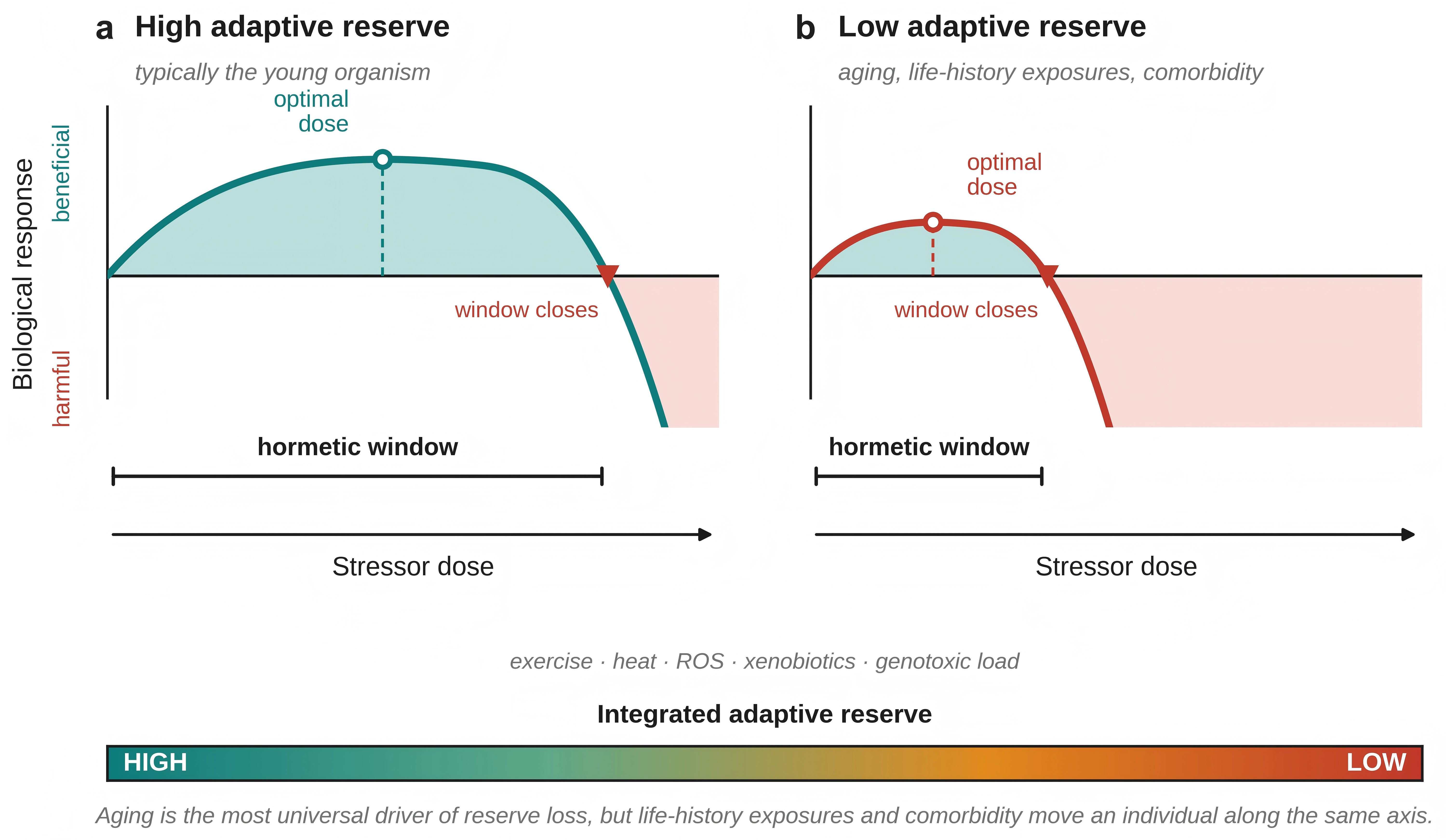
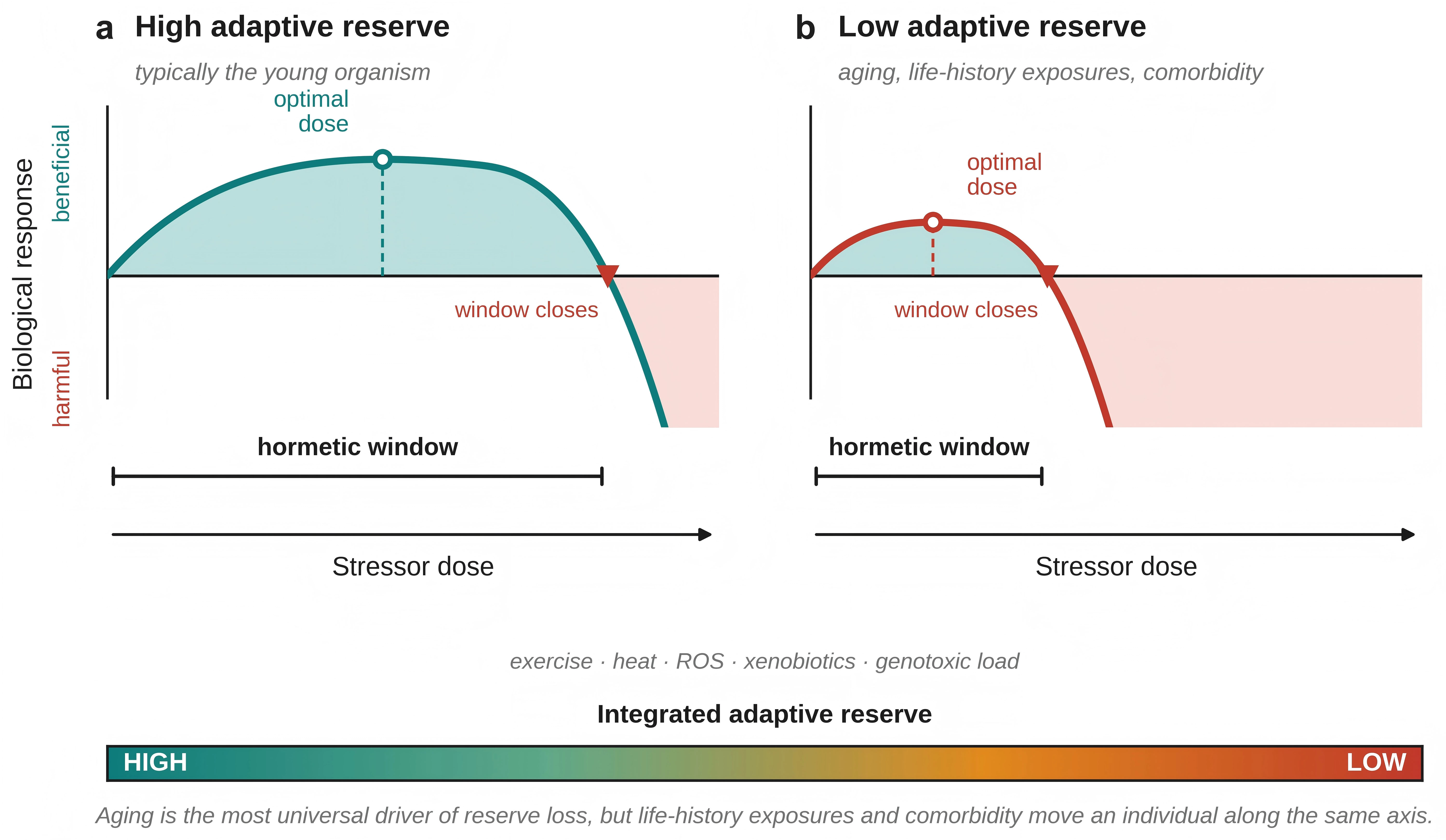
Figure 1. Narrowing of the hormetic window with loss of adaptive reserve. Schematic representation of the hormetic dose-response relationship. The x-axis indicates increasing stressor dose (exercise, heat, reactive oxygen species, xenobiotics, genotoxic load) and the y-axis the biological response, from harmful (below the neutral line) to beneficial (above it). (a) With high adaptive reserve, typically but not exclusively the young organism, the window is broad: a wide range of doses elicits a beneficial response, the optimal dose lies at intermediate intensity, and the harm threshold at which the window closes is reached only at high doses; (b) With low adaptive reserve, the window contracts, the optimum shifts toward lower doses, and the window closes earlier, so that doses which remain adaptive in (a) fall outside it. The gradient bar depicts the continuum of integrated adaptive reserve. Aging is the most universal driver of movement along this axis, but cumulative life-history exposures and comorbidity burden displace individuals along it independently of chronological age, so that (a) and (b) may correspond to two individuals of the same age. ROS: reactive oxygen species.
This distinction matters clinically because it identifies what to measure before prescribing a hormetic intervention. It also aligns the hormesis literature with the direction geroscience has already taken, in which resilience and reserve, rather than age itself, are treated as the actionable variables[7], and with the parallel effort in geriatrics to characterize intrinsic capacity as a multidimensional and modifiable quantity[8,9]. Figure 2 summarizes the framework developed here: the three inputs that set adaptive reserve, the reserve systems through which they act, the integrated reserve they produce, and the resulting dose-response.
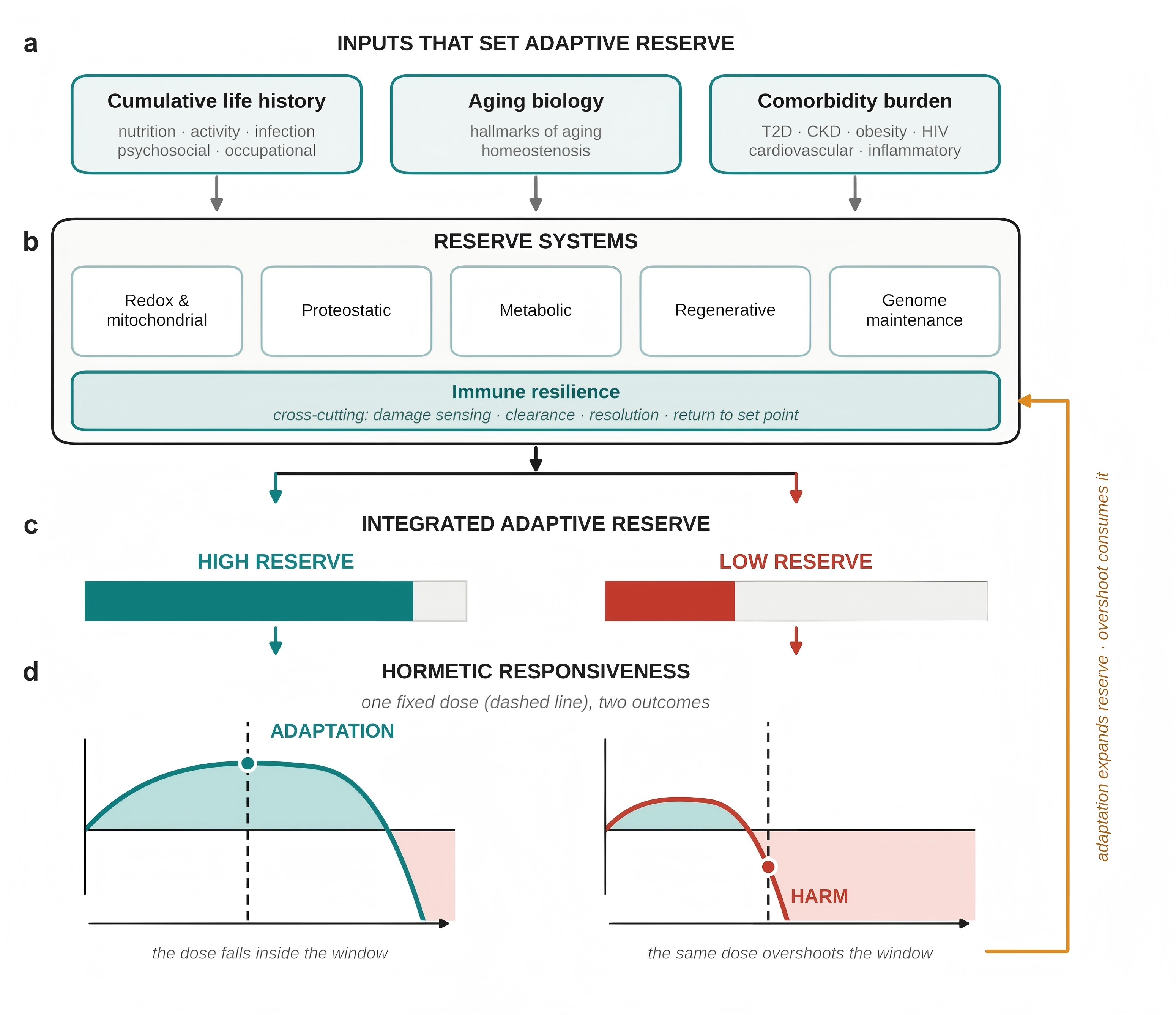
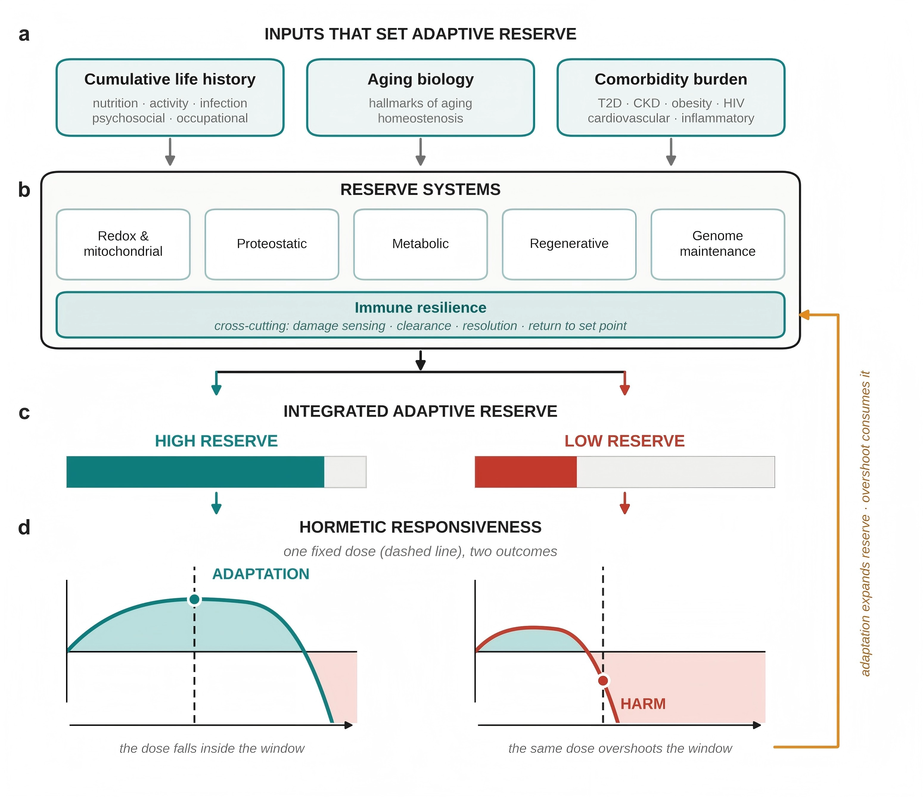
Figure 2. Adaptive reserve as the determinant of hormetic responsiveness. (a) Three classes of input set adaptive reserve, namely cumulative life-history exposures, aging biology and comorbidity burden; (b) These inputs act on six partly independent reserve systems; immune resilience is drawn as a band spanning the other five because it participates at every stage of the adaptive arc, from damage sensing through clearance and resolution to the return to a stable set point; (c) The aggregate output of these systems is the integrated adaptive reserve, shown as bar width for a high-reserve and a low-reserve individual; (d) The reserve determines the hormetic dose-response. A single fixed dose (dashed line) falls within the window of the high-reserve individual and produces adaptation, but beyond the harm threshold of the low-reserve individual, in whom the identical stimulus produces harm. The feedback arrow indicates that the relationship is dynamic: an adaptive response expands reserve whereas overshoot consumes it, so the window is a trajectory rather than a fixed patient characteristic. DDR: DNA damage response; T2D: type 2 diabetes; CKD: chronic kidney disease.
The practical relevance of the distinction is that it is routinely ignored. Investigations of hormetic activation are conducted predominantly in young or middle-aged models, and the protocols established there are subsequently extrapolated to older populations with minimal modification, under the implicit assumption that the dose-response relationship is invariant. The evidence assembled below indicates that it is not.
This review does not question hormesis, whose beneficial effects are well established; it argues that we must incorporate an explicit age axis into how we interpret and prescribe hormetic interventions. It begins by examining the fundamental signaling pathways and then analyzes how aging, cumulative life-history exposures, and comorbidity erode them. It next compares five classical paradigms in young and aged organisms: mitohormesis, caloric restriction, thermal stress, exercise, and genotoxic stress. Finally, it examines a further set of interventions whose efficacy is explicitly conditional on the recipient’s state rather than on the stimulus alone: senolytics, nicotinamide adenine dinucleotide (NAD+) precursors, partial epigenetic reprogramming, B-cell lymphoma-extra large (Bcl-xL) overexpression in T cells, and stem-cell-derived extracellular vesicles[10]. These act at different points along the adaptive arc, removing an accumulated burden, replenishing a depleted co-substrate, restoring a regulatory state, reinforcing an intracellular effector, or supplying an extrinsic signal; together, they probe the same principle from complementary directions. The review closes with the implications for geriatric trial design and clinical practice.
2. The Molecular Architecture of Hormesis
At the mechanistic level, stressor-induced hormesis converges on a small number of transcriptional and metabolic programs. Low-dose oxidants, mild heat, nutrient scarcity, and mechanical load act on partially overlapping sensors, and the downstream effectors largely coincide with the networks that maintain cellular housekeeping[1,11]. The KEAP1/NRF2 axis translates mild electrophilic or oxidative cues into phase II antioxidant and detoxifying gene expression[12]. AMPK senses energy stress and redirects metabolism toward catabolic and autophagic reprogramming[13]. Sirtuins, particularly sirtuin-1 (SIRT1) and SIRT3, couple NAD+ availability to chromatin state and mitochondrial function[14]. FOXO factors integrate growth-factor signaling with oxidative-stress resistance and stem-cell quiescence[15]. HSF1 activates the heat-shock response, with immediate effects on protein folding capacity[16]. Beyond this transcriptional scaffold, autophagy provides a mechanical clearance mechanism for damaged organelles and misfolded proteins[17].
A sixth arm of this scaffold, largely absent from hormesis reviews written from a metabolic perspective, is the genome-maintenance and DNA damage response (DDR) system, in which telomeres occupy a privileged position. Telomeres are not merely passive counters of replicative history; they are stress-responsive structures whose length, chromatin state, and associated shelterin composition are modified by oxidative, metabolic, psychosocial, and nutritional stress and, in turn, signal to the rest of the cell. Jacome Burbano and Gilson have formalized this as the telo-hormesis hypothesis: low-intensity, transient telomeric signaling can trigger adaptive responses that improve subsequent stress tolerance, whereas the same signal beyond a threshold drives persistent DDR activation, senescence, and tissue decline[18]. The proposal extends the logic of hormesis into the genome-maintenance compartment and supplies a mechanistic bridge between the classical hallmarks of telomere attrition and cellular senescence and the adaptive-response framework used throughout this review.
Most “gold standard” geroscience interventions map onto this scaffold. Caloric restriction extends lifespan across taxa and recruits AMPK, sirtuins, and mechanistic target of rapamycin (mTOR) inhibition[19,20]. Moderate exercise induces mitochondrial biogenesis and systemic anti-inflammatory adaptations[21]. Repeated mild heat exposure acts through HSF1, delaying senescence in cultured human cells[22]. Polyphenols such as resveratrol, sulforaphane, or curcumin act as xenohormetins, activating sirtuins or NRF2 at doses that would be toxic if sustained[23,24]. Rapamycin, administered late in life in heterogeneous mice, still extends lifespan by partially inhibiting mTOR complex 1 (mTORC1)[25]. The common feature is sublethal stress, followed by an adaptive response that raises the threshold for future damage.
This is a compact and useful picture, but it says nothing about the condition of the responder. It treats the adaptive machinery as a fixed system parameter, when in fact it is the variable most affected by aging[3].
3. Adaptive Reserve and the Determinants of the Hormetic Window
3.1 Aging erodes the adaptive machinery
The original hallmarks of aging and their recent update describe progressive alterations in the same pathways that hormetic stimuli try to mobilize[6,26,27]. From a hormetic perspective, each hallmark further reduces the signal-to-response ratio. Mitochondrial dysfunction increases basal reactive oxygen species (ROS) and lowers the ceiling for a useful oxidative pulse. Loss of proteostasis fills the chaperone pool with pre-existing substrates, so that a new heat challenge has fewer resources to work with. Dysregulated nutrient sensing limits the autophagic flux that caloric restriction tries to unlock. Inflammaging compromises the resolution phase of any acute response and pushes recovery into a chronic, low-grade state[28,29]. Stem-cell exhaustion reduces the regenerative output expected following tissue stress[30]. NAD+ levels fall with age and limit sirtuin activity[14]; NRF2 induction is blunted in several tissues[31].
The most direct demonstration of this last point comes from Suh and colleagues, who showed in old rats that nuclear NRF2 binding to the antioxidant response element and the resulting de novo glutathione synthesis are substantially reduced compared with young animals, and that lipoic acid supplementation only partially restores both[32]. The pattern is not specific to NRF2: the same age-dependent flattening of the inducible response has been documented for HSF1-driven chaperone induction[33], FOXO-dependent stress resistance[15], and AMPK activation in skeletal muscle[34]. Heydari and colleagues showed that transcriptional induction of HSP70 after a heat challenge is markedly reduced in hepatocytes from old rats compared with young animals, and that the deficit occurs at the level of HSF1 binding to the heat-shock element rather than upstream signal generation[33]. Martins and colleagues, reviewing the FOXO literature across model organisms, documented age-dependent loss of FOXO nuclear translocation and target-gene activation in multiple tissues, with downstream weakening of antioxidant, autophagic, and proteostatic stress responses[15]. Reznick and colleagues showed that, in rat skeletal muscle, the magnitude and duration of AMPK phosphorylation in response to a contractile or pharmacological stimulus are sharply reduced in old animals, accompanied by parallel deficits in peroxisome proliferator-activated receptor gamma coactivator 1-alpha (PGC-1α) induction and mitochondrial biogenesis[34]. Taken together, these data suggest that the narrowing of the hormetic window is best understood as the cumulative consequence of parallel attenuation across all major adaptive arms rather than the failure of any single pathway.
The clinical counterpart of this molecular picture is frailty[7]. Frailty is, essentially, a state in which the organism has lost adaptive reserve. Minor insults such as an infection, surgery, or a medication change that a younger individual would absorb with little perturbation, now trigger disproportionate decompensation. At the tissue level, the same logic applies to hormetic stimuli: the headroom is smaller, so the dose that used to be hormetic can now push the system into the zone where damage exceeds repair.
3.2 Life-history exposures and comorbidity as modifiers of the window
If the hormetic window is the operational expression of adaptive reserve, then anything that alters reserve will narrow the window, whether or not it results from chronological aging. This has two implications that the narrowing-window hypothesis must accommodate.
The first concerns cumulative life history. Adaptive reserve at any given age is the integral of everything the responder system has been exposed to. Lifelong physical activity expands mitochondrial and cardiovascular reserve; chronic undernutrition or protein insufficiency contracts anabolic reserve; repeated or persistent infection consumes immune reserve; chronic psychosocial stress and adverse socioeconomic circumstances accelerate biological aging by measurable margins[35]; and cumulative environmental and occupational exposures load the detoxification and genome-maintenance systems that hormetic stimuli must subsequently recruit[36]. None of these is captured by date of birth. Two 70-year-olds of the same chronological age may sit at opposite ends of the reserve distribution, and the practical consequence is that the same exercise, fasting, or thermal protocol may be comfortably hormetic for one and frankly injurious for the other.
The second, and clinically more tractable, concerns comorbidity burden. Chronic diseases should be regarded not as background noise in geroscience trials but as explicit modifiers of the hormetic window, because they impair precisely the pathways on which hormetic adaptation depends. Type 2 diabetes and obesity compromise metabolic flexibility, mitochondrial function, and autophagic flux[37], thereby blunting responses to caloric restriction and exercise that operate through those routes. Chronic kidney disease reduces the capacity to buffer metabolic and volume perturbations and is associated with accelerated cellular senescence[38]. Cardiovascular disease constrains the cardiopulmonary reserve that exercise and thermal protocols draw on and directly narrows the safe dose range. Chronic inflammatory and autoimmune disorders occupy the inflammatory-resolution machinery that any acute adaptive response must eventually engage. Chronic infection is a particularly instructive case: people living with HIV, even with sustained virological suppression, show premature immunosenescence, persistent inflammation and accelerated epigenetic aging, and constitute a population in which the window is demonstrably narrowed decades ahead of chronological schedule[8,39].
The general point is that biological responsiveness to hormetic interventions may depend as much on disease burden as on chronological age[40]. Trials that exclude comorbid participants in order to obtain clean signals therefore select for the subgroup with the widest windows, and their results generalize poorly to the multimorbid older patients who make up most of geriatric practice. Conversely, trials that include comorbid participants without stratifying by disease burden will average across radically different dose-response curves and risk reporting a null result.
3.3 Immune resilience as a determinant of hormetic responsiveness
Among the reserve systems that determine whether a hormetic stimulus is successfully translated into adaptation, the immune system deserves particular attention, because it participates in nearly every stage of the adaptive arc. Damage sensing, recruitment and clearance of injured cells, resolution of inflammation, and restoration of tissue homeostasis are all immune-dependent. A stimulus that a competent immune system processes as a transient, self-limiting perturbation may, in an immunologically compromised host, become a persistent inflammatory lesion. In this sense, immunosenescence and inflammaging are not simply two hallmarks among many; they determine whether the other hallmarks can be adaptively engaged at all.
The concept of immune resilience formalizes this. Ahuja and colleagues defined immune resilience as the capacity to preserve or rapidly restore immune functions that promote disease resistance while simultaneously controlling inflammation, and quantified it with two peripheral-blood metrics: the balance of cluster of differentiation 8-positive (CD8+) and CD4+ T-cell levels, and gene-expression signatures tracking longevity-associated immunocompetence versus mortality-associated inflammation. Profiling approximately 48,500 individuals, they showed that some people resist alterations in immune resilience during aging and when challenged with varied inflammatory stressors, and that preserved optimal immune resilience tracked a lower risk of HIV acquisition, acquired immunodeficiency syndrome (AIDS) progression, and symptomatic influenza infection, along with longer survival[8]. Crucially, immune resilience was not a simple function of age: inflammatory challenge altered it at any age, and some individuals preserved it into late life.
At the level of a single organ system, this is a direct analog of the argument made throughout this review, and it supplies a measurable readout of one component of adaptive reserve. It also offers a mechanistic account of two otherwise puzzling clinical observations: why the same exercise or thermal stimulus produces slower and less complete recovery in older adults even when the acute response is intact, and why individuals with a high lifetime burden of infection or chronic inflammation respond poorly to hormetic protocols independently of their chronological age. Senescent immune cells acquire a proinflammatory senescence-associated secretory phenotype that amplifies inflammaging, and the resulting failure to resolve inflammatory responses propagates damage across neurodegenerative, oncological, infectious, and autoimmune domains[41-44]. The resolution phase, rather than the initiation phase, is where the aged system most often fails[45].
In practical terms, this argues for including immune-resilience metrics alongside the metabolic and proteostatic readouts conventionally used in geroprotection trials, and for treating a history of chronic infection or inflammatory disease as a stratification variable rather than an exclusion criterion. Practically, this argues for including immune-resilience metrics alongside the metabolic and proteostatic readouts conventionally used in geroprotection trials, and for treating a history of chronic infection or inflammatory disease as a stratification variable rather than an exclusion criterion. The two constructs are conceptually distinct: intrinsic capacity is an integrated functional readout at the whole-organism level, whereas immune resilience is a mechanistic measure of one contributing system. Whether they index the same reserve or separable parts of it has not been tested directly, and combining them in a single cohort would be a straightforward addition to existing screening infrastructure. Section 6.1 returns to this point[9].
3.4 Hormesis and antagonistic pleiotropy
The narrowing of the hormetic window invites an evolutionary reading. Antagonistic pleiotropy, as formulated by Williams, holds that alleles conferring fitness benefits early in life can be positively selected even if they impose costs later, because the force of natural selection declines with age[46]. Many of the pathways discussed here fit that description with unusual precision. Robust inflammatory responses are essential to survive infection during reproductive life and become inflammaging in old age. The DDR and senescence programs suppress malignancy in young tissue and accumulate senescent cells that degrade tissue function later[18]. mTOR-driven anabolism supports growth and reproduction and contributes to the loss of proteostasis and autophagic flux with age. Even the ROS signaling that underlies mitohormesis is a case in point: the retrograde signal that primes a young mitochondrial network operates against a basal oxidative load that has itself risen with age.
The two frameworks are complementary rather than competing, and the distinction is worth stating precisely. Antagonistic pleiotropy is an evolutionary explanation of why adaptive programs are optimized for early life and become maladaptive later. The narrowing hormetic window is a physiological description of the consequence: because those programs are tuned for a young responder, the dose-response relationship they generate shifts and compresses as the responder changes. Antagonistic pleiotropy therefore predicts what the narrowing window observes and explains why the shift should be systematic rather than random, with programs degrading in the direction of their early-life optimization rather than arbitrarily.
The framework also generates a testable prediction with translational content. If the narrowing of the window reflects the late-life expression of early-life-optimized programs, then interventions that selectively remove the late-life cost without abolishing the early-life benefit, with senolytics as the clearest example, should show the age-conditional efficacy profile described in Section 5, with negligible effects in young organisms and substantial effects in old ones. That is broadly what is observed, and it constitutes indirect support for reading the narrowing window through an evolutionary lens.
3.5 The window is tissue-specific, and it moves
One point often glossed over is that the window’s narrowing is not uniform across tissues or pathways. Skeletal muscle retains a remarkable capacity to respond to exercise well into old age, with training responses that remain qualitatively similar to those in young adults[47,48]. However, this preservation is not absolute: while basal muscle protein synthesis rates remain identical between young and old (~1.35% day-1), resistance training increases synthesis in young individuals (to 1.61% day-1) but not in older adults (1.49% day-1), reflecting an “anabolic resistance” involving deficits in ribosomal biogenesis and translational efficiency[49,50]. The immune system, by contrast, is often the first compartment to lose adaptive flexibility, for the reasons set out in Section 3.3[41-43]. Even within single organs, hormetic interventions produce heterogeneous responses. The hippocampus and other brain regions differ markedly in their responses to caloric restriction, with significant regional effects in hippocampal energy metabolism that are age- and region-specific[51,52]. Caloric restriction suppresses age-dependent gene signatures specifically in the hippocampal CA1 region, demonstrating that even within a single structure, subregions respond differently to the same intervention[53].
These observations argue against the simplification, implicit in the schematic of Figure 1 and in most of the hormesis literature, that an organism possesses a single window. It is more accurate to say that an organism possesses a set of coupled, organ-specific windows: immune, musculoskeletal, metabolic, cardiovascular, and cognitive windows, that decline on different timetables and which interact. The coupling is not incidental. Immune failure propagates into muscle through inflammatory catabolism; metabolic dysfunction constrains cardiovascular reserve; and cognitive impairment limits the behavioral adherence on which exercise and dietary protocols depend. A multimodal stimulus such as exercise engages several windows at once, which is a strength because it distributes adaptive demand, and a limitation because the narrowest window sets the safe ceiling for the whole intervention. The organism-wide window, in other words, is closer to the intersection of the component windows than to their average, and a useful geromedicine will need to be tissue-aware as well as age-aware[3,54,55].
A second simplification worth correcting is temporal. Biological age and adaptive capacity are not static properties but trajectories, and they can move in both directions: an intercurrent illness, a hospitalization, or a period of immobility can contract the window over weeks, while successful rehabilitation, nutritional repletion, or treatment of an inflammatory condition can widen it again. The hormetic window is therefore a moving target rather than a fixed patient characteristic. This has direct methodological consequences, developed in Section 6.2: a single baseline assessment cannot define the appropriate dose for an intervention delivered over months, and trial designs that fix the dose at randomization implicitly assume a stability that the underlying biology does not possess.
4. Classical Hormetic Interventions Across the Lifespan
The five paradigms discussed below have all been tested in young and aged organisms, and they all show the same general pattern: a clear net benefit early in life, and a compressed or inverted dose-response later on.
4.1 Mitohormesis
The case for mitohormesis rests on the observation that a mild rise in mitochondrial ROS acts as a retrograde signal that promotes mitochondrial biogenesis, improved respiratory efficiency, and resistance to subsequent oxidative insults[56,57]. In young tissues, the loop is clean. Inducible antioxidant responses neutralize a transient ROS peak, and the system settles at a slightly higher level of defense. In aged tissues, the loop is already running close to its limit. Basal ROS is elevated, mitochondria are partially dysfunctional, NRF2-driven antioxidant induction is attenuated, and clearance mechanisms are slower. A ROS rise that would have primed a young cell may tip an old one into damage[11]. Work by Enríquez and others on respiratory supercomplex assembly has clarified the structural basis for elevated basal ROS in aged tissues[58,59]. Lapuente-Brun and colleagues showed that assembling complexes I, III, and IV into stable respiratory supercomplexes determines electron flux through the mitochondrial respiratory chain, and that reduced supercomplex stoichiometry redirects electrons to increase ROS leak[60]. Latorre-Pellicer and colleagues later showed in conplastic mice (animals carrying nuclear and mitochondrial genomes from genetically distant sources) that even a subtle mismatch between nuclear-encoded and mitochondrial-encoded respiratory-chain subunits accelerates aging and increases oxidative stress, while a matched pairing extends healthspan[61]. These findings reframe the mitohormetic margin mechanistically: the structural baseline of the aged respiratory chain (decreased supercomplex stability and gradual accumulation of mitochondrial DNA (mtDNA) heteroplasmy with imperfect nuclear-mitochondrial coupling) determines the extent of the window for a useful ROS pulse before it becomes damaging.
The well-known Ristow experiment showed that oral antioxidant supplementation abolishes the metabolic benefits of exercise in healthy young subjects by blunting the ROS signal[62]. A controlled trial of vitamin C supplementation during endurance training reached the same conclusion: modest antioxidant doses suppressed exercise-induced mitochondrial biogenesis and abrogated the training-induced gain in maximal oxygen uptake[63], and the effect was most pronounced in subjects with the highest training stimulus. If the margin for useful ROS signaling is that tight in healthy young humans, there is no reason to expect it to widen with age. Two specific findings frame the age-dependence of this loop. In Caenorhabditis elegans, glucose restriction extends lifespan by transiently increasing mitochondrial respiration and ROS, an effect abolished by antioxidant co-administration, indicating that the ROS pulse signals rather than being a side effect of the metabolic shift[64]. A second finding frames the downstream side of the same loop. Houtkooper and colleagues showed that the stoichiometric balance between nuclear- and mitochondrially-encoded subunits of the respiratory chain predicts lifespan across genetically diverse mice, and that interventions which perturb that balance extend lifespan through induction of the mitochondrial unfolded protein response, establishing the retrograde arm as a determinant of longevity rather than a passive readout of mitochondrial state[65]. Since the structural baseline of the aged respiratory chain is already imbalanced, as described above, the same perturbation is delivered to a system with less remaining margin, and the adaptive value of the retrograde signal should be correspondingly smaller. Together, these findings converge on the same point: even when the upstream ROS or proteostatic stimulus is preserved, the downstream amplification machinery that turns it into an adaptive response is the rate-limiting step that aging erodes first. Both findings converge on the same point: even when the upstream ROS or proteostatic stimulus is preserved, the downstream amplification machinery that turns it into an adaptive response is the rate-limiting step that aging erodes first.
4.2 Caloric restriction
Caloric restriction is the most thoroughly characterized hormetic intervention in biogerontology. It extends lifespan in yeast, worms, flies, and rodents, and at least modestly in non-human primates[19,20]. The mechanistic basis is solid: energetic deficit activates AMPK, inhibits mTORC1, and unlocks autophagy. When the same intervention is tested in older adults, the picture is different. Older adults on calorie restriction tend to lose lean mass faster than they should; bone turnover is compromised, and immune function can be impaired. Sarcopenia and frailty replace metabolic modulation as the dominant outcomes[66]. The window that is wide in youth becomes narrow in old age, and the narrow band is also shifted: protein intake that would have been safely reduced at 30 can no longer be reduced at 75 without clinical cost. Two studies sharpen this picture. Forster and colleagues showed that the survival benefit of 40% caloric restriction in mice depends strongly on age at onset and genetic background, with late-onset protocols extending median but not maximum lifespan and producing strain-dependent toxicity[67]. A more comprehensive analysis of nearly 1,500 mice across multiple genetic backgrounds, sexes, and calorie restriction (CR) regimens established that the magnitude and even the sign of CR effects on healthspan markers are strongly modulated by sex and strain, with some combinations showing benefit and others showing harm at identical levels of restriction[68], a degree of inter-individual variability that closely resembles what is seen clinically when restriction is applied to older adults. In humans, the Comprehensive Assessment of the Long-term Effects of Reducing Intake of Energy 2 (CALERIE-2) randomized trial demonstrated that two years of moderate (about 12%) caloric restriction in healthy non-obese adults aged 21-50 years improved cardiometabolic markers without compromising lean mass or bone mineral density[69]; the trial deliberately excluded older adults and individuals with low BMI, so its safety margins cannot be transferred to the geriatric population without further evidence. Consensus recommendations for older adults set a protein floor of 1.0-1.2 g·kg-1·day-1[70], a threshold below which generic caloric restriction protocols would routinely fall.
4.3 Thermal stress
Repeated mild heat exposure induces HSF1-driven chaperone upregulation and delays senescence-associated changes in cultured cells[22]. This does not translate well into the geriatric clinic because the proteostatic buffer in aged tissues is already partially occupied by accumulated aggregates and by the loss of chaperone-mediated autophagy[17]. A heat challenge that a young chaperone network would absorb can, in an older one, displace existing substrates and fail to produce a net folding benefit. The epidemiology of heat waves is consistent with this view: older people are overrepresented in heat-wave mortality, not because they lack a heat-shock response, but because their cardiovascular, thermoregulatory, and proteostatic reserves have been eroded. The molecular basis of this attrition is well documented. In C. elegans, reducing hsf-1 activity accelerates tissue aging and shortens lifespan; hsf-1 is required for the longevity conferred by reduced insulin/insulin-like growth factor-1 (IGF-1) signaling, and its overexpression is sufficient to extend lifespan, placing the heat-shock response upstream of the proteostatic contribution to longevity rather than alongside it[71]. What erodes with age is not that requirement but the capacity to meet it: as set out in Section 3.1, the inducible arm of the response is itself attenuated in aged tissue, at the level of HSF1 binding to the heat-shock element rather than in upstream signal generation, so that an identical heat dose yields a smaller chaperone response in older animals[33]. In humans, observational data from a middle-aged Finnish cohort show a dose-dependent association between sauna frequency and reduced cardiovascular and all-cause mortality[72], while clinical appraisal of heat stress in older individuals and in patients with common chronic diseases locates the limiting factor in cardiovascular and thermoregulatory reserve rather than in the heat-shock response itself, with comorbidity and medication compounding the deficit[73]. These findings suggest that the heat-shock paradigm retains some benefit at the population level past midlife, but the dose-response curve flattens with age, so blanket extrapolation of sauna or heat-acclimation protocols from younger cohorts to frail older adults is not warranted. Cold exposure follows the same logic and should be acknowledged here, even though it has been studied less extensively in geriatric populations: Brown adipose tissue activation declines with age[74]. Whether the noradrenergic stress response is similarly attenuated, and whether this translates into cardiovascular instability under cold exposure in older patients, has not been directly tested; both remain plausible concerns rather than established outcomes, and the safe range of cold-acclimation protocols across the lifespan remains poorly defined.
4.4 Physical exercise
Exercise is the paradigmatic multimodal hormetic stimulus and, in older adults, remains arguably the single most effective geroprotector available[75]. It is worth emphasizing that the narrowing-window message is not that exercise stops working in old age. It does work. What changes is the dose-response. In young and middle-aged subjects, the range of useful exercise intensities is broad, and sessions that produce transient soreness are followed by full recovery and clear adaptive gains[21,76]. In older adults, the same intensity results in slower recovery, incomplete resolution of post-exercise inflammation, and a narrower margin before overuse begins to drive structural damage. Individual variation is also much wider than in younger cohorts, so the same protocol can be moderate for one 80-year-old and clearly excessive for another. Prescriptions must be individualized based on current functional status, not chronological age.
The clinical evidence supports both points. The LIFE Study, a randomized trial of structured physical activity in 1,635 sedentary adults aged 70-89 years, demonstrated that a multicomponent moderate-intensity program reduced major mobility disability by 18% over a median 2.6-year follow-up[77], with benefit preserved in the oldest and most vulnerable participants. At the cellular level, the same resistance-training protocol produces equivalent acute mTORC1 activation in young and old human muscle, but cumulative protein synthesis over six weeks is roughly 10% lower in older adults because ribosomal biogenesis is impaired, an “anabolic resistance” that is not overcome by simply increasing training intensity[49]. Together, these data suggest that the exercise window does not close in old age but narrows and steepens, with smaller margins between an effective stimulus and one that overshoots into damage or fails to translate into adaptation. Animal models show the same dissociation. Lifelong spontaneous wheel-running in mice initiated in young adulthood improves a panel of healthspan markers (motor coordination, grip strength, cognitive performance, and mitochondrial function in skeletal muscle) but does not extend maximal lifespan[78], a result that fits the narrowing-window hypothesis: exercise compresses late-life morbidity into a shorter terminal interval rather than postponing the upper limit of viable function. Read together, the Pahor and García-Valles datasets converge on the same conclusion across species: the exercise stimulus retains its protective signal in old age, but its translation into a life-extending benefit probably erodes because the downstream adaptive machinery no longer operates with the headroom available in younger systems.
4.5 Genotoxic stress: Telomeres and the DNA damage response
The four paradigms above are metabolic, thermal, or mechanical. A fifth, less frequently integrated into hormesis reviews, is genotoxic stress, and it displays the same age-conditional geometry with an instructive twist.
The classical adaptive response to DNA damage is well established: sublethal genotoxic exposure upregulates repair capacity and confers resistance to a subsequent, larger challenge. The telo-hormesis hypothesis adds that telomeres act as privileged sensors and transducers of this response, rather than passive counters of replicative history[18]. Telomeric chromatin is unusually susceptible to oxidative lesions, is inefficiently repaired relative to the rest of the genome, and is a preferential site of persistent DDR foci during both replicative and stress-induced senescence[79]. Low-intensity telomeric signaling can therefore act as an early-warning system that mobilizes protective programs before damage becomes generalized, while the same signal sustained beyond a threshold converts into permanent DDR activation and senescence. Jacome Burbano and Gilson review evidence that stress alters telomere length, structure, and shelterin composition in ways that feedback on cellular physiology, and propose that these changes carry hormetic value in the low-dose range[18]. A concrete illustration is the behavior of oxidative guanine lesions at telomeres, where low levels of 8-oxoguanine promote telomerase-dependent lengthening whereas high levels drive telomere loss and chromosomal aberrations, a biphasic dose-response of exactly the shape the hormesis literature describes[80-82].
The age-dependence follows directly from the structure of the argument, and it is more severe than for the metabolic paradigms. First, the substrate is progressively consumed: telomere reserve length declines across the lifespan, so the margin between an informative telomeric signal and one that triggers irreversible senescence contracts monotonically. Second, the downstream repair machinery is itself attenuated with age, so the adaptive arm of the response weakens as the stimulus arm becomes more easily saturated. Third, and unlike the metabolic paradigms, the damaging outcome is not merely a failure to adapt but the generation of senescent cells, which then export the failure to neighboring tissue through the senescence-associated secretory phenotype and consume the immune reserve required to clear them[41-43]. Genotoxic hormesis in an aged organism therefore risks converting a local, transient stimulus into a systemic and persistent inflammatory burden.
This has a practical corollary that deserves emphasis in a clinical review. Interventions that deliberately impose genotoxic stress, including low-dose radiation protocols occasionally proposed as hormetic therapies, carry an age-conditional risk profile that is qualitatively different from that of exercise or heat. The relevant safety margin is not defined by acute tolerability but by the senescent-cell burden generated, which accumulates and is poorly reversible. Until that burden can be measured in vivo in humans, genotoxic hormesis should be regarded as the paradigm with the least favorable risk-benefit ratio in older adults, and the one where extrapolation from young models is least defensible.
Table 1 summarizes the five interventions in young and age organisms along with their main limiting factors.
Table 1. Age-dependent divergence of five classical hormetic interventions.
| Intervention | Young organism | Aged organism | Main limiting factor |
| Mitohormesis | Transient ROS peak triggers mitochondrial biogenesis and NRF2/phase II antioxidant induction[56,57]; in C. elegans, glucose-restriction-induced ROS pulse extends lifespan and is abolished by antioxidant co-administration[64]; in healthy young humans, vitamin C or N-acetylcysteine supplementation suppresses exercise-induced mitochondrial biogenesis and gains in VO2max, confirming the ROS pulse is the signal[62,63]. | Pre-existing mitochondrial dysfunction and elevated basal ROS[11,27]; nuclear NRF2 binding to ARE and de novo glutathione synthesis decline substantially in aged tissues[31,32]; mtUPR retrograde signalling is activated by mitonuclear protein imbalance and is an established longevity-regulating mechanism[65], although whether this response is itself attenuated with age remains to be directly tested; the same ROS rise that primes a young cell tips the system into oxidative damage rather than adaptation[11]. | Saturated basal ROS; attenuated inducible NRF2 and mtUPR responses; slower mitophagic clearance machinery[11,31,32,65]. |
| Caloric restriction | AMPK activation, mTORC1 inhibition and autophagy induction[13,19]; lifespan extension across yeast, worms, flies, rodents and modestly in non-human primates[19,20]; CALERIE-2 randomised trial: two years of ~12% CR in healthy adults aged 21-50 improves cardiometabolic markers without compromising lean mass or bone mineral density[69]. | Accelerates sarcopenia and bone loss; impairs immune recovery; outcomes dominated by frailty rather than metabolic modulation[66]. In mice, survival benefit highly dependent on age at onset, sex and strain—some combinations show benefit, others harm at identical restriction levels[67,68]. In humans, consensus recommendations for older adults set a protein floor of 1.0-1.2 g·kg-1·day-1[70]. | Low anabolic reserve; limited autophagic flux; nutritional and immune vulnerability; high inter-individual variability driven by sex, genetic background and frailty status[66-68,70]. |
| Thermal stress | HSF1 trimerisation drives chaperone induction and delays cellular senescence in vitro[16,22]; hsf-1 is required for normal lifespan and for insulin/IGF-1-pathway longevity in C. elegans, and sufficient to extend lifespan when overexpressed[71]; sauna frequency is dose-dependently associated with reduced cardiovascular and all-cause mortality in middle-aged Finnish men[72]. | Saturated proteostatic buffer from accumulated aggregates and loss of chaperone-mediated autophagy[17]; HSP70 induction after a heat challenge markedly reduced in aged tissue, at the level of HSF1 binding to the heat-shock element[33]; limiting factor is reduced cardiovascular/thermoregulatory reserve worsened by comorbidity and medication[73]; cardiovascular and thermoregulatory decompensation underlies excess heat-wave mortality[73]. | Pre-existing aggregate load; reduced HSF1 inducibility; diminished thermoregulatory and cardiovascular reserve; inflammaging-driven recovery deficit[18,28,33,71,73]. |
| Physical exercise | Broad range of useful intensities; mitochondrial biogenesis, antioxidant gene upregulation and PGC-1α induction[21,76]; ROS-mediated adaptive arc fully intact, with antioxidant supplementation blunting mitochondrial biogenesis and VO2max gains[62,63]; clear cardiometabolic and cognitive gains across cohorts. | Benefit is preserved, but the window is narrower and steeper. LIFE Study (n = 1635, ages 70-89): structured moderate-intensity activity reduces major mobility disability ~18% over 2.6 years[77]; anabolic resistance, equivalent acute mTORC1 activation but ~10% lower cumulative protein synthesis vs. young, traced to ribosomal biogenesis deficit[49]; in mice, lifelong spontaneous exercise improves healthspan markers without extending maximal lifespan[78]. | Inflammaging and delayed inflammatory resolution[7,28]; ribosomal anabolic resistance to training[49]; high heterogeneity tied to frailty status; healthspan extension without lifespan extension when started lifelong[78]. |
| Genotoxic stress (telomeres and DDR) | Sublethal DNA damage upregulates repair capacity and confers resistance to subsequent challenge; telomeres act as stress-responsive sensors whose low-intensity signalling mobilises adaptive programmes (telo-hormesis)[18]; biphasic dose-response of oxidative telomeric lesions, with low 8-oxoG promoting lengthening and high levels driving loss and aberration[18]. | Telomere reserve is progressively consumed, contracting the margin between an informative signal and irreversible senescence[79]. DDR and repair capacity are attenuated with age, and persistent telomeric DDR foci accumulate[79,81,82]. This damaging outcome generates senescent cells that propagate injury via SASP[66]; the proposed link between telomere-driven damage and hormetic responsiveness remains a conceptual hypothesis rather than an established mechanism[18]. Senescent cells more broadly consume immune reserve and contribute to immunosenescence and inflammaging[41-43]. | Depleted telomere reserve; attenuated repair machinery; poorly reversible senescent-cell accumulation; least favourable risk-benefit ratio of the five paradigms in older adults[18,41-43]. |
ARE: antioxidant response element; AMPK: adenosine monophosphate (AMP)-activated protein kinase; CR: caloric restriction; DDR: DNA damage response; HSF1: heat shock factor 1; HSP70: heat shock protein 70; mTORC1: mechanistic target of rapamycin complex 1; mtUPR: mitochondrial unfolded protein response; NRF2: nuclear factor erythroid 2-related factor 2; PGC-1α: peroxisome proliferator-activated receptor gamma coactivator 1-alpha; ROS: reactive oxygen species; SASP: senescence-associated secretory phenotype; VO2max: maximal oxygen uptake; CALERIE-2: Comprehensive Assessment of the Long-term Effects of Reducing Intake of Energy 2; IGF-1: insulin-like growth factor-1.
5. Interventions Whose Effects Diverge Between Young and Old
The narrowing-window argument predicts that the same molecular intervention should produce different effects in young and aged organisms, and that the difference should indicate which step of the adaptive arc is rate-limiting at each age. This prediction can be tested across interventions that differ in mechanism but share age-conditional efficacy.
5.1 Age-conditional efficacy across the intervention landscape
Senolytics. Senolytic compounds provide the cleanest illustration of the principle, because their target, i.e., the senescent cell, is essentially absent in young tissue. The intervention therefore has no substrate to act on early in life and an expanding substrate thereafter, predicting a dose-response that is flat in youth and rises with age. Clinical translation has followed this logic: first-in-human work with dasatinib plus quercetin was conducted in idiopathic pulmonary fibrosis, an age-related disease characterized by high senescent-cell burden, and reported improvement in physical function measures in an open-label pilot[83]; a parallel study in diabetic kidney disease demonstrated that intermittent dosing reduced senescent-cell abundance in adipose tissue and skin[84]. A subsequent randomized, placebo-controlled pilot in the same fibrotic indication confirmed feasibility and tolerability but was not powered for efficacy, and appropriately framed itself as informing rather than establishing the next stage of trial design[85]. The relevant point for this review is not that senolytics work, but that their expected effect size depends on the recipient’s senescent-cell burden, a component of biological rather than chronological age. Recruitment based on date of birth therefore samples that burden blindly, and a null result cannot distinguish an inactive compound from a cohort with little substrate for it to act on.
NAD+ precursors. The rationale for NAD+ repletion is explicitly deficit-based: NAD+ falls with age, sirtuin and PARP activity depend on it, and supplementation should therefore benefit those with the deficit[14]. The clinical data illustrate both the promise and the difficulty of the age-conditional logic. Chronic nicotinamide riboside supplementation reliably and substantially elevates blood NAD+ in middle-aged and older adults and is well tolerated, with preliminary signals on blood pressure and arterial stiffness[86]. Yet trials with functional endpoints have been more equivocal: a randomized placebo-controlled trial in older adults with mild cognitive impairment doubled blood NAD+ without producing significant cognitive improvement[87], and a systematic appraisal of NAD+ precursors for skeletal muscle mass and function in older adults has concluded that benefits are modest, inconsistent, and possibly restricted to specific subgroups[88]. This dissociation between a robust biochemical response and an inconsistent functional one is itself informative in this framework: raising the concentration of a co-substrate does not restore adaptive capacity if the downstream enzymatic and transcriptional machinery is the rate-limiting step. It is the pharmacological analog of the mitohormetic finding that a preserved upstream stimulus does not guarantee a preserved response.
Partial epigenetic reprogramming. Transient expression of Yamanaka factors reverses age-associated molecular signatures without loss of cell identity, and the age-dependence of its effects is particularly instructive because it has been examined systematically. In physiologically aging wild-type mice, long-term partial reprogramming produced beneficial effects in kidney and skin and at the organismal level, with the duration of treatment determining the extent of benefit, accompanied by reversion of the epigenetic clock and reduced expression of inflammation, senescence, and stress-response genes; short-term regimens produced weaker effects[89]. Optic-nerve work had earlier shown that Oct4, Sox2, and Klf4 (OSK) expression can restore youthful epigenetic information and visual function in aged animals[90]. Most directly relevant here, systemic adeno-associated virus (AAV) delivery of an inducible OSK cassette to 124-week-old mice, an age equivalent to the human ninth decade, extended median remaining lifespan and improved frailty scores[91]. Partial reprogramming is thus, at present, the intervention with the strongest evidence that a late-life intervention can widen rather than merely preserve the window. That said, the safety profile in humans is unestablished, the delivery problem is unsolved, and the oncogenic risk inherent in reprogramming factors remains the principal barrier to translation.
Taken together, these three classes share the property that the magnitude, and in the senolytic case arguably the sign, of the effect depends on the biological state of the recipient rather than on the intervention alone. They also probe different points along the adaptive arc: senolytics remove an accumulated burden, NAD+ precursors replenish a depleted co-substrate, and partial reprogramming attempts to restore the regulatory state that generates the response. The two interventions developed in our group, described below, add an intracellular effector and an extrinsic regulatory signal to the same map.
5.2 Two age-conditional examples: Reinforcing the effector and supplying the signal
Our interest in Bcl-xL began with a functional transcriptomic comparison of human centenarians, septuagenarians and young adults, in which the centenarian profile resembled the young one more closely than the intermediate-age one, with Bcl-xL among the key differentially expressed transcripts[92]. Bcl-xL is an anti-apoptotic Bcl-2 family member at the outer mitochondrial membrane whose expression in T cells declines with age[93]. In mice overexpressing human Bcl-xL selectively in T cells under the Lck promoter, regulatory T cells accumulated in skeletal muscle, infiltration by interferon-γ (IFN-γ)-producing effector cells fell, muscle mitochondrial function was preserved, and the onset of frailty was delayed[94]. The age-conditionality is the point here. Young transgenic and wild-type littermates were indistinguishable in grip strength, motor coordination, and endurance, and genotype separated the Valencia frailty score only beyond 24 months, with plasma IFN-γ, fat mass, and fiber cross-sectional area following the same shape. The pattern is not uniform across readouts, however: several T-cell features already differ at young and adult ages, indicating that the narrowing window operates at the integrative level of inflammation, muscle architecture and frailty rather than at every level at once.
One caveat applies here and to most genetic models in the field. Constitutive lines express the modification from development onward, so they show that lifelong reinforcement of an effector preserves function into old age, but cannot tell us whether the same intervention started late would rescue an already aged phenotype. The late-life literature is consistent on this point: late-life initiation of rapamycin from 600 days extends lifespan in mice[25]. Senolytic regimens have shown feasibility and tolerability in early-phase human trials[83-85], though these are pilot studies in patients with specific age-related conditions rather than direct tests of benefit in aged cohorts generally. NAD+ repletion in aged cohorts is well tolerated and reliably raises NAD+ levels, but clinical outcomes have been modest and heterogeneous[86,87]; whether this reflects a smaller benefit relative to lifelong protocols specifically, rather than a general limitation of late-life NAD+ repletion, has not been directly tested. Across all three interventions, direct comparative evidence between late-life and lifelong exposure remains limited. Separating prevention from rescue requires age-conditional designs in older animals, an approach still unevenly applied across the geroscience literature.
A second example moves the analysis from the single cell to the exchange between cells. Extracellular vesicles (EVs) secreted by stem cells reprogram recipient-cell phenotypes through horizontal transfer[95], and both their biogenesis and cargo are redox-sensitive, so the donor’s redox state is written into the signal that reaches the recipient[10]. Small EVs released by human dental pulp stem cells made senescent by culture at 21% O2, rather than at 3% physioxia, were enriched in manganese superoxide dismutase (MnSOD) and glutathione peroxidase (GPx) mRNA and, when applied to young cells, induced MnSOD, catalase, and GPx, improved proliferation and migration, and reduced apoptosis[10]. The response came at a cost: maximal respiration and spare respiratory capacity fell, leaving less bioenergetic headroom for a subsequent challenge. We examined a recipient with intact induction machinery; whether one whose antioxidant induction is already attenuated could respond the same way was not tested, and this framework predicts it could not, as the same preparation would then arrive as an oxidative load rather than a signal. The reciprocal transfer has been reported in vivo, where small EVs from young adipose-derived stem cells reduced frailty and lowered predicted epigenetic age in old mice[96]. In both directions, the determining variable is the recipient’s state, not the vesicle’s identity.
Read together with the interventions of Section 5.1, these examples describe three recurrent geometries. In the Bcl-xL and senolytic cases, the difference is one of time scale: the dose-response curve is flat early and steepens once the endogenous deficit or burden emerges. In the EV case, it lies in the recipient rather than in the signal. The NAD+ case adds a cautionary third, in which a robust biochemical response is not matched by a functional response because the corrected deficit was not the limiting one. Inferring the effect of an intervention in old animals from its effect in young ones is therefore unsafe in both directions. None of this argues for deprioritizing classical hormetic interventions: moderate exercise remains the intervention with the most consistent evidence of healthspan benefit across the lifespan[77,78]. Each occupies a region of the age-by-dose spectrum, and comprehensive geromedicine will need to combine them based on where a given recipient falls within it.
6. Practical Implications for Geromedicine
6.1 Characterizing the responder: Comprehensive geriatric assessment and molecular biomarkers
If the hormetic window is set by adaptive reserve rather than by age, then the practical question becomes how to measure reserve in an individual patient before prescribing a stimulus. Geriatric medicine has already developed tools for this purpose.
Comprehensive geriatric assessment (CGA) is the multidimensional, interdisciplinary evaluation of physical function, comorbidity and medication burden, cognition, mood, nutrition, sensory capacity, and social circumstances, conducted to generate a coordinated care plan. It is, in effect, a structured measurement of adaptive reserve across precisely the domains identified in Section 3 as organ-specific windows. Its components map onto them directly: gait speed and the Short Physical Performance Battery index musculoskeletal and cardiopulmonary reserve; nutritional assessment indexes anabolic reserve; cognitive and mood assessment indexes both a reserve domain in its own right and the capacity for adherence; and comorbidity and polypharmacy indices capture the disease burden that Section 3.2 identified as a modifier of the window. The parallel construct of intrinsic capacity, operationalized in the WHO Integrated Care for Older People (ICOPE) framework, provides a screening-level version of the same multidimensional assessment suitable for primary care[97,98].
This review suggests using CGA and molecular biomarkers together rather than as alternatives, because they complement each other in a specific and useful way. CGA measures the output of adaptive reserve, i.e., what the organism can currently do, with excellent clinical validity but limited mechanistic resolution. Molecular readouts measure the machinery, i.e., epigenetic age, inflammatory tone, mitochondrial function, NAD+ concentration, immune-resilience metrics[8], with mechanistic specificity but uncertain clinical calibration. A patient with preserved CGA and altered molecular markers is at a different point on the curve from one with the reverse profile, and the two should not receive the same prescription. The geroscience community has begun to converge on this position: the International Conference on Frailty and Sarcopenia Research (ICFSR) and Geroscience Task Force have explicitly recommended developing intrinsic-capacity and frailty trials under a geroscience framework, with biomarker-informed stratification[9].
Empirical evidence supports the approach. In a community-dwelling geriatric cohort, plasma markers of oxidative damage such as lipid peroxidation, protein carbonyls and oxidized glutathione correlated strongly with frailty status but not with chronological age or sex, identifying a substantial subgroup of biologically aged but chronologically “younger-old” individuals who would be expected to fare poorly under an aggressive hormetic protocol[99]. That is precisely the subgroup that age-based inclusion criteria fail to identify.
6.2 A moving target: Longitudinal monitoring and adaptive designs
Section 3.5 argued that the hormetic window moves. If so, a single baseline characterization is insufficient for any intervention delivered over months, and the methodological implications are concrete.
Repeated rather than baseline assessment. Reassess frailty status, intrinsic capacity, and functional reserve at pre-specified intervals during an intervention, not only at entry and exit. An intercurrent hospitalization or infection may contract a participant’s window mid-trial to the point that the assigned dose has moved from the adaptive to the damaging side of the curve; under a fixed-dose design, this appears as an adverse event or a dropout rather than the dose-response information it actually represents.
Longitudinal biomarker trajectories. Single measurements of epigenetic age or inflammatory tone are noisy and are more informative as trajectories than as values. The rate of change of a biomarker under an intervention may better index preserved adaptive capacity than its absolute level, and this is testable within existing trial infrastructure by adding intermediate sampling points[100].
Adaptive and dose-titration designs. Fixed-dose designs assume a stability the biology does not possess. Designs already standard in oncology and critical care, i.e., dose escalation to a pre-defined tolerability or response ceiling, response-adaptive randomization, and pre-specified dose modification rules triggered by functional or biomarker thresholds, are directly applicable and are, to our knowledge, almost unused in geroprotection trials. An exercise trial that titrates intensity to a maintained functional response, rather than prescribing a fixed percentage of baseline maximum, tests the narrowing-window hypothesis rather than assuming its irrelevance.
N-of-1 and sequential designs. Where inter-individual heterogeneity is the dominant source of variance, as Section 6.5 argues, a series of N-of-1 trials with within-participant crossover can extract dose-response information that parallel-group designs average away.
6.3 Safety thresholds and monitoring
The clinical corollary of a narrowed window is a smaller margin for error, which warrants more explicit guidance than the hormesis literature currently offers. The following principles are proposed and deliberately framed as a starting point for consensus development rather than as validated thresholds.
a. Titrate from below. In older or frail recipients, set the starting dose of any hormetic stimulus at a fraction of the dose established in younger cohorts and escalate based on measured response, rather than starting at the adult-derived target and reducing it if problems appear. The asymmetry is justified because the cost of an overshoot (a fall, a decompensation, a period of immobility) is itself reserve-consuming and may be irreversible, whereas the cost of an undershoot is a delay.
b. Define stopping rules on recovery, not on peak response. The evidence reviewed here indicates that the aged system most often fails at the resolution phase rather than at initiation. Monitoring should therefore emphasize the time course of recovery, including resolution of post-exercise inflammation, return of function to baseline, and normalization of sleep and appetite, rather than tolerance of the acute stimulus. Failure to return to baseline within a pre-specified interval should trigger dose reduction irrespective of how well the session itself was tolerated.
c. Set absolute floors for nutritional interventions. Caloric restriction protocols in adults over 65 should not reduce protein intake below 1.0-1.2 g·kg-1·day-1[70], and should be accompanied by monitoring of lean mass and bone density rather than weight alone. Weight loss that is not accompanied by lean-mass preservation should be treated as an adverse outcome, not a successful one.
d. Screen cardiovascular and thermoregulatory reserve before thermal protocols. Given the reduced cardiovascular and thermoregulatory reserve documented in older individuals and in patients with common chronic diseases[73], and the epidemiology of heat-related mortality, sauna and heat-acclimation protocols in this group require baseline cardiovascular assessment, supervised initial exposures, explicit hydration protocols, and shorter exposure durations than published younger-adult regimens. Comparable caution applies to cold exposure, where the safe range across the lifespan is even less well defined[74].
e. Treat comorbidity as a dose modifier. Patients with the conditions identified in Section 3.2 should start at the conservative end of any range, and protocols should state disease-specific contraindications explicitly rather than leaving them to clinical judgment.
f. Apply distinct standards to genotoxic interventions. As argued in Section 4.5, interventions that impose genotoxic stress generate a poorly reversible senescent-cell burden and should not be evaluated against acute-tolerability endpoints alone. Without validated in vivo measures of senescent-cell burden in humans, these interventions do not currently meet a reasonable risk-benefit threshold for use in older adults outside carefully controlled trials.
g. Report adverse events against the reserve of the recipient. An adverse event rate reported for a whole cohort is uninterpretable if the cohort spans a wide reserve distribution. Safety reporting should be stratified by frailty status at minimum.
6.4 Implications for trial design and preclinical practice
Preclinical data obtained from young animals must be re-evaluated with age-related transferability in mind. A protocol that extends lifespan when started at weaning may not maintain its benefit-risk balance when initiated in older populations. Late-life initiation groups should become standard in preclinical studies, and regulatory assessment of geroprotectors should require age-stratified endpoints. The scarcity of octogenarian and nonagenarian cohorts in intervention studies is no longer a minor methodological issue; it is a primary cause of translational failure.
The therapeutic repertoire should be expanded to include interventions whose effects are explicitly age-dependent. Anti-apoptotic mitochondrial stabilizers such as Bcl-xL mimetics, senolytics and senomorphics, NAD+ precursors, chaperone inducers, and stem-cell-derived EV therapies all show benefits that vary with the recipient’s biological age, typically strongest in older groups where the endogenous deficit is greatest. These are potential options for older patients and can supplement, rather than replace, exercise- and nutrition-based approaches.
Sex requires explicit treatment. Hormetic responses differ between males and females across paradigms: caloric restriction, exercise, and pharmacological mimetics all display sex-dependent effects, and in some instances opposite effects on healthspan markers[68,101,102]. Trials that pool male and female participants without separate reporting will conceal clinically relevant dose-response relationships. If the window narrows with age and is also modulated by sex, then the joint distribution determines who falls on which side of the curve. Sex-disaggregated analysis should be standard rather than optional in geroprotective trials.
This analysis suggests several concrete design changes. Recruitment should stratify by frailty status and at least one biomarker of biological age, since date of birth alone does not adequately separate populations within a narrow window. Primary endpoints should include molecular readouts of the targeted adaptive response (mitochondrial function, chaperone induction, autophagic flux, NAD+ concentration, immune-resilience metrics) alongside clinical outcomes, so a null clinical result can be attributed to either an absent stimulus or an absent response[103]. Parallel young and old arms with pre-specified age- and frailty-conditioned analyses should be included wherever feasible, so that an intervention is neither dismissed as inactive in the wrong cohort nor generalized from one group to another in which it acts on a different substrate.
6.5 Limitations, and what would be required to overcome them
The limitations of this framework are not merely theoretical caveats; several constitute substantial obstacles to clinical translation and deserve to be stated as such.
The heterogeneity problem is the most serious. If adaptive reserve varies as much within an age stratum as between strata, as the frailty and oxidative-stress data suggest[99], then conventional parallel-group trials in older adults are structurally underpowered for the effects they seek. Averaging a positive response in high-reserve participants with a negative response in low-reserve participants produces a null, and the null is then read as evidence that the intervention does not work in older adults. This may already be the case in the geroprotection literature. Three approaches could realistically address it. First, enrichment designs that recruit within a pre-specified reserve band rather than an age band, accepting reduced generalizability in exchange for interpretability, and then extending stepwise to adjacent bands. Second, stratified randomization with pre-specified interaction analyses, which preserves generalizability but requires substantially larger samples and honest pre-specification to avoid post hoc subgroup mining. Third, within-participant designs (N-of-1 series, sequential dose titration) that make heterogeneity the object of study rather than a nuisance parameter. None is without cost, and the choice depends on whether the immediate goal is mechanistic proof or population-level effectiveness.
Biomarkers of adaptive reserve are not yet fit for purpose. The framework requires measurable reserve, and current candidates fall short. Epigenetic clocks have good population-level associations but wide individual confidence intervals and unclear responsiveness to intervention over trial-relevant timescales[104]. Inflammatory markers are sensitive but non-specific. Immune-resilience metrics[8] are promising but remain unvalidated as intervention-responsive endpoints. What is needed is a validation program establishing, for a small number of candidate measures, that they change in response to known reserve-modifying exposures, that the change predicts subsequent functional outcome, and that they perform consistently across sexes and comorbidity strata. Until that exists, biomarker-based stratification is a reasonable research strategy but not a clinical tool[105].
The evidence base is skewed toward the non-frail. Almost all of the clinical evidence reviewed here comes from relatively robust older adults; CALERIE-2 excluded participants over 50[69], and the LIFE Study, while including vulnerable participants, still required the ability to complete a 400-meter walk[77]. Extrapolation to the frailest quartile, i.e., the group with the narrowest windows and the one in which the framework makes its strongest predictions, is therefore speculative. Dedicated trials in this group are required, and their design will need to accept functional and quality-of-life endpoints in place of mortality or long-term disability outcomes.
The framework is descriptive, not yet predictive. The narrowing window is at present an organizing principle that accommodates existing observations rather than a quantitative model that forecasts them. It does not specify how much the window narrows per unit of reserve loss, nor how the organ-specific windows of Section 3.5 combine into a whole-organism constraint. Converting it into a predictive model would require dose-response data at multiple stimulus intensities in age- and reserve-stratified groups, which essentially no existing trial provides, since almost all test a single dose. Systematically incorporating at least two intensity arms into geroprotection trials would be the single most informative change to current practice.
Scope. The five-paradigm structure was selected for clinical relevance and does not cover the entire hormetic literature; xenohormetic compounds, pharmacological AMPK and sirtuin activators, hypoxic and hyperbaric protocols, and dietary-composition interventions independent of caloric level are treated only briefly or not at all.
7. Conclusion
Hormesis is a powerful organizing principle for geroscience, but its dose-response depends on the responder’s state. The central claim of this review is that the relevant state variable is integrated adaptive reserve, i.e., the aggregate capacity to detect, transduce, resolve, and recover from a perturbation, and that this reserve is shaped by aging biology, by cumulative life-history exposures and by comorbidity burden, of which chronological age is a useful but imprecise proxy. Because reserve declines unevenly across organ systems and fluctuates over time, the hormetic window is neither singular nor static: it is a set of coupled, organ-specific ranges that narrow, shift toward lower intensities, and move.
The practical solution is not to abandon hormesis but to recognize that the same intervention can have different effects, in magnitude or kind, depending on the recipient’s reserve. The examples discussed here make the point from several directions: senolytics act on a burden that barely exists in youth; NAD+ precursors correct a deficit that may not be the limiting one; partial reprogramming attempts to restore the regulatory state itself; Bcl-xL overexpression is silent in young tissue and protective in old; and extracellular vesicles shed under oxidative stress are converted into an adaptive antioxidant response by recipients whose defences are still inducible. Immune resilience emerges across these examples as a determinant of whether any of them can be converted into adaptation.
Calibrating trial design and clinical practice to reserve rather than to age is therefore not a refinement but a prerequisite. Comprehensive geriatric assessment combined with molecular readouts offers a practical route to that calibration; longitudinal monitoring accommodates the fact that the target moves; and explicit safety thresholds are warranted because the margin for error has narrowed. In this reserve-dependent context, assessing the hormetic dose should precede translating geroscience findings into interventions with predictable effectiveness.
Acknowledgements
Claude Sonnet 4.8 was used for language editing of the manuscript and for figure preparation. The authors reviewed, revised, and approved the final manuscript and take full responsibility for its content
Authors contribution
The author contributed solely to the article.
Conflicts of interest
The author declares no conflicts of interest.
Ethical approval
Not applicable.
Consent to participate
Not applicable.
Consent for publication
Not applicable.
Availability of data and materials
Not applicable.
Funding
This work was supported by Ministerio de Ciencia, Innovación y Universidades (Grant Nos. PID2020-113839RB-I00 and PID2024-156346OB-I00), Conselleria de Educación, Cultura, Universidades (Grant No. CIAICO/2022/190), VCL-Bioclinic (Grant No. PI-2023-004), and VLC-Biomed to CB (Grant No. AP2024VLC-08). The Generalitat Valenciana has funded part of the equipment used in this work and co-financed it with ERDF funds (OP ERDF of Comunitat Valenciana 2014-2020).
Copyright
© The Author(s) 2026.
References
-
3. Rattan SIS. Hormesis in aging. Ageing Res Rev. 2008;7(1):63-78.[DOI]
-
6. López-Otín C, Blasco MA, Partridge L, Serrano M, Kroemer G. Hallmarks of aging: An expanding universe. Cell. 2023;186(2):243-278.[DOI]
-
8. Ahuja SK, Manoharan MS, Lee GC, McKinnon LR, Meunier JA, Steri M, et al. Immune resilience despite inflammatory stress promotes longevity and favorable health outcomes including resistance to infection. Nat Commun. 2023;14(1):3286.[DOI]
-
12. Suzuki T, Yamamoto M. Molecular basis of the Keap1-Nrf2 system. Free Radic Biol Med. 2015;88:93-100.[DOI]
-
20. Mattison JA, Colman RJ, Beasley TM, Allison DB, Kemnitz JW, Roth GS, et al. Caloric restriction improves health and survival of rhesus monkeys. Nat Commun. 2017;8:14063.[DOI]
-
23. Baur JA, Pearson KJ, Price NL, Jamieson HA, Lerin C, Kalra A, et al. Resveratrol improves health and survival of mice on a high-calorie diet. Nature. 2006;444(7117):337-342.[DOI]
-
24. Surh YJ. Xenohormesis mechanisms underlying chemopreventive effects of some dietary phytochemicals. Ann N Y Acad Sci. 2011;1229(1):1-6.[DOI]
-
25. Harrison DE, Strong R, Sharp ZD, Nelson JF, Astle CM, Flurkey K, et al. Rapamycin fed late in life extends lifespan in genetically heterogeneous mice. Nature. 2009;460(7253):392-395.[DOI]
-
26. López-Otín C, Blasco MA, Partridge L, Serrano M, Kroemer G. The hallmarks of aging. Cell. 2013;153(6):1194-1217.[DOI]
-
27. Sun N, Youle RJ, Finkel T. The mitochondrial basis of aging. Mol Cell. 2016;61(5):654-666.[DOI]
-
28. Franceschi C, Garagnani P, Parini P, Giuliani C, Santoro A. Inflammaging: A new immune-metabolic viewpoint for age-related diseases. Nat Rev Endocrinol. 2018;14(10):576-590.[DOI]
-
29. Viña J, Borrás C, Mas-Bargues C. Free radicals in Alzheimer’s disease: From pathophysiology to clinical trial results. Free Radic Biol Med. 2024;225:296-301.[DOI]
-
35. Petrovic D, Carmeli C, Sandoval JL, Bodinier B, Chadeau-Hyam M, Schrempft S, et al. Life-course socioeconomic factors are associated with markers of epigenetic aging in a population-based study. Psychoneuroendocrinology. 2023;147:105976.[DOI]
-
39. Schoepf IC, Esteban-Cantos A, Thorball CW, Rodés B, Reiss P, Rodríguez-Centeno J, et al. Epigenetic ageing accelerates before antiretroviral therapy and decelerates after viral suppression in people with HIV in Switzerland: A longitudinal study over 17 years. Lancet Healthy Longev. 2023;4(5):e211-e218.[DOI]
-
40. Talifu Z, Ren Z, Chen C, Guo S, Wu Y, Li Y, et al. The association between accelerated biological aging and the physical, psychological, and cognitive multimorbidity and life expectancy: Cohort study. Aging Cell. 2025;24(9):e70142.[DOI]
-
42. Ajoolabady A, Pratico D, Tang D, Zhou S, Franceschi C, Ren J. Immunosenescence and inflammaging: Mechanisms and role in diseases. Ageing Res Rev. 2024;101:102540.[DOI]
-
44. Franck M, Tanner KT, Tennyson RL, Daunizeau C, Ferrucci L, Bandinelli S, et al. Nonuniversality of inflammaging across human populations. Nat Aging. 2025;5(8):1471-1480.[DOI]
-
46. Williams GC. Pleiotropy, natural selection, and the evolution of senescence. Evolution. 1957;11(4):398-411.[DOI]
-
47. Chodzko-Zajko WJ, Proctor DN, Fiatarone Singh MA, Minson CT, Nigg CR, Salem GJ, et al. Exercise and physical activity for older adults. Med Sci Sports Exerc. 2009;41(7):1510-1530.[DOI]
-
48. Koopman R, van Loon LJ. Aging, exercise, and muscle protein metabolism. J Appl Physiol. 2009;106(6):2040-2048.[DOI]
-
55. Calabrese EJ, Osakabe N, Di Paola R, Siracusa R, Fusco R, D’Amico R, et al. Hormesis defines the limits of lifespan. Ageing Res Rev. 2023;91:102074.[DOI]
-
56. Yun J, Finkel T. Mitohormesis. Cell Metab. 2014;19(5):757-766.[DOI]
-
61. Latorre-Pellicer A, Moreno-Loshuertos R, Lechuga-Vieco AV, Sánchez-Cabo F, Torroja C, Acín-Pérez R, et al. Mitochondrial and nuclear DNA matching shapes metabolism and healthy ageing. Nature. 2016;535(7613):561-565.[DOI]
-
68. Mitchell SJ, Madrigal-Matute J, Scheibye-Knudsen M, Fang E, Aon M, González-Reyes JA, et al. Effects of sex, strain, and energy intake on hallmarks of aging in mice. Cell Metab. 2016;23(6):1093-1112.[DOI]
-
75. Izquierdo M, Merchant RA, Morley JE, Anker SD, Aprahamian I, Arai H, et al. International exercise recommendations in older adults (ICFSR): Expert consensus guidelines. J Nutr Health Aging. 2021;25(7):824-853.[DOI]
-
76. Gomez-Cabrera MC, Domenech E, Viña J. Moderate exercise is an antioxidant: Upregulation of antioxidant genes by training. Free Radic Biol Med. 2008;44(2):126-131.[DOI]
-
78. Garcia-Valles R, Gomez-Cabrera MC, Rodriguez-Mañas L, Garcia-Garcia FJ, Diaz A, Noguera I, et al. Life-long spontaneous exercise does not prolong lifespan but improves health span in mice. Longev Healthspan. 2013;2(1):14.[DOI]
-
85. Nambiar A, Kellogg D, Justice J, Goros M, Gelfond J, Pascual R, et al. Senolytics dasatinib and quercetin in idiopathic pulmonary fibrosis: Results of a phase I, single-blind, single-center, randomized, placebo-controlled pilot trial on feasibility and tolerability. EBioMedicine. 2023;90:104481.[DOI]
-
88. Prokopidis K, Moriarty F, Bahat G, McLean J, Church DD, Patel HP. The effect of nicotinamide mononucleotide and riboside on skeletal muscle mass and function: A systematic review and meta-analysis. J Cachexia Sarcopenia Muscle. 2025;16(3):e13799.[DOI]
-
92. Borras C, Abdelaziz KM, Gambini J, Serna E, Inglés M, de la Fuente M, et al. Human exceptional longevity: Transcriptome from centenarians is distinct from septuagenarians and reveals a role of Bcl-xL in successful aging. Aging. 2016;8(12):3185-3208.[DOI]
-
97. World Health Organization. Integrated care for older people (ICOPE): Guidance for person-centred assessment and pathways in primary care. 1st ed. Geneva: World Health Organization; 2019. Available from: https://iris.who.int/server/api/core/bitstreams/f16bdf52-c991-4beb-badf-dfbd47359307/content
-
104. Higgins-Chen AT, Thrush KL, Wang Y, Minteer CJ, Kuo PL, Wang M, et al. A computational solution for bolstering reliability of epigenetic clocks: Implications for clinical trials and longitudinal tracking. Nat Aging. 2022;2(7):644-661.[DOI]
-
105. Moqri M, Herzog C, Poganik JR, Ying K, Justice JN, Belsky DW, et al. Validation of biomarkers of aging. Nat Med. 2024;30(2):360-372.[DOI]
Copyright
© The Author(s) 2026. This is an Open Access article licensed under a Creative Commons Attribution 4.0 International License (https://creativecommons.org/licenses/by/4.0/), which permits unrestricted use, sharing, adaptation, distribution and reproduction in any medium or format, for any purpose, even commercially, as long as you give appropriate credit to the original author(s) and the source, provide a link to the Creative Commons license, and indicate if changes were made.
Publisher’s Note
Science Exploration remains a neutral stance on jurisdictional claims in published
maps
and institutional affiliations. The views expressed in this article are solely those
of
the author(s) and do not reflect the opinions of the Editors or the publisher.
Share And Cite


Science Exploration Style
Borrás C. The narrowing hormetic window in aging: Adaptive reserve as the determinant of hormetic responsiveness. Geromedicine. 2026;2:202632. https://doi.org/10.70401/Geromedicine.2026.0038
Tips
Copy completed.
Submit a Manuscript
Author Instructions
Cite this Article
Article Metrics
0
View
0
Download
Cited
Article Updates

- Abstract
- Keywords
- 1. Introduction
- 2. The Molecular Architecture of Hormesis
- 3. Adaptive Reserve and the Determinants of the Hormetic Window
- 4. Classical Hormetic Interventions Across the Lifespan
- 5. Interventions Whose Effects Diverge Between Young and Old
- 6. Practical Implications for Geromedicine
- 7. Conclusion
- Acknowledgements
- Authors contribution
- Conflicts of interest
- Ethical approval
- Consent to participate
- Consent for publication
- Availability of data and materials
- Funding
- References
- Copyright
 Get citation
Science Exploration Style

Borrás C. The narrowing hormetic window in aging: Adaptive reserve as the determinant of hormetic responsiveness. Geromedicine. 2026;2:202632. https://doi.org/10.70401/Geromedicine.2026.0038
copy
Share Link
copy