Exploring mortality-linked proteins: Understanding their association with biological aging, disease phenotypes, signaling pathways, and their implications for longevity

Nicholas Garvey
1

,
Sanish Sathyan
2,3,*
,
Toshiko Tanaka
4
,
Cassandra Joynes
5
,
Hamilton Se-Hwee Oh
6,7
,
Tina Gao
1
,
Keenan A. Walker
5
,
Tony Wyss-Coray
6,7
,
Joe Verghese
2
,
Luigi Ferrucci
4
,
Sofiya Milman
1,8
,
Nir Barzilai
1,8,*
*Correspondence to:
Nir Barzilai, Department of Medicine, Institute for Aging Research, Albert Einstein College of Medicine, Bronx, NY 10461, USA.
E-mail: nir.barzilai@einsteinmed.edu
Sanish Sathyan, Department of Neurology, Renaissance School of Medicine, Stony Brook University, Stony Brook, NY 11794, USA; Department of Molecular Biology, Kannur University, Kannur, Keralam 670661, India. E-mail: sanishsathyan@kannuruniv.ac.in
Sanish Sathyan, Department of Neurology, Renaissance School of Medicine, Stony Brook University, Stony Brook, NY 11794, USA; Department of Molecular Biology, Kannur University, Kannur, Keralam 670661, India. E-mail: sanishsathyan@kannuruniv.ac.in
Geromedicine. 2027;3:202609. 10.70401/Geromedicine.2026.0042
Received: February 13, 2026Accepted: October 09, 2026Published: October 09, 2026
Abstract
Aims: To identify plasma proteins associated with all-cause mortality and determine their relationships with chronic disease burden and biological aging at both the organismal and organ-specific levels, thereby providing insights into the biological mechanisms underlying healthy aging and longevity.
Methods: We conducted a prospective cohort study of 1,025 older adults from the LonGenity study, analyzing 4,265 plasma proteins. Associations between protein levels and all-cause mortality were evaluated using Cox proportional hazards regression over a mean follow-up of 8.05 years. Mortality-associated proteins were further examined for their relationships with chronic disease burden and proteome-derived measures of organismal and organ-specific biological aging. Findings were independently validated in 1,124 participants from the Baltimore Longitudinal Study of Aging (BLSA).
Results: Seventeen plasma proteins were significantly associated with all-cause mortality. Higher levels of epidermal growth factor receptor (ERBB1) and anthrax toxin receptor 2 (ANTXR2) were associated with lower mortality risk and younger biological age across multiple organ systems. Conversely, higher levels of macrophage inhibitory cytokine 1 (MIC-1) (growth differentiation factor 15 (GDF15)), human epididymis protein 4 (HE4), and insulin-like growth factor-binding protein 2 (IGFBP-2) were associated with increased mortality and older biological age across multiple organ systems. Specifically, elevated MIC-1 was associated with older organismal biological age, whereas ERBB1 was associated with younger liver biological age. These associations were independently replicated in the BLSA cohort.
Conclusion: Mortality-associated plasma proteins reflect key biological pathways involved in biological aging, physiological homeostasis, and systemic stress. These biomarkers may have potential utility for identifying individuals at increased mortality risk and advancing the development of biomarkers for healthy aging and longevity.
Keywords
Mortality, proteomics, SomaScan® assay, death, biological aging
1. Introduction
Death represents the culmination of multiple overlapping pathological processes, and understanding the biological factors that ultimately contribute to mortality remains a central challenge in aging and biomedical research. A wide range of mechanisms have been implicated in shaping mortality risk, including, telomere shortening[1], mitochondrial dysfunction, cellular senescence[2], stem cell exhaustion[3], chronic inflammation[4] and genomic instability[5].These hallmarks of aging are believed to drive variation in biological aging trajectories, influence lifespan, and contribute to differences in health outcomes and mortality risk[6,7]. Proteomics has emerged as a powerful approach for understanding the biological underpinnings of aging and mortality, given that proteins are the principal executors of cellular function and physiological processes. Unlike genomic or transcriptomic data, proteomic profiles more closely reflect real-time cellular activity and functional states. This distinction is critical, as mRNA abundance often correlates poorly with protein expression levels, and genomic data alone cannot account for post-transcriptional and post-translational modifications that ultimately govern protein function and biological outcomes[8]. As such, the identification and characterization of proteins associated with mortality are essential for uncovering the key molecular mechanisms that drive aging, disease susceptibility, and interindividual differences in lifespan.
Proteomic studies have already begun to reveal important insights into the biology of mortality[9,10]. Established biomarkers, such as C-reactive protein (CRP), have been consistently validated, and several novel protein markers associated with mortality risk have also been identified[9,10]. One of the largest proteomic studies conducted using the SOMAscan assay in 22,913 Icelandic participants, with a mean follow-up of 13.7 years and 7,061 deaths recorded, identified a broad repertoire of proteins associated with mortality, with growth differentiation factor 15 (GDF-15) emerging as the top hit out of 4,684 plasma proteins[9]. However, findings across studies and populations regarding mortality associated proteins have varied. For instance, some have drawn attention to proteins related to the regulation of insulin-like growth factor (IGF) transport and uptake by IGF proteins[11], while others have emphasized proteins involved in immune response, coagulation, and complement activation[12], among many other pathways. Given the diversity of proteomic pathways linked to mortality, further investigation is necessary to both validate previous findings and to uncover novel associations, ultimately expanding our knowledge of the molecular determinants of aging and mortality. Biological age, derived from molecular and physiological measurements, has been proposed as a more informative measure than chronological age for predicting mortality, disease risk, and overall health[13]. Our prior work has shown that both biological aging and organ-specific aging measures, derived from plasma proteomic aging clocks, are associated with mortality and incident diseases[14,15]. Building on this evidence, we hypothesize that proteins associated with mortality may play a key role in influencing the biological aging process.
In this study, we leverage the comprehensive proteomic dataset generated using the SOMAscan assay in the LonGenity cohort to identify plasma proteins associated with incident mortality. After identifying these mortality-linked proteins, we systematically investigate their biological relevance by assessing their correlations with clinical phenotypes and disease states, determining the directionality of these associations, and mapping their involvement in key biological pathways across diverse physiological systems. We further evaluate their relationships with established markers of biological aging and organ-specific aging signatures. We hypothesize that this subset of proteins is not only predictive of mortality risk but also reflects underlying biological aging and pathogenic processes. By elucidating the molecular networks and functional pathways linked to these proteins, we aim to uncover mechanisms that influence longevity and healthspan, ultimately offering new insights into the molecular drivers of aging and mortality and possibly identifying potential targets for future intervention.
2. Methods
2.1 LonGenity cohort
The LonGenity study, initiated in 2007, recruited community-dwelling Ashkenazi Jewish (AJ) adults aged 65 and older. Participants were categorized into two groups: offspring of parents with exceptional longevity (OPEL), defined as having at least one parent who lived to age 95 or older, and offspring of parents with usual survival (OPUS), defined as having neither parent survive to age 95. The primary objective of the study is to identify genotypic and phenotypic factors associated with exceptional longevity and healthy aging[16]. Participants were systematically recruited using public records, as well as through contacts at synagogues, community organizations, and advertisements in Jewish newspapers in the New York City area. Potential participants were contacted by telephone to assess their interest and eligibility. Exclusion criteria include the following: a score of > 8 on the Blessed Memory-Information-Concentration test and > 2 on the AD8[17] at the initial screening interview, severe visual impairment, and having a sibling in the study. At baseline and during annual follow-up visits, participants underwent a detailed medical history evaluation and cognitive testing. All participants provided written informed consent for study assessments and genetic testing prior to enrollment. The study protocol was approved by the Albert Einstein College of Medicine Institutional Review Board (Protocol number 2007-272). During annual visits, participants completed neuropsychological tests evaluating memory, language, visuospatial functioning, attention, and executive function under the supervision of the study neuropsychologist.
LonGenity participants received telephone reminders before scheduled annual follow-up visits, with additional follow-up calls conducted every three months. If a participant could not be reached, study personnel contacted a previously designated significant other, and a reminder letter was sent by mail. Several Institutional Review Board (IRB)-approved retention strategies were implemented, including assignment of a primary study staff contact to each participant and reinforcement of the study objectives and value of continued participation at each visit. For participants unable to attend in person, survey packets were mailed to their homes for completion. Between visits, participant engagement was maintained through quarterly newsletters, sharing of study findings, and periodic appreciation symposiums. Study personnel were also available to address questions or concerns from participants and their families. These retention strategies resulted in high follow-up rates, with 86% and 93% of participants returning for Waves 2 and 3, respectively.
2.2 Baltimore longitudinal study of aging (BLSA)
The BLSA is a prospective cohort study that primarily enrolled community-dwelling adults residing in the Baltimore-Washington, DC region[18]. At enrollment, participants were required to be free of major chronic illnesses (with the exception of controlled hypertension) and without cognitive or functional impairments. Once enrolled, participants are followed at varying intervals based on their age (4 years for participants younger than 60 years, every 2 years between 60 and 79 years, and every year for participants older than 80 years). Demographic characteristics, including age, sex, and years of education, are obtained in a structured interview by study staff. Self-reported race was grouped into three categories of White, Black, and, due to very small numbers, other races (American Indian or Alaska Native, Chinese, Filipino, Japanese, Hawaiian, other Asian or Pacific Islander, not classifiable, or other non-white) were grouped together. In BLSA, there were 1,242 participants with proteomic data; of these, 118 didn’t have follow up data and were dropped from the analysis. There were no missing covariates in BLSA. This analysis included 1,124 participants with data on plasma proteomics and vital status. The study protocol (Protocol number 03-AG-0325) was approved by the National Institutes of Health Intramural Research Program Institutional Review Board, and informed consent was obtained from participants at each visit.
2.3 Mortality
Frequent interactions with participants and their families allowed for effective tracking of life events in the LonGenity cohort. Participant deaths were primarily ascertained through reports from designated contacts. For those lost to follow-up, study staff supplemented mortality information using obituary searches. Mortality data for LonGenity participants were collected through December 31, 2023. In BLSA, mortality ascertainment of inactive participants was done by telephone follow-up, correspondence from relatives, and annual searches of the National Death Index through January 24, 2025. All-cause mortality was defined as death from any cause.
2.4 Ascertainment of other variables
Age and sex were determined based on self-report. In the LonGenity cohort, self-reported physician diagnoses were used to identify the presence of depression, diabetes, heart failure, hypertension, myocardial infarction, stroke, Parkinson’s disease (PD), chronic obstructive pulmonary disease (COPD), cancer, and arthritis. Objective measures included hand grip strength, which was assessed at baseline using a Jamar handgrip dynamometer on the dominant hand. Gait speed (cm/s) was measured with an 8.5-meter computerized walkway embedded with pressure sensors (GAITRite; CIR Systems, PA). Body mass index (BMI) was calculated as weight in kilograms divided by height in meters squared (kg/m2), with a BMI of 30.0 or higher classified as obesity[19]. Frailty index was defined in the LonGenity cohort using the cumulative deficit index[20] as previously described[21]. In BLSA, demographic characteristics recorded included age and sex. Prevalent health conditions, including cancer (excluding basal and squamous cell skin cancers), hypertension, diabetes, heart disease, and heart failure, were ascertained using standardized definitions based on self-report, medication use, clinical examinations, and laboratory measurements, as appropriate. Gait speed was assessed during a 6-m walk at usual pace, and grip strength was measured using a hand-held dynamometer. Frailty in both studies was defined using the Rockwood cumulative frailty index, the construction of which has been described previously[21].
2.5 Proteomic assessment
The LonGenity study utilized the SomaScan® Assay V4, a platform that initially contained 5,284 SOMAmer reagents. The vast majority of these (5,209) targeted human proteins, while the remainder consisted of 22 non-human proteins and various controls, including 12 hybridization control elutions, 10 non-biotinylated reagents, 4 non-cleavable reagents, 7 deprecated proteins, and 20 spuriomers. SomaScan data standardization was carried out as previously described[22]. Following quality control (QC) checks, 960 sequences that failed QC were removed. After the additional exclusion of all non-human proteins, deprecated markers, and other control reagents, a final dataset of 4,265 SOMAmer reagents was made available for the proteomic analysis[15,23]. Proteomic profiling in BLSA was conducted using the 7k SomaScan assay v4.1, following a protocol that has been detailed elsewhere[24]. The proteins, expressed as relative fluorescence units, were natural log transformed before analysis.
2.6 Biological aging signature: Organ specific clocks
Machine learning models were used to derive individual-level predictors of biological age from comprehensive plasma proteomic datasets[14,15]. This framework was further extended to compute organ-specific biological ages for eleven organs: adipose tissue, artery, brain, heart, immune system, intestine, kidney, liver, lung, muscle, and pancreas[14]. These organ-specific age estimates were calculated using a panel of organ-enriched proteins. A protein was defined as ‘organ-enriched’ if its corresponding gene expression, identified through bulk RNA sequencing data from the Genotype-Tissue Expression (GTEx) project, was at least four times higher in one organ compared to any other, based on criteria from the Human Protein Atlas. From the approximately 5,000 proteins measured in the SomaScan assay, a total of 856 were identified as organ-enriched and used to derive the organ-specific age estimates.
For each individual, the model calculates an ‘age gap,’ which reflects the difference between their predicted biological age and chronological age, thereby quantifying the rate of age acceleration or deceleration. In addition to organ-specific assessments, overall aging was evaluated using two distinct metrics: a “Conventional” aging score, which incorporates all measured proteins, and an “Organismal” aging score, which is computed by excluding organ-enriched proteins to specifically reflect systemic aging processes. The detailed methodology for this approach has been extensively described[14]. Among the 17 mortality-associated proteins identified in our study, 4 of the 856 proteins were included (Thrombospondin-2 in the artery clock, N-terminal pro-BNP in the heart clock, iduronate 2-sulfatase (IDS) in the brain clock, and Angiostatin in the liver clock). In this study, we focused on the impact of mortality-associated proteins on heart, brain, immune, kidney, and liver aging.
2.7 Pathway analysis
Pathway and network enrichment analyses were conducted using the proteins associated with mortality. Network analysis was performed with Qiagen’s Ingenuity® Pathway Analysis (IPA®, QIAGEN Redwood City, www.qiagen.com/ingenuity)[25]. For the initial analysis, we included the top 200 proteins that showed associations with mortality. The IPA network analysis output consisted of a list of biological functions, a set of proteins, and a score (p-score = -log10 (p-value)) based on the fit of the protein set. We also explored the top diseases and biofunctions linked to mortality.
2.8 Statistical analysis
2.8.1 Association of proteins with incident mortality
Cox proportional hazard models were used to compute hazard ratios (HRs) with 95% CIs to predict incident mortality based on protein expression. Models were adjusted for age, sex, and parental longevity. Prior to Cox proportional hazards regression, plasma protein expression values (log-transformed SomaScan relative fluorescence units) were standardized to z-scores (mean = 0, standard deviation = 1). Hazard ratios therefore represent the relative change in mortality risk associated with a one standard deviation increase in protein abundance. Statistical significance was defined as a false discovery rate (FDR)-adjusted p-value (pFDR < 0.05), using the Benjamini-Hochberg procedure for multiple testing correction. The proportional hazards assumption was evaluated for each Cox proportional hazards model using Schoenfeld residuals implemented in the cox.zph function of the R survival package. Both protein-specific and global tests were examined.
To account for the health status of participants, a sensitivity analysis was carried out with significant proteins further adjusted for the global health score. The Global Health Score (GHS) was constructed as a summary measure of baseline disease burden (range 0-6). The GHS was derived by assigning 1 point each for the presence of (i) diabetes, (ii) heart disease (myocardial infarction, angina, or congestive heart failure), (iii) hypertension, (iv) stroke, (v) Parkinson’s disease, and (vi) arthritis/degenerative orthopedic disease. For the heart disease domain, a single point was assigned if any condition was present. All disease indicators were coded as binary (0/1), and missing values were treated as absence (0). The total score was calculated as the sum across domains, with higher values reflecting greater multimorbidity[26,27].
We next examined whether the proteins identified in the LonGenity study showed consistent associations with mortality in the BLSA. Using plasma proteomic data from 1,124 BLSA participants, we assessed each protein’s association with mortality using Cox proportional hazards models adjusting for age and sex.
Further, a comparative analysis was carried out to evaluate the status of the proteins associated with mortality in our study against findings from one of the largest proteomic investigations, which used Cox regression analysis to examine associations of 4,684 proteins with death in the Icelandic population (22,913 participants, mean follow-up 13.7 years, 7,061 deaths, age range = 18-101 years)[9].
2.8.2 Association of mortality associated proteins with clinical and biological measures
To evaluate the biological relevance of proteins associated with incident mortality, we assessed their cross-sectional associations with a broad range of clinical phenotypes and chronic conditions at baseline. These phenotypes included metabolic markers (e.g., LDL, HDL, triglycerides, insulin, fasting blood glucose), cardiovascular risk factors (e.g., hypertension, diabetes, cardiovascular conditions, total cholesterol, waist circumference), inflammatory markers (e.g., CRP), mental health indicators (e.g., depression), and aging-related outcomes (e.g., frailty index, gait velocity, low physical activity, BMI, and cancer, excluding skin cancer). Logistic regression was used to examine the association between protein expression and dichotomous disease phenotypes. Multiple linear regression was applied to assess the associations between mortality-associated proteins and clinical, physical, and cognitive measures. In this exploratory analysis, statistical significance was categorized based on p-value thresholds of p < 0.05, p < 0.01, and p < 0.001.
2.8.3 Association of proteins with organ aging signatures
Linear regression analyses were conducted to examine the associations between mortality-associated proteins and biological age acceleration measures across multiple organ systems (heart, brain, immune, kidney, and liver). Additionally, associations were assessed with overall aging as captured by “Conventional” and systemic “Organismal” aging metrics.
3. Results
3.1 Study population
Of the 1,025 eligible participants in the LonGenity cohort with available proteomic data, 506 individuals (49.4%) were classified as OPEL, defined as having at least one parent who lived to age 95 or older. The remaining participants were categorized as OPUS, meaning neither parent reached age 95. Baseline demographic and clinical characteristics are summarized in Table 1. The mean age at enrollment was 75.8 ± 6.7 years, with women comprising 55.7% of the cohort. The average age of male participants was 76.0 ± 6.8 years, compared to 75.6 ± 6.7 years for female participants. Of the 1,025 participants, 11 had missing mortality/follow-up data and were excluded from the analysis. Among the remaining 1,014 participants, 258 died during follow-up and 756 were censored, including 142 participants who were lost to follow-up.
Table 1. Baseline Characteristics of LonGenity and BLSA.
| Variables | LonGenity | Alive | Deaths |
| Participants | 1,025 | 756 | 258 |
| Age, mean ± SD, years | 75.8 ± 6.7 | 74.1 ± 6.1 | 80.7 ± 6.4 |
| Women, n (%) | 573 (55.7) | 453 (59.9) | 113 (43.8) |
| OPEL, n (%) | 506 (49.4) | 404 (54.4) | 93 (36.0) |
| Education, mean ± SD, years | 17.4 ± 2.9 | 17.6 ± 2.9 | 17.0 ± 3.0 |
| Stroke, % | 3.3 | 2.0 | 7.0 |
| Diabetes, % | 8.9 | 7.5 | 12 |
| Myocardial Infarction, % | 5.3 | 3.7 | 9.3 |
| Hypertension, % | 43.2 | 39.3 | 53.5 |
| Depression, % | 15.9 | 14.9 | 18.2 |
| Frailty Index (mean ± SD) | 0.16 ± 0.09 | 0.15 ± 0.08 | 0.21 ± 0.09 |
| Variables | BLSA | Alive | Deaths |
| Participants | 1124 | 749 | 375 |
| Age, mean ± SD, years | 68.76 ± 13.74 | 64.04 ± 13.62 | 78.18 ± 7.92 |
| Women, n (%) | 583 (51.9) | 423 (56.5) | 160 (42.7) |
| OPEL, n (%) | |||
| Education, mean ± SD, years | 17.31 ± 2.51 | 17.5 ± 2.43 | 16.84 ± 2.66 |
| Stroke, % | 7.4 | 6.1 | 13.2 |
| Diabetes, % | 9.9 | 7.9 | 19.2 |
| Heart Disease, % | 6.5 | 4.4 | 16.6 |
| Hypertension, % | 44.2 | 39.5 | 66.2 |
| Depression, % | 10.4 | 9.6 | 13.9 |
| Frailty Index (mean ± SD) | 0.09 ± 0.06 | 0.08 ± 0.05 | 0.13 ± 0.08 |
BLSA: Baltimore Longitudinal Study of Aging; OPEL: offspring of parents with exceptional longevity.
3.2 Association analysis with incident mortality
Among the 1,014 participants in the LonGenity cohort, 258 deaths were reported over a mean follow-up period of 8.05 ± 4.13 years. 17 of the 4,265 proteins were significantly associated with mortality after adjusting for chronological age, sex, and parental longevity. The top protein associated with reduced mortality risk included epidermal growth factor receptor (EGFR) (HR = 0.66; 95% CI: 0.57-0.77). Other proteins that also had inverse associations with mortality included anthrax toxin receptor 2 (ANTR2) (HR = 0.72; 95% CI: 0.63-0.83), Complement C1q Tumor Necrosis Factor-Related Protein 3 (CTRP3 or C1QT3) (HR = 0.75; 95% CI: 0.66-0.86), hyaluronidase-1 (HYAL1) (HR = 0.76; 95% CI: 0.66-0.87), Growth/Differentiation Factor 11/8 (GDF-11/8) (HR = 0.75; 95% CI: 0.66-0.87), and Growth Hormone Receptor (GHR) (HR = 0.75; 95% CI: 0.65-0.87) (Table 2 and Figure 1).
Table 2. Proteins associated with incident mortality in LonGenity cohort. Models were adjusted for age, sex and parental longevity.
| Seq Id | UniProt | Target | Target Full Name | HR (95% CI) | p value | FDR |
| 2677-1 | P00533 | ERBB1 | Epidermal growth factor receptor | 0.660(0.566-0.770) | 1.12E-07 | 0.0005 |
| 11178-21* | Q4LDE5 | SVEP1 | Sushi, von Willebrand factor type A, EGF and pentraxin domain-containing protein 1 | 1.414(1.230-1.627) | 1.18E-06 | 0.0020 |
| 11109-56* | Q4LDE5 | SVEP1 | Sushi, von Willebrand factor type A, EGF and pentraxin domain-containing protein 1 | 1.412(1.227-1.625) | 1.43E-06 | 0.0020 |
| 15559-5 | P58335 | ANTR2 | Anthrax toxin receptor 2 | 0.724(0.632-0.830) | 3.40E-06 | 0.0036 |
| 8252-2 | Q6P988 | NOTUM | Palmitoleoyl-protein carboxylesterase NOTUM | 0.745(0.652-0.851) | 1.52E-05 | 0.0115 |
| 7251-64 | Q9BXJ4 | C1QT3 | Complement CTRP3 | 0.755(0.665-0.858) | 1.62E-05 | 0.0115 |
| 8309-12 | Q12794 | HYAL1 | Hyaluronidase-1 | 0.759(0.665-0.866) | 4.05E-05 | 0.0247 |
| 3339-33 | P35442 | TSP2 | Thrombospondin-2 | 1.319(1.152-1.511) | 6.21E-05 | 0.0327 |
| 11388-75 | Q14508 | HE4 | WAP four-disulfide core domain protein 2 | 1.365(1.170-1.591) | 7.41E-05 | 0.0327 |
| 4374-45 | Q99988 | MIC-1 | Growth/differentiation factor 15 | 1.379(1.175-1.617) | 7.88E-05 | 0.0327 |
| 2765-4 | O95390 O14793 | GDF-11/8 | Growth/differentiation factor 11/8 | 0.755(0.657-0.869) | 8.61E-05 | 0.0327 |
| 7655-11 | P16860 | N-terminal pro-BNP | N-terminal pro-BNP | 1.325(1.151-1.526) | 9.20E-05 | 0.0327 |
| 8469-41 | P18065 | IGFBP-2 | Insulin-like growth factor-binding protein 2 | 1.333(1.150-1.544) | 1.35E-04 | 0.0400 |
| 13113-7 | P10451 | Osteopontin | Osteopontin | 1.293(1.133-1.475) | 1.41E-04 | 0.0400 |
| 3198-4 | P22304 | IDS | Iduronate 2-sulfatase | 0.787(0.696-0.891) | 1.46E-04 | 0.0400 |
| 2948-58 | P10912 | Growth hormone receptor | Growth hormone receptor | 0.755(0.653-0.873) | 1.50E-04 | 0.0400 |
| 3710-49 | P0074 | Angiostatin | Angiostatin | 0.763(0.662-0.880) | 1.95E-04 | 0.0488 |
Hazard ratios (HRs) represent the relative risk of incident all-cause mortality per one standard deviation (SD) increase in log-transformed plasma protein abundance. Cox proportional hazards models were adjusted for age, sex, and parental longevity status (OPEL vs. OPUS). *In the SomaScan assay, SVEP1 is measured using two aptamers (11109-56 and 11178-21); both measurements are reported in the table to facilitate future validation of this finding. HRs: hazard ratios;FDR: false discovery rate; OPEL: offspring of parents with exceptional longevity; OPUS: offspring of parents with usual survival.
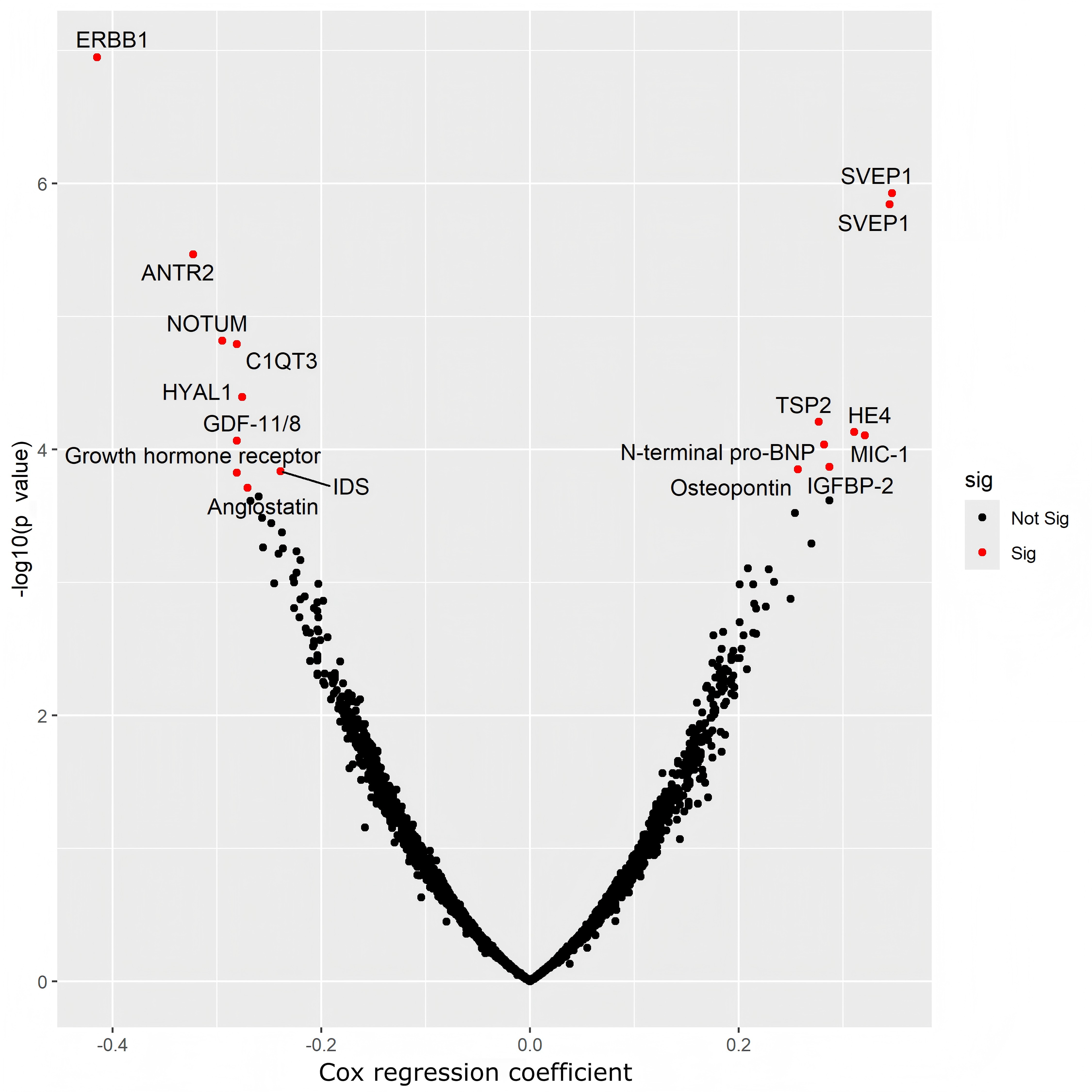
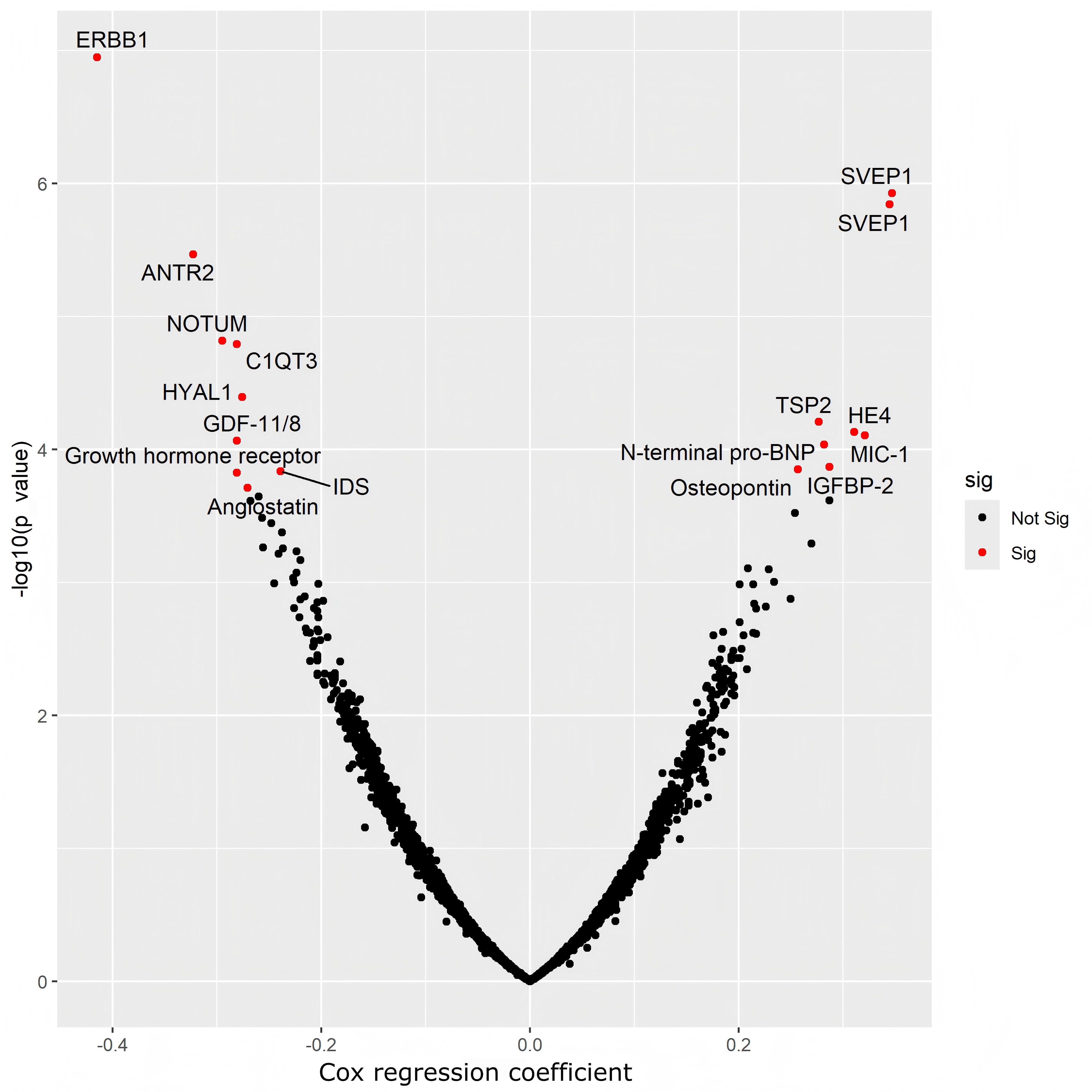
Figure 1. Association of proteins with incident mortality. Volcano plot showing associated proteins as red dots (p-value < 1.0 × 10⁻⁵). The x-axis shows the standardized Cox regression coefficient (β), representing the association between a one standard deviation increase in protein abundance and all-cause mortality after adjustment for age, sex, and cohort (OPEL/OPUS). Red points denote proteins meeting the predefined significance threshold. OPEL: offspring of parents with exceptional longevity; OPUS: offspring of parents with usual survival; ERBB1: epidermal growth factor receptor; ANTXR2: anthrax toxin receptor 2; IDS: iduronate 2-sulfatase; IGFBP-2: insulin-like growth factor-binding protein 2; MIC-1: macrophage inhibitory cytokine 1; GDF-11/8: growth differentiation factor-11/8; HYAL1: hyaluronidase-1; HE4: human epididymis protein 4; TSP2: thrombospondin-2; SVEP1: sushi, von Willebrand factor type A, EGF, and pentraxin domain-containing protein 1.
Conversely, elevated levels of several proteins were positively associated with mortality. These included sushi, von Willebrand factor type A, EGF and pentraxin domain-containing protein 1 (SVEP1), represented by two SOMAmers (HR = 1.41; 95% CI: 1.23-1.63 and 1.41; 95% CI: 1.28-1.63), thrombospondin-2 (TSP2) (HR = 1.32; 95% CI: 1.15-1.51), growth/differentiation factor 15 (GDF15) (HR = 1.38; 95% CI: 1.17-1.62), Osteopontin (HR = 1.29; 95% CI: 1.13-1.47), and insulin-like growth factor-binding protein 2 (IGFBP-2) (HR = 1.33; 95% CI: 1.15-1.54). Notably, N-terminal pro-BNP, a recognized marker of cardiovascular stress, was also positively associated with mortality (HR = 1.32; 95% CI: 1.15-1.53) (Table 2 and Figure 1). Full summary statistics for all the top proteins are provided in Table S1. In the SomaScan assay, SVEP1 is measured using two independent aptamers (11109-56 and 11178-21) targeting different domains of the protein: Sushi domains 15-18 (11109-56) and EGF-like domains 4-6 (11178-21). Both measurements are reported in the table to facilitate future validation of this finding. In downstream analyses, the two aptamers were represented separately as SVEP1 (11109-56) and SVEP1.1 (11178-21), respectively.
The proportional hazards assumption was evaluated for each of the 17 mortality-associated proteins using Schoenfeld residuals. None of the protein-specific tests or corresponding global tests showed statistically significant evidence of violating the proportional hazards assumption (all p > 0.05), supporting the validity of the estimated hazard ratios. The associations remained similar after adjusting for global health score for all of the associated proteins (Table S2). A detailed overview of the possible functionality of proteins with respect to aging and age-associated phenotypes is provided in Table S3.
IPA identified pathways related to cell death and survival, cellular growth and proliferation, cancer, organismal injury and abnormalities, cardiovascular system development and function, and organismal development and survival to be the top pathways related to incident mortality (Table S4). Cancer, organismal injury and neurological disease were the top networks associated with incident mortality (Table S4).
Replication: In BLSA, 11 of the 17 proteins demonstrated significant associations with mortality after FDR correction, including SVEP1, TSP2, macrophage inhibitory cytokine 1 (MIC-1), N-terminal pro-BNP, HE4, Epidermal Growth Factor Receptor (ERBB1), Angiostatin, Osteopontin, C1QT3, and IGFBP-2. Others, such as NOTUM, ANTR2, IDS, HYAL1, and GDF-11/8, did not show statistically significant associations after FDR correction (Table S5).
In comparative analysis with the Eiriksdottir et al., study, 13 of the 17 proteins demonstrated strong and highly significant associations with mortality at the predefined threshold of p < 1×10⁻⁵[9]. Many of these proteins, including SVEP1, GDF15, and ANTR2, showed extremely low p-values, reflecting highly robust and reproducible associations across studies. Only proteins C1q and tumor necrosis factor-related protein 3 (CTRP3), IDS, Angiostatin, and HYAL1 did not meet the stringent cut-off (p ≥ 1 × 10⁻⁵) (Table S6). Apart from Hyaluronidase-1 (p = 0.074), all remaining proteins showed nominal associations with mortality (p < 0.05).
3.3 Association of mortality associated proteins with chronic conditions and clinical measures
Based on the analysis of associations between the 17 mortality-linked proteins and various chronic conditions and clinical measures, a clear hierarchy of associative strength was identified. The results, summarized in Table 3, show that ANTXR2 and ERBB1 had the greatest number of associations in the most stringent significance category (p < 0.001), with 13 and 12 significant associations, respectively. Growth hormone receptor and MIC-1 had 8 associations each in this category, followed by IGFBP-2 and GDF-11/8 with 7 each. HYAL1 and NOTUM each had 6 associations, while the remaining had fewer.
Table 3. Ranked Summary of Associations for Mortality-Linked Proteins with Chronic Conditions and Clinical Measures.
| Target | p < 0.05 | p < 0.01 | p < 0.001 | Total |
| ERBB1 | 2 | 2 | 12 | 16 |
| ANTR2 | 0 | 2 | 13 | 15 |
| IGFBP-2 | 3 | 4 | 7 | 14 |
| Growth hormone receptor | 2 | 1 | 8 | 11 |
| MIC-1 | 2 | 1 | 8 | 11 |
| GDF-11/8 | 0 | 4 | 7 | 11 |
| HYAL1 | 2 | 2 | 6 | 10 |
| TSP2 | 4 | 1 | 5 | 10 |
| NOTUM | 1 | 2 | 6 | 9 |
| IDS | 4 | 1 | 4 | 9 |
| N-terminal pro-BNP | 4 | 2 | 2 | 8 |
| HE4 | 2 | 3 | 1 | 6 |
| SVEP1.1 | 3 | 2 | 1 | 6 |
| Angiostatin | 1 | 1 | 3 | 5 |
| C1QT3 | 2 | 0 | 2 | 4 |
| Osteopontin | 1 | 2 | 1 | 4 |
| SVEP1 | 2 | 2 | 0 | 4 |
Values represent the number of significant associations observed for each protein across all tested chronic conditions and clinical measures at nominal significance thresholds (p < 0.05, p < 0.01, and p < 0.001). These analyses are exploratory and hypothesis-generating. p-values were not adjusted for multiple testing. ERBB1: epidermal growth factor receptor; ANTXR2: anthrax toxin receptor 2; IDS: iduronate 2-sulfatase; IGFBP-2: insulin-like growth factor-binding protein 2; MIC-1: macrophage inhibitory cytokine 1; GDF-11/8: growth differentiation factor 11/8; HYAL1: hyaluronidase-1; HE4: human epididymis protein 4; ; C1QT3: C1q and tumor necrosis factor-related protein 3; TSP2: thrombospondin-2; SVEP1: sushi, von Willebrand factor type A, EGF, and pentraxin domain-containing protein 1.
In a complementary analysis, we examined which chronic conditions and clinical measures exhibited the greatest number of correlations with the set of mortality associated proteins, as summarized in Figure S1. This approach provides insight into which phenotypes may be most broadly implicated in proteomic pathways linked to mortality and disease. Notably, the Frailty Index demonstrated the strongest pattern of association, correlating with 13 out of the 17 mortality-associated proteins. In contrast, depression exhibited the fewest associations, with only 5 proteins showing significant links. This ranking helps highlight which phenotypes and clinical measures may serve as the most robust indicators of overall health risk and mortality, suggesting that markers like frailty may reflect a convergence of multiple biological aging processes. The number of participants available for association analyses varied across disease-related phenotypes and clinical measures because of variable-specific missingness; the number and percentage of participants with available data for each measure are provided in Table S7.
3.4 Association of mortality associated proteins with biological aging
3.4.1 LonGenity
In the analysis of the relationship between mortality-associated proteins and overall biological aging (Conventional), several proteins demonstrated highly significant associations. The results indicated that higher levels of MIC-1 (GDF15) (p = 4.13E-47), HE4 (p = 7.00E-38), N-terminal pro-BNP (p = 1.10E-29), and IGFBP-2 (p = 1.43E-26) were positively correlated with an older biological age captured by the conventional aging clock. Significant positive associations were also observed for TSP2 (p = 5.11E-14), SVEP1 (Two aptamers: p = 4.19E-11, p = 1.55E-09) and Osteopontin (p = 3.03E-11)(Figure 2 and Table S8). Conversely, a number of proteins were inversely associated with biological aging, where higher protein levels correlated with a younger biological age. The most significant negative associations were seen with ERBB1 (p = 1.50E-21), Growth hormone receptor (p = 1.75E-16), Angiostatin (p = 2.11E-11), and NOTUM (p = 3.01E-09). Other proteins, including GDF-11/8 (p = 1.33E-07), ANTR2 (p = 1.62E-04), and C1QT3 (p = 1.38E-03), also showed a significant negative correlation. In contrast, IDS and HYAL1 did not show a statistically significant association with biological aging in this analysis.
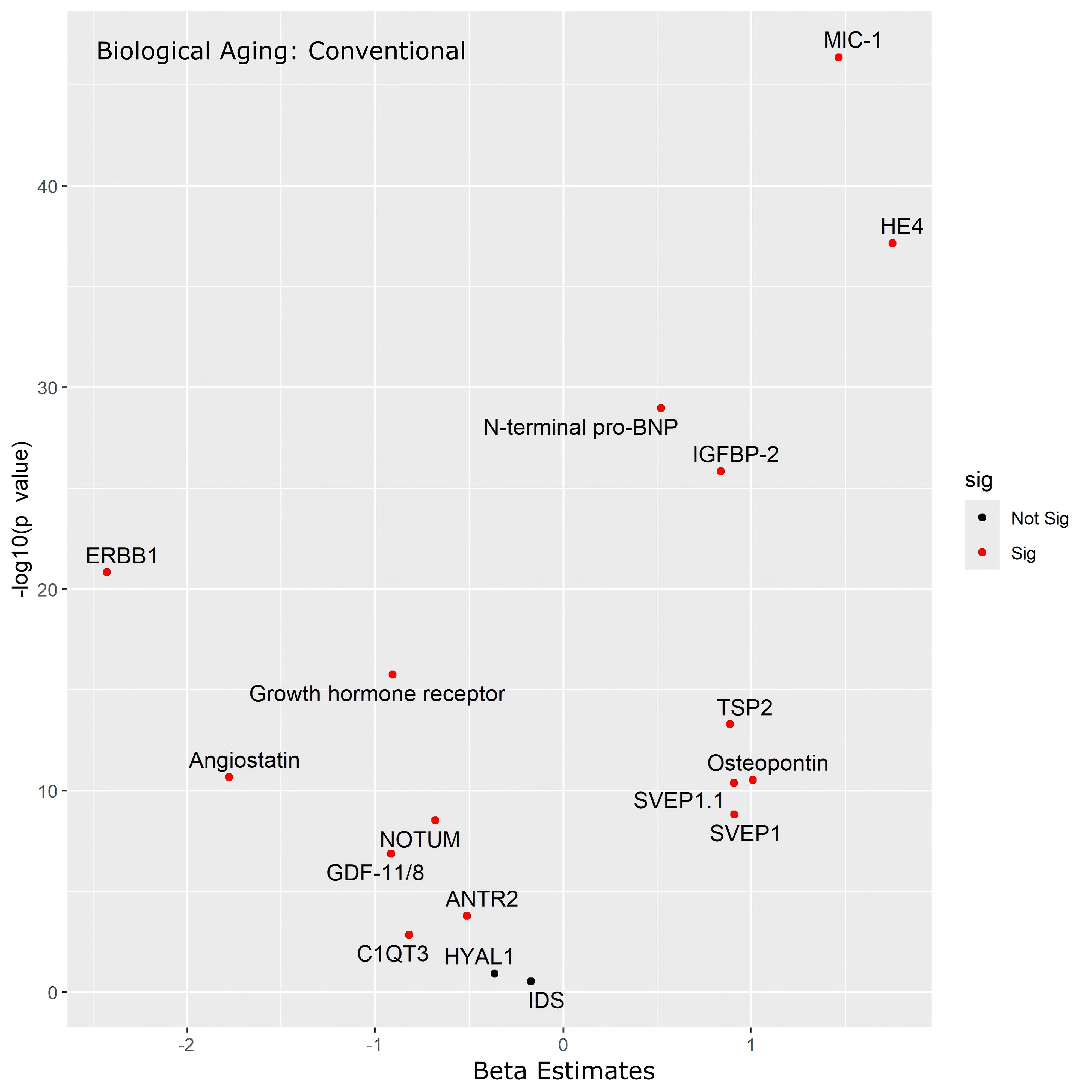
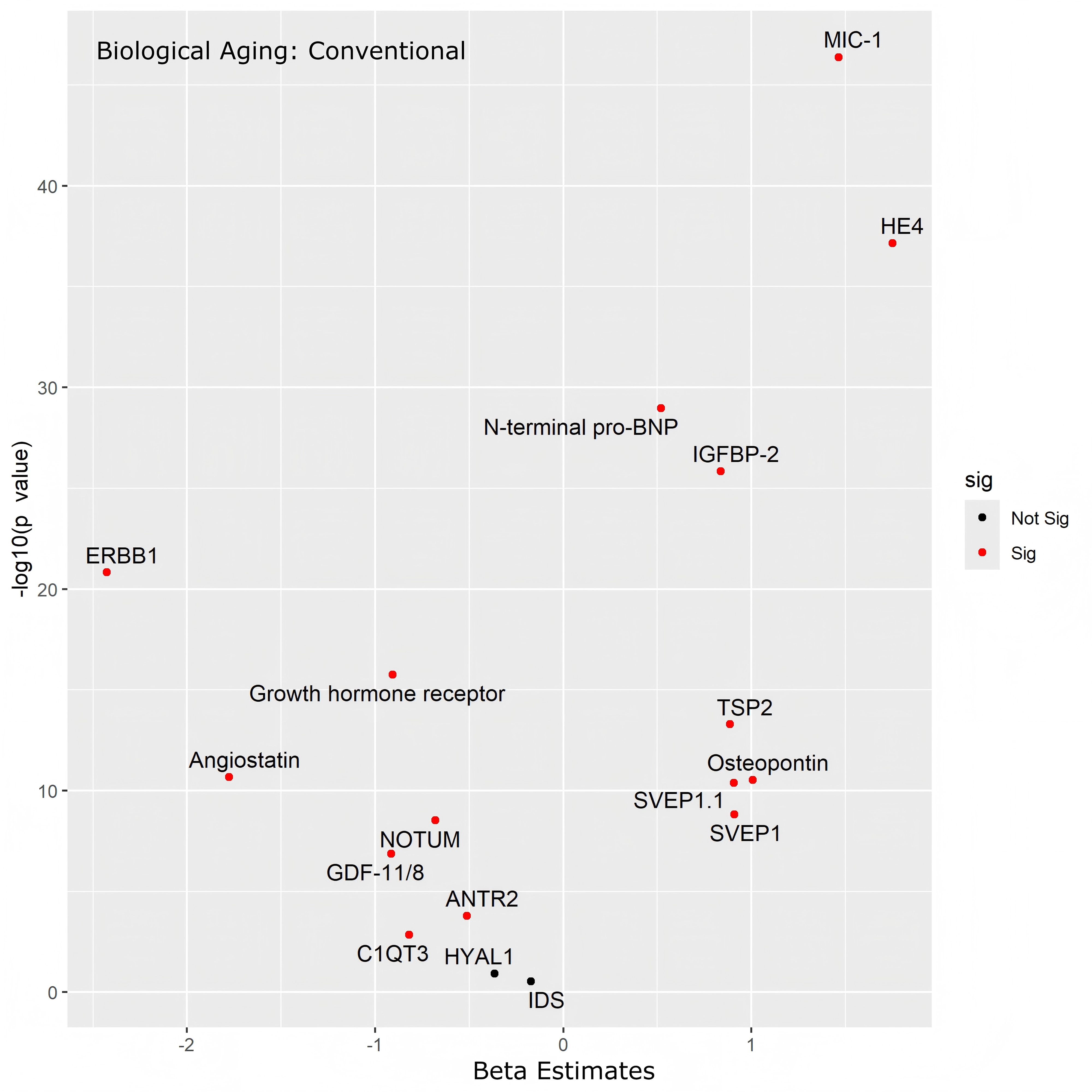
Figure 2. Mortality-associated proteins and conventional biological aging estimated using a proteomic aging clock. x-axis denotes the beta estimate coefficients, and y-axis, the significance level presented as -log10 (p-value) from a linear regression model adjusted for age, sex, and parental longevity status. Topmost hit proteins have been marked. ERBB1: epidermal growth factor receptor; ANTXR2: anthrax toxin receptor 2; IDS: iduronate 2-sulfatase; IGFBP-2: insulin-like growth factor-binding protein 2; MIC-1: macrophage inhibitory cytokine 1; GDF-11/8: growth differentiation factor-11/8; HYAL1: hyaluronidase-1; HE4: human epididymis protein 4; TSP2: thrombospondin-2; SVEP1: sushi, von Willebrand factor type A, EGF, and pentraxin domain-containing protein 1; C1QT3: C1q and tumor necrosis factor-related protein 3.
Further analysis of aging in five individual organ systems (heart, brain, immune, kidney, and liver) and the systemic “Organismal” score revealed distinct patterns (Figure 3 and Table S8). A core set of proteins showed consistent associations across systems. Higher levels of MIC-1 and HE4 were significantly associated with older age across all six measures. The strongest associations for MIC-1 was the organismal score (p = 1.05E-54), and for HE4 with brain aging (p = 3.44E-63). Similarly, IGFBP-2 and N-terminal pro-BNP were robustly correlated with older age across most measures, including the heart (IGFBP-2, p = 8.68E-36; N-terminal pro-BNP, p < 2.1E-100) and the systemic organismal aging (IGFBP-2, p = 8.05E-29; N-terminal pro-BNP, p = 2.99E-29). Conversely, higher levels of ERBB1 and NOTUM showed significant negative correlations with aging across all six measures evaluated, with particularly strong associations for ERBB1 with liver (p = 5.84E-32) and brain (p = 2.36E-22) aging, and for NOTUM with liver (p = 8.44E-18) and heart (p=8.06E-14) aging. Angiostatin and the Growth hormone receptor were also strongly associated with younger age across most systems, though the association was not significant for the kidney (p = 6.50E-02 and p = 1.69E-01, respectively). Some aptamers displayed more organ-specific patterns; for example, one of the SVEP1 capturing aptamers (SVEP1.1) showed a particularly strong positive association with heart aging (p = 6.70E-21) but was not significantly associated with immune (p = 5.13E-02) or kidney (p = 3.10E-01) aging. Kidney age showed the least number of proteins associations, with 8 out of 17 proteins not associated (p > 0.05) (Figure 3 and Table S8).
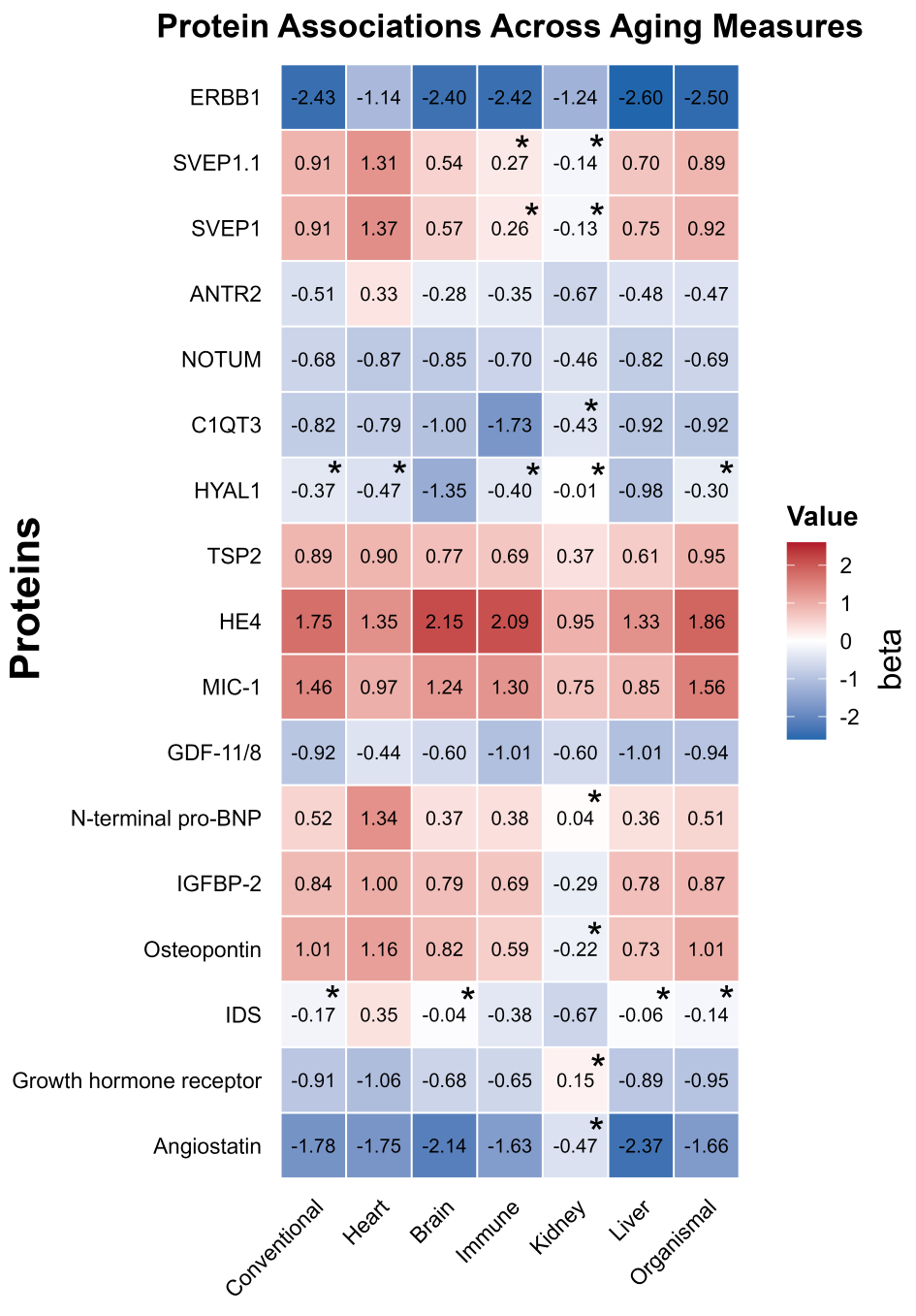
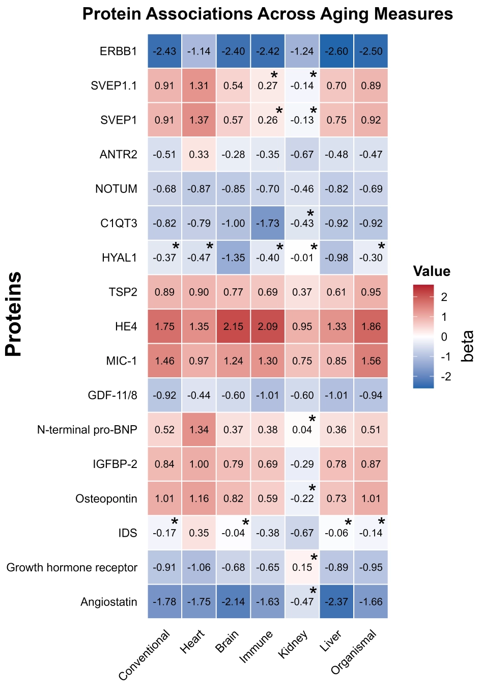
Figure 3. Heatmap of associations between mortality-associated plasma proteins and biological aging measures. Values represent standardized regression coefficients (β). Red indicates positive associations (accelerated aging) and blue indicates negative associations (younger biological age). * indicates that the association with the corresponding biological aging measure was not statistically significant (p ≥ 0.05). All other associations were statistically significant. Brain and Immune aging measures show particularly strong cross-cohort agreement, especially for HE4, MIC-1, ERBB1, NOTUM, and IGFBP-2. We observed distinct organ-specific patterns, validating the biological specificity of the aging measures. N-terminal pro-BNP, a well-established cardiac marker, demonstrated the strongest association specifically with heart aging in both LonGenity (β = 1.34, p < 2.1E-100) and BLSA (β = 0.94, p < 2E-16) cohorts, while showing non-significant or weaker associations with kidney aging in these models. ERBB1: epidermal growth factor receptor; ANTXR2: anthrax toxin receptor 2; IDS: iduronate 2-sulfatase; IGFBP-2: insulin-like growth factor-binding protein 2; MIC-1: macrophage inhibitory cytokine 1; GDF-11/8: growth differentiation factor-11/8; HYAL1: hyaluronidase-1; HE4: human epididymis protein 4; C1QT3: C1q and tumor necrosis factor-related protein 3; TSP2: thrombospondin-2; SVEP1: sushi, von Willebrand factor type A, EGF, and pentraxin domain-containing protein 1.
3.4.2 BLSA
We validated these findings in an independent cohort using the BLSA. The associations observed in the primary analysis were largely replicated, with strong concordance in both the direction and magnitude of effects. MIC-1 (GDF15; p = 2.59E-61) and HE4 (p = 1.42E-41) exhibited the largest effect sizes, followed by SVEP1 (both aptamers: p = 3.40E-21 and p = 2.43E-20), TSP2 (p = 3.21E-22), and N-terminal pro-BNP (β = 0.25, p = 1.52E-17) with the conventional aging measure (Table S9). Osteopontin, IDS, IGFBP-2, and C1QT3 were all positively associated with the conventional aging measure (p < 0.05). Proteins negatively associated with conventional aging included ERBB1 (p = 4.55E-12), GDF-11/8 (p = 3.26E-6), NOTUM (p = 1.38E-6), and ANTR2 (p = 4.78E-6). Growth hormone receptor levels also declined with age, showing a nominal association (p = 0.034). HYAL1 and Angiostatin were not significantly associated with the conventional aging measure.
4. Discussion
This study identified proteins significantly associated with all-cause mortality in a cohort of older adults. Our subsequent analyses revealed that this signature is not simply a collection of disparate risk factors, but rather a coherent molecular fingerprint closely linked to the burden of chronic diseases and the pace of biological aging across multiple organ systems. When additionally adjusted for the GHS, which summarizes overall health and comorbidity burden, all proteins retained significant associations. This finding suggests that the associations we observe are not solely explained by global health status or accumulated deficits but instead reflect possible independent biological processes that contribute to mortality risk. Comparison with thr BLSA study and the Eiriksdottir study further emphasizes the influence of age in shaping proteomic associations[9]. While most of the proteins identified in our analysis were also associated with mortality in those studies, four markers, including CTRP3, IDS, Angiostatin, and HYAL1, did not replicate in the Eiriksdottir study. A key difference lies in the age distribution: our cohort was restricted to adults aged 65-95 years, whereas the Eiriksdottir study included participants spanning 18-80+ years. This broader range likely captured age-related biological processes that differ in their relevance to mortality risk across the life course, whereas our findings specifically highlight proteomic signatures of late-life mortality. Similarly, in BLSA, we found associations between most of the top proteins and mortality. The findings provide a systems-level perspective on the biological processes that underpin healthspan and longevity, highlighting key pathways related to growth, tissue maintenance, and systemic stress.
Together, the 17 mortality-associated proteins may represent interconnected biological processes involved in aging and longevity beyond their individual associations. Several proteins, including Thrombospondin-2 (THBS2), Osteopontin (SPP1), Angiostatin (PLG), HYAL1, SVEP1, and ANTXR2, are involved in extracellular matrix (ECM) organization, tissue remodeling, and cell-matrix interactions, supporting the emerging concept that age-related alterations in the circulating matrisome are associated with biological aging and disease risk[28]. Other proteins, including ERBB1, GHR, GDF11/8, and IGFBP-2, participate in growth factor signaling pathways that regulate tissue maintenance, regeneration, and metabolic homeostasis[29,30]. In contrast, MIC-1 (GDF15), HE4 (WFDC2), and N-terminal pro-BNP (NPPB) primarily reflect systemic stress responses, chronic inflammation, cardiovascular dysfunction, and end-organ injury[31]. Together, these findings suggest that mortality in older adults is associated with coordinated dysregulation of extracellular matrix homeostasis, intercellular signaling, tissue repair, and systemic stress responses rather than perturbation of a single biological pathway.
Importantly, the identified proteins should primarily be viewed as circulating biomarkers associated with mortality risk rather than direct mediators of aging or longevity. Many are secreted or extracellular proteins released during tissue remodeling, or induced in response to inflammation, chronic tissue injury or cellular stress. Although several of these proteins have been implicated in senescence-associated secretory pathways and age-related inflammatory signaling, the present observational analyses cannot distinguish causal mechanisms from downstream biological responses. Accordingly, the identified protein signature is best interpreted as reflecting the cumulative physiological consequences of biological aging and chronic disease, while also highlighting candidate pathways for future mechanistic investigation.
A key finding of this work is the identification of proteins whose circulating levels are inversely associated with mortality. The role of ERBB1 (EGFR) is particularly noteworthy given its well-established role in promoting cellular proliferation, which is often linked to cancer[32]. However, our data demonstrate that higher ERBB1 levels are also strongly associated with a younger biological age across all organ systems evaluated. This suggests that in the context of aging, maintaining robust ERBB1 signaling may be essential for homeostatic processes such as tissue repair and cellular turnover, and that these benefits may outweigh its oncogenic potential in an elderly population. Similarly, the effect of ANTR2, a protein critical for ECM adhesion and vascular morphogenesis[33], aligns with our pathway analysis implicating a significant role for ECM organization in mortality. Loss of ANTR2 function in mouse models is associated with tissue-specific ECM dysregulation or fibrosis[34,35], underscoring that its sustained expression is vital for maintaining tissue integrity and preventing the widespread fibrotic changes characteristic of aging.
This analysis also reinforces the importance of the somatotropic and related growth factor axes in aging, though with some complexity. Greater levels of GHR and GDF-11/8 were associated with reduced mortality and a younger biological age. The finding for GHR appears to contrast with studies in model organisms where reduced GH signaling extends lifespan, supporting the concept of a trade-off between somatic growth and longevity[36]. In humans, particularly in later life, our results may suggest that the anabolic and supportive functions of the GH axis are critical for preventing frailty and sarcopenia, making its preservation beneficial for survival. The association of GDF-11/8 is noteworthy given its proposed role as a rejuvenation factor that has been reported in some models to improve age-related phenotypes, although conflicting evidence regarding its effects on aging remains[37]. Our findings suggest that circulating GDF-11/8 levels are associated with aging-related health phenotypes, although the observational nature of our study precludes conclusions regarding a causal beneficial effect[38].
Conversely, several proteins demonstrated strong positive associations with both mortality and advanced biological age. Among these, elevated MIC-1 (GDF15) and HE4 were the strongest predictors of advanced biological age across all organ systems. This aligns with their known biology; MIC-1 is a well-documented marker of cellular stress, inflammation, and all-cause mortality[31], while HE4, best known as a biomarker for gynecological cancers[39], is also elevated in non-malignant chronic conditions of aging such as chronic kidney disease and heart failure[40]. Their consistent associations across multiple organ systems highlight their role as indicators of systemic decline rather than organ-specific decline.
The paradoxical nature of IGFBP-2 was also evident. While some studies associate it with favorable metabolic profiles in healthy individuals[41], our findings align with growing evidence that, in the context of chronic disease and advanced age, elevated IGFBP-2 reflects a catabolic and inflammatory state that is strongly associated with increased mortality[30,42]. Its positive correlation with both mortality and biological age in our cohort reinforces its role as an integrative biomarker of severe physiological stress. Our study also reaffirms SVEP1 as a key risk factor. Its strong association with increased mortality and older biological age, particularly within the heart, is consistent with genetic evidence identifying SVEP1 as a causal driver of atherosclerosis and vascular inflammation[43].
This study demonstrates key strengths, such as employing a large-scale proteomic panel and validation and further analysis in an independent cohort. We also acknowledge the limitations related to the use of a proteomic panel based on the same technology and the need for replicating using other platforms like Olink or mass spectrometry. Additionally, deriving biological age from a single technology, such as the SomaScan panel, raises concerns about generalizability. Cross-platform validation would also help confirm target specificity and assess the reproducibility of the identified mortality-associated proteins across different proteomic technologies. Further studies are warranted in this direction. This study is observational in nature, and the associations observed should be interpreted as hypothesis-generating, and further experimental studies, genetic approaches like Mendelian randomization, and mechanistic investigations are needed to determine whether these proteins play causal roles in aging and longevity. Another limitation of this study is that the discovery cohort consisted exclusively of older Ashkenazi Jewish adults[15], which may limit the generalizability of the findings to other populations. However, the LonGenity cohort has served as the discovery cohort for several plasma proteomic biomarkers of aging, including the proteomic frailty index[21], proteomic aging clock[15], and organ-specific aging clocks[14], which have subsequently been validated in independent cohorts with diverse participant characteristics. These observations support the utility of this well-characterized cohort for biomarker discovery. Nevertheless, because circulating proteomic profiles may differ across ancestral groups, further validation of the present findings in larger, multi-ethnic populations is warranted.
In conclusion, this study moves beyond the identification of individual mortality-associated proteins and reveals a cohesive proteomic signature of systemic health and biological aging. This signature reflects the status of fundamental biological axes such as growth signaling, extracellular matrix integrity, and inflammatory stress. The varied associations of these proteins with mortality and biological age highlight the complex trade-offs involved in preserving health later in life.
Supplementary materials
The supplementary material for this article is available at: Supplementary materials.
Acknowledgements
ChatGPT (OpenAI) was used solely for language polishing and grammar check during the preparation of this manuscript. No AI tools were used for any other aspect including data analysis, interpretation of results, or generation of scientific conclusions.
The sponsors had no role in the design and conduct of the study; collection, management, analysis, and interpretation of the data; and preparation, review, or approval of the manuscript. The authors reviewed, revised, and approved the final manuscript and take full responsibility for its content.
Authors contribution
Garvey N, Sathyan S: Conceptualization, data curation, formal analysis, investigation, writing-original draft.
Tanaka T, Oh HSH, Joynes C, Gao T, Wyss-Coray T, Walker KA: Data curation, formal analysis.
Verghese J, Ferrucci L, Milman S, Barzilai N: Conceptualization, data curation, funding acquisition, supervision.
All the authors approved the final version of the manuscript and agree to be accountable for all aspects of the work.
Conflicts of interest
Keenan A. Walker is an Associate Editor for Alzheimer’s & Dementia: The Journal of the Alzheimer’s Association, Alzheimer’s & Dementia: Translational Research and Clinical Interventions (TRCI), a member of the Editorial Board of Annals of Clinical and Translational Neurology. Keenan Walker is on the Board of Directors of the National Academy of Neuropsychology. Keenan Walker has given unpaid presentations and seminars on behalf of SomaLogic. Keenan Walker is a co-founder of Centia Bio. The work presented in this manuscript was conducted independently of Centia Bio and without financial support from the company. The other authors declare no competing interests.
Ethical approval
LonGenity cohort: The study protocol was approved by the Albert Einstein College of Medicine Institutional Review Board (Protocol number 2007-272).
BLSA cohort: The study protocol was approved by the National Institutes of Health Intramural Research Program Institutional Review Board (Protocol number 03-AG-0325).
Consent to participate
LonGenity cohort: All participants provided written informed consent for study assessments and genetic testing prior to enrollment.
BLSA cohort: The informed consent was obtained from participants at each visit.
Consent for publication
Not applicable.
Availability of data and materials
LonGenity proteomic data used in this study are available upon request. Please contact the corresponding author for further information. BLSA data is available upon request and proposal and submit a pre-analysis plan for approval (https://blsa.nia.nih.gov/how-apply).
Funding
LonGenity study and work was supported by grants from the National Institutes of Health (NIH; Grant Nos. P01AG021654, R01AG046949, and R01AG057909; R01AG044829, R01AG057548, R01AG044829, and R01AG061155), the Nathan Shock Center of Excellence for the Biology of Aging (Grant No. P30AG038072), and the Glenn Center for the Biology of Human Aging (Paul Glenn Foundation Grant). Baltimore Longitudinal Study of Aging and work was supported by the National Institute on Aging, Intramural Research Program (Grant Nos. ZIAAG000015 and ZIAAG000191).
Copyright
© The Author(s) 2026.
References
-
1. Herrmann W, Herrmann M. The importance of telomere shortening for atherosclerosis and mortality. J Cardiovasc Dev Dis. 2020;7(3):29.[DOI]
-
3. Kumar N, Walia HK. Cellular senescence and stem cell exhaustion: Implications for aging and regeneration. In: Qamar I, Maurya PK, editors. Cellular senescence, age-related disorders, and emerging treatments. Singapore: Springer; 2025. p. 103-133.[DOI]
-
5. Niedernhofer LJ, Gurkar AU, Wang Y, Vijg J, Hoeijmakers JHJ, Robbins PD. Nuclear genomic instability and aging. Annu Rev Biochem. 2018;87:295-322.[DOI]
-
6. López-Otín C, Blasco MA, Partridge L, Serrano M, Kroemer G. The hallmarks of aging. Cell. 2013;153(6):1194-1217.[DOI]
-
7. López-Otín C, Blasco MA, Partridge L, Serrano M, Kroemer G. Hallmarks of aging: An expanding universe. Cell. 2023;186(2):243-278.[DOI]
-
8. Payne SH. The utility of protein and mRNA correlation. Trends Biochem Sci. 2015;40(1):1-3.[DOI]
-
9. Eiriksdottir T, Ardal S, Jonsson BA, Lund SH, Ivarsdottir EV, Norland K, et al. Predicting the probability of death using proteomics. Commun Biol. 2021;4:758.[DOI]
-
13. Earls JC, Rappaport N, Heath L, Wilmanski T, Magis AT, Schork NJ, et al. Multi-omic biological age estimation and its correlation with wellness and disease phenotypes: A longitudinal study of 3,558 individuals. J Gerontol Ser A. 2019;74(Supplement_1):S52-S60.[DOI]
-
18. Shock NW, Greulich RC, Andres R, Arenberg D, Costa PT, Lakatta EG, et al. Normal human aging: The Baltimore longitudinal study of aging. Washington: U.S. Department of Health and Human Services, Public Health Service, National Institutes of Health, National Institute on Aging, Gerontology Research Center; 1984. Available from: https://books.google.com/books?hl=zh-CN&lr=&id=8OxqAAAAMAAJ&oi=fnd&pg=PR5&dq=Normal+human+aging:+The+Baltimore+longitudinal+study+of+aging&ots=0RyLulJHq8&sig=mlPP_smIU9xv9kTyosCa-okAcPk#v=onepage&q=Normal%20human%20aging%3A%20The%20Baltimore%20longitudinal%20study%20of%20aging&f=false
-
19. Sathyan S, Milman S, Ayers E, Gao T, Verghese J, Barzilai N. Plasma proteomic profile of abdominal obesity in older adults. Obesity. 2024;32(5):938-948.[DOI]
-
20. Rockwood K, Mitnitski A. Frailty in relation to the accumulation of deficits. J Gerontol Ser A Biol Sci Med Sci. 2007;62(7):722-727.[DOI]
-
22. Candia J, Cheung F, Kotliarov Y, Fantoni G, Sellers B, Griesman T, et al. Assessment of variability in the SOMAscan assay. Sci Rep. 2017;7:14248.[DOI]
-
24. Candia J, Daya GN, Tanaka T, Ferrucci L, Walker KA. Assessment of variability in the plasma 7k SomaScan proteomics assay. Sci Rep. 2022;12:17147.[DOI]
-
29. Dehkhoda F, Lee CMM, Medina J, Brooks AJ. The growth hormone receptor: Mechanism of receptor activation, cell signaling, and physiological aspects. Front Endocrinol. 2018;9:35.[DOI]
-
30. van den Beld AW, Carlson OD, Doyle ME, Rizopoulos D, Ferrucci L, van der Lely AJ, et al. IGFBP-2 and aging: A 20-year longitudinal study on IGFBP-2, IGF-I, BMI, insulin sensitivity and mortality in an aging population. Eur J Endocrinol. 2019;180(2):109-116.[DOI]
-
31. Wiklund FE, Bennet AM, Magnusson PKE, Eriksson UK, Lindmark F, Liyun W, et al. Macrophage inhibitory cytokine-1 (MIC-1/GDF15): A new marker of all-cause mortality. Aging Cell. 2010;9(6):1057-1064.[DOI]
-
32. Roskoski R Jr. The ErbB/HER family of protein-tyrosine kinases and cancer. Pharmacol Res. 2014;79:34-74.[DOI]
-
34. Huang X, Zhang L, Luo W, Zeng Y, Li X, Yang N, et al. Endothelial anthrax toxin receptor 2 plays a protective role in liver fibrosis. Front Cell Dev Biol. 2024;11:1278968.[DOI]
-
37. Machelak W, Szczepaniak A, Jacenik D, Zielińska M. The role of GDF11 during inflammation-an overview. Life Sci. 2023;322:121650.[DOI]
-
38. Bartke A. Growth hormone and aging: Updated review. World J Mens Health. 2019;37(1):19.[DOI]
-
39. Hertlein L, Stieber P, Kirschenhofer A, Fürst S, Mayr D, Hofmann K, et al. Human epididymis protein 4 (HE4) in benign and malignant diseases. Clin Chem Lab Med. 2012;50(12):2181-2188.[DOI]
-
42. Barutaut M, Fournier P, Peacock WF, Evaristi MF, Caubère C, Turkieh A, et al. Insulin-like growth factor binding protein 2 predicts mortality risk in heart failure. Int J Cardiol. 2020;300:245-251.[DOI]
Copyright
© The Author(s) 2027. This is an Open Access article licensed under a Creative Commons Attribution 4.0 International License (https://creativecommons.org/licenses/by/4.0/), which permits unrestricted use, sharing, adaptation, distribution and reproduction in any medium or format, for any purpose, even commercially, as long as you give appropriate credit to the original author(s) and the source, provide a link to the Creative Commons license, and indicate if changes were made.
Publisher’s Note
Science Exploration remains a neutral stance on jurisdictional claims in published
maps
and institutional affiliations. The views expressed in this article are solely those
of
the author(s) and do not reflect the opinions of the Editors or the publisher.
Share And Cite


Science Exploration Style
Garvey N, Sathyan S, Tanaka T, Joynes C, Oh H, Gao T, et al. Exploring mortality-linked proteins: Understanding their association with biological aging, disease phenotypes, signaling pathways, and their implications for longevity. Geromedicine. 2027;3:202609. https://doi.org/10.70401/Geromedicine.2026.0042
Tips
Copy completed.
Submit a Manuscript
Author Instructions
Cite this Article
Article Metrics
0
View
0
Download
Cited
Article Updates

 Get citation
Science Exploration Style

Garvey N, Sathyan S, Tanaka T, Joynes C, Oh H, Gao T, et al. Exploring mortality-linked proteins: Understanding their association with biological aging, disease phenotypes, signaling pathways, and their implications for longevity. Geromedicine. 2027;3:202609. https://doi.org/10.70401/Geromedicine.2026.0042
copy
Share Link
copy