Fibroblast growth factor 21 (FGF21) in healthy aging and aging-related diseases

Angeliki Vakka
1,#
,
Frederick Sherman
2,#
,
Kajol Thapa
2
,
Konstantinos Tsioufis
1
,
Konstantinos Drosatos
2,*

*Correspondence to:
Konstantinos Drosatos, Metabolic Biology Laboratory, Cardiovascular Center, Department of Biomedical Sciences, University of Cincinnati College of Medicine, Cincinnati, OH 45267, USA.
E-mail: k.drosatos@uc.edu
Geromedicine. 2027;3:202622. 10.70401/Geromedicine.2026.0045
Received: April 04, 2026Accepted: October 22, 2026Published: October 26, 2026
Abstract
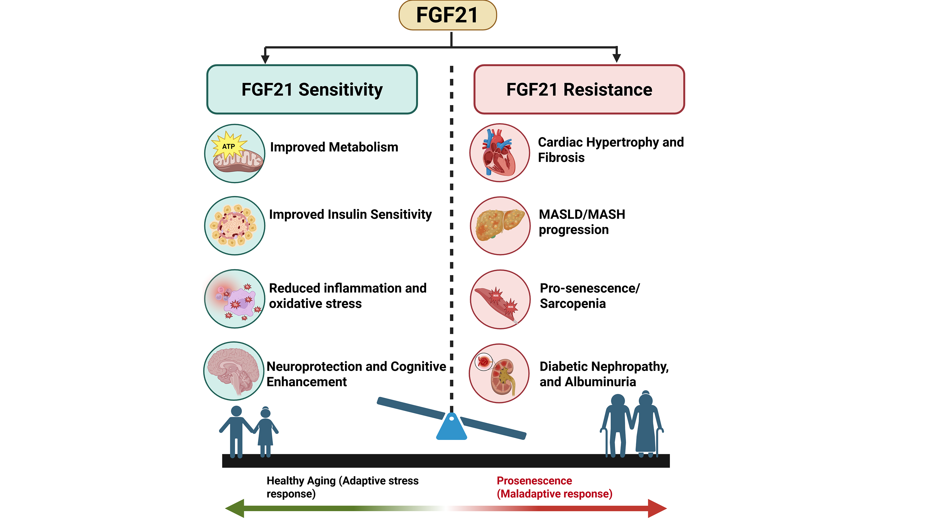
The global human population is aging rapidly, and the prevalence of aging-related diseases is increasing. Thus, understanding the molecular pathways involved in the hallmarks of aging is essential for the development of therapies that promote healthy aging and extend lifespan. Fibroblast growth factor 21 (FGF21) has emerged as a promising therapeutic candidate due to its pleiotropic beneficial roles in metabolism, stress response, and tissue repair through pathways that regulate oxidative stress and cellular senescence. Preclinical studies have associated FGF21 with beneficial effects against metabolic syndrome, liver steatosis, kidney injury, and neurodegenerative disorders that occur in aged individuals. Conversely, FGF21 has also been linked to skeletal muscle loss and concentric cardiac hypertrophy. This review summarizes current evidence on FGF21 signaling and its effects on either healthy aging or aging-related diseases, highlighting its tissue-, time-, origin-, and stress-dependent effects.
Graphical Abstract
Keywords
Aging, FGF21, organ dysfunction, metabolism, interorgan communication
1. Introduction
Aging is a complex biological process associated with the progressive deterioration of physiological functions and increased vulnerability to organ dysfunction and remodeling, ultimately leading to death[1] (Figure 1). With recent technological advances and improvements in modern medicine, global life expectancy has risen significantly[2]. This benefit largely stems from more effective medical therapies introduced in recent decades; however, it has been accompanied by a rising prevalence of diseases with onset at older ages. According to the World Health Organization, by 2030, one in six people will be aged sixty or older, and by 2050, that number will almost double[2]. Statistical models also predict that by 2050, the global fertility rate will fall to 1.83, continuing to decline well below the replacement level of 2.1[3]. This demographic transition, coupled with the rising prevalence of age-related conditions, such as cardiovascular and neurodegenerative diseases, presents new challenges for health care systems and research communities worldwide, as the demand for treatment facilities and medications continues to expand[4].
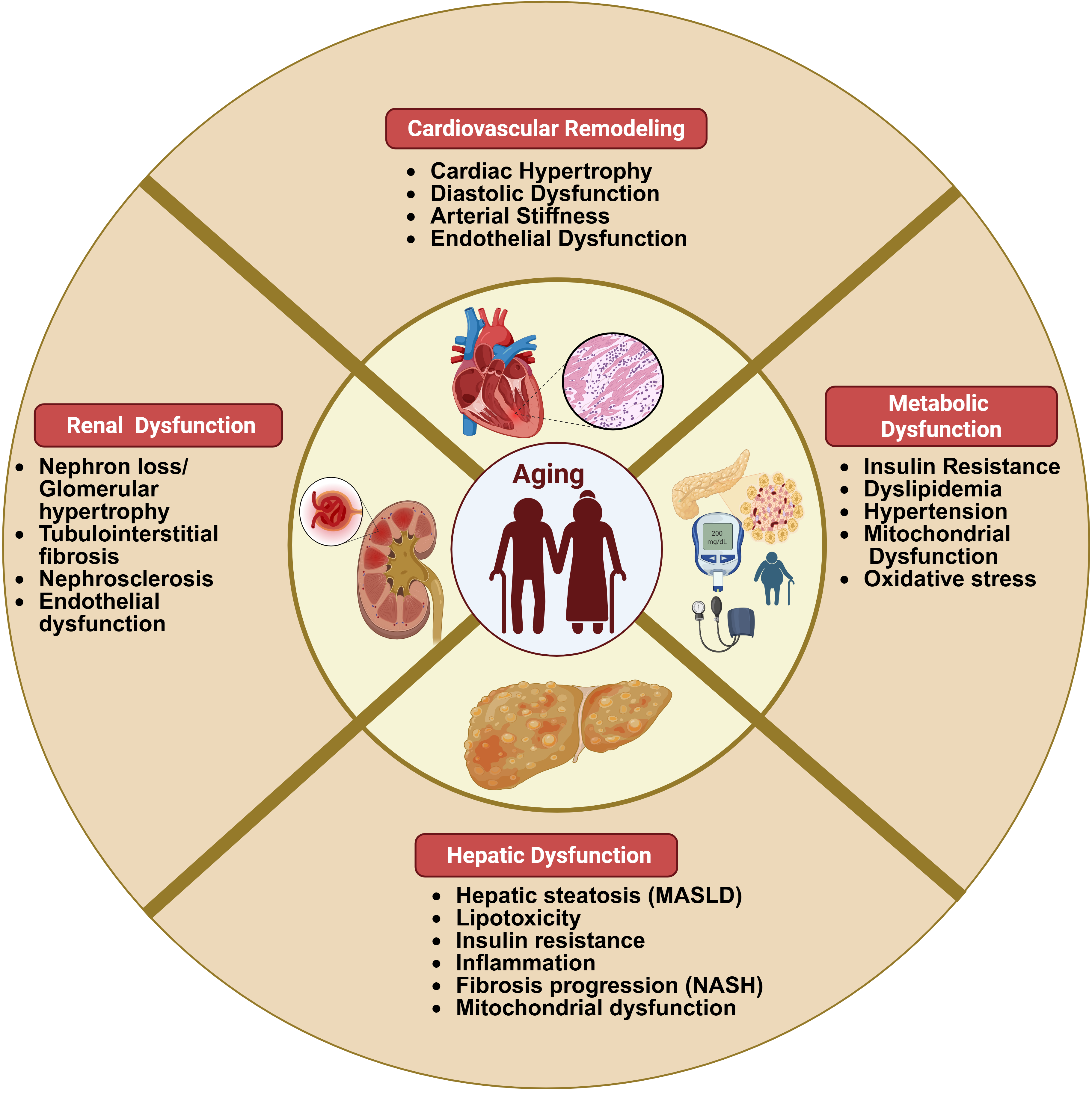
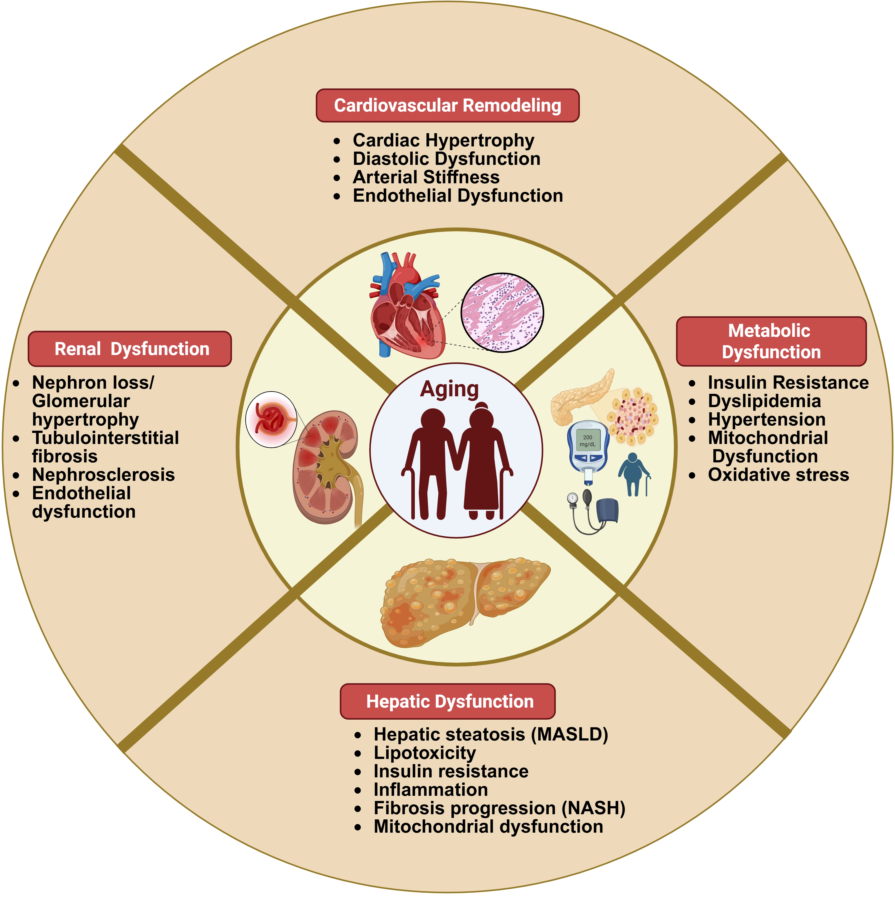
Figure 1. Aging-associated organ dysfunction and remodeling: Summary of the effects of aging in multiple organ systems, including cardiovascular, renal, metabolic, and hepatic systems, leading to progressive functional decline. Created in BioRender. Thapa, K. (2026) https://BioRender.com/zyooutd. MASLD: metabolic dysfunction-associated steatotic liver disease.
The process of aging at the cellular level involves oxidative stress, mitochondrial dysfunction, cellular senescence, reduced autophagy, and chronic inflammation[5]. Understanding the molecular pathways involved in these major hallmarks of aging and identifying those that hold therapeutic potential will enable the development of novel therapies that promote healthy aging and extend lifespan. Approaches such as caloric restriction, inhibition or knockout of specific genes, and pharmacological modulation have shown promise in delaying or reversing aging-associated disorders[6-9]. Among the molecular factors of interest, fibroblast growth factor 21 (FGF21) has emerged as a prominent candidate due to its pleiotropic roles in metabolism, stress response, and tissue repair[10].
This review article summarizes the effects of FGF21 in healthy aging and aging-related diseases that affect multiple organs and discusses its potential therapeutic implications.
2. FGF21
FGF21 is a peptide hormone that belongs to the endocrine FGF19 subfamily. The FGF family consists of 22 members that are classified into six subfamilies based on sequence homology and phylogenetic relationships. The endocrine FGF19 subfamily includes FGF19 (FGF15 in mice), FGF21, and FGF23[11,12]. These members are characterized by the absence of a heparin-binding domain, a feature that facilitates paracrine FGF ligand-receptor interaction[12,13]. The human FGF21 preprotein consists of 209 amino acids, including a 28-amino-acid signaling sequence at the N-terminus[14]. Following proteolysis, FGF21 is secreted as an 181-amino-acid protein[12]. The murine FGF21, which shares 79% similarity to the human ortholog, is encoded by a gene on chromosome 7 and consists of three coding exons, whereas the human FGF21 gene is located on chromosome 19 and contains three coding exons and one non-coding exon[12]. FGF21 is highly conserved among humans’ closest relatives, differing by only one or two amino acids between humans and chimpanzees and between humans and gorillas[15]. Human FGF21 consists of a 13-residue N-terminus, a central β-trefoil core domain, and a 40-residue C-terminus[16]. The C-terminal tail of FGF21 mediates binding to its co-receptor β-Klotho, whereas the N-terminal region is critical for activating the FGF receptor component of the signaling complex[16]. FGF21 requires β-Klotho to activate the following FGF receptors (FGFR): the ‘c’ splice isoforms of FGFR1 (FGFR1c), FGFR2 (FGFR2c), FGFR3 (FGFR3c), and FGFR4, though it exhibits the highest binding affinity for FGFR1[12,13]. FGF21 binds β-Klotho, facilitating the interaction of its N-terminus with FGFR, which subsequently activates multiple downstream intracellular signaling pathways[8].
FGF21 is primarily produced and secreted by the liver, and expression levels vary extensively based on the physiological conditions of an organism[14]. When subjected to physiologically stressful conditions, such as dietary restrictions, cardiomyopathy and obesity, FGF21 is also expressed in other organs, including adipose tissue, cardiac muscle, and skeletal muscle[17-19]. Furthermore, FGF21 has been identified as an adipokine, cardiomyokine, and myokine[10].
Under standard conditions, FGF21 plays an imperative role in maintaining metabolic balance. It enhances insulin sensitivity, which promotes glucose uptake by tissues. FGF21 also facilitates lipolysis, thus increasing circulating fatty acid content[14,17]. Furthermore, FGF21 induces fatty acid beta-oxidation, which increases energy usage and prevents triglyceride accumulation[20]. Overall, in stable conditions, FGF21 serves as an endocrine hormone that closely regulates energy balance and metabolism.
3. FGF21 in Physiology and Aging
In humans, plasma FGF21 levels increase incrementally from 5 to 80 years of age, independently of body composition[21]. Whether elevation of FGF21 compromises health or not is under investigation. Increased serum FGF21 levels in elderly individuals older than 70 years correlate with higher serum glucose levels, insulin resistance, and HOMA-IR[22]. FGF21 is considered a mitokine (mitochondrial stress response molecule), which, along with other mitokines, such as growth differentiation factor 15 (GDF15) and humanin (HN), is significantly increased with age, with the highest levels found in centenarians[23]. Elevation of these mitokines is associated with higher BMI, higher plasma levels of CRP, triglycerides, and uric acid in individuals aged 61-99 years, and decreased survival in individuals aged 82 years and above[23]. Disease-free centenarians have significantly lower plasma FGF21 concentrations compared to healthy controls of 70-80 years[24]. In the Polsenior2 study, higher plasma FGF21 levels were a predictor of all-cause mortality among 3,512 subjects aged 60 years or older during a follow-up of 4.19 years, independently of co-morbidities and blood parameters[25]. Plasma levels of FGF21 have also been associated with the presence of frailty in older community-dwelling adults[26,27]. Thus, various studies have associated plasma FGF21 abundance with worse aging-related outcomes, implying a detrimental role.
The higher levels of circulating FGF21 are accompanied by enhanced FGF21 signaling. FGF21 mainly signals through the FGFR1/β-Klotho receptor complex to activate downstream signaling, including extracellular signal-regulated kinase (ERK), phosphatidylinositol 3-kinase (PI3K)-AKT, and AMP-activated protein kinase (AMPK)-sirtuin 1 (SIRT1) pathways, leading to metabolic adaptations that enhance glucose utilization, fatty acid oxidation, mitochondrial function, and cell survival while suppressing inflammation[28-30] (Figure 2). This was shown in a study that found elevated β-Klotho levels, unchanged FGFR1 levels, and activation of the ERK 1/2 signaling pathway in subcutaneous adipose tissue from individuals above 70 years old[22]. Aged mice also have higher circulating plasma FGF21 levels and normal levels of expression of FGF21 signaling mediators[22]. In the adipose tissue from older mice, the mRNA levels of FGF21 are also significantly higher compared to those of younger mice[31]. Further, adipose tissue explants from young and aged mice that were treated with FGF21 have similar responsiveness to FGF21[22]. Thus, elevation of circulating FGF21 levels does not seem to result from an FGF21-resistance state. Moreover, it remains unclear whether elevated FGF21 levels constitute a serendipitous aging-related effect or a critical element of the pathophysiology of aging-related diseases.
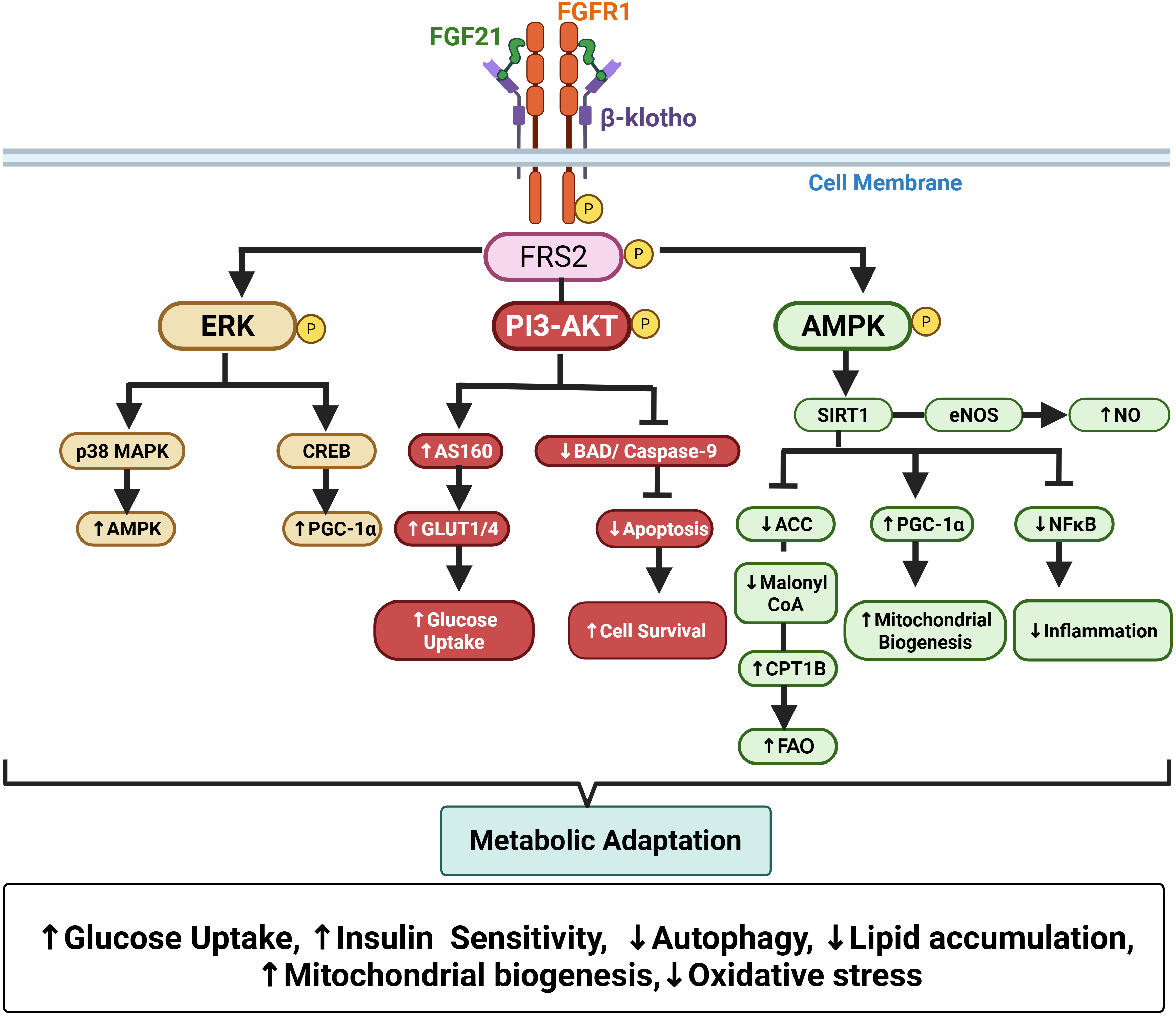
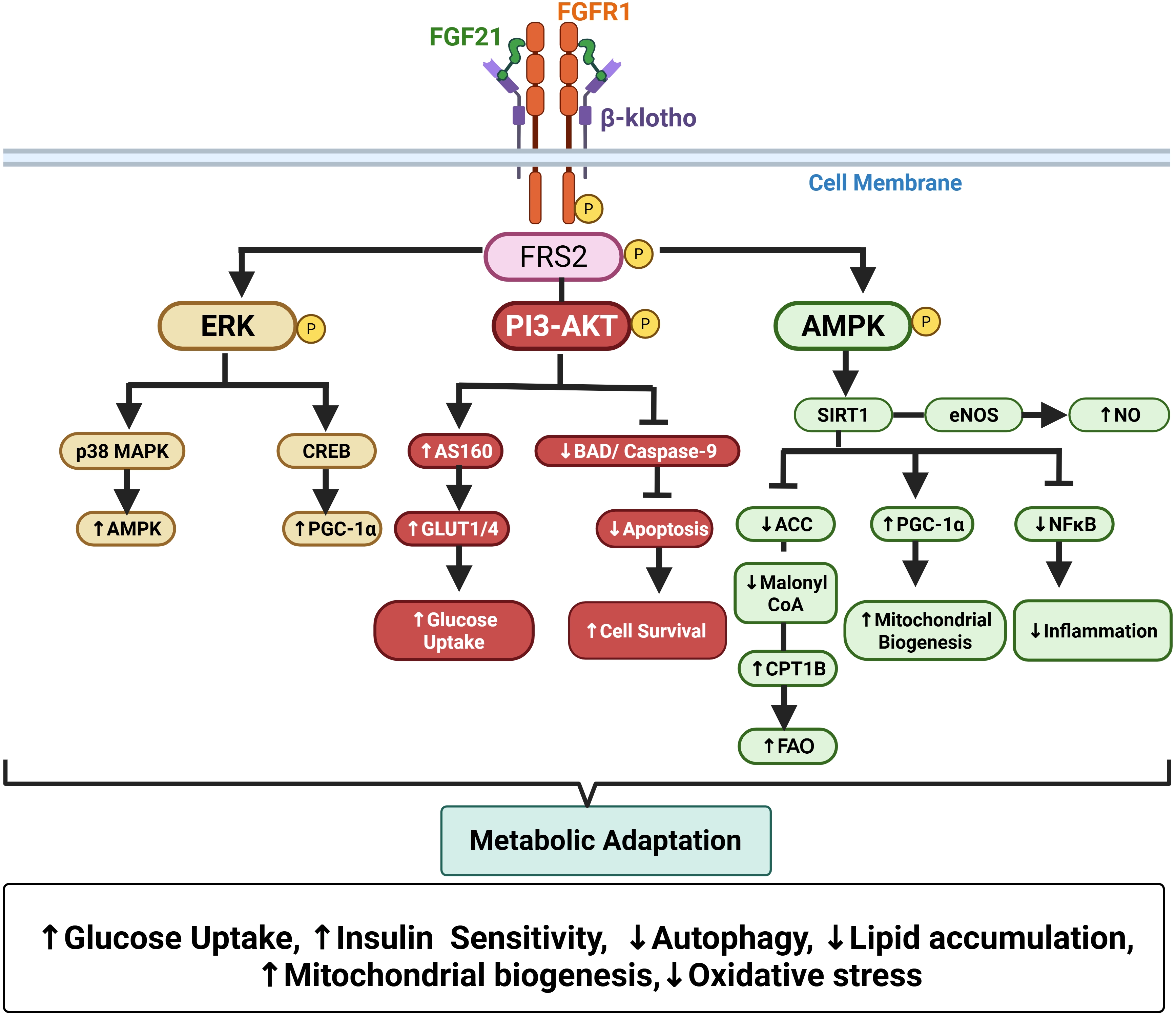
Figure 2. FGF21 signaling pathways that mediate metabolic adaptation: Activation of FGFR by FGF21 recruits the adaptor protein FRS2 and initiates three major downstream signaling pathways: ERK/MAPK, PI3K-AKT, and AMPK. The ERK/MAPK pathway activates p38 MAPK and CREB to promote PGC-1α-dependent transcriptional programs that support mitochondrial biogenesis and oxidative metabolism. The PI3K-AKT pathway enhances glucose uptake through AS160 (TBC1D4) mediated translocation of GLUT4 while activating mTORC1 signaling to promote protein synthesis and cell survival. Similarly, the AMPK pathway increases fatty acid oxidation via inhibition of ACC and activation of CPT1B and stimulates mitochondrial biogenesis through SIRT1-PGC-1α pathways. In addition, the AMPK-SIRT1-eNOS axis enhances NO production, thus suppressing oxidative and inflammatory stress. Together, these integrated signaling networks enhance glucose uptake, mitochondrial function, autophagy, and fatty acid oxidation while reducing oxidative and inflammatory stress, thereby promoting metabolic adaptation. Created in BioRender. Thapa, K. (2026) https://BioRender.com/r9emd2t. FGFR: fibroblast growth factor receptor; FRS2: FGFR substrate 2; ERK: extracellular signal-regulated kinase; PI3K: phosphatidylinositol 3-kinase; AMPK: AMP-activated protein kinase; ACC: acetyl-CoA carboxylase; CPT1B: carnitine palmitoyltransferase 1B; SIRT1: sirtuin 1; eNOS: endothelial nitric oxide synthase; NO: nitric oxide; FGF21: fibroblast growth factor 21.
Several preclinical studies that aimed to address this have yielded controversial findings. Mouse studies have shown that FGF21 gain of function extends health span and lifespan[32,33], suggesting a protective role for FGF21 in the aging process. Specifically, it has been shown that transgenic mice that overexpress FGF21 in hepatocytes had a 36% increase in the median survival time that was accompanied by lower plasma insulin-like growth factor (IGF)-1 levels, increased glucose tolerance, and improved whole-body insulin sensitivity. The improvement in survival was even higher in female mice compared to male mice (88% vs 65%, respectively)[33]. The pro-survival effect of hepatic FGF21 activation has been attributed to mimicking selective caloric restriction substances, that regulate a relevant subset of genes, such as Flavin-containing monooxygenase 3 (FMO3) (Fmo3), insulin like growth factor binding protein acid labile subunit (Igfals), hairy and enhancer of split 6 (Hes6), 5-aminolevulinate synthase 2 (Alas2), cytochrome P450, family 4, subfamily a, polypeptide 12B (Cyp4a12b), major urinary protein 4 (Mup4), serpin family A member 12 (Serpina12), and hydroxy-delta-5-steroid dehydrogenase, 3 beta and steroid delta-isomerase 5 (Hsd3b5)[33]. In a similar manner to hepatocytes, FGF21 overexpression in adipocytes of mice fed on a high-fat diet (HFD) extended lifespan to 3.3 years, while it prevented obesity and liver steatosis, improved insulin sensitivity, and reduced levels of inflammatory immune cells and lipotoxic ceramides in the visceral adipose tissue[32]. Additionally, muscle-directed adeno-associated virus (AAV)-mediated overexpression of FGF21 increased midlife survival by 20.5% in aged male and female mice, and it improved glucose homeostasis, reversed the age-associated overexpression of fibrotic markers in the heart, liver, and kidney, and enhanced cognition by activating AMPK signaling, enhancing mitochondrial function, restoring proteostasis, and reversing inflammation[34]. FGF21 through β-Klotho can also increase sulfide signaling and enhance the unfolded protein response, a cellular mechanism that preserves homeostasis in response to protein folding stress, thereby integrating stress response and possibly promoting healthy aging[35,36]. In summary, in contrast to clinical observations, various preclinical studies have identified beneficial effects of FGF21 in longevity.
However, other preclinical studies have shown moderate or even detrimental effects of FGF21 in aging. Although AAV-mediated overexpression of FGF21 in the adipose tissue increased relative lean mass and energy expenditure, high circulating levels of FGF21 did not improve muscle performance and liver steatosis in aged female mice[37]. Moreover, muscle-specific FGF21 deletion in a mouse model of skeletal muscle aging (skeletal muscle knockout of optic atrophy 1; OPA1) reversed several aspects of the aging phenotype, such as liver steatosis, hyperglycemia, and inflammation[38].
FGF21 has been linked to both harmful and beneficial outcomes in obesity and heart failure, creating ongoing controversy. Several studies associate higher FGF21 with reduced adipocyte lipolysis[39], increased adiposity or fat mass[40-42], and insulin resistance[43], suggesting a maladaptive role. In contrast, other work interprets elevated circulating FGF21 in obesity as a compensatory, protective response[44]. Consistent with this latter view, pharmacologic FGF21 analogues or receptor agonists have been tested in animal models and humans with obesity/diabetes, improving glycemia and lipid profiles, reducing hepatic steatosis, and promoting weight loss, supporting beneficial metabolic effects of FGF21 signaling[45-50].
In the cardiovascular system, findings are similarly mixed. A clinically tested FGF21 analogue increased blood pressure in humans[51], raising safety concerns. In mice, cardiac FGF21 expression rises across multiple heart failure models, suggesting an association with disease, yet global FGF21 deletion can worsen some hypertrophic responses, implying protection in certain contexts[52]. Other studies show transient benefits of FGF21 overexpression or treatment after ischemic injury[53,54], and exercise benefits in diabetic cardiomyopathy appear blunted in FGF21-null mice[55]. However, in humans, myocardial and circulating FGF21 levels are elevated in end-stage heart failure and correlate with diastolic dysfunction and mortality[56]. Overall, short-term FGF21 signaling may be cardioprotective, whereas prolonged or tissue-specific FGF21 signaling may contribute to adverse remodeling and dysfunction.
4. FGF21 Signaling
FGF21 exerts its metabolic actions through signaling pathways that influence senescence mechanisms (Figure 2). β-Klotho is an essential component of FGF21 signaling as it serves as a co-factor of the FGF21 receptor. In contrast to α-klotho, which is considered a longevity gene[57], the effects of β-Klotho on aging are FGF21-dependent.
Binding of FGF21 to the FGFR1-β-klotho receptor complex induces tyrosine phosphorylation of fibroblast growth factor receptor substrate 2 (FRS2). FRS2 is a docking protein that organizes downstream signaling complexes. Phosphorylated FRS2 recruits GRB2-SOS and SHP2, which eventually facilitate activation of the RAS-RAF-MEK-ERK cascade[58]. In parallel, GRB2 couples FRS2 to GAB1, the phosphorylation of which stimulates binding of the p85 regulatory subunit of PI3K, thus activating the PI3K-AKT pathway[59]. Some of the main effects of FGF21 include acute insulin-sensitizing actions and the chronic actions on body composition by suppressing weight gain, while increasing the percent of lean mass and decreasing the percent of fat mass in mice[60]. In adipocytes, the FGF21-FRS2 complex enhances mitochondrial function by activating AMPK through LKB1 protein kinase, which increases cellular NAD+ levels and thus activates SIRT1, a nicotinamide adenine dinucleotide (NAD+)-dependent class III histone deacetylase (HDAC)[29]. Through SIRT1-mediated deacetylation, FGF21 induces peroxisome proliferator-activated receptor-γ coactivator-1α (PGC-1α) activity[29]. Moreover, FGF21 increases adiponectin secretion, leading to improvement of insulin sensitivity and attenuation of hepatic steatosis and intramuscular triglyceride accumulation in mice[61].
FGF21 may also promote white adipose tissue expansion through downstream mechanisms that include induction of PPARγ expression[62], as well as enhancement of its transcriptional activity through reduced inhibitory sumoylation[63]. In parallel, FGF21 has been shown to suppress hepatic GH signaling, leading to decreased IGF-1 production and reduced lipolysis in adipose tissue[64,65]. Attenuation of GH signaling is known to favor adiposity and hepatic lipid accumulation[66-69].
Apart from metabolic actions, FGF21 also contributes to the prevention of major hallmarks of cellular aging, such as oxidative stress, inflammation, and decreased autophagy. FGF21 reduces the expression of pro-inflammatory cytokines even in highly pro-inflammatory conditions, such as in LPS-stimulated macrophages[70]. Specifically, FGF21 lowers the mRNA expression of tumor necrosis factor (TNF)-α, interleukin (IL)-1β, IL-6, and interferon-γ (IFN-γ), and increases the levels of IL-10 in a dose-dependent manner. Furthermore, FGF21 protects against immunosenescence by delaying age-related thymic involution and preserving naive T-cell diversity in mice[71,72]. Conversely, FGF21 deficiency accelerates thymic aging and impairs T-cell reconstitution following hematopoietic stem-cell transplantation[71]. Mechanistic studies have identified locally produced FGF21 from thymic epithelial cells and adipocytes, rather than hepatic endocrine FGF21, as a key mediator of thymic protection[73]. Enhanced epithelial FGF21 signaling increases naive CD8 T cells, whereas epithelial β-klotho deletion accelerates thymic involution, establishing the importance of local receptor signaling[73]. It also augments the activity of superoxide dismutase (SOD) and glutathione peroxidase (GSH-Px), possibly by enhancing nuclear factor-erythroid 2-related factor 2 (NRF2)/heme oxygenase-1 (HO-1) and by suppressing nuclear factor-κB (NF-κB) signaling pathways, in LPS-stimulated macrophages[70]. Furthermore, FGF21 administration inhibits accumulation of senescence markers P53 and P16 in hepatocytes and promotes transition of type 1 macrophages (M1) to type 2 macrophages (M2) by enhancing AMPKα phosphorylation and inhibiting the NF-κB pathway in the livers of aging mice[74]. Finally, administration of FGF21 in obese mice enhances hepatic autophagy via Jumonji-D3 (JMJD3/KDM6B) histone demethylase[75].
5. Metabolic Diseases
Metabolic syndrome is a constellation of metabolic abnormalities including abdominal obesity, hyperglycemia, dyslipidemia, and hypertension[76]. According to the United States National Health and Nutrition Examination Survey (NHANES), between 1999 and 2018, the prevalence of metabolic syndrome increased from 50.78 to 63.02%, and so did the prevalence of diabetes (from 16% to 25.1%) in subjects aged 65 years or older[76,77]. In addition to their high prevalence in older adults, metabolic syndrome, diabetes, and increased BMI are also associated with accelerated biological aging[78-84].
Serum FGF21 levels are independently associated with metabolic syndrome and its related complications. Overweight and obese individuals exhibit significantly higher circulating FGF21 levels compared with lean individuals[85]. Similarly, patients with type 2 diabetes mellitus (T2DM) have elevated serum FGF21 concentrations relative to nondiabetic subjects[86]. Both circulating FGF21 levels and the FGF21-to-adiponectin ratio have been proposed as biomarkers of T2DM[87,88]. In patients with T2DM, plasma FGF21 levels correlate independently with glycemic control and insulin resistance indices, including HbA1c, fasting insulin, and the homeostasis model assessment of insulin resistance[89].
Beyond glycemic abnormalities, elevated FGF21 levels are associated with diabetic complications. Serum FGF21 concentrations are significantly higher in patients with increased urinary albumin excretion, suggesting a potential link to diabetic nephropathy[90]. Patients with T2DM and carotid artery plaques also display higher FGF21 levels than diabetic individuals without plaque formation[91]. In addition, men with T2DM-related erectile dysfunction have elevated FGF21 levels compared with those who maintain normal erectile function[92]. Importantly, increased circulating FGF21 may have prognostic value, as higher serum levels have been associated with an increased risk of cardiovascular morbidity and mortality over a two-year follow-up in patients with T2DM[93].
The metabolic activity of FGF21 was first described in 2005, when it was found that it stimulates glucose uptake in mouse 3T3-L1 and primary human adipocytes, as well as that subcutaneous administration of FGF21 lowers plasma glucose and triglyceride levels in db/db mice and obese rats[94]. In accordance with these findings, FGF21 knockout mice have increased insulin resistance without changes in glycemia, while they also exhibit islet hyperplasia with distorted morphology and hyperinsulinemia as compensatory responses, possibly due to enhanced GH sensitivity[95]. These effects are accompanied by suppression of glucose-stimulated insulin secretion (GSIS)[96]. On the other hand, pancreatic islet‐specific overexpression of FGF21 in db/db mice enhances GSIS and decreases β-cell apoptosis by activating PI3K/Akt[96]. Moreover, subcutaneous infusion of FGF21 in db/db mice for 8 weeks increases plasma insulin and normalizes blood glucose levels[97]. The same study showed that FGF21 increases the number of islets in the pancreas and the number of β cells per islet. This effect was attributed to protective effects of FGF21 on glucolipotoxicity and cytokine-induced apoptosis[97]. FGF21 also ameliorates inflammation, as shown by lower concentrations of CRP, IL-1β, IL-6, and TNF-α in the serum and suppressed expression of IL-6, TNF-α, and monocyte chemoattractant protein-1 (MCP-1) in the white adipose tissue of db/db mice[98]. Furthermore, treatment with FGF21 increases energy expenditure and fat utilization in diet-induced obese and ob/ob mice, and induces the browning of the subcutaneous white adipose tissue in obese mice[50,99]. However, another study revealed that FGF21 attenuates lipolysis in human adipocytes and 3T3-L1 cells after a 3-day incubation, with no short-time effect on lipolysis[39].
The striking pre-clinical findings showing alleviation of metabolic complications by FGF21 led to the use of recombinant FGF21, FGF21 analogues, and FGFR agonists in Phase I and II clinical studies[45-48,100,101] for treating obesity and metabolic syndrome-related complications. Double-blind, randomized, controlled phase 1 and 2 trials recruiting humans with T2DM or hypertriglyceridemia, showed that FGF21 analogues incur significant improvements in blood lipid profile, whereas they have modest effects on fasting glucose reduction[45-47,101,102]. Specifically, administration of FGF21 or FGF21 analogues in obese humans normalized plasma glucose and lipid levels, and lowered liver steatosis and body weight[45-48].
The biological pathways through which FGF21 influences body weight regulation remain only partially defined. Evidence indicates that the weight-modulating actions of FGF21 involve signaling not only in adipose tissue but also in non-adipose tissue[103,104]. The contribution of adiponectin to these effects remains unresolved, as both adiponectin-dependent and adiponectin-independent mechanisms have been reported[61,105]. Whether elevated FGF21 levels in metabolic diseases indicate an FGF21 resistance state or a compensatory mechanism is still unclear. Expression levels of β-Klotho and phospho-ERK1/2 are reduced in the visceral fat from obese patients, despite the elevated FGF21 serum levels, showing a possible reduced sensitivity to FGF21 in the adipose tissue of obese people[106]. Accordingly, mice with diet-induced obesity are also characterized by decreased phosphorylation of ERK1/2 in white adipose tissue[44]. While chronic HFD suppresses FGFR1 and β-Klotho expression in the adipose tissue of mice, no changes are observed in the liver, pancreas, skeletal muscle, and hypothalamus[107]. This suggests that FGF21 resistance due to prolonged HFD may be an adipose-specific effect. Mice overexpressing β-Klotho in the adipose tissue have increased energy expenditure and are protected from diet-induced obesity, showing increased sensitivity to endogenous FGF21[108]. However, other studies support the finding that FGF21 sensitivity is unchanged in obese mice, despite the downregulation of β-Klotho and FGFR-1c expression levels[109].
Notably, the physiological consequences of elevated FGF21 appear to depend on the tissue source of its production. Studies in non-human primates exposed to HFD have demonstrated divergent responses, with diet-sensitive animals exhibiting increased FGF21 expression across multiple tissues, whereas diet-resistant animals showed more restricted induction, primarily in the liver, pancreas, and select adipose depots[41]. Furthermore, cardiomyocyte-derived FGF21 seems to induce faster white adipocyte expansion[40]. These findings support the concept that FGF21 exerts tissue-specific actions, and that its systemic metabolic effects may vary by site of origin and disease state (Figure 3).
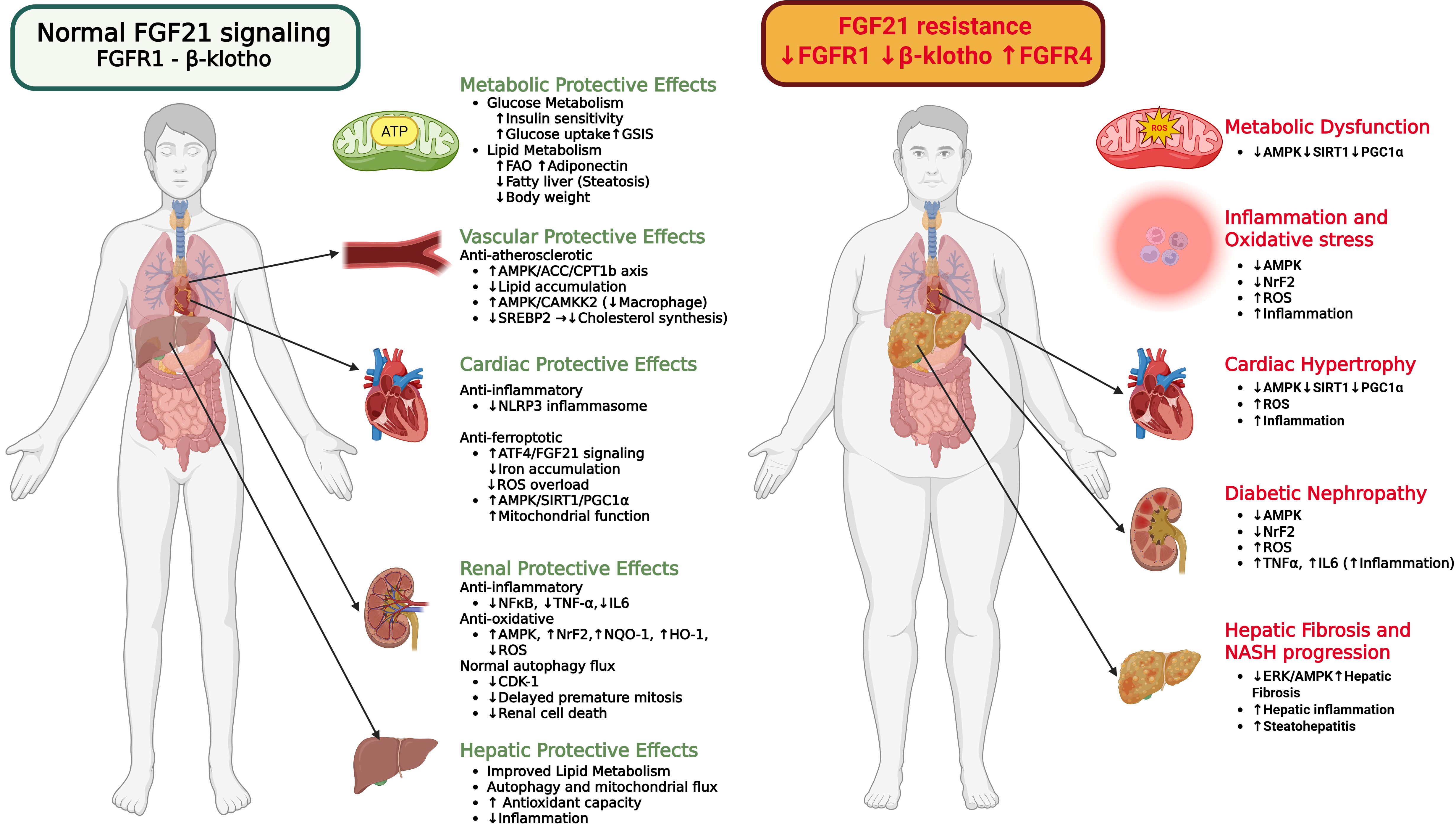
Figure 3. Organ-specific FGF21 signaling and resistance across health and disease states: Metabolic associated diseases are associated with elevated circulating FGF21, which initially known to exert protective effects on glucose metabolism (enhanced insulin sensitivity, glucose uptake, and GSIS), lipid metabolism (increased fatty acid oxidation, adiponectin, and reduced steatosis and body weight), and inflammation (reduced CRP, IL-1β, IL-6, MCP-1, and TNFα) thereby providing cardiac, renal, and hepatic protection. Chronic metabolic stress that occurs with aging, induces FGF21 resistance, characterized by decreased β-klotho/FGFR1 signaling, increased FGFR4 signaling and reduced tissue sensitivity to FGF21 and downstream AMPK signaling, which may contribute to metabolic dysfunction, inflammation, oxidative stress, diabetic nephropathy, albuminuria, hepatic fibrosis and progression to NASH. Created in BioRender. Thapa, K. (2026) https://BioRender.com/xdgp7p5. FGF21: fibroblast growth factor 21; GSIS: glucose-stimulated insulin secretion; IL: interleukin; MCP-1: monocyte chemoattractant protein-1; TNF: tumor necrosis factor; AMPK: AMP-activated protein kinase; ACC: acetyl-CoA carboxylase; CPT: carnitine palmitoyltransferase; NLRP3: NOD-like receptor family pyrin domain containing 3; SIRT1: sirtuin 1; NRF2: nuclear factor-erythroid 2-related factor 2; CDK1: cyclin-dependent kinase 1; ERK: extracellular signal-regulated kinase.
6. Liver Disease
Age affects hepatocytes and increases the risk of developing chronic liver diseases, such as metabolic dysfunction-associated steatotic liver disease (MASLD)[110]. Although aged livers in rats have lower FGF21 expression[111], increased hepatic fat content, as in humans with MASLD, is accompanied by higher serum levels of FGF21 compared with healthy subjects[112]. Accordingly, serum FGF21 levels increase progressively with the increase of hepatic fat content in patients with mild or moderate MASLD, while patients with severe MASLD have decreased FGF21 levels, possibly due to hepatic inflammation and damage[113]. Accordingly, mice with metabolic dysfunction-associated steatohepatitis (MASH) induced by a methionine- and choline-deficient diet (MCD) have elevated levels of hepatic FGF21 mRNA[114]. Serum FGF21 levels change proportionately to hepatic fat accumulation at middle-age (12 months) and old age (24 and 28 months)[115]. Two preclinical studies have displayed an association between elevated serum FGF21 levels and declining expression of β-Klotho in mouse livers, suggesting that FGF21 resistance might be the cause of high plasma FGF21 concentrations[115,116]. However, this was not confirmed in other studies[106].
FGF21 mainly has a protective role for hepatocytes against stress. Fgf21-null mice have greater hepatic triglyceride content and ER stress compared with the wild-type mice one week after the MCD treatment[114]. Palmitate, an ER stress inducer, increases hepatocyte FGF21 independent of PPARα, possibly to attenuate lipid-derived ER stress[114]. FGF21 expression is also induced by iron overload in primary hepatocytes and the liver of mice, and suppresses iron overload-induced ferroptosis via HO-1 ubiquitination and degradation and NRF2 activation[117]. Specifically, mice with hepatocyte-specific FGF21 overexpression are protected from iron overload-induced liver injury and fibrosis[117]. Exogenous FGF21 treatment reduces acute liver injury by inducing SIRT1 expression and SIRT1-mediated autophagy[118]. Moreover, FGF21 has a protective role against drug-induced hepatotoxicity by inducing PGC-1α/NRF2-mediated liver antioxidant mechanisms, as shown by the effect of recombinant FGF21, which reverses acetaminophen-induced liver injury in FGF21 knockout mice[119].
Aged FGF21 knockout mice are characterized by abnormal accumulation of hepatic lipids[120]. In mice with MASLD, FGF21 improves hyperglycemia and hyperlipidemia, and alleviates hepatic steatosis. Specifically, in obese mice with MASLD, treatment with recombinant murine FGF21 for 5 weeks increases autophagic flux, reduces body weight, improves insulin sensitivity, and reverses liver steatosis, with a better therapeutic effect compared with fenofibrate[121]. Furthermore, hepatocyte-specific FGF21 overexpression in APOE*3-Leiden-CETP mice that are fed on HFD alleviates liver lipotoxicity and inflammation by attenuating activation of Kupffer cells and macrophages[122]. Administration of long-standing FGF21 in mice with choline-deficient HFD-induced MASH for 15 days increases mitochondrial β-oxidation of fatty acids in the liver, and reduces hepatic steatosis and inflammation through a pathway that entails adiponectin and IL17A[123]. FGF21 also suppresses hepatic de novo lipogenesis by engaging the central nervous system that increases sympathetic nerve activity to the liver[124]. Finally, the FGF21/FGFR1 signaling pathway seems to mediate the anti-MASLD effect of sulforaphane and metformin[125,126].
FGF21 analogues are currently being tested for the resolution of MASLD. Efruxifermin, a long-acting Fc-FGF21 fusion protein, reduces hepatic fat and fibrosis as shown in two randomized, double-blind, placebo-controlled trials with patients with MASH[127-129]. Recently, efruxifermin was tested as an add-on to existing GLP1-receptor agonist treatment in the SYMMETRY Cohort D, which consisted of 31 patients with MASH and T2DM, and showed a reduction in hepatic fat fraction and noninvasive markers of fibrosis[130]. Pegozafermin, a long-acting glycopegylated recombinant FGF21 analogue, also reduced liver fibrosis and had a benefit regarding MASH resolution[48].
Thus, stress increases FGF21 secretion from the liver. FGF21 treatment alleviates liver steatosis in both preclinical and clinical studies, possibly by reducing oxidative stress and inflammation and inducing autophagy.
7. Cardiovascular Diseases
Cardiovascular diseases (CVD) constitute the leading cause of death worldwide, and their prevalence increases with age[131]. According to the American Heart Association 2019 Heart Disease and Stroke Statistics, the prevalence of CVD (comprising coronary heart disease, heart failure, stroke, and hypertension) is over 75% in both males and females aged 60-79 years old, and higher than 90% for individuals of at least 80 years old[132]. Structural and functional deterioration of the cardiovascular system occurs progressively with aging. Specifically, age-associated changes include cardiac hypertrophy, diastolic dysfunction, myocardial fibrosis, arterial stiffness, and endothelial dysfunction in the vasculature[133].
The effects of FGF21 in cardiac physiology vary. Regarding the vasculature, senescent human umbilical vein endothelial cells (HUVECs) and aged mouse aortas exhibit markedly lower expression of β-Klotho along with an upregulation of ADAM10, a protease responsible for β-Klotho degradation, and lower expression of mitochondrial ribosomal protein (MRP) genes[8]. FGF21 treatment upregulates the expression of MRP genes in the senescent cells[8]. In mice with either T1DM or T2DM, FGF21 treatment improves aortic endothelial function, as it mitigates oxidative stress and enhances endothelial nitric oxide synthase (eNOS) activity, leading to aortic vasodilation through activation of the CaMKK2-AMPKα signaling pathway[134]. Also in diabetic mice, FGF21 overexpression inhibits NOD-like receptor family pyrin domain containing 3 (NLRP3) inflammasome activation, which leads to activation of PPARγ and an eventual increase in catalase, a decrease in collagen accumulation, and relaxation of the aortas of diabetic mice[135]. Moreover, administration of dichloroacetate, a general pyruvate dehydrogenase kinase (PDK) inhibitor that ameliorates the development of atherosclerotic plaques and hyperlipidemia, increases the hepatic expression of FGF21 via AMPK activation and FGF21 signaling in brown adipose tissue in APOE-/- mice fed a western diet[136]. The anti-atherosclerotic effect of FGF21 has also been attributed to inhibition of pyroptosis[137], as well as to adiponectin production that ignites a cascade of events that includes reduced macrophage inflammation and neointima formation along with lower hepatic expression of sterol regulatory element-binding-protein-2 and cholesterol synthesis reduction[138]. Thus, FGF21 may have protective effects on endothelial function and antiatherosclerotic properties.
As for myocardial ischemia, FGF21 expression is upregulated in the liver and adipose tissue of mice in response to myocardial ischemia/reperfusion injury, and FGF21-/- mice exhibit greater myocardial injury, greater left ventricular dysfunction, increased caspase-3 activity, and enhanced cellular apoptosis at 24 hours after myocardial ischemia/reperfusion injury compared to wild-type mice[139]. Administration of FGF21 reverses these pathological effects[139]. FGF21 treatment is also protective in isolated rat hearts with global ischemia[54]. However, according to another study, FGF21 overexpression in the liver or muscle of mice improves systolic function 2 weeks postmyocardial ischemia but not beyond that time point[53].
FGF21 is also involved in hypertension, as patients with hypertension and angiotensin II-induced hypertensive mice are characterized by elevated circulating FGF21 concentrations[140]. However, knockout of FGF21 significantly exacerbates angiotensin II-induced hypertension and vascular inflammation in mice, whereas AAV-mediated FGF21 overexpression suppresses angiotensin II-induced hypertension and vascular dysfunction[140]. Despite these promising preclinical data, one of the metabolically beneficial FGF21 analogues that is used in clinical trials increases systolic and diastolic blood pressure in a dose- and time-related manner in humans[51].
Moreover, in preclinical models with diabetic cardiomyopathy, data are conflicting. Overexpression of FGF21 alleviates ferroptosis and improves cardiac function in mice with diabetic cardiomyopathy[141]. Furthermore, FGF21 prevents diabetic lipotoxicity-induced cardiomyopathy through both AMPK-AKT2-NRF2 antioxidative pathway and AMPK- acetyl-CoA carboxylase (ACC)- carnitine palmitoyl transferase 1 (CPT1) pathway-mediated lipid lowering effect in the heart[30]. Within STZ-induced T1DM mice fed a HFD, FGF21 deficiency exacerbates diabetic cardiomyopathy by lessening AMPK activation and paraoxonase 1 (PON1) expression, whereas AAV-mediated overexpression of FGF21 ameliorates diabetic cardiomyopathy[142]. Similarly, FGF21 knockout mice with T1DM exhibit earlier and more severe cardiac dysfunction, increased oxidative stress, and cardiac lipid accumulation, possibly mediated by cardiac NRF2-driven CD36 upregulation, compared with wild-type T1DM mice[143]. Cardiac hypertrophy, lipid accumulation in the heart, and blockage of cardiac autophagy also occur in Fgf21-/- mice fed a HFD without diabetes[144]. Moreover, an FGF21 analogue with enhanced capacity to bind FGFR1c, named FGF21D2D3, leads to a greater amelioration of diabetic cardiomyopathy and inhibition of cardiac oxidative stress via FGFR1-AMPK activation in T2DM mice[145]. A study in diabetic mice[55] also showed that exercise training did not improve heart function and mitochondrial biology in global FGF21-/- mice, suggesting a protective role for FGF21 in diabetic cardiomyopathy. However, it was not addressed why exercise-induced benefits were associated with lower plasma FGF21 levels[55]. In contrast to these predominantly protective findings, sustained FGF21-FGFR4 signaling has been associated with concentric cardiac hypertrophy in both diabetic and wild-type mice at 24 weeks of age[146]. Specifically, FGF21 transgenic mice with elevated hepatic FGF21 expression under the control of the apolipoprotein E promoter exhibit reduced body weight and blood glucose levels, but also left ventricular hypertrophy and increased relative wall thickness[146]. Similarly, recombinant FGF21 protein administration in wild-type mice results in increased left ventricular anterior wall thickness and mass[146]. Treatment with the pan-FGFR inhibitor PD173074 in ob/ob mice, which typically have elevated FGF21 levels and develop cardiac hypertrophy, inhibits the development of cardiac hypertrophy associated with diabetic cardiomyopathy[146]. This study differs from the preceding studies on diabetic cardiomyopathy in the duration of FGF21 exposure and the receptor pathway implicated. Controversy regarding the role of FGF21 exists not only in the field of diabetic cardiomyopathy, but also in other types of heart failure. According to a study with N[w]-nitro-L-arginine methyl ester (L-NAME)-induced HFpEF mice fed a HFD, global or adipose FGF21 deficiency leads to exacerbation of cardiac diastolic dysfunction and exercise intolerance, while AAV-mediated Fgf21 overexpression ameliorates these effects by activating PI3K/AKT signals[147]. Similarly, Fgf21-/- mice exhibit greater cardiac hypertrophy, dysfunction, mitochondrial damage, and mitophagy impairment after four weeks of TAC surgery-mediated pressure overload compared with wild-type mice, whereas FGF21 administration reduces cardiac hypertrophy and promotes mitophagy via activation of PTEN-induced putative kinase 1 (PINK1)[148]. However, in mice with heart failure induced by three different conditions (isoproterenol infusion, transverse aortic constriction, ischemia), increased cardiac FGF21 expression is reported[52], implying a detrimental association of FGF21 with heart failure. To add to the confusion, the same study showed that global FGF21 deletion worsens isoproterenol-induced cardiac hypertrophy[52]. Although the effect of FGF21 ablation was tested over a short-term period and not in all heart failure models, this study suggested that FGF21 is protective. On the contrary, another study showed that CM-specific constitutive FGF21 expression suppresses cardiac expression of fatty acid oxidation-related genes[42], similar to the maladaptive transcriptional changes that characterize increased pressure-overload and left ventricular hypertrophy[149], as well as right ventricular hypertrophy[150]. Accordingly, mice with global Fgf21 deletion are protected from pregnancy-induced cardiomyopathy[151]. Moreover, inhibition of hepatocyte FGF21 is protective against cardiac hypertrophy due to pressure overload, and inhibition of cardiomyocyte FGF21 also prevents cardiac hypertrophy, independently of systemic FGF21 levels[152]. According to the same study, the stimulation of cardiomyocyte FGF21 expression that leads to hypertrophy occurs two weeks after the increase in hepatic and plasma FGF21 levels, showing that possibly chronic FGF21 upregulation exacerbates cardiac dysfunction[152].
Regarding patients with heart failure, FGF21 staining is increased in heart biopsies of patients with end-stage heart failure[153]. Higher plasma FGF21 levels are associated with diastolic dysfunction and higher mortality in patients with heart failure with preserved ejection fraction (HFpEF)[56], atrial remodeling in patients with atrial fibrillation[154], and increased risk of mortality and hospital readmission in patients with heart failure with reduced ejection fraction (HFrEF)[155]. As these studies are cross-sectional, they establish an association, not causation. Thus, these findings could indicate that prolonged FGF21 signaling may worsen cardiac dysfunction in several phenotypes of heart failure, or reflect compensatory secretion, and/or reduced target-tissue responsiveness.
Thus, FGF21 seems to have anti-aging effects on the vasculature, while its role in aging-related conditions of the heart requires further research, as current data are conflicting and its role may be time-, tissue-, and stress-dependent. FGF21 may exert beneficial effects during mild mitochondrial dysfunction, whereas sustained or chronic expression could contribute to concentric hypertrophy[146,152]. Also, it may oppose eccentric hypertrophy and promote concentric hypertrophy[146]. Additional studies have also revealed that the effects of FGF21 are influenced by the timing and the tissue of FGF21 origin, as FGF21 may become detrimental regarding cardiac hypertrophy when signaling is constitutive or prolonged, is derived from the liver, and activates FGFR4[146,152].
8. Chronic Kidney Disease (CKD)
As kidney function declines with age[156], serum FGF21 levels increase progressively[157,158]. Higher serum FGF21 levels were associated with renal pathology in patients with CKD[159,160]. Further, a high mortality rate was shown in patients with end-stage renal disease and higher serum FGF21 levels[161,162].
Aged mice with FGF21 deletion in kidney proximal tubular epithelial cells (PTEC) have severe tubular injury and atrophy, enlarged lysosomes and autophagic degradation compared to aged control mice[163]. Although FGF21 deficiency increases autophagy in the PTECs of young mice, it does the opposite in aged mice due to lysosomal overburden[163]. Moreover, FGF21 ablation exacerbates tubular injury, inflammation and fibrosis in 3-22 month old mice that consume a low-protein diet[164].
Diabetic db/db mice have higher renal cortical FGF21 expression and decreased ERK phosphorylation compared to non-diabetic mice, suggesting an FGF21-resistant state in the diabetic kidney[165]. Daily FGF21 injections improving kidney function and reduced fibrosis, while also improved insulin sensitivity and serum lipids[165]. In mice with T1DM that have developed diabetic nephropathy, subcutaneous injections of FGF21 for 4 weeks improve renal function, reduce proteinuria, decrease the amount of glycogen deposition in the kidney, as well as reduce fasting blood glucose, total cholesterol, serum levels of inflammatory components and oxidative stress that accompanies the metabolism of glycolipids[166]. FGF21 administration also reduces the expression of cyclin-dependent kinase 1 (CDK1) in the renal tissue of those mice. As CDK1 induces cell death by causing premature entry into mitosis, FGF21 delays this phenomenon, and thus the aggravation of diabetic nephropathy[166]. According to the same study, treatment of diabetic db/db mice with FGF21 for 8 weeks also ameliorates diabetic nephropathy by decreasing glomerular fibrosis and glycogen deposition[166]. After treatment with adriamycin (ADR), which is an anthracycline chemotherapeutic agent with potential nephrotoxic effects, HFD-fed mice show increased renal FGF21 expression, while they display suppression of FGFR1, p-FGFR1, and β-Klotho expression, downregulation of ERK phosphorylation and decreased expression of cFos and Egr1 mRNA in kidney tissue[167]. Thus, ADR-injected mice that were fed with HFD develop renal FGF21 resistance[167]. Exogenous supplementation of FGF21 in those mice decreases renal lipid accumulation and improves renal function, while similar benefits also arise in diabetic (STZ, db/db) mice after FGF21 treatment[167].
Moreover, deoxycorticosterone acetate (DOCA)-salt-induced hypertensive mice have increased circulating FGF21 levels and higher renal FGF21 expression[168]. This seems to be a protective response, as DOCA-salt treatment of FGF21 knockout mice causes even greater elevation of blood pressure and renal damage compared with wild-type mice. Treatment with recombinant human FGF21 decreases blood pressure values and attenuates salt-sensitive hypertension-induced renal injury, possibly by promoting AMPK activation and, in this way, reducing the nuclear translocation of NF-κB p65 and increasing the nuclear accumulation of NRF2[168]. A more stable form of recombinant human FGF21, which has higher kidney retention, displays even greater therapeutic effects regarding improving kidney function and insulin sensitivity, and alleviating hyperlipidemia and inflammation in diet-induced obese mice with diabetic nephropathy[169].
Thus, aged kidneys that lack FGF21 have a more robust aging phenotype. In diabetic, hypertensive, and ADR-induced nephropathy, renal FGF21 resistance may be present. Supplementation of FGF21 improves renal functional and morphological abnormalities.
9. Sarcopenia
As the largest tissue in the human body by mass and a major metabolic organ, skeletal muscle requires substantial amounts of energy during activity[170]. Consequently, an extensive and functional mitochondrial network is essential for proper muscular performance. With advancing age, mitochondria, along with other cellular structures, undergo functional decline due to degeneration of the recycling (mitophagy and fusion-fission cycles) process. FGF21 is a key regulator of complex mechanisms that mitigate mitochondrial deterioration over time. However, FGF21’s effects in skeletal muscle appear to depend on the duration of its induction: acute FGF21 induction is associated with mitochondrial adaptation, whereas chronic FGF21 elevation, as occurs during aging and persistent mitochondrial stress, is linked to altered mitochondrial quality control, muscle atrophy, and systemic pro-senescence effects[38,170,171] (Figure 4). Nevertheless, adults and children with mitochondrial disorders that affect skeletal muscle have higher serum or plasma FGF21 levels[172].
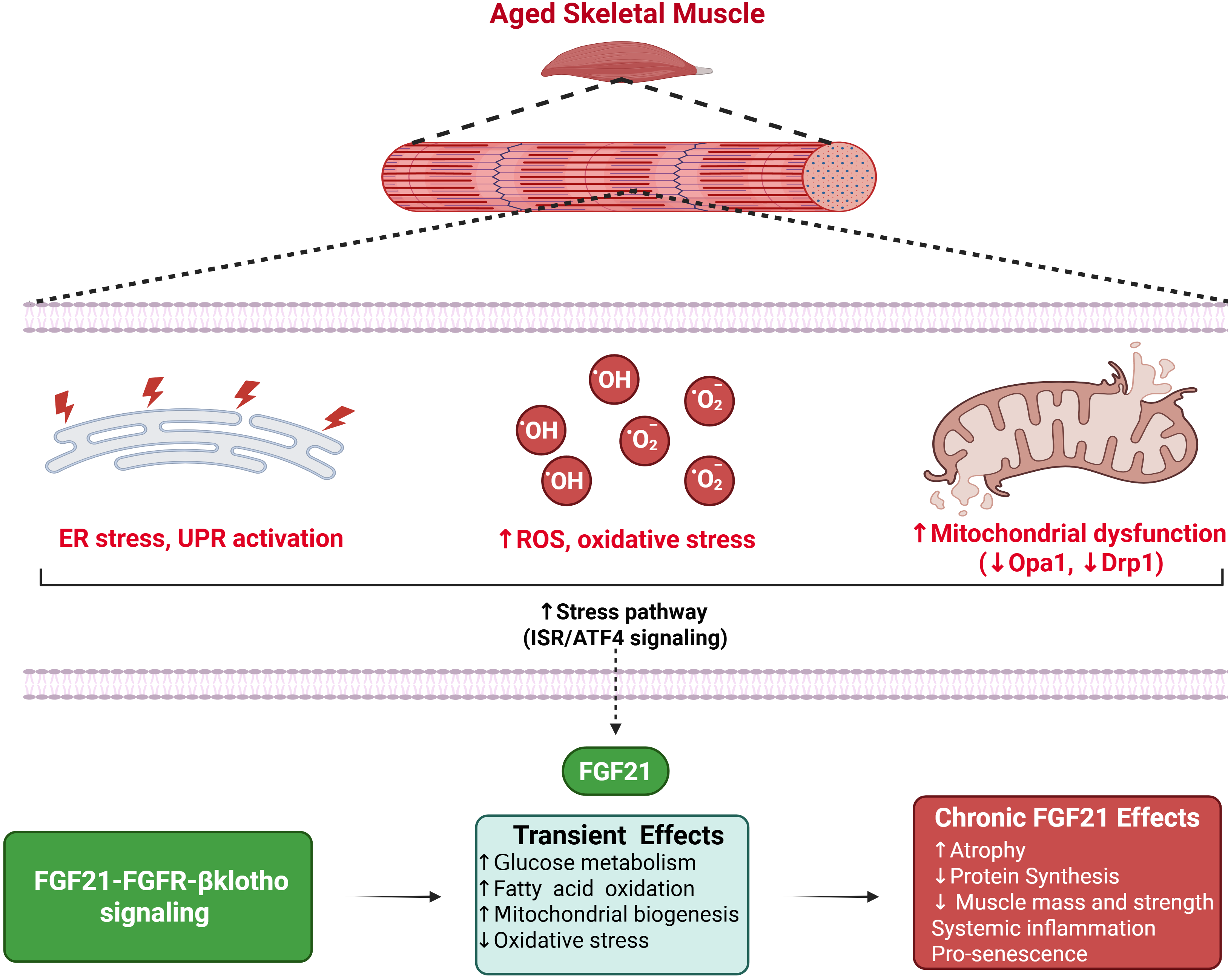
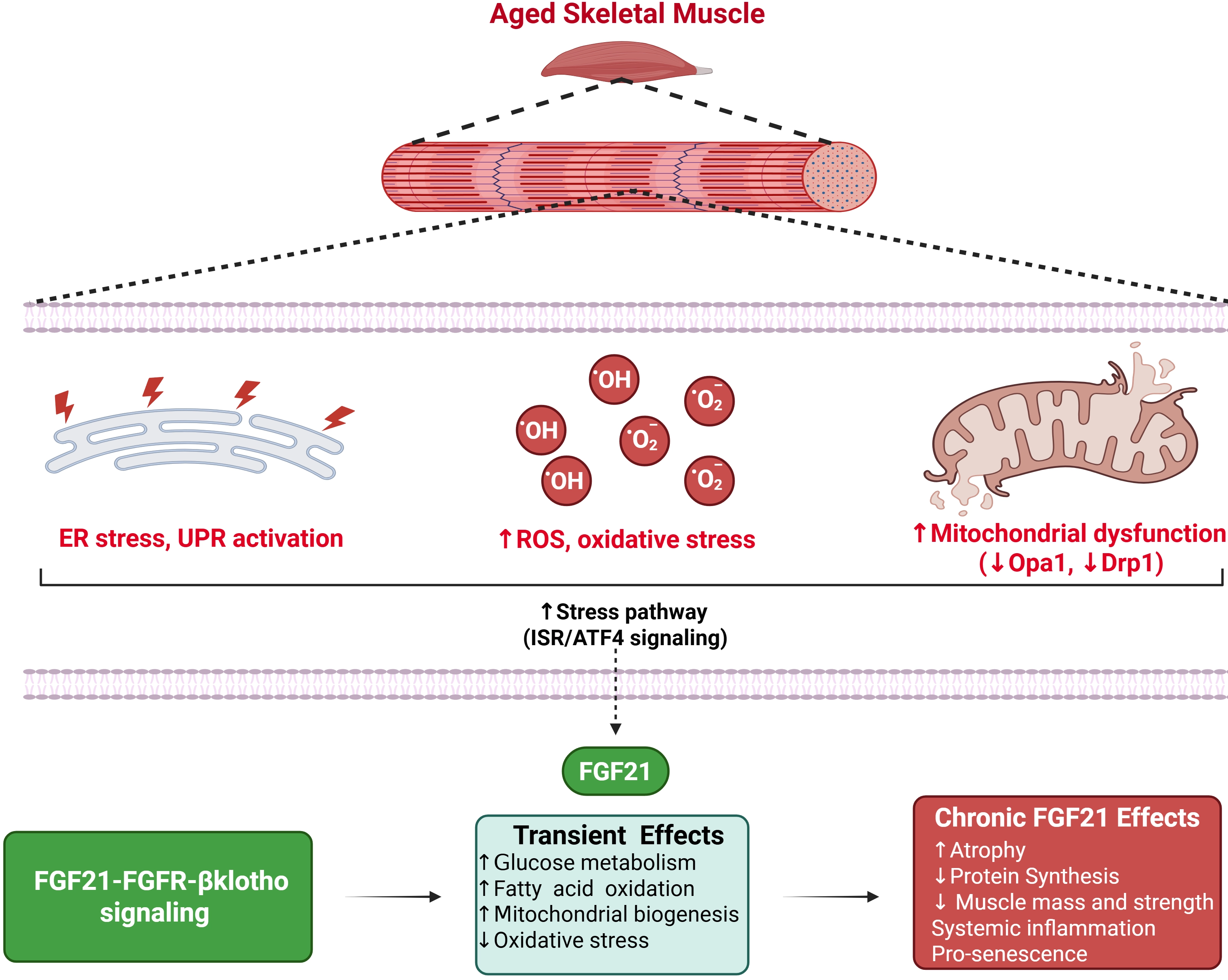
Figure 4. FGF21 in aging skeletal muscle: Aging-associated mitochondrial dysfunction, ER stress, and ROS activate the ISR, leading to ATF4-dependent induction of FGF21. Secreted FGF21 signals via FGFR1c-β-Klotho to regulate systemic metabolism. Acute FGF21 promotes adaptive responses, including mitochondrial biogenesis, fatty acid oxidation, and reduced oxidative stress. In contrast, chronic FGF21 elevation is associated with activation of atrophy pathways and aging-related muscle decline highlighting the context dependent role of FGF21. Created in BioRender. Thapa, K. (2026) https://BioRender.com/ss8su3m. FGF21: fibroblast growth factor 21; ISR: integrated stress response; FGFR: FGF receptors.
Despite the existing evidence about the beneficial role of FGF21 in aging-related repair mechanisms, several studies have also associated it with negative outcomes in aged skeletal muscle tissue. Among humans above 60 years of age, patients with cachexia have significantly higher total FGF21 levels than healthy subjects[173,174]. Moreover, a study of 386 adults aged 70-84 years found that high plasma FGF21 levels were negatively associated with strength proportionately. Higher FGF21 levels have been associated with lower bone mineral density[175] and more severe bone loss[176], although this effect of FGF21 seems to be lost in obese mice[177]. Although a study in 125 older adults suggested an independent association of higher serum FGF21 with sarcopenia, low muscle mass, and weak grip strength[178], additional studies did not identify an independent association between FGF21 and muscle mass[179,180]. Sedentary humans exhibit a decline in OPA1, and this decline is linked to muscle loss[38]. In mice, deletion of FGF21 in muscle tissue offsets the loss of OPA1[38]. Accordingly, inhibition of dynamin-related protein-1 (Drp1), the complementary fission component to OPA1’s fusion mechanism, has been shown to alleviate skeletal muscle senescence by suppressing FGF21[170]. Similar results were observed in mice with fasting-induced muscle atrophy, where FGF21 knockout animals were protected from muscle loss[181]. Conversely, muscle OPA1 deficiency induces FGF21 secretion that protects against obesity and insulin resistance[182].
Data from both clinical and preclinical studies revealed that acute exercise usually results in an increase in circulating FGF21[183], consistent with its role as an acute metabolic stress signal. Long-term exercise training (≥ 4 weeks) decreases circulating FGF21 levels and enhances components of the FGF21 signaling pathway, as shown by increased expression of both FGF receptors and β-Klotho in adipose tissue, liver, and skeletal muscle[184]. Specifically, human subjects ranging from 62 to 70 years of age, who underwent resistance training, had increased muscle mass and decreased FGF21 expression in plasma[185]. Regarding the impact of resistance training in elderly patients with T2DM, both individuals with T2DM and those without T2DM had a significant reduction in FGF21 levels[186]. However, another study suggested that the FGF21 response to exercise may vary according to baseline metabolic status. A 10-week resistance exercise program decreased circulating FGF21 levels in women with metabolic syndrome, whereas FGF21 levels increased in women without metabolic syndrome[187].
Thus, transient FGF21 induction following acute exercise may represent an adaptive metabolic response, whereas chronically elevated FGF21 may reflect detrimental effects in aging skeletal muscle, as limited preclinical and clinical studies have demonstrated negative associations between FGF21 and skeletal muscle mass and function. Since a direct causal relationship has not been established yet, future studies should determine whether elevated FGF21 is a mediator of muscle deterioration or a compensatory biomarker of stress, while distinguishing acute from chronic exposure and accounting for metabolic status and exercise modality.
10. Aging-Related Neurological Diseases
With an aging population, the prevalence of neurodegenerative diseases such as Parkinson’s disease (PD) and Alzheimer’s disease (AD) has increased significantly, with projections indicating that PD diagnoses may double within the next thirty years[4,188]. FGF21 has been identified as a promising therapeutic target; although demonstrated primarily in preclinical models, these studies strongly suggest its potential to protect against neurodegenerative disorders and support overall brain health during human aging.
PD is a debilitating neurodegenerative disorder characterized by the depletion of dopaminergic neurons in the substantia nigra (a midbrain region) and the pathological accumulation of α-synuclein, leading to hallmark symptoms such as resting tremor, postural instability, and rigidity[189]. A Mendelian-randomization analysis suggests that FGF21 levels may be associated with PD risk through a phosphatidylcholine-dependent pathway[188]. Furthermore, FGF21 administration in mice reduced both motor symptoms and cognitive impairments associated with 1-methyl-4-phenyl-1,2,3,6-tetrahydropyridine (MPTP)-induced PD[190]. Using both in vitro and in vivo approaches, the authors identified a distinct mechanism through which FGF21 enacts protective effects against PD-related dysfunction via activation of the SIRT1/NF-κB pathway-mediated microglial polarization[190].
The beneficial effects of FGF21 pertain to a wide range of neurological and metabolic adaptations, including mitochondrial function[191], proteostasis and clearance[192], antioxidant defence[191,193], cell survival[194,195] and cognitive enhancement[192,196] (Figure 5). Infusion of FGF21 improved cognitive performance and prevented hippocampal neuron loss and apoptosis in mice with diabetes-induced cognitive decline[197]. FGF21 also protected against angiotensin II-induced cerebrovascular aging by inhibiting p53 through an AMPK axis, causing an improvement in mitochondrial generation[198]. Further, FGF21 protected mouse brains against D-galactose-induced aging via attenuation of reactive oxygen species and advanced glycation end products[199]. In the brains of D-galactose-treated mice, FGF21 also reduced inflamm-aging by preventing inhibitor κBα (IκBα) degradation and NF-κB p65 nuclear translocation products[199]. In the brains of diabetic and aged mice, FGF21 reduced the expression of NF-κB, Iba1, IL6 and IL8, and activated AMPKα/AKT pathways[192]. Recombinant human FGF21 (rhFGF21) reversed D-galactose-induced cognitive deficits by reducing oxidative stress, neuroinflammation, and cellular senescence in the cortex and hippocampus, and was associated with increased FGFR1 phosphorylation and restored SIRT1 expression[200]. FGF21 can also mediate protective effects on Alzheimer-like pathologies through protein phosphatase 2A/mitogen-activated protein kinases/hypoxia-inducible factor-1α pathways[201]. Conversely, endogenous deletion of FGF21 induced learning and memory deficits, anxiety-like behaviors, and neuronal degeneration, and transcriptomic profiling shows associated disruption of neuroprotective pathways, including downregulation of PPARγ signaling and its downstream targets[202]. Current evidence suggests that FGF21 may promote synaptic plasticity through pathways involving AMPK activation, improved neuronal metabolism, and reduced neuroinflammation, with potential contributions from PI3K/AKT signaling[201-203]. A recent study that subjected FGF21-/- mice to various behavioral tests demonstrated that FGF21 deletion caused deficits in learning and memory, anxiety-like behaviors, and neuronal degeneration[202]. This finding suggests that FGF21 may have a preventive role for cognitive impairment-related diseases. Interestingly, clinical data present a more complex picture; in a cross-sectional study of groups ranging from healthy controls to patients with AD, serum FGF21 concentration was inversely associated with cognitive performance[204], highlighting the need to better understand systemic FGF21 resistance or compensatory upregulation in human pathology.
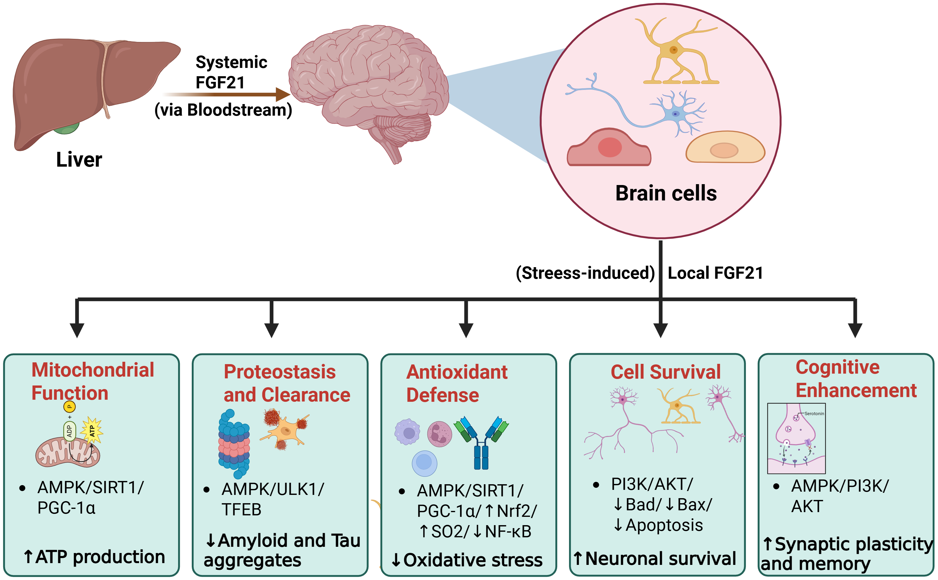
Figure 5. FGF21-mediated neuroprotection: FGF21, derived from both circulating (hepatic) and local (brain) sources, signals through FGFR1-β-Klotho to activate multiple protective pathways in the brain. The AMPK-SIRT1-PGC-1α axis enhances mitochondrial function and ATP production. FGF21 also promotes protein homeostasis by activating AKT and AMPK signaling and suppressing NF-κB, IL6 which is associated with reduced amyloid-β and tau aggregation. FGF21 also activates NRF2-dependent antioxidant pathway and suppresses NF-κB-mediated inflammation, leading to reduced oxidative stress. In parallel, PI3K-AKT signaling inhibits GSK3β, that attenuates tau phosphorylation and apoptosis, thereby supporting neuronal survival. Additionally, AMPK-PI3K-AKT signaling enhances synaptic plasticity and improves cognitive function. Created in BioRender. Thapa, K. (2026) https://BioRender.com/b52dmvb. FGF21: fibroblast growth factor 21; FGFR: FGF receptors; AMPK: AMP-activated protein kinase; SIRT1: sirtuin 1; NRF2: nuclear factor-erythroid 2-related factor 2; IL: interleukin; PI3K: phosphatidylinositol 3-kinase; TFEB: transcription factor EB.
Following acute stroke, FGF21 has also been shown to exert neuroprotective effects. In rats with acute intracerebral hemorrhage, FGF21 enhanced the differentiation of mesenchymal stem cells into neural stem cells through the FGFR1-SIRT1-MTOR signaling[205]. In transient middle cerebral artery occlusion (tMCAO) mice, FGF21 suppresses astrocyte activation, thereby preventing astrocyte-mediated inflammatory responses and promoting neurotrophic responses[206]. Accordingly, FGF21 knockout in tMCAO mice exacerbated infarct size and cognitive deficits[206]. Research utilizing tMCAO models of ischemic stroke demonstrated that the administration of blood plasma from healthy mice protected the blood-brain barrier (BBB), reduced neuronal apoptosis, and suppressed neuroinflammation, while FGF21 knockout effectively reversed these neuroprotective effects[207]. Similarly, in distal middle cerebral artery occlusion (dMCAO) models of ischemic stroke, FGF21 was shown to induce autophagy and reduce both neural apoptosis and infarct size[208]. These cerebrovascular benefits are likely facilitated by the ability of FGF21 to cross the BBB in a non-saturable manner, making it one of the few fibroblast growth factors capable of doing so[206,209]. However, the exact transport mechanisms and the effects of aging on FGF21 permeability remain to be fully elucidated.
Nutritional interventions and aging models further underscore the role of FGF21 in neuroprotection. Dietary protein restriction extends lifespan and improves metabolic health, physical performance, and frailty in male mice through FGF21-dependent mechanisms[210,211]. Conversely, FGF21 deficiency abolishes these benefits and, under protein restriction, leads to earlier age-related weight loss, impaired physical performance, increased frailty, and reduced lifespan[210]. It has been shown that non-aging-related effects of dietary protein restriction and other interventions are mainly dependent on FGF21 signaling in the brain[212-214]. In ApoE-deficient mice, long-term caloric restriction was associated with Fgf21-related activation of the AMPK/mTOR pathway and neuroprotective effects[6]. This contributes to a decrease in rapamycin signaling activity and inhibition of tau-hyperphosphorylation, resulting in the reduction of neurofibrillary tangles, which are a prominent biomarker for Alzheimer’s disease[6]. Similarly, dietary methionine restriction increased circulating FGF21 and improved cognitive outcomes in aged mice; these benefits were attenuated by AAV-mediated Fgf21 knockdown[193,215]. Mechanistically, in aged APP/PS1 mice, late-life methionine restriction selectively induced hepatic FGF21, which acted on brain FGFR1 to activate AMPKα and suppress NF-κB-mediated neuroinflammation[216]. FGFR1 knockdown abolished these effects, demonstrating that FGF21 is the essential mediator of MR-induced anti-inflammatory signaling[216]. However, although FGF21 mediates MR-induced increases in energy expenditure, adipose thermogenesis, and insulin sensitivity, reductions in body weight and adiposity also occur in FGF21-deficient mice, and these findings separate the anti-aging effect of FGF21 from its anti-obesogenic effect[217]. In other aging models, dietary lycopene restored cognitive performance by activating hepatic FGF21, which subsequently reduced neuronal degeneration, mitochondrial dysfunction, and synaptic damage[218]. Knockdown of hepatic FGF21 abolished the observed benefits, indicating that the neuroprotective benefits of lycopene were FGF21- dependent[218]. Moreover, treadmill exercise, that reduced cognitive decline and neuroinflammation, also increased hepatic and serum FGF21 protein levels via the AMPK- transcription factor EB (TFEB) axis in aged mice, while hepatic Fgf21 knockdown reduced FGF21 levels in hippocampal tissue and impaired the activation of exercise-mediated mitophagy[219]. Dietary isoleucine restriction extends lifespan and reduces frailty in genetically heterogeneous mice, with greater longevity benefits in males[220]. Although a low isoleucine diet activates FGF21 signaling[221], whether FGF21 is required for the geroprotective effect of dietary isoleucine restriction remains to be established.
Conclusively, FGF21 exerts significantly beneficial effects on neurodegeneration and brain function during aging. However, additional studies involving human cohorts are still required to confirm and clarify FGF21’s therapeutic perspective.
11. Summary and Conclusions
FGF21 exerts its metabolic actions through signaling pathways that regulate oxidative stress, inflammation, autophagy, and senescence. Elevated levels of circulating FGF21 are present in older adults and aged preclinical models and have been associated with parameters of metabolic syndrome, frailty and mortality, suggesting that FGF21 may represent either a compensatory response to aging or a critical element of the pathophysiology of aging-related diseases. Preclinical studies suggest that FGF21 has beneficial effects in diabetes and obesity, possibly in a tissue- and origin-dependent manner. FGF21 also has a protective role against liver steatosis, kidney injury, and neurodegenerative disorders. Although short-term FGF21 signaling may be protective against myocardial ischemia and heart failure, long-term effects on heart function remain unclear. Detrimental effects of FGF21 have also been documented in cardiac muscle tissue due to induction of concentric cardiac hypertrophy after exposure to excessive stress, as well as in skeletal muscle tissue by causing skeletal muscle loss in preclinical models.
Whether FGF21 signaling will be beneficial or detrimental may be driven by differences in the formation of the receptor-co-receptor complexes. Protective FGF21 signaling in the heart usually entails β-Klotho and FGFR1c, whereas an FGF21-FGFR4 interaction that does not require β-Klotho is associated with concentric cardiac hypertrophy. Accordingly, chronic metabolic stress lowers β-Klotho and FGFR1 abundance in adipose tissue and kidney, whereas β-Klotho degradation by ADAM-10 has been identified as a proximate mechanism. Notably, a progressive loss of the co-receptor redistributes FGF21 signaling rather than eliminating it.
The duration of exposure to FGF21 is also critical for defining whether FGF21 will incur toxicity. A transient FGF21 increase, which is induced with acute exercise or mild mitochondrial dysfunction, has been reported to be beneficial for two weeks but not thereafter. On the other hand, sustained exposure becomes detrimental as cardiomyocyte-restricted constitutive expression suppresses fatty acid oxidation, while overexpression for more than 24 weeks causes left ventricular hypertrophy. Similarly, this temporal dimorphism defines the effect in skeletal muscle, with acute induction being associated with mitochondrial adaptation while chronic elevation is accompanying atrophy.
Tissue of origin also seems to matter independently of systemic concentration. Hepatic and adipose FGF21 overexpression extends lifespan and improves metabolic parameters, whereas cardiomyocyte-derived FGF21 promotes hypertrophy and adipocyte expansion even when systemic levels are unchanged.
While FGF21 has been investigated in both human and animal studies, these studies answer different questions. Human studies are mainly observational, mostly measuring circulating FGF21, thus correlating with age, adiposity, renal impairment and mortality without resolving causality or association. On the other hand, preclinical studies offer opportunities to study causality and identify differential effects depending on tissue of origin and receptor route. However, in various cases, these studies rely on supraphysiologic overexpression, global rather than tissue-specific ablation, and artificial ways of mimicking aging. This asymmetry has produced some conflicting results, such as an association of increased endogenous FGF21 with mortality and frailty in older adults, whereas overexpression in mouse preclinical studies prolongs lifespan. Accordingly, although FGF21 lowers blood pressure in hypertensive mice, a clinical analogue raised it. These discrepancies are consistent with the receptor- and duration-dependent framework proposed above, since pharmacologic agonism is not equivalent to endogenous elevation in tissues with suppressed co-receptor expression.
Thus, further research, in which the time-, tissue-, origin-, and stress-dependent effects of FGF21 will be considered, is needed to elucidate the effects of FGF21 on aging processes and aging-related diseases, especially nowadays that FGF21 analogues are studied extensively for the treatment of T2DM, hypertriglyceridemia and MASLD.
Authors contribution
Vakka A, Sherman F, Thapa K: Writing-original draft.
Tsioufis K: Writing-review & editing.
Drosatos K: Supervision, writing-review & editing.
Conflicts of interest
Not applicable.
Ethical approval
Not applicable.
Consent to participate
Not applicable.
Consent for publication
Not applicable.
Availability of data and materials
Not applicable.
Funding
This study was supported by the National Heart, Lung, and Blood Institute (HL175251; KD) and the American Heart Association Supporting Undergraduate Research Experience (25SURE1512008; FS).
Copyright
© The Author(s) 2026.
References
-
1. Li Q, Xiao N, Zhang H, Liang G, Lin Y, Qian Z, et al. Systemic aging and aging-related diseases. FASEB J. 2025;39(5):e70430.[DOI]
-
2. Ageing and health [Internet]. Available from: https://www.who.int/newsroom/fact-sheets/detail/ageing-and-health
-
3. Bhattacharjee NV, Schumacher AE, Aali A, Abate YH, Abbasgholizadeh R, Abbasian M, et al. Global fertility in 204 countries and territories, 1950–2021, with forecasts to 2100: A comprehensive demographic analysis for the Global Burden of Disease Study 2021. Lancet. 2024;403(10440):2057-2099.[DOI]
-
5. López-Otín C, Blasco MA, Partridge L, Serrano M, Kroemer G. Hallmarks of aging: An expanding universe. Cell. 2023;186(2):243-278.[DOI]
-
9. Godoy-Lugo JG, Young KE, Vaddi P, Batamack Y, Aldridge J, Fernandez-Kim SO, et al. Low protein-induced-FGF-21 signaling remodels adipose tissue on reduced markers of senescence during aging. GeroScience. 2026;48(1):55-75.[DOI]
-
10. Arias-Calderón M, Casas M, Balanta-Melo J, Morales-Jiménez C, Hernández N, Llanos P, et al. Fibroblast growth factor 21 is expressed and secreted from skeletal muscle following electrical stimulation via extracellular ATP activation of the PI3K/Akt/mTOR signaling pathway. Front Endocrinol. 2023;14:1059020.
-
14. Liu T, Li J, Yang Z, Wei J. Synergistic pathways in Parkinson’s disease: The promise of FGF21 and ACE2. Ageing Res Rev. 2025;110:102804.[DOI]
-
15. Fisher FM, Maratos-Flier E. Understanding the physiology of FGF21. Annu Rev Physiol. 2016;78:223-241.[DOI]
-
16. Phan P, Saikia BB, Sonnaila S, Agrawal S, Alraawi Z, Kumar TKS, et al. The Saga of endocrine FGFs. Cells. 2021;10(9):2418.[DOI]
-
20. Huang Z, Xu A, Cheung BMY. The potential role of fibroblast growth factor 21 in lipid metabolism and hypertension. Curr Hypertens Rep. 2017;19(4):28.[DOI]
-
23. Conte M, Ostan R, Fabbri C, Santoro A, Guidarelli G, Vitale G, et al. Human aging and longevity are characterized by high levels of mitokines. J Gerontol A Biol Sci Med Sci. 2019;74(5):600-607.[DOI]
-
24. Sanchis-Gomar F, Pareja-Galeano H, Santos-Lozano A, Garatachea N, Fiuza-Luces C, Venturini L, et al. A preliminary candidate approach identifies the combination of chemerin, fetuin-A, and fibroblast growth factors 19 and 21 as a potential biomarker panel of successful aging. Age (Dordr). 2015;37(3):9776.
-
26. Conte M, Sevini F, Conte G, Tognocchi M, Ciurca E, Trofarello L, et al. The combination of GDF15, FGF21, sRAGE and NfL plasma levels can identify frailty in community-dwelling people across old age. Mech Ageing Dev. 2025;226:112077.[DOI]
-
30. Yang H, Feng A, Lin S, Yu L, Lin X, Yan X, et al. Fibroblast growth factor-21 prevents diabetic cardiomyopathy via AMPK-mediated antioxidation and lipid-lowering effects in the heart. Cell Death Dis. 2018;9:227.[DOI]
-
34. Jimenez V, Sacristan V, Garcia M, Jambrina C, Casana E, Muñoz S, et al. AAV-mediated FGF21 gene therapy promotes health span extension by whole-body tissue-specific adaptations. Mol Ther. 2026;34(8):4546-4568.[DOI]
-
37. Anderson JM, Arnold WD, Huang W, Ray A, Owendoff G, Cao L. Long-term effects of a fat-directed FGF21 gene therapy in aged female mice. Gene Ther. 2024;31(3):95-104.[DOI]
-
39. Arner P, Pettersson A, Mitchell PJ, Dunbar JD, Kharitonenkov A, Rydén M. FGF21 attenuates lipolysis in human adipocytes-A possible link to improved insulin sensitivity. FEBS Lett. 2008;582(12):1725-1730.[DOI]
-
41. Nygaard EB, Møller CL, Kievit P, Grove KL, Andersen B. Increased fibroblast growth factor 21 expression in high-fat diet-sensitive non-human primates (Macaca mulatta). Int J Obes. 2014;38(2):183-191.[DOI]
-
43. Chavez AO, Molina-Carrion M, Abdul-Ghani MA, Folli F, DeFronzo RA, Tripathy D. Circulating fibroblast growth factor-21 is elevated in impaired glucose tolerance and type 2 diabetes and correlates with muscle and hepatic insulin resistance. Diabetes Care. 2009;32(8):1542-1546.
-
47. Sanyal A, Charles ED, Neuschwander-Tetri BA, Loomba R, Harrison SA, Abdelmalek MF, et al. Pegbelfermin (BMS-986036), a PEGylated fibroblast growth factor 21 analogue, in patients with non-alcoholic steatohepatitis: A randomised, double-blind, placebo-controlled, phase 2a trial. Lancet. 2019;392(10165):2705-2717.
-
50. Coskun T, Bina HA, Schneider MA, Dunbar JD, Hu CC, Chen Y, et al. Fibroblast growth factor 21 corrects obesity in mice. Endocrinology. 2008;149(12):6018-6027.[DOI]
-
51. Kim AM, Somayaji VR, Dong JQ, Rolph TP, Weng Y, Chabot JR, et al. Once-weekly administration of a long-acting fibroblast growth factor 21 analogue modulates lipids, bone turnover markers, blood pressure and body weight differently in obese people with hypertriglyceridaemia and in non-human Primates. Diabetes Obes Metab. 2017;19(12):1762-1772.
-
53. Joki Y, Ohashi K, Yuasa D, Shibata R, Ito M, Matsuo K, et al. FGF21 attenuates pathological myocardial remodeling following myocardial infarction through the adiponectin-dependent mechanism. Biochem Biophys Res Commun. 2015;459(1):124-130.[DOI]
-
60. Ding X, Boney-Montoya J, Owen BM, Bookout AL, Coate KC, Mangelsdorf DJ, et al. βKlotho is required for fibroblast growth factor 21 effects on growth and metabolism. Cell Metab. 2012;16(3):387-393.[DOI]
-
67. Völzke H, Nauck M, Rettig R, Dörr M, Higham C, Brabant G, et al. Association between hepatic steatosis and serum IGF1 and IGFBP-3 levels in a population-based sample. Eur J Endocrinol. 2009;161(5):705-713.[DOI]
-
72. Wedemeyer SA, Jones NE, Raza IGA, Green FM, Xiao Y, Semwal MK, et al. Paracrine FGF21 dynamically modulates mTOR signaling to regulate thymus function across the lifespan. Nat Aging. 2025;5(4):588-606.[DOI]
-
78. McCarthy K, O’Halloran AM, Fallon P, Kenny RA, McCrory C. Metabolic syndrome accelerates epigenetic ageing in older adults: Findings from The Irish Longitudinal Study on Ageing (TILDA). Exp Gerontol. 2023;183:112314.[DOI]
-
86. Cheng X, Zhu B, Jiang F, Fan H. Serum FGF-21 levels in type 2 diabetic patients. Endocr Res. 2011;36(4):142-148.[DOI]
-
91. An SY, Lee MS, Yi SA, Ha ES, Han SJ, Kim HJ, et al. Serum fibroblast growth factor 21 was elevated in subjects with type 2 diabetes mellitus and was associated with the presence of carotid artery plaques. Diabetes Res Clin Pract. 2012;96(2):196-203.[DOI]
-
93. Lenart-Lipińska M, Matyjaszek-Matuszek B, Gernand W, Nowakowski A, Solski J. Serum fibroblast growth factor 21 is predictive of combined cardiovascular morbidity and mortality in patients with type 2 diabetes at a relatively short-term follow-up. Diabetes Res Clin Pract. 2013;101(2):194-200.
-
94. Kharitonenkov A, Shiyanova TL, Koester A, Ford AM, Micanovic R, Galbreath EJ, et al. FGF-21 as a novel metabolic regulator. J Clin Invest. 2005;115(6):1627-1635.[DOI]
-
98. Wang N, Xu TY, Zhang X, Li JY, Wang YX, Guo XC, et al. Improving hyperglycemic effect of FGF-21 is associated with alleviating inflammatory state in diabetes. Int Immunopharmacol. 2018;56:301-309.[DOI]
-
105. Holland WL, Adams AC, Brozinick JT, Bui HH, Miyauchi Y, Kusminski CM, et al. An FGF21-adiponectin-ceramide axis controls energy expenditure and insulin action in mice. Cell Metab. 2013;17(5):790-797.[DOI]
-
107. Geng L, Liao B, Jin L, Huang Z, Triggle CR, Ding H, et al. Exercise alleviates obesity-induced metabolic dysfunction via enhancing FGF21 sensitivity in adipose tissues. Cell Rep. 2019;26(10):2738-2752.e4.[DOI]
-
110. Radonjić T, Dukić M, Jovanović I, Zdravković M, Mandić O, Popadić V, et al. Aging of liver in its different diseases. Int J Mol Sci. 2022;23(21):13085.[DOI]
-
111. Wang W, Qian J, Shang M, Qiao Y, Huang J, Gao X, et al. Integrative analysis of the transcriptome and metabolome reveals the importance of hepatokine FGF21 in liver aging. Genes Dis. 2024;11(5):101161.[DOI]
-
114. Tanaka N, Takahashi S, Zhang Y, Krausz KW, Smith PB, Patterson AD, et al. Role of fibroblast growth factor 21 in the early stage of NASH induced by methionine- and choline-deficient diet. Biochim Biophys Acta Mol Basis Dis. 2015;1852(7):1242-1252.[DOI]
-
119. Ye D, Wang Y, Li H, Jia W, Man K, Lo CM, et al. Fibroblast growth factor 21 protects against acetaminophen-induced hepatotoxicity by potentiating peroxisome proliferator-activated receptor coactivator protein-1α-mediated antioxidant capacity in mice. Hepatology. 2014;60(3):977-989.[DOI]
-
121. Zhu S, Wu Y, Ye X, Ma L, Qi J, Yu D, et al. FGF21 ameliorates nonalcoholic fatty liver disease by inducing autophagy. Mol Cell Biochem. 2016;420(1):107-119.[DOI]
-
128. Harrison SA, Frias JP, Neff G, Abrams GA, Lucas KJ, Sanchez W, et al. Safety and efficacy of once-weekly efruxifermin versus placebo in non-alcoholic steatohepatitis (HARMONY): A multicentre, randomised, double-blind, placebo-controlled, phase 2b trial. Lancet Gastroenterol Hepatol. 2023;8(12):1080-1093.[DOI]
-
129. Noureddin M, Frias JP, Neff GW, Lucas KJ, Behling C, Bedossa P, et al. Safety and efficacy of once-weekly efruxifermin versus placebo in metabolic dysfunction-associated steatohepatitis (HARMONY): 96-week results from a multicentre, randomised, double-blind, placebo-controlled, phase 2b trial. Lancet. 2025;406(10504):719-730.[DOI]
-
130. Harrison SA, Frias JP, Lucas KJ, Reiss G, Neff G, Bollepalli S, et al. Safety and efficacy of efruxifermin in combination with a GLP-1 receptor agonist in patients with NASH/MASH and type 2 diabetes in a randomized phase 2 study. Clin Gastroenterol Hepatol. 2025;23(1):103-113.[DOI]
-
131. Pedretti RFE, Asteggiano R, Gevaert AB, Bowen TS, Caselli S, Cornelissen VA, et al. Cardiovascular risk factors management in older adults: A clinical consensus statement from the European Association of Preventive Cardiology of the ESC and the ESC Council for Cardiology Practice. Eur J Prev Cardiol. 2025.[DOI]
-
132. Benjamin EJ, Muntner P, Alonso A, Bittencourt MS, Callaway CW, Carson AP, et al. Heart disease and stroke statistics-2019 update: A report from the American Heart Association. Circulation. 2019;139(10):e56-e528.[DOI]
-
134. Ying L, Li N, He Z, Zeng X, Nan Y, Chen J, et al. Fibroblast growth factor 21 ameliorates diabetes-induced endothelial dysfunction in mouse aorta via activation of the CaMKK2/AMPKα signaling pathway. Cell Death Dis. 2019;10:665.[DOI]
-
146. Yanucil C, Kentrup D, Li X, Grabner A, Schramm K, Martinez EC, et al. FGF21-FGFR4 signaling in cardiac myocytes promotes concentric cardiac hypertrophy in mouse models of diabetes. Sci Rep. 2022;12:7326.[DOI]
-
150. Gomez-Arroyo J, Mizuno S, Szczepanek K, Van Tassell B, Natarajan R, dos Remedios CG, et al. Metabolic gene remodeling and mitochondrial dysfunction in failing right ventricular hypertrophy secondary to pulmonary arterial hypertension. Circ Heart Fail. 2013;6(1):136-144.[DOI]
-
151. Redondo-Angulo I, Mas-Stachurska A, Sitges M, Tinahones FJ, Giralt M, Villarroya F, et al. Fgf21 is required for cardiac remodeling in pregnancy. Cardiovasc Res. 2017;113(13):1574-1584.[DOI]
-
154. Han X, Chen C, Cheng G, Xie C, Yang M, Shou X, et al. Serum fibroblast growth factor 21 levels are increased in atrial fibrillation patients. Cytokine. 2015;73(1):176-180.[DOI]
-
157. Lee CH, Hui EYL, Woo YC, Yeung CY, Chow WS, Yuen MMA, et al. Circulating fibroblast growth factor 21 levels predict progressive kidney disease in subjects with type 2 diabetes and normoalbuminuria. J Clin Endocrinol Metab. 2015;100(4):1368-1375.[DOI]
-
162. Li M, Jiang LQ, Zhang MY, Liu SS, Sawh RRR, Zheng J, et al. Elevated serum FGF21 is an independent predictor for adverse events in hemodialysis patients from two large centers: A prospective cohort study. Ren Fail. 2023;45(2):2256414.[DOI]
-
167. Li J, Tang Y, Lu G, Liu Q, Guo Y, Wang J, et al. Elevation of p53 sensitizes obese kidney to adriamycin-induced aberrant lipid homeostasis via repressing HNF4α-mediated FGF21 sensitivity. J Adv Res. 2025;72:165-180.[DOI]
-
169. Zhao L, Wang H, Xie J, Chen Z, Li X, Niu J. Potent long-acting rhFGF21 analog for treatment of diabetic nephropathy in db/db and DIO mice. BMC Biotechnol. 2017;17(1):58.[DOI]
-
173. Franz K, Ost M, Otten L, Herpich C, Coleman V, Endres AS, et al. Higher serum levels of fibroblast growth factor 21 in old patients with cachexia. Nutrition. 2019;63:81-86.[DOI]
-
174. Cai X, Xu T, Ma R, Meng W, Gao S, Abulizi Y, et al. YAP1-driven pathogenic fibro-adipogenic progenitors secrete IL-6 and FGF21 to mediate muscle-bone crosstalk and promote bone loss. Adv Sci. 2026;13(58):e76788.[DOI]
-
175. Liu J, Jiang J, Li Y, Chen Q, Yang T, Lei Y, et al. Effects of FGF21 overexpression in osteoporosis and bone mineral density: A two-sample, mediating Mendelian analysis. Front Endocrinol. 2024;15:1439255.[DOI]
-
178. Jung HW, Park JH, Kim DA, Jang IY, Park SJ, Lee JY, et al. Association between serum FGF21 level and sarcopenia in older adults. Bone. 2021;145:115877.[DOI]
-
179. Liu H, He X, Deng XY, Yan JL. Exploring the correlation between serum fibroblast growth factor-21 levels and Sarcopenia: A systematic review and meta-analysis. BMC Musculoskelet Disord. 2023;24(1):533.[DOI]
-
182. Pereira RO, Tadinada SM, Zasadny FM, Oliveira KJ, Pires KMP, Olvera A, et al. OPA1 deficiency promotes secretion of FGF21 from muscle that prevents obesity and insulin resistance. EMBO J. 2017;36(14):2126-2145.[DOI]
-
186. Shabkhiz F, Khalafi M, Rosenkranz S, Karimi P, Moghadami K. Resistance training attenuates circulating FGF-21 and myostatin and improves insulin resistance in elderly men with and without type 2 diabetes mellitus: A randomised controlled clinical trial. Eur J Sport Sci. 2021;21(4):636-645.
-
187. Li L, Jeong D, Jeong H, Choi J, Kang S. Exercise-induced change in FGF21 and adiponectin and their association with metabolic syndrome in older women: A randomized controlled trial. J Nutr Health Aging. 2026;30(9):100923.[DOI]
-
189. Zafar S, Lui F, Yaddanapudi SS. Parkinson disease. In: StatPearls [Internet]. Treasure Island (FL): StatPearls Publishing; 2025.[PubMed]
-
192. Kang K, Xu P, Wang M, Chunyu J, Sun X, Ren G, et al. FGF21 attenuates neurodegeneration through modulating neuroinflammation and oxidant-stress. Biomed Pharmacother. 2020;129:110439.[DOI]
-
195. Zheng W, Matei N, Pang J, Luo X, Song Z, Tang J, et al. Delayed recanalization at 3 days after permanent MCAO attenuates neuronal apoptosis through FGF21/FGFR1/PI3K/Caspase-3 pathway in rats. Exp Neurol. 2019;320:113007.[DOI]
-
200. Wu J, Wang X, Zheng S, Xu Y, Qiu R, Ye S, et al. RhFGF21 mitigated D-gal-triggered learning and memory dysfunction in mice by inhibiting oxidative stress, inflammation and senescence in cortex and hippocampus via modulating FGFR1 and SIRT1. Mol Neurobiol. 2026;63(1):577.[DOI]
-
202. Jiang ZH, Tian XX, Sun MX, Yuan HZX, Xiang SR, Zhang HF, et al. Investigation of the effects and mechanisms of endogenous deletion of FGF21 on cognitive functions. Physiol Behav. 2026;304:115174.[DOI]
-
204. Dallaire-Théroux C, Denis HL, Cottez R, Tremblay C, Provencher A, Valentin-Escalera J, et al. Distinct alterations of adiponectin, fibroblast growth factor 21 (FGF-21), and insulin-like growth factor binding protein 2 (IGFBP-2) link dysmetabolism with cognitive decline across the Alzheimer’s disease spectrum. Alzheimers Dement. 2026;22(2):e71097.[DOI]
-
207. Mamtilahun M, Jiang L, Song Y, Shi X, Liu C, Jiang Y, et al. Plasma from healthy donors protects blood–brain barrier integrity via FGF21 and improves the recovery in a mouse model of cerebral ischaemia. Stroke Vasc Neurol. 2021;6(4):561-571.[DOI]
-
214. Flippo KH, Jensen-Cody SO, Claflin KE, Potthoff MJ. FGF21 signaling in glutamatergic neurons is required for weight loss associated with dietary protein dilution. Sci Rep. 2020;10:19521.[DOI]
-
219. Li P, Wu T, Chen M, Wang N, Jia X, Chen Z, et al. Exercise alleviates neuroinflammation and cognitive decline in aged mice via hepatic FGF21-driven microglial mitophagy. Brain Behav Immun. 2026;138:106935.[DOI]
-
221. Yu D, Richardson NE, Green CL, Spicer AB, Murphy ME, Flores V, et al. The adverse metabolic effects of branched-chain amino acids are mediated by isoleucine and valine. Cell Metab. 2021;33(5):905-922.e6.[DOI]
Copyright
© The Author(s) 2026. This is an Open Access article licensed under a Creative Commons Attribution 4.0 International License (https://creativecommons.org/licenses/by/4.0/), which permits unrestricted use, sharing, adaptation, distribution and reproduction in any medium or format, for any purpose, even commercially, as long as you give appropriate credit to the original author(s) and the source, provide a link to the Creative Commons license, and indicate if changes were made.
Publisher’s Note
Science Exploration remains a neutral stance on jurisdictional claims in published
maps
and institutional affiliations. The views expressed in this article are solely those
of
the author(s) and do not reflect the opinions of the Editors or the publisher.
Share And Cite


Science Exploration Style
Vakka A, Sherman F, Thapa K, Tsioufis K, Drosatos K. Fibroblast growth factor 21 (FGF21) in healthy aging and aging-related diseases. Geromedicine. 2027;3:202622. https://doi.org/10.70401/Geromedicine.2026.0045
Tips
Copy completed.
Submit a Manuscript
Author Instructions
Cite this Article
Article Metrics
0
View
0
Download
Cited
Article Updates

- Abstract
- Keywords
- 1. Introduction
- 2. FGF21
- 3. FGF21 in Physiology and Aging
- 4. FGF21 Signaling
- 5. Metabolic Diseases
- 6. Liver Disease
- 7. Cardiovascular Diseases
- 8. Chronic Kidney Disease (CKD)
- 9. Sarcopenia
- 10. Aging-Related Neurological Diseases
- 11. Summary and Conclusions
- Authors contribution
- Conflicts of interest
- Ethical approval
- Consent to participate
- Consent for publication
- Availability of data and materials
- Funding
- References
- Copyright
 Get citation
Science Exploration Style

Vakka A, Sherman F, Thapa K, Tsioufis K, Drosatos K. Fibroblast growth factor 21 (FGF21) in healthy aging and aging-related diseases. Geromedicine. 2027;3:202622. https://doi.org/10.70401/Geromedicine.2026.0045
copy
Share Link
copy