Nuclear condensates in aging-related diseases: The roles and diagnostic potential

Ruofei Li
1,#
,
Su Wu
2,#
,
Guang Shi
3,*
,
Hui Huang
1,*
,
Feng Liu
3,*

*Correspondence to:
Guang Shi, School of Life Sciences, Sun Yat-sen University, Guangzhou 510275, Guangdong, China.
E-mail: shguang@mail.sysu.edu.cn
Hui Huang, The Eighth Affiliated Hospital, Sun Yat-sen University, Shenzhen 518033, Guangdong, China. E-mail: huangh8@mail.sysu.edu.cn
Feng Liu, School of Life Sciences, Sun Yat-sen University, Guangzhou 510275, Guangdong, China. E-mail: liufeng23@mail.sysu.edu.cn
Hui Huang, The Eighth Affiliated Hospital, Sun Yat-sen University, Shenzhen 518033, Guangdong, China. E-mail: huangh8@mail.sysu.edu.cn
Feng Liu, School of Life Sciences, Sun Yat-sen University, Guangzhou 510275, Guangdong, China. E-mail: liufeng23@mail.sysu.edu.cn
Geromedicine. 2027;3:202646. 10.70401/Geromedicine.2026.0046
Received: July 27, 2026Accepted: October 28, 2026Published: November 09, 2026
Abstract
Aging is a major risk factor for chronic diseases, and reliable biomarkers are urgently needed to improve healthspan. Nuclear condensates (NCs), membraneless organelles formed via liquid-liquid phase separation (LLPS), regulate essential nuclear functions including transcription, DNA repair, and RNA processing. This review summarizes recent advances in NC biology, focusing on their dynamic changes during cellular senescence and their dysregulation in major aging-related diseases, including neurodegeneration, cancer, progeroid syndromes, and cardiovascular diseases. Canonical NCs such as nucleoli, Cajal bodies, PML-NBs, nuclear speckles, paraspeckles, and heterochromatin domains undergo structural and functional remodeling with aging, and their dysregulation, manifested as morphological alterations, aberrant phase transitions, or mislocalization of components, contributes to disease pathogenesis. We also discuss the emerging, though still largely exploratory, potential of NCs as diagnostic biomarkers, highlighting both imaging and molecular strategies for clinical translation as well as the substantial challenges that remain. Finally, we outline key unanswered questions and future directions, with an emphasis on the need for standardized methodologies, causal mechanistic studies, and validated clinical assays. Collectively, this review suggests that NCs may offer new conceptual and therapeutic angles on aging-related diseases.
Keywords
Nuclear condensates, aging-related diseases, diagnostic potential
1. Introduction
Aging results from accumulated molecular and cellular damage, leading to functional decline, increased disease risk, and ultimately death. It is a heterogeneous process influenced by genetic, environmental, and lifestyle factors[1]. Global population aging is accelerating. By 2030, one in six people will be aged 60 or older, reaching 2.1 billion by 2050, with the population aged 80 years and above tripling to 426 million[2]. Although this trend reflects longer life expectancy, it raises challenges due to rising aging-related diseases, such as diabetes, cardiovascular disorders, cancer, and neurodegeneration[3], highlighting the need for reliable biomarkers and interventions to extend healthspan.
Eukaryotic cells contain membrane-bound organelles (e.g., nucleus, mitochondria) and membraneless organelles (MLOs)[4-6]. Nuclear condensates (also known as nuclear bodies), including nucleoli (NO), Cajal bodies (CBs), and promyelocytic leukemia nuclear bodies (PML-NBs), among others, are MLOs that regulate essential nuclear processes such as transcription, DNA repair, and RNA processing by spatially organizing nuclear biochemistry[7,8]. Highly dynamic nuclear condensates respond to cellular stimuli through structural remodeling across spatiotemporal scales, and their dysfunction contribute to aging-related diseases[9,10]. For example, the nucleolus exhibits age-related architectural changes, suggesting its potential as an aging biomarkers[1,11]. However, the precise roles of nuclear condensates in aging and age-related diseases remain unclear. While recent reviews have comprehensively summarized the molecular mechanisms, biophysical principles, and broad disease associations of biomolecular condensates and MLOs[4-7], they have largely focused on fundamental biology and general disease pathogenesis. In contrast, this review adopts an aging-centric and translational perspective, focusing on how nuclear condensates contribute to cellular senescence and multiple aging-related diseases, and critically evaluating their potential as diagnostic biomarkers and therapeutic targets.
In this review, we first outline the fundamental principles of NC biology, then examine their roles in cellular senescence and multiple aging-related diseases. We further discuss the emerging yet still largely exploratory potential of nuclear condensates as diagnostic biomarkers, highlighting both the opportunities and the substantial challenges that remain for clinical translation. Finally, we outline key unanswered questions and future directions, with the aim of stimulating further investigation into how nuclear condensates may inform our understanding and management of aging-related diseases.
2. Mechanisms of Major Nuclear Condensate Formation and Functional Implications
Nuclear condensates are dynamic macromolecular assemblies formed via liquid-liquid phase separation (LLPS), primarily driven by multivalent, weak interactions among biomolecules such as proteins, and sometimes nucleic acids (DNA, RNA) are also involved[12,13]. For example, paraspeckles and heterochromatin domains rely on RNA or DNA as integral architectural components, whereas PML-NBs are composed predominantly of proteins and form largely independently of nucleic acids[5].
Based on their phase separation capacity, the components of nuclear condensates are classified as scaffolds or clients. Scaffolds, such as proteins with intrinsically disordered regions (IDRs), oligomerization domains, or structured modular domains, nucleate and drive condensate formation, whereas clients are recruited through interactions with scaffolds or other condensate components and can modulate condensate composition and function[14]. For example, NPM1 and SMN1 act as scaffolds in the nucleolus and nuclear gems (GEMs), respectively[15,16], while long non-coding RNAs like NEAT1 act as scaffolds in paraspeckles[17]. Meanwhile, client incorporation facilitates coalescence and functional maturation of nuclear condensates[18,19]. The assembly and disassembly of nuclear condensates are dynamically regulated by cellular signals, including post-translational modifications and environmental stresses (e.g., heat shock). These signals can alter multivalent interactions, local component concentrations, and the recruitment and exchange of components between the surrounding nucleoplasm and condensates, thereby modulating condensate formation, maintenance, and dissolution[4,20-22]. Spatially, nuclear condensates localize non-randomly, sometimes associating with specific genomic loci to exert localized control over nuclear processes[7]. Below, we highlight several well-characterized nuclear condensates, with their key features summarized in Table 1.
Table 1. Key morphological and molecular features of major nuclear condensates.
| Nuclear condensate | Number per nucleus | Typical size (μm) | Morphological features | Signature components/markers | Key functions | Major associated diseases when dysregulated | Refs |
| NO | 2-5 | 0.5-8.0 | Spherical, multiphase layered structure (FC, DFC, PDFC, GC, NR) | NPM1, RNA Pol I, rRNA, UBF | Ribosome biogenesis, stress response, genome organization, telomere maintenance | Ribosomopathies, cancer, progeroid syndromes (HGPS) | [5,6,23] |
| CB | 0-6 | 0.1-2.0 | Spherical or irregular | p80/coilin, SMN, sn/snoRNAs, scaRNAs | snRNP modification and assembly, telomerase biogenesis, transcription regulation | SMA, ALS | [5,24,25] |
| GEM | 0-6 | 0.1-1.0 | Closely associated with CBs, sometimes indistinguishable | SMN complex (SMN1, GEMIN2-8), ZPR1 | snRNP biogenesis, DNA repair, stress response | SMA, ALS | [26-29] |
| PML-NB | 5-30 | 0.1-1.0 | Ring-like or core-shell structure | PML, SUMO, SIM-containing partners (e.g., DAXX, SP100) | Signaling hub, oxidative stress sensing, DNA damage response, tumor suppression | APL, prostate/colon/lung cancers, progeroid syndromes (HGPS) | [5,30-32] |
| NS | 25-50 | 0.18-1.8 | Irregular, highly dynamic | SRRM2, SON, MALAT1, splicing factors | Splicing factor storage/recycling, gene expression regulation, co-transcriptional splicing | Neurodegenerative disorders (AD, Huntington’s disease), multiple cancers | [5,33-35] |
| PS | 2-20 | 0.2-0.5 | Irregular, located in interchromatin space | NEAT1, NONO, SFPQ, PSPC1 | RNA nuclear retention, transcriptional regulation, stress response | ALS/FTD, multiple cancers, viral infections | [21,36-38] |
| HD | Variable | Variable | Condensed, often associated with nuclear lamina | HP1 (HP1α/β/γ), H3K9me2/3, H3K27me3, SUV39H1 | Transcriptional silencing, genome stability, chromosome segregation | Progeroid syndromes (Werner syndrome, HGPS), cardiovascular diseases, cancer | [39-42] |
AD: Alzheimer’s disease; ALS: amyotrophic lateral sclerosis; APL: acute promyelocytic leukemia; CB: Cajal body; DFC: dense fibrillar component; FC: fibrillar center; FTD: frontotemporal dementia; GC: granular component; GEM: nuclear gem; HD: heterochromatin domain; HGPS: Hutchinson-Gilford progeria syndrome; NR: nucleolar rim; NS: nuclear speckle; PDFC: periphery of DFC; PML-NB: promyelocytic leukemia protein nuclear body; PS: paraspeckle; SIM: SUMO-interacting motif; SMA: spinal muscular atrophy; SMN: survival of motor neuron; snRNA: small nuclear RNA; snoRNA: small nucleolar RNA; snRNP: small nuclear ribonucleoprotein; SUMO: small ubiquitin-like modifier; NO: nucleolus.
2.1 Nucleolus (NO)
The nucleolus, the largest and first-identified nuclear condensate, typically appears as spherical structures around active rDNA loci[6]. It drives ribosome biogenesis through the coordinated transcription, modification, and processing of pre-rRNA across several well-defined sub-compartments, including the fibrillar center (FC), dense fibrillar component (DFC), FC-DFC interface, periphery of DFC (PDFC), granular component (GC), and nucleolar rim (NR)[43,44]. Its multiphase organization segregates the sequential maturation steps of small and large ribosomal subunit particles. Notably, rRNA not only constitutes the product but also serves as an essential scaffold for the nucleolus, directly contributing to the formation and organization of nucleolar multiphases[23]. Despite its established role in ribosome assembly, proteomic studies show that only ~30% of nucleolar proteins are ribosome-related, indicating broad non-canonical functions[5]. Indeed, the nucleolus regulates genome architecture[45], stress responses[46], DNA repair, recombination[47,48], and telomere maintenance[49]. Its structural plasticity reflects the cellular state, making it a sensitive indicator of aging and disease. Disruption of the nucleolus leads to ribosomopathies, impaired immunity, and increased cancer risk[50], underscoring its essential role as a central regulatory hub in cellular homeostasis (Table 1).
2.2 Cajal body (CB)
First observed by Ramón y Cajal in neurons, the Cajal body exists in neurons, stem cells, and transformed cell lines[24,25]. Cajal bodies are enriched at active histone and small nuclear/small nucleolar RNA (sn/snoRNA) gene clusters, where they are proposed to regulate transcription[24,51]. CB formation occurs at these loci through a two-step mechanism, initiated by a seeding event that is followed by self-assembly. This process depends critically on the scaffold protein p80/coilin, which recruits IDRs-containing proteins and RNA complexes, including snRNAs, snoRNAs, and small Cajal body-specific RNAs (scaRNAs)[5,24]. However, coilin self-interaction is necessary but not sufficient for CB assembly[52]. Instead, sn/snoRNA transcripts and their interactions with RNA-binding proteins play important roles in CB formation and stability[53]. CB formation is further modulated by post-translational modifications. Phosphorylation, SUMOylation (small ubiquitin-like modifier, SUMO), and arginine methylation of p80/coilin modulate CB structure and interactions[24,26,54,55]. Functionally, CBs serve as major sites for the modification and assembly of small nuclear ribonucleoproteins (snRNPs) and telomerase, thereby influencing pre-mRNA splicing, gene expression, and genome organization[51,56]. Although Drosophila tolerates Cajal body loss, p80/coilin deficiency causes embryonic lethality in zebrafish and mice, and growth defects in plants, highlighting evolutionary divergence but essential roles in most eukaryotes[57-59]. Cajal bodies also respond to stresses. For example, UV irradiation and viral infection induce Cajal body fragmentation or proliferation, suggesting a role in cellular defense[60,61] (Table 1).
2.3 GEMs
Also known as Gemini of coiled bodies, GEMs closely associate with, and are sometimes indistinguishable from, CBs under the microscope[62]. This association is primarily governed by SMN-COIL interactions, with asymmetric versus symmetric arginine demethylation determining the spatial segregation or fusion of the two structures[26]. Despite their spatial proximity, GEMs are compositionally distinct from CBs[6]. GEMs lack snRNPs and are primarily composed of the SMN complex (SMN1 and GEMIN2-8) and ZPR1[62]. Although the SMN complex plays a crucial role in snRNP biogenesis and RNA polymerase II transcription/processing, the precise function of GEMs remains under investigation. They have been proposed to support CBs in snRNP biogenesis and to participate in splicing regulation[27]. In addition, GEMs are implicated in cellular stress responses. SMN1-GEMIN2 is involved in homologous recombination repair following DNA double-strand breaks[28]. Upon UV exposure, GEMs relocalize to the nucleolar periphery, and cells lacking SMN1 exhibit reduced tolerance to UV stress[47]. Our group revealed that GEMs are involved in human telomerase RNA (hTR) maturation and telomerase biogenesis via crosstalk with CBs, and regulate replication-dependent histone pre-mRNA processing by cooperating with histone locus bodies (HLB)[8,29]. Notably, depletion or disruption of GEM structure is a signature feature of motor neuron diseases, including spinal muscular atrophy (SMA) and amyotrophic lateral sclerosis (ALS)[63,64] (Table 1).
2.4 Promyelocytic leukemia protein nuclear body (PML-NB)
PML-NBs are scaffolded by the tumor suppressor protein PML[65,66], and exhibit a layered structure, comprising a polymeric PML shell (~100 nm thick) surrounding a protein-dense core[5]. PML-NB assembly begins with PML self-oligomerization via the RING-Box-Coiled Coil (RBCC) domain, followed by the SUMOylation-dependent recruitment of partner proteins through multivalent SUMO-SIM (SUMO-interacting motif) interactions, which are mediated by the SIM domain at the PML C-terminus[66-68]. PML-NBs act as signaling hubs, regulating SUMOylation, phosphorylation, acetylation, and ubiquitination[31,69,70]. PML-NBs modulate transcription by directly binding DNA, organizing heterochromatin, or sequestering regulators[71]. Functionally, PML-NBs sense oxidative stress[67,72], respond to DNA damage[32], regulate mitochondrial biogenesis[73], and influence senescence, apoptosis, and stem cell renewal[71]. Dysregulation of PML-NBs is strongly linked to cancer. Notably, in acute promyelocytic leukemia (APL), the t(15;17) translocation generates a PML-RARα fusion protein that displaces PML from normal PML-NBs onto nuclear microspeckles. Arsenic trioxide and retinoic acid (RA) target PML-RARα for degradation, thereby restoring PML-NB integrity and demonstrating the therapeutic relevance of PML-NBs[71,74] (Table 1).
2.5 Nuclear speckle (NS)
Nuclear speckles are enriched in splicing factors, snRNPs, polyadenylated RNAs, and the lncRNA MALAT1[33,34,75,76]. Structurally, they exhibit a protein-rich core containing the two non-redundant scaffold proteins SRRM2 and SON, surrounded by an RNA-rich shell of nascent transcripts[34,75,76]. The assembly of NSs was proposed by Zhang et al.: SRRM2 nucleates through homotypic RS-domain oligomerization, then co‑condenses with mRNA non‑selectively via its NCK region and recruits client proteins into the dense phase; finally, SRRM2 condensates form immiscible multiphases with SON, giving rise to fully mature and functional NSs[77]. Although transcriptionally inert, nuclear speckles associate with active genes at their periphery, where chromatin is decondensed[78,79]. The proposed functions of nuclear speckles include acting as “rest stops” where mRNAs associate with RNA-binding proteins (RBPs)[35], serving as reservoirs for the recycling of splicing factors[35,80], and functioning as gene expression hubs that promote transcription and co-transcriptional splicing through the local concentration of regulatory factors[75,81,82]. Nuclear speckles also accumulate pre-mRNA, controlling its rate of splicing and export[83]. Moreover, nuclear speckles are implicated in different neurodegeneration disorders, whose dynamics are disrupted by Tau aggregation[84]. In Huntington’s disease, CAG RNA foci are sequestered in NSs[85]. In addition, MALAT1 is overexpressed in multiple cancers[86], and viruses such as HSV-1 and HIV reorganize nuclear speckles to the nuclear periphery to facilitate replication[35] (Table 1).
2.6 Paraspeckle (PS)
Paraspeckles are irregularly sized subnuclear bodies that are restricted to mammalian cells, observed in the interchromatin space near to but distinct from nuclear speckles. These bodies vary in number across different cell types. For example, there are about 10-20 paraspeckles per nucleus in HeLa cells[36]. Like nuclear speckles, the scaffolds of paraspeckles are formed by the architectural long ncRNA NEAT1 and several mammalian DBHS (Drosophila melanogaster behavior, human splicing) proteins, such as PSPC1, P54NRB/NONO and PSF/SFPQ. NEAT1, whose expression is restricted to mammals, is critical for paraspeckle formation[37,87-90]. The functions of paraspeckles are proposed to include nuclear retention of RNA[38,91], regulation of gene transcription[92], and responses to cellular stimuli[21,93]. Mice engineered to lack paraspeckles are viable and fertile, but paraspeckles have been considered central hubs in stress response and tumor formation[94]. It is reported that the neurodegeneration-related protein TDP-43 colocalizes with paraspeckles[21] and regulates paraspeckle condensation to execute neuroprotective roles[95,96]. In addition, paraspeckles have also been reported to be involved in viral infections[97] (Table 1).
2.7 Heterochromatin domains (HD)
Heterochromatin domains are a conserved feature of eukaryotic chromosomes, distinguished from euchromatin by their condensed morphology and transcriptional silencing[39,40]. They are classified into constitutive and facultative types. Constitutive heterochromatin, which preferentially associates with repetitive elements such as centromeres and telomeres, is generally stably propagated. Facultative heterochromatin is more dynamic, capable of assembly or disassembly in response to developmental or environmental signals, enabling rapid reprogramming of gene expression[39]. Heterochromatin domains are hallmarked by H3K9 methylation and the binding partner HP1, and they exhibit dynamics that are characteristic of LLPS[40]. Their condensation is influenced by DNA methylation, histone modifications, transcriptional states, and other chromatin binding proteins[41]. Notably, LLPS driven by interactions between H3K9me3 and chromodomain-containing proteins (e.g., HP1 and SUV39H1, a reader of H3K9 methylation) has been shown to play a critical role in constitutive heterochromatin formation[42]. Functionally, heterochromatin domains are essential for epigenetic regulation of gene expression, maintenance of genome stability, and proper chromosome segregation[39] (Table 1).
Collectively, the nuclear condensates described above, despite their distinct molecular compositions and structural features, share common principles: they are formed through LLPS, regulated by multivalent interactions and post-translational modifications, and they organize nuclear biochemistry in space and time. Their structural and functional integrity is closely linked to cellular homeostasis, and their dysregulation has been increasingly recognized in various human diseases. In the following section, we discuss how these condensates undergo dynamic changes during cellular senescence, a process that lies at the heart of organismal aging.
3. The Dynamics of Nuclear Condensates during Cellular Senescence
Accumulating evidence indicates that LLPS and MLOs are profoundly involved in cellular senescence and organismal aging, though the precise mechanisms remain incompletely understood[98]. Nuclear condensates undergo substantial architectural and functional remodeling during senescence, positioning them as promising biomarkers of aging that warrant further validation[1].
3.1 Remodeling of canonical nuclear condensates in senescent cells
Several well-characterized nuclear condensates exhibit distinct alterations in senescent cells. The nucleolus, for instance, becomes enlarged in both naturally senescent cells and those derived from patients with Hutchinson-Gilford progeria syndrome (HGPS)[11,99,100]. Notably, nucleolar size inversely correlates with lifespan across diverse species, including nematodes, fruit flies, and mice[101]. Functionally, the nucleolus regulates multiple aging-related processes, including ribosome biogenesis, genome organization, stress response, telomere maintenance, and sequestration of misfolded proteins[1,102]. Consistently, mutations in nucleolar proteins accelerate premature aging in both yeast and humans[50]. PML-NBs also undergo dramatic reorganization during senescence, increasing in both number and size[103]. In HGPS fibroblasts, they shift from dot-like to thread-like structures[104]. Functionally, PML-NBs act as signaling hubs through interactions with diverse proteins and directly regulate senescence primarily via the p53/p21 and p16/Rb pathways[71,105]. Moreover, PML contributes to alternative lengthening of telomeres (ALT), DNA repair, and transcriptional regulation, further linking it to aging[1,106].
3.2 Heterochromatin remodeling and phase separation in aging
Heterochromatin undergoes profound remodeling during senescence. Heterochromatin loss, characterized by erosion of epigenetic marks (e.g., H3K9me2, H3K9me3, H3K27me3) and loss of gene silencing, drives the senescence process[107]. This loss typically correlates with decreased expression of HP1 and lamin B1, and the activation of certain retrotransposons[107,108]. The consequences of heterochromatin loss are multifaceted. Constitutive heterochromatin, which is generally enriched in H3K9me3 and H4K20me3, undergoes loss that leads to instability of telomeres, centromeres, and adjacent regions, along with derepression of transposons and developmentally restricted genes, triggering a DNA damage response and culminating in cell senescence. In parallel, facultative heterochromatin, marked by H3K27me3 and varying across cell types and states, forms aberrantly during senescence as senescence-associated heterochromatin foci (SAHF), which silence genes that promote cell division[109,110]. Notably, treatment of senescent human mesenchymal progenitor cells (hMPCs) with reverse transcriptase inhibitors, vitamin C, gallic acid (GA), or low-dose chloroquine (CQ) elevates heterochromatin-associated marks such as H3K9me3 and HP1, leading to rescue of cellular senescence and tissue aging phenotypes[1]. Mechanistically, HP1α, a key heterochromatin protein, forms droplets that selectively partition nucleosomes and DNA, thereby bridging LLPS with chromatin remodeling in aging[90,111]. In invertebrates, HP1α levels positively correlate with both lifespan and heterochromatin integrity[112].
3.3 Emerging roles of less-characterized condensates in senescence
Beyond these canonical nuclear condensates, emerging studies highlight roles of less-characterized condensates in senescence. For example, BuGZ, a mitotic coacervating protein, forms nuclear condensates in Drosophila intestinal stem cells (ISCs) during interphase, particularly under conditions of aging or injury. These condensates promote ISC proliferation, facilitating gut repair but accelerating aging, while inhibiting BuGZ condensation extends fly lifespan[113]. DNA damage, a central driver of senescence, triggers the formation of 53BP1 nuclear condensates that activate p53 signaling to maintain genome stability and heterochromatin integrity. Disruption of either 53BP1 expression or its phase separation capacity impairs this protective response and promotes senescence[114,115]. Similarly, SGF29, a component of the Spt-Ada-Gcn5 acetyltransferase (SAGA) transcriptional coactivator complex, forms liquid-like condensates in senescent hMPCs and fibroblasts. SGF29 condensates localize to specific genomic loci and drive the expression of senescence-associated regulators such as CDKN1A[116].
3.4 Proteostasis, stress responses, and the aging condensates
Mechanistically, the age-related decline in proteostasis, a hallmark of aging[117], is tightly intertwined with LLPS and stress-response pathways. Protein misfolding, aggregation, and supersaturation can drive aberrant maturation of LLPS condensates, causing them to transition from dynamic liquid states into rigid, gel- or solid-like assemblies with increased cytotoxicity[118]. This irreversible transformation contributes to cellular dysfunction and accelerates aging[119]. Conversely, LLPS may also confer adaptive benefits during aging by enabling efficient spatial organization of cellular components[120]. Indeed, many age-associated proteins exhibit an enhanced propensity for phase separation[121], and in aged Caenorhabditis elegans (C. elegans), MLOs either become hyper-insoluble or dissolve uncontrollably[122]. Under proteostatic stress, proteins such as HP1α, FUS, C9orf72, and DAXX form unusual pathological nuclear condensates linked to age-related diseases[98].
Additionally, stress response pathways further intersect with aging and nuclear condensate dynamics. For instance, nuclear stress bodies (nSBs) assemble in response to thermal, chemical, or proteotoxic stress[123]. Their scaffold components, HSF1 and non-coding HSatIII RNA, play key roles in aging regulation. In C. elegans, HSF1 overexpression extends lifespan, whereas its knockdown shortens it[124,125]. HSF1 also modulates aging through insulin/IGF-1 signaling and dietary restriction pathways[123,126]. Importantly, nuclear condensates are not randomly distributed. They often associate with specific genomic regions[5]. Age-related chromosomal aberrations and epigenetic changes may drive the reorganization of nuclear condensates, though this relationship warrants further investigation[1].
Collectively, these findings indicate that nuclear condensates not only respond to but also actively regulate cellular senescence, at least in part through modulating chromatin architecture, epigenetic landscapes, transcriptional programs, and signaling pathways. In the following section, we examine how dysregulation of these condensates contributes specifically to major aging-related diseases.
4. Nuclear Condensates in Aging-Related Diseases
Nuclear condensates play pivotal roles in aging and age-related pathologies. Dysregulation of LLPS is increasingly linked to disease mechanisms, highlighting nuclear condensates as both potential diagnostic biomarkers and therapeutic targets in aging-associated disorders[98]. In this section, we discuss the involvement of nuclear condensates in four major categories of aging-related diseases, including neurodegenerative diseases, cancer, progeroid syndromes, and cardiovascular diseases (CVDs).
4.1 Neurodegenerative diseases
MLOs are central to neurodegeneration, where disrupted LLPS contributes to pathological protein aggregation in ALS, frontotemporal dementia (FTD), Parkinson’s disease (PD), Alzheimer’s disease (AD), and Huntington’s disease[4]. GEMs are diminished or absent in SMA and ALS patient cells[127-129]. SMA is primarily caused by SMN1 gene mutations, leading to reduced SMN protein levels that correlate with disease severity. In ALS models, TDP-43 or SOD1 mutations impair GEM integrity, linking GEM dysfunction to neurodegeneration[63,130].
Paraspeckle components such as FUS and TDP-43 are implicated in ALS and FTD. Pathological phosphorylation and aggregation of TDP-43 are hallmark features[4]. Normally involved in mRNA splicing and stress granule (SG) dynamics, TDP-43 forms nuclear condensates under physiological conditions. However, disease-linked mutations in some cases disrupt its phase separation, promoting cytoplasmic inclusions with increased viscosity and impaired transport[131-133]. Oligomerization and RNA binding are critical for TDP-43 function and pathology[134], and optogenetic strategies targeting RNA interactions have shown therapeutic promise in preclinical settings[135]. Similarly, FUS mutations cause cytoplasmic mislocalization, contributing to ALS[136]. The C9orf72 hexanucleotide repeat expansion, a major genetic cause of ALS, promotes DAXX phase separation and abnormal nuclear accumulation, driving epigenetic dysregulation in motor neurons[137].
In AD, Tau pathology intersects with nuclear condensate dynamics. While predominantly cytoplasmic, nuclear Tau isoforms associate with ribosomal DNA (rDNA) and nucleolar organizer regions (NORs), where they suppress rDNA transcription[138,139]. Nuclear Tau aggregates colocalize with nuclear speckles, altering their composition and promoting mislocalization of splicing factors like SRRM2, MSUT2, and PABPN1 to cytoplasmic aggregates, thereby facilitating Tau propagation[139,140]. Moreover, Tau interacts with nucleoporins, disrupting nuclear pore complex (NPC) function and impairing nucleocytoplasmic transport in AD and human Tau (hTau) mouse models[141].
4.2 Cancer
Cancer involves genetic, immune, and metabolic dysregulation, with nuclear condensates contributing to tumorigenesis and progression[6]. Classical nuclear condensates are thought to provide “microenvironments” that facilitate normal biological reactions in the nucleus. Notably, altered morphology of these nuclear structures is found in many cancers, which may relate to so-called “nuclear atypia” in histological examinations[142]. Nuclear speckles and paraspeckles are frequently altered across cancers. Dysregulated splicing factor expression, such as elevated SRSF1 in breast, colorectal, and lung tumors, promotes oncogenic splicing and tumor progression[143,144]. The lncRNA MALAT1, enriched in nuclear speckles, is overexpressed in lung and breast cancers, enhancing proliferation, invasion, and metastasis[86].
The nucleolus is enlarged in cancers including Hodgkin lymphoma and lung carcinoma, reflecting heightened ribosome biogenesis to support rapid proliferation[145]. Emerging evidence points to “onco-ribosomes”, ribosomes with altered composition due to mutations in rRNA processing or assembly factors, which drive oncogenic translation and metabolic reprogramming[146]. Such defects underlie ribosomopathies, which increase cancer risk. Meanwhile, PML-NBs are especially involved in APL, in which structure loss or alteration of key components occurs. APL is associated with the t(15;17) translocation, which generates a PML-RARα fusion protein between PML and RARα, a nuclear receptor for RA. PML-RARα undergoes LLPS, displaces PML from normal PML-NB, and co-assembles with BRD4 into nuclear microspeckles, thereby driving transcriptional dysregulation and leukemogenesis[74,147]. Importantly, arsenic trioxide and retinoic acid target PML-RARα for degradation, restore PML-NB integrity, and are effective therapies in APL[74]. Targeting PML-NBs has therefore emerged as an effective therapeutic strategy in leukemia[148]. In addition, PML acts as a tumor suppressor, and its loss increases susceptibility to prostate, colon, and lung cancers and correlates with poor prognosis[70,71,149].
Furthermore, altered LLPS leads to dysregulation of nuclear events and epigenetics, often resulting in tumorigenesis and tumor progression. A series of hotspot mutations in the histone acetylation reader eleven-nineteen-leukemia (ENL) have been reported to enhance its intrinsic propensity to form aberrant condensates, driving oncogenic gene activation and inducing aberrant expansion of myeloid progenitors, ultimately triggering rapid onset of aggressive AML[150,151]. These studies provided new insights into cancer-associated condensates, supporting an oncogenic mechanism of aberrant nuclear condensates. Oncogenic extrachromosomal circular DNA elements (ecDNA) represent a prevalent alteration in cancer genomes associated with aggressive cancer behavior and poor outcome, and are also regarded as an integrated part of nuclear condensates. Disruption of ecDNA hubs displaces MED1 binding and inhibits oncogenic transcription, leading to cancer cell death[152,153]. In breast cancer, forkhead (FKH) box protein M1 (FOXM1) undergoes LLPS and compartmentalizes the transcription apparatus, sustaining chromatin accessibility and super-enhancer (SE) landscapes crucial for tumor metastatic outgrowth[154].
4.3 Progeroid syndromes
Progeroid syndromes (PSs) comprise a subset of single-gene mutations that impact multiple aspects of the complex senescent phenotype. For example, Werner syndrome is caused by homozygous null mutations in the WRN gene, which encodes a member of the RECQ family of DNA helicases. WRN normally localizes to nucleoli and translocates to the nucleoplasm upon DNA damage. WRN mutations impair its nuclear/nucleolar localization, including failure of nuclear entry due to disrupted nuclear localization signals, leading to genomic instability syndromes characterized by multiple features consistent with accelerated aging[155,156]. WRN also associates with heterochromatin proteins SUV39H1 and HP1α, as well as the nuclear lamina-heterochromatin anchoring protein LAP2β, to maintain heterochromatin stability. Differentiation of WRN-null ESCs to mesenchymal stem cells (MSCs) recapitulates features of premature cellular aging, a global loss of H3K9me3, and changes in heterochromatin architecture[157]. Meanwhile, cellular phenotypes of Werner syndrome are precipitated by exhaustion of telomere reserves in mice, and telomere dysfunction elicits a classical Werner-like premature aging syndrome typified by premature death, hair graying, alopecia, osteoporosis, type II diabetes, and cataracts. The capacity of enforced telomerase expression to rescue premature senescence of cultured cells from individuals with Werner syndrome has been demonstrated[158].
Another progeroid syndrome, HGPS, is caused by aberrant splicing of the LMNA gene and produces a mutant protein product named progerin[159]. Progerin accumulation leads to nuclear morphological abnormalities and genomic instability, including abnormal gene expression, defects in DNA repair, and telomere shortening, all of which limit cellular proliferation and cause premature aging[100,160]. Cells from patients with HGPS have expanded nucleoli and a decreased number of nucleoli, indicating increased protein synthesis[11]. Also, the classical dot-like PML-NBs are reorganized into thread-like structures in HGPS patient fibroblasts, perturbing DNA repair and gene expression processes[104].
4.4 Cardiovascular diseases
CVDs are the leading cause of death in the elderly, with incidence and prevalence increasing with age[161,162]. Components of nuclear condensates have been demonstrated to play significant roles in several cardiovascular diseases. Cardiac fibrosis is a pathological process of cardiac remodeling driven by excessive collagen and extracellular matrix deposition, leading to distorted cardiac structure and impaired function[163]. Under pathological conditions, cardiac fibroblasts are activated and differentiate into myofibroblasts to mediate cardiac fibrosis[163]. The mechanosensitive protein VGLL3 is specifically expressed in myofibroblasts. Under high substrate stiffness, VGLL3 translocates to the nucleus and forms condensates with NONO (paraspeckle component) that are distinct from canonical paraspeckles, and suppresses miR-29b, which targets collagen mRNA, thereby promoting collagen production and cardiac fibrosis[164]. VGLL3-knockout mice show reduced fibrosis, underscoring its pathogenic role[163,164]. The SE component BRD4, a transcription coactivator with LLPS propensity, has been identified as an efficient target for attenuating cardiac fibrosis[163]. Upon transforming growth factor β (TGFβ) stimulation, BRD4 undergoes genome-wide redistribution and becomes specifically enriched at a subset of enhancers and super-enhancers, driving efficient transcription of pro-fibrotic target genes[165].
Heart failure is a clinical syndrome characterized by cardiac systolic and/or diastolic dysfunction[163]. In the nucleus of cardiomyocytes, Runx2 condensates colocalize with MED-1, a subunit of the transcriptional mediator complex, on the EGFR promoter and promote EGFR expression. Inhibition of Runx2 LLPS can reverse pathological cardiac remodeling due to heart failure[166], suggesting that condensates may represent a revolutionary target of precision medicine in heart failure[163]. Dilated cardiomyopathy (DCM) is a heterogeneous nonischemic cardiac muscle disease characterized by abnormal left ventricular or biventricular dilation and sustained contractile dysfunction[163]. The muscle-specific splicing factor RBM20 is mainly distributed in the nucleus. However, the pathogenic R636S variant of RBM20 displays LLPS properties, accumulates abnormally in the sarcoplasm, and fuses with stress granules, leading to DCM[167,168].
Atherosclerosis is characterized by abnormal autophagy and chronic inflammation of the arterial wall[163]. Oxidized low-density lipoprotein (ox-LDL) induces abnormal crosstalk between autophagy and inflammation by causing macrophage autophagy deficiency, accompanied by TFEB cytoplasmic accumulation and increased reactive oxygen species generation. This in turn activates BRD4-dependent super-enhancers associated with LLPS at the regulatory regions of inflammatory genes, thereby contributing to atherogenesis[169]. Vascular calcification is characterized by progressive medial calcification and degeneration, accompanied by intimal thickening. NONO, downregulated in calcified arteries of chronic kidney disease patients and mice, acts as a protective factor. Loss of NONO enhances vascular calcification, positioning NONO as a novel regulator of cardiovascular aging[170]. Depletion of the repressive heterochromatin marks H3K9me3 and H3K27me3 is an early hallmark of vascular aging, induced by both nuclear lamina dysfunction and dysregulated mineral metabolism, and promotes calcification[171]. While definitive proof of nuclear condensate involvement in CVDs is still emerging, the observed aberrant LLPS behaviors and condensate-associated regulators across multiple disease contexts provide compelling circumstantial evidence. Despite the nascent stage of research linking nuclear condensates to CVDs, deciphering their pathogenic roles may open new diagnostic and therapeutic avenues by uncovering novel dynamically regulated condensates and mechanisms.
Collectively, the studies reviewed above demonstrate that nuclear condensate dysregulation is a recurring theme across diverse aging-related diseases, spanning neurodegeneration, cancer, progeroid syndromes, and cardiovascular disorders (Table 1). While the mechanistic understanding varies across disease contexts, common threads include aberrant phase transitions, altered condensate morphology, and mislocalization of condensate components. In the following section, we discuss the emerging potential of these condensate-associated alterations for diagnostic applications.
5. Emerging Role of Nuclear Condensates in the Diagnosis of Aging-Related Diseases
Age-related molecular and cellular alterations have been increasingly recognized across diverse nuclear condensates, suggesting their dysregulation as a fundamental hallmark of aging and age-related diseases[1,11,99,103]. While most studies reviewed herein are proof-of-concept and conducted in cultured cells or animal models, we focus on the emerging evidence that extends beyond these settings and discuss the translational potential of nuclear condensates as diagnostic biomarkers.
5.1 Categories of condensate-associated alterations amenable to detection
We propose three categories of condensate-associated alterations that are conceptually amenable to detection in aging and disease. First, we consider morphological remodeling of canonical nuclear bodies, including alterations in size, shape, number, and loss of structural integrity. Second, we consider the formation of aberrant condensates by individual proteins under pathological conditions, such as SGF29 phase separation in senescence and BuGZ condensation in aged intestinal stem cells[113,116]. Third, we consider the mislocalization of specific proteins to or from nuclear condensates, such as RBM20 R636S mutants aberrantly accumulating in the sarcoplasm, nuclear Tau aggregates in nuclear speckles in Alzheimer’s disease[84,167,168], PML displaced from PML-NBs onto nuclear microspeckles by the PML-RARα fusion protein in APL[74,147], and the failure of mutant WRN proteins in Werner syndrome fail to enter the nucleus due to disrupted nuclear localization signals[156]. (Figure 1a).
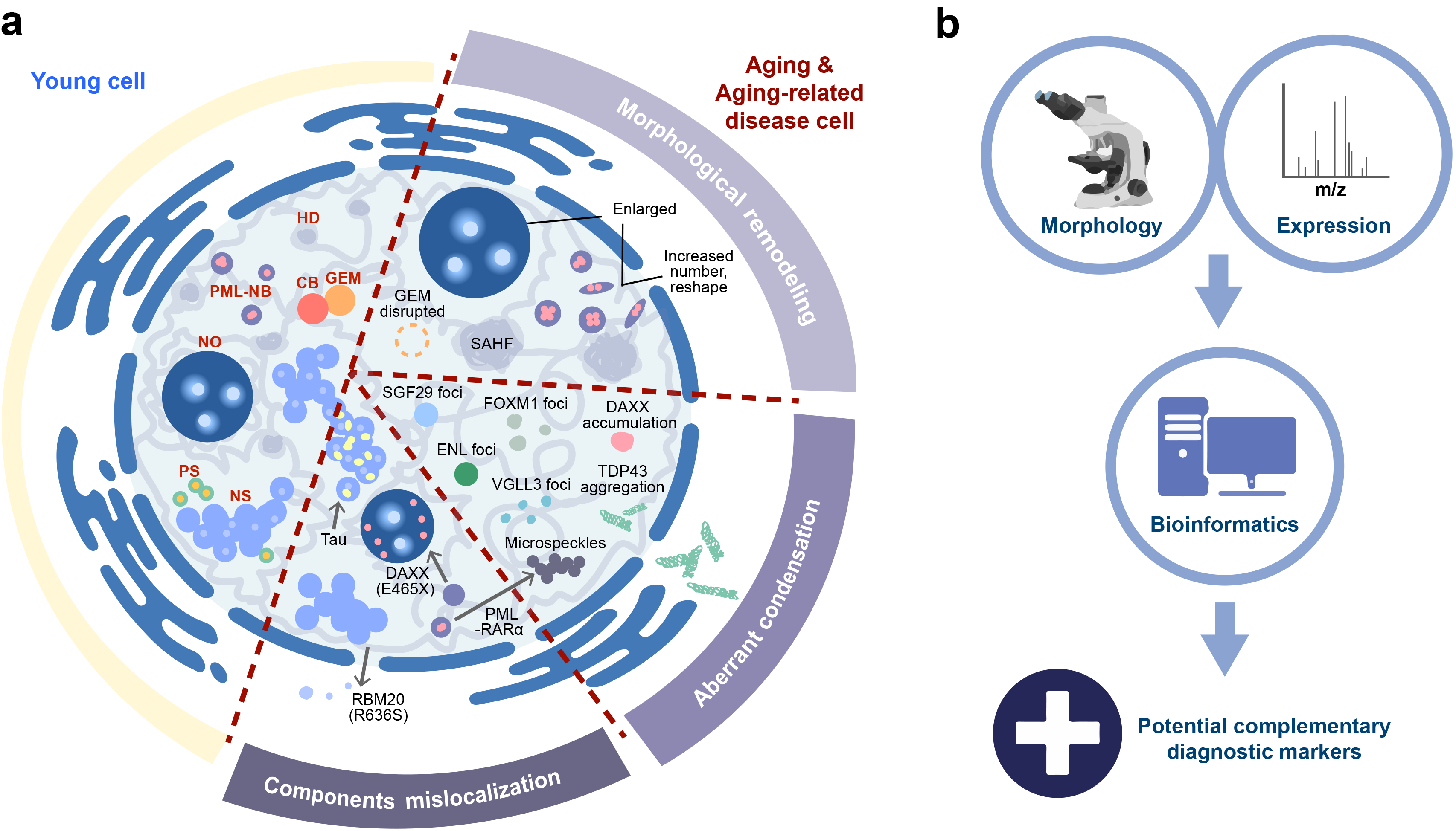
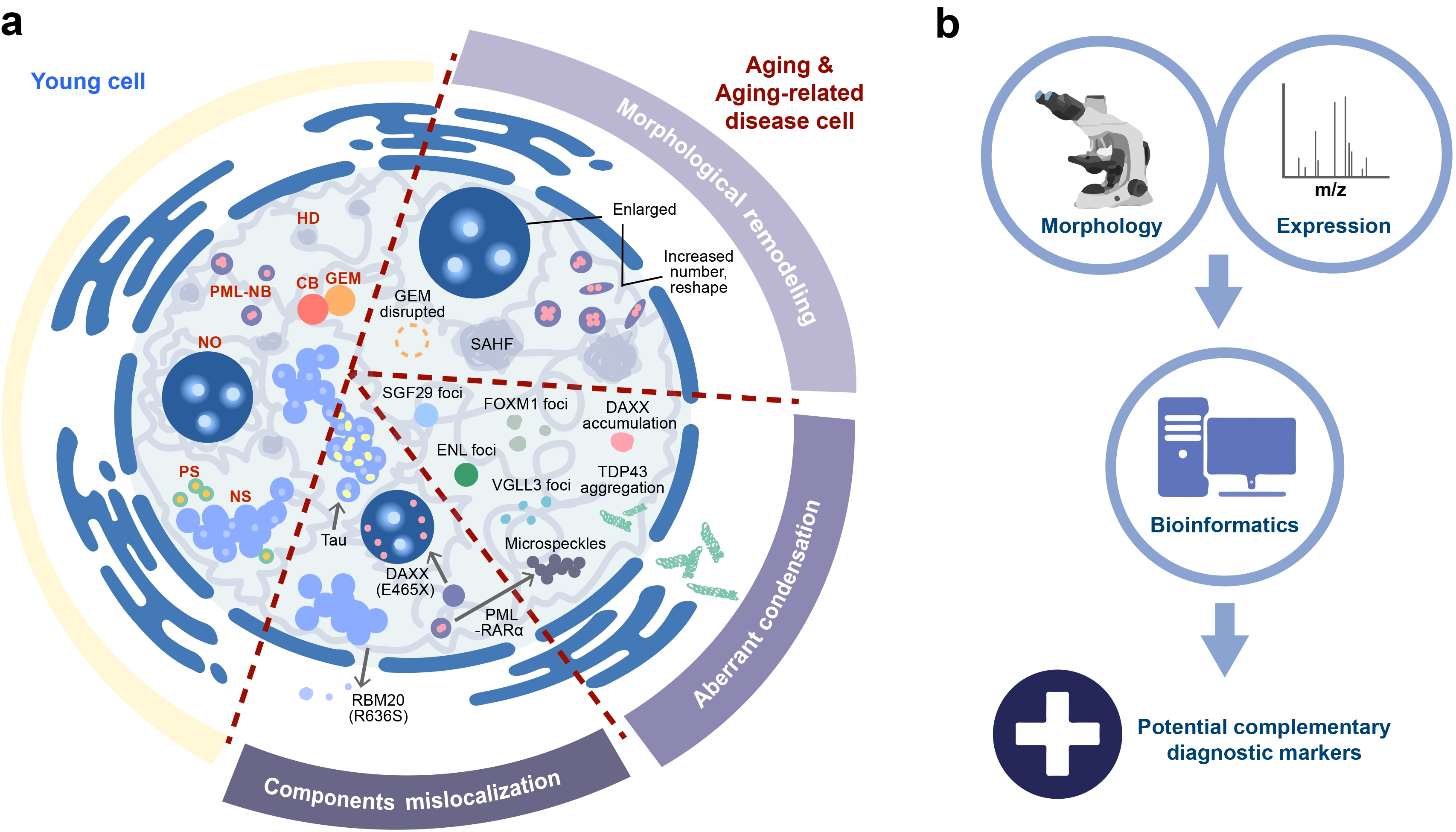
Figure 1. Changes in nuclear condensates during aging and associated disorders. (a) Comparison of nuclear condensates in young cells versus those undergoing senescence or aging-associated pathology, highlighting three major categories of alterations; (b) Toward clinical translation, an integrative framework is proposed in which morphological and compositional data from high-resolution imaging, biochemical assays, and omics are combined with computational prediction. Selected illustrations in Figure 1b were created with BioGDP.com[172]. These multimodal inputs are expected to feed into AI-driven data integration platforms, ultimately enabling precise diagnosis. CB: Cajal body; NO: nucleolus; GEM: nuclear gem; NS: nuclear speckle; PS: paraspeckle; PML-NB: PML nuclear body; HD: heterochromatin domain; SAHF: senescence-associated heterochromatin foci; SGF29: Spt-Ada-Gcn5 acetyltransferase transcriptional coactivator complex associated factor 29; FOXM1: forkhead box protein M1; ENL: histone acetylation reader eleven-nineteen-leukemia; TDP43: TAR DNA-binding protein 43, a paraspeckle component; VGLL3: transcription cofactor vestigial-like protein 3, a mechanosensitive protein; DAXX: death domain-associated protein 6, component of PML-NB; PML-RARα: fusion protein between PML and RARα, a nuclear receptor for retinoic acid; RBM20: RNA-binding protein 20, a muscle-specific splicing factor.
These categories underscore a principle often overlooked in conventional biomarker discovery: protein function is regulated not only by abundance but also by spatial distribution[173,174]. For example, ~90% of hundreds of proteins undergo subcellular relocalization upon viral infection without changes in total abundance[175]. This is exemplified by ALS/FTD-associated TDP-43 and the RBM20 R636S variant, in which aberrant subcellular localization may play a more prominent role than changes in protein expression[174,176]. Protein mislocalization also plays important roles in cancer, where genes frequently undergo mutations. For instance, redistribution of the tumor suppressor SPOP from nuclear speckles to SPOP/DAXX condensates driven by its mutations interferes with LLPS and compromises its adaptor function for the E3 ubiquitin ligase, ultimately contributing to solid tumors[177]. Deacetylation of HP1γ promotes aberrant nuclear condensation, driving drug resistance in myeloma[178]. These observations reveal that spatial dynamics and phase behavior constitute regulatory dimensions beyond protein abundance, offering diagnostic potential complementary to conventional abundance-based markers.
Moreover, the same protein can exhibit distinct subnuclear localization patterns depending on the specific mutation type. DAXX, for instance, is mutated in ~30% of pancreatic neuroendocrine tumors (PanNETs). Different mutations lead to alternative localization, e.g., the R306X mutant loses PML-NB localization, whereas the E465X mutant is mislocalized to the nucleolus[8,179]. Such mutation-specific spatial signatures may help distinguish aggressive subtypes and guide therapeutic decisions, extending the utility of nuclear condensate biomarkers beyond binary detection toward precise disease classification and personalized intervention.
Beyond spatial regulation, nuclear condensates exhibit rapid and reversible assembly and altered proteomic composition in response to cellular stimuli[8,175]. Their dynamics reflect upstream regulatory events that precede overt changes in gene expression[5], positioning them as potential sensors of incipient pathological perturbations, and offering a window for early diagnosis. In this context, NC alterations are cell-intrinsic and may occur earlier than downstream tissue changes. In cardiovascular diseases, for example, extracellular alterations such as fibrosis and vascular calcification reflect late-stage tissue remodeling rather than the upstream NC changes themselves[163,170]. Therefore, extracellular and NC markers should be viewed as complementary: extracellular markers report organ-level burden, whereas NC markers report early cellular dysfunction. Integrating these orthogonal readouts may ultimately improve diagnostic specificity and prognostic stratification.
5.2 Strategies for clinical translation: Imaging and molecular detection
Encouragingly, nuclear condensate-associated abnormalities are not confined to diseased tissues. In neurodegenerative diseases, altered TDP-43-associated protein complexes and SMN protein changes can be detected in PBMCs from patients with ALS or SMA[180,181]. In cancer, the nucleolar protein NLC is significantly elevated in PBMC lysates of prostate cancer patients[182]. Furthermore, PML-NBs and γH2AX foci have been directly quantified in PBMCs from cancer patients and healthy individuals across ages, revealing age-dependent changes[183]. Collectively, these studies demonstrate that circulating cells and circulating tumor cells can report on nuclear condensate abnormalities, offering a minimally invasive window into condensate dysregulation without requiring tissue biopsies.
Two complementary and synergistic strategies are emerging to translate nuclear condensate biology into clinical diagnostics (Figure 1b). First, high-resolution imaging, such as super-resolution microscopy and cryo-electron tomography, enables precise quantification of nuclear condensate morphology, including size, number, spatial distribution, and phase state (e.g., transitions from dynamic liquid condensates toward rigid aggregates)[184-186]. Age- or disease-linked shifts may mirror functional impairments in genomic regulation and stress response, offering morphological readouts of patient-derived cellular states[6]. High-resolution imaging has been used to detect Alzheimer’s disease-associated protein aggregates and DNA organization alterations[187,188], demonstrating the feasibility of capturing disease-related structural changes at the nanoscale and supporting the potential integration of condensate imaging into digital pathology pipelines for objective, quantitative assessment[185]. However, it is important to acknowledge that imaging-based NC quantification in solid tissues (e.g., brain, heart, or solid tumors) currently requires biopsied specimens, which are invasive to obtain. For routine screening, this approach remains largely confined to research settings or cases where tissue biopsy is already clinically indicated.
Second, molecular detection of condensate components or their post-translational modifications in biofluids offers a complementary approach[6]. EcDNA is present predominantly in cancers and rarely in normal cells, and can be detected in urine and plasma[189]. Clinically, ecDNA detection correlates with tumor stage, metastases, and shorter overall survival, showing strong concordance with tumor samples in urothelial carcinoma[190,191]. For neurodegenerative diseases, however, the blood-brain barrier (BBB) presents a critical challenge. While BBB disruption has been reported in Alzheimer’s disease and amyotrophic lateral sclerosis, its timing and extent vary across disease contexts. Thus, the detection of NC-associated proteins (e.g., TDP-43) in peripheral blood, including PBMCs[180], may offer opportunities for early biomarker development, although their utility for early diagnosis remains to be established. For cardiovascular diseases, NC-based readouts may complement established extracellular markers such as fibrosis imaging and calcification scores, potentially enabling earlier detection of cellular dysfunction before irreversible tissue remodeling occurs. Moreover, although urine and saliva represent theoretically attractive non-invasive sources, no direct evidence has yet been reported for NC-associated biomarkers in these fluids, and their utility remains speculative at present. Given that the low abundance of NC-associated biomarkers in body fluids remains a challenge, standardized and highly sensitive detection methods remain a future research need for clinical translation.
In summary, the NC-based biomarker strategy is conceptually complementary to conventional empirical markers (e.g., PSA, CA-125, pTau181/217). While those markers report on protein abundance or modification status, NC-associated readouts offer mechanistic insight into subcompartmental dysfunction, phase-state transitions, and spatial mislocalization, information that abundance-based assays alone cannot capture. Integrating these orthogonal data types may ultimately improve diagnostic specificity and prognostic stratification in aging-related diseases. Nevertheless, it is important to recognize that most of the evidence reviewed here remains at the preclinical stage, and substantial validation in large, diverse patient cohorts is required before nuclear condensate-based biomarkers can be considered for routine clinical application.
6. Conclusions and Future Directions
The nucleus, long recognized as the command center of the cell, is now emerging as a central hub in the aging process, where MLOs known as nuclear condensates act as intrinsic “sensors” of cellular senescence and physiological integrity. In this review, we have summarized recent advances in the biology of nuclear condensates, with a focus on their dynamic changes during cellular senescence and their dysregulation in major aging-related diseases. We have also discussed the emerging potential of nuclear condensates as diagnostic biomarkers, highlighting both the opportunities and the substantial challenges that remain for clinical translation.
Several key themes have emerged from the studies reviewed here. First, nuclear condensates are not static structures but highly dynamic assemblies that respond to cellular cues through LLPS, post-translational modifications, and spatial reorganization. Second, their structural and functional integrity is closely linked to cellular homeostasis, and their dysregulation, manifested as morphological alterations, aberrant phase transitions, or mislocalization of components, is a recurring feature across diverse aging-related diseases. Third, despite the promising conceptual framework for using nuclear condensates as biomarkers, most evidence remains preclinical, and substantial gaps exist in our understanding of their disease specificity, temporal dynamics, and detectability in clinically accessible samples.
Significant challenges remain on the path toward clinical translation. First, the key alterations in most nuclear condensates across different diseases, particularly age-related disorders, are still incompletely characterized. Second, high-throughput and standardized methodologies for identifying condensate morphology changes in the absence of protein expression alterations are lacking. Third, distinguishing shared abnormalities of nuclear condensates that occur across multiple conditions from those unique to individual diseases remains a major challenge. On the technical side, real-time monitoring of nuclear condensate dynamics in living cells is difficult, and standardized, quantitative methods for assessing condensate properties are yet to be established. Meanwhile, the low abundance of multiple disease-related condensate biomarkers in biofluids restricts their clinical application. Although emerging therapeutic strategies aim to target nuclear condensates or their component proteins, a fundamental question endures: can interventions that restore normal condensate structure or phase behavior reverse not only diagnostic markers but also the underlying disease phenotypes?
Looking ahead, we propose several specific directions that may accelerate the translation of nuclear condensate biology into clinical applications. First, the development of novel probes and biosensors for real-time, live-cell tracking of condensate dynamics would enable a deeper understanding of their behavior in physiologically relevant contexts. Genetically encoded reporters or small-molecule probes that report on condensate assembly, composition, or physical state could provide powerful tools for both basic research and drug discovery. Second, the establishment of standardized and quantitative methods for assessing condensate properties, including size, number, morphology, and phase state, is urgently needed to enable cross-study comparisons and to facilitate the incorporation of condensate readouts into clinical workflows. Current practices vary widely across laboratories, hampering reproducibility and clinical validation. Third, distinguishing causation from correlation remains a fundamental question: are the condensate changes observed in aging and disease drivers of pathology, or merely bystander phenomena? Addressing this will require longitudinal studies, genetic perturbation experiments, and, where possible, intervention studies that test whether modulating condensate behavior can alter disease outcomes. Fourth, the development of condensate-targeting small molecules that modulate LLPS, either by stabilizing normal condensates or dissolving pathological ones, represents an emerging frontier in drug discovery[192]. Initial proof-of-concept studies have demonstrated the feasibility of this approach, but much work remains to establish specificity and safety.
In parallel, the integration of multimodal data, including imaging phenotypes, molecular profiles, and clinical parameters, with AI-driven analytical platforms may represent a promising but still unproven strategy for developing robust diagnostic and prognostic tools[193,194], although its performance remains to be validated in large and diverse patient cohorts. Such platforms may eventually support disease stratification, progression monitoring, and the prediction of therapeutic responses, thereby contributing to personalized medicine. Additionally, the construction of comprehensive nuclear condensate proteomic maps could facilitate the capture of multi-scale dynamics of condensate composition, spatial organization, and phase behavior across aging and disease trajectories[8,195,196], which could help identify candidate biomarkers that are difficult to characterize using conventional methods, although their sensitivity, specificity, and clinical utility remain to be established.
In summary, nuclear condensates are more than fascinating membraneless organelles. They are functionally integrated into the cellular machinery that governs aging and disease (Figure 1). While the path to clinical application is long and fraught with challenges, the continued exploration of their biology, from fundamental mechanisms to translational strategies, holds promise for redefining how we understand, diagnose, and ultimately treat the diseases of aging. Addressing the outstanding questions outlined above will require interdisciplinary collaboration and sustained technological innovation, bridging the gap between basic research and medical practice.
Authors contribution
Li R, Wu S: Writing-original draft.
Shi G, Huang H, Liu F: Conceptualization, writing-review & editing.
Conflicts of interest
The authors declare no conflicts of interest.
Ethical approval
Not applicable.
Consent to participate
Not applicable.
Consent for publication
Not applicable.
Availability of data and materials
Not applicable.
Funding
This work was supported by the National Key R&D Program of China (Grant No. 2025YFA1805302 to F.L.); the National Natural Science Foundation of China (Grant Nos. NSFC 32470794 to F.L., 82603087 to R.L., 32670991 to S.W.); the Shenzhen Medical Research Fund (Grant No. SMRF, A2603013 to R.L.); and the China Postdoctoral Science Foundation (Grant No. 2026M792552 to R.L.).
Copyright
© The Author(s) 2026.
References
-
2. World Health Organization. Ageing and health [Internet]. Available from: https://www.who.int/news-room/fact-sheets/detail/ageing-and-health
-
3. Kroemer G, Maier AB, Cuervo AM, Gladyshev VN, Ferrucci L, Gorbunova V, et al. From geroscience to precision geromedicine: Understanding and managing aging. Cell. 2025;188(8):2043-2062.[DOI]
-
5. Hirose T, Ninomiya K, Nakagawa S, Yamazaki T. A guide to membraneless organelles and their various roles in gene regulation. Nat Rev Mol Cell Biol. 2023;24(4):288-304.[DOI]
-
8. Li R, Li Y, Wu S, Zhou Z, Hou X, Yang P, et al. Proximal proteomics reveals a landscape of human nuclear condensates. Nat Cell Biol. 2025;27(12):2198-2213.[DOI]
-
9. Wang Y, Yu C, Pei G, Jia W, Li T, Li P. Dissolution of oncofusion transcription factor condensates for cancer therapy. Nat Chem Biol. 2023;19(10):1223-1234.[DOI]
-
11. Buchwalter A, Hetzer MW. Nucleolar expansion and elevated protein translation in premature aging. Nat Commun. 2017;8:328.[DOI]
-
21. Wang C, Duan Y, Duan G, Wang Q, Zhang K, Deng X, et al. Stress induces dynamic, cytotoxicity-antagonizing TDP-43 nuclear bodies via paraspeckle LncRNA NEAT1-mediated liquid-liquid phase separation. Mol Cell. 2020;79(3):443-458.e7.[DOI]
-
24. Staněk D. Coilin and Cajal bodies. Nucleus. 2023;14(1):2256036.[DOI]
-
26. Courchaine EM, Barentine AES, Straube K, Lee DR, Bewersdorf J, Neugebauer KM. DMA-Tudor interaction modules control the specificity of in vivo condensates. Cell. 2021;184(14):3612-3625.[DOI]
-
27. Makarov EM, Owen N, Bottrill A, Makarova OV. Functional mammalian spliceosomal complex E contains SMN complex proteins in addition to U1 and U2 snRNPs. Nucleic Acids Res. 2012;40(6):2639-2652.[DOI]
-
29. Wang C, Chen W, Li R, Yang Y, Wu J, Tian Y, et al. Disruption of ZC3H15 compromises telomere length maintenance by entrapping telomerase within Cajal bodies. Cell Biosci. 2025;15(1):107.[DOI]
-
36. Fox AH, Lam YW, Leung AKL, Lyon CE, Andersen J, Mann M, et al. Paraspeckles: A novel nuclear domain. Curr Biol. 2002;12(1):13-25.[DOI]
-
41. Fujishiro S, Sasai M, Maeshima K. Chromatin domains in the cell: Phase separation and condensation. Curr Opin Struct Biol. 2025;91:103006.[DOI]
-
48. Liu R, Xuan J, Cerra C, Stojnic N, Pan J, Chelliah SS, et al. RAD54L coordinates the nucleolar DNA damage response to maintain rDNA stability. EMBO J. 2026;45(17):6299-6319.[DOI]
-
50. Menjivar-Vallecillo MJ, Padilla-Claros NV, Gavarrete-Garrido P, León-Castañeda SA, Ramos-Zaldívar HM. The nucleolus and its associated pathologies. WIREs Mech Dis. 2025;17(5):e70003.[DOI]
-
53. Noguchi K, Suzuki H, Abe R, Horiuchi K, Onoguchi-Mizutani R, Akimitsu N, et al. Multi-omics analysis using antibody-based in situ biotinylation technique suggests the mechanism of Cajal body formation. Cell Rep. 2024;43(9):114734.[DOI]
-
54. Lyon CE, Bohmann K, Sleeman J, Lamond AI. Inhibition of protein dephosphorylation results in the accumulation of splicing snRNPs and coiled bodies within the nucleolus. Exp Cell Res. 1997;230(1):84-93.[DOI]
-
61. White L, Erbay B, Blair GE. The Cajal body protein p80-coilin forms a complex with the adenovirus L4-22K protein and facilitates the nuclear export of adenovirus mRNA. mBio. 2023;14(5):e01459-23.[DOI]
-
62. Matera AG, Frey MR. Coiled bodies and gems: Janus or Gemini? Am J Hum Genet. 1998;63(2):317-321.[DOI]
-
63. Yamazaki T, Chen S, Yu Y, Yan B, Haertlein TC, Carrasco MA, et al. FUS-SMN protein interactions link the motor neuron diseases ALS and SMA. Cell Rep. 2012;2(4):799-806.[DOI]
-
68. Lallemand-Breitenbach V, Zhu J, Puvion F, Koken M, Honoré N, Doubeikovsky A, et al. Role of promyelocytic leukemia (PML) sumolation in nuclear body formation, 11S proteasome recruitment, and As2O3-induced PML or PML/retinoic acid receptor alpha degradation. J Exp Med. 2001;193(12):1361-1371.
-
69. Yang S, Kuo C, Bisi JE, Kim MK. PML-dependent apoptosis after DNA damage is regulated by the checkpoint kinase hCds1/Chk2. Nat Cell Biol. 2002;4(11):865-870.[DOI]
-
72. Niwa-Kawakita M, Ferhi O, Soilihi H, Le Bras M, Lallemand-Breitenbach V, de Thé H. PML is a ROS sensor activating p53 upon oxidative stress. J Exp Med. 2017;214(11):3197-3206.[DOI]
-
79. Khanna N, Hu Y, Belmont AS. HSP70 transgene directed motion to nuclear speckles facilitates heat shock activation. Curr Biol. 2014;24(10):1138-1144.[DOI]
-
81. Chen Y, Belmont AS. Genome organization around nuclear speckles. Curr Opin Genet Dev. 2019;55:91-99.[DOI]
-
91. Prasanth KV, Prasanth SG, Xuan Z, Hearn S, Freier SM, Bennett CF, et al. Regulating gene expression through RNA nuclear retention. Cell. 2005;123(2):249-263.[DOI]
-
93. Imamura K, Imamachi N, Akizuki G, Kumakura M, Kawaguchi A, Nagata K, et al. Long noncoding RNA NEAT1-dependent SFPQ relocation from promoter region to paraspeckle mediates IL8 expression upon immune stimuli. Mol Cell. 2014;53(3):393-406.[DOI]
-
98. Wang H, Tang J, Yan S, Li C, Li Z, Xiong Z, et al. Liquid-liquid phase separation in aging: Novel insights in the pathogenesis and therapeutics. Ageing Res Rev. 2024;102:102583.[DOI]
-
101. Tiku V, Jain C, Raz Y, Nakamura S, Heestand B, Liu W, et al. Small nucleoli are a cellular hallmark of longevity. Nat Commun. 2017;8:16083.[DOI]
-
110. Bell O, Burton A, Dean C, Gasser SM, Torres-Padilla ME. Heterochromatin definition and function. Nat Rev Mol Cell Biol. 2023;24(10):691-694.[DOI]
-
113. Zhang Q, Deng K, Liu M, Yang S, Xu W, Feng T, et al. Phase separation of BuGZ regulates gut regeneration and aging through interaction with m6A regulators. Nat Commun. 2023;14:6700.[DOI]
-
117. López-Otín C, Blasco MA, Partridge L, Serrano M, Kroemer G. Hallmarks of aging: An expanding universe. Cell. 2023;186(2):243-278.[DOI]
-
119. Alberti S, Carra S. Quality control of membraneless organelles. J Mol Biol. 2018;430(23):4711-4729.[DOI]
-
120. Bai Y, Ma T, Zhao S, Li S, Wang X, Li J, et al. Mitochondria-associated condensates maintain mitochondrial homeostasis and promote lifespan. Nat Aging. 2025;5(10):1983-2002.[DOI]
-
122. Mukherjee P, Panda P, Kasturi P. A comparative meta-analysis of membraneless organelle-associated proteins with age related proteome of C. elegans. Cell Stress Chaperones. 2022;27(6):619-631.[DOI]
-
128. Ishihara T, Ariizumi Y, Shiga A, Kato T, Tan CF, Sato T, et al. Decreased number of Gemini of coiled bodies and U12 snRNA level in amyotrophic lateral sclerosis. Hum Mol Genet. 2013;22(20):4136-4147.[DOI]
-
130. Gertz B, Wong M, Martin LJ. Nuclear localization of human SOD1 and mutant SOD1-specific disruption of survival motor neuron protein complex in transgenic amyotrophic lateral sclerosis mice. J Neuropathol Exp Neurol. 2012;71(2):162-177.[DOI]
-
132. Alami NH, Smith RB, Carrasco MA, Williams LA, Winborn CS, Han SSW, et al. Axonal transport of TDP-43 mRNA granules is impaired by ALS-causing mutations. Neuron. 2014;81(3):536-543.[DOI]
-
135. Mann JR, Gleixner AM, Mauna JC, Gomes E, DeChellis-Marks MR, Needham PG, et al. RNA binding antagonizes neurotoxic phase transitions of TDP-43. Neuron. 2019;102(2):321-338.e8.[DOI]
-
136. Moens TG, Da Cruz S, Neumann M, Shelkovnikova TA, Shneider NA, Van Den Bosch L. Amyotrophic lateral sclerosis caused by FUS mutations: Advances with broad implications. Lancet Neurol. 2025;24(2):166-178.[DOI]
-
138. Bukar Maina M, Al-Hilaly YK, Serpell LC. Nuclear tau and its potential role in Alzheimer’s disease. Biomolecules. 2016;6(1):9.[DOI]
-
139. Chinnathambi S, Velmurugan G, Suresh S, Adithyan A, Chandrashekar M. Nuclear Tau accumulation in Alzheimer's disease. Adv Protein Chem Struct Biol. 2025;143:323-337.[DOI]
-
140. Younas N, Saleem T, Younas A, Zerr I. Nuclear face of Tau: An inside player in neurodegeneration. Acta Neuropathol Commun. 2023;11(1):196.[DOI]
-
142. Nozawa RS, Yamamoto T, Takahashi M, Tachiwana H, Maruyama R, Hirota T, et al. Nuclear microenvironment in cancer: Control through liquid-liquid phase separation. Cancer Sci. 2020;111(9):3155-3163.[DOI]
-
144. Das S, Anczuków O, Akerman M, Krainer AR. Oncogenic splicing factor SRSF1 is a critical transcriptional target of MYC. Cell Rep. 2012;1(2):110-117.[DOI]
-
146. Babaian A, Rothe K, Girodat D, Minia I, Djondovic S, Milek M, et al. Loss of m1acp3Ψ ribosomal RNA modification is a major feature of cancer. Cell Rep. 2020;31(5):107611.[DOI]
-
154. Xie F, Zhou X, Ran Y, Li R, Zou J, Wan S, et al. Targeting FOXM1 condensates reduces breast tumour growth and metastasis. Nature. 2025;638(8052):1112-1121.[DOI]
-
156. Xiong H, Gao H, Wan J, Xiao J, Luo X, Dong X, et al. Whole exome sequencing identifies a novel frameshift mutation of the WRN gene in a Werner syndrome family and functional analysis. Mol Genet Genomic Med. 2025;13(6):e70118.[DOI]
-
164. Horii Y, Matsuda S, Toyota C, Morinaga T, Nakaya T, Tsuchiya S, et al. VGLL3 is a mechanosensitive protein that promotes cardiac fibrosis through liquid–liquid phase separation. Nat Commun. 2023;14:550.[DOI]
-
170. Lu Y, Meng L, Ren R, Wang X, Sui W, Xue F, et al. Paraspeckle protein NONO attenuates vascular calcification by inhibiting bone morphogenetic protein 2 transcription. Kidney Int. 2024;105(6):1221-1238.[DOI]
-
172. Jiang S, Li H, Zhang L, Mu W, Zhang Y, Chen T, et al. Generic Diagramming Platform (GDP): A comprehensive database of high-quality biomedical graphics. Nucleic Acids Res. 2025;53(D1):D1670-D1676.[DOI]
-
174. Sigaeva A, Hutchings C, Cesnik A, Lilley KS, Lundberg E. Subcellular localization as a driver of protein function. Nat Rev Mol Cell Biol. 2026;27(7):493-510.[DOI]
-
175. Hein MY, Peng D, Todorova V, McCarthy F, Kim K, Liu C, et al. Global organelle profiling reveals subcellular localization and remodeling at proteome scale. Cell. 2025;188(4):1137-1155.e20.[DOI]
-
177. Bouchard JJ, Otero JH, Scott DC, Szulc E, Martin EW, Sabri N, et al. Cancer mutations of the tumor suppressor SPOP disrupt the formation of active, phase-separated compartments. Mol Cell. 2018;72(1):19-36.e8.[DOI]
-
180. Yu SF, Michon M, Lingappa AF, Paulvannan K, Solas D, Staats K, et al. An ALS assembly modulator signature in peripheral blood mononuclear cells: Implications for ALS pathophysiology, therapeutics, and diagnostics. Clin Proteomics. 2025;22(1):16.[DOI]
-
181. Kolb SJ, Gubitz AK, Olszewski RF, Ottinger E, Sumner CJ, Fischbeck KH, et al. A novel cell immunoassay to measure survival of motor neurons protein in blood cells. BMC Neurol. 2006;6(1):6.[DOI]
-
182. Miranda A, Santos T, Carvalho J, Alexandre D, Jardim A, Caneira CF, et al. Aptamer-based approaches to detect nucleolin in prostate cancer. Talanta. 2021;226:122037.[DOI]
-
184. Stein J, Ericsson M, Nofal M, Magni L, Aufmkolk S, McMillan RB, et al. Cryosectioning-enhanced super-resolution microscopy for single-protein imaging across cells and tissues. Proc Natl Acad Sci U S A. 2025;122(32):e2504578122.[DOI]
-
192. Mitrea DM, Mittasch M, Gomes BF, Klein IA, Murcko MA. Modulating biomolecular condensates: A novel approach to drug discovery. Nat Rev Drug Discov. 2022;21(11):841-862.[DOI]
Copyright
© The Author(s) 2026. This is an Open Access article licensed under a Creative Commons Attribution 4.0 International License (https://creativecommons.org/licenses/by/4.0/), which permits unrestricted use, sharing, adaptation, distribution and reproduction in any medium or format, for any purpose, even commercially, as long as you give appropriate credit to the original author(s) and the source, provide a link to the Creative Commons license, and indicate if changes were made.
Publisher’s Note
Science Exploration remains a neutral stance on jurisdictional claims in published
maps
and institutional affiliations. The views expressed in this article are solely those
of
the author(s) and do not reflect the opinions of the Editors or the publisher.
Share And Cite


Science Exploration Style
Li R, Wu S, Shi G, Huang H, Liu F. Nuclear condensates in aging-related diseases: The roles and diagnostic potential. Geromedicine. 2027;3:202646. https://doi.org/10.70401/Geromedicine.2026.0046
Tips
Copy completed.
Submit a Manuscript
Author Instructions
Cite this Article
Article Metrics
0
View
0
Download
Cited
Article Updates

- Abstract
- Keywords
- 1. Introduction
- 2. Mechanisms of Major Nuclear Condensate Formation and Functional Implications
- 3. The Dynamics of Nuclear Condensates during Cellular Senescence
- 4. Nuclear Condensates in Aging-Related Diseases
- 5. Emerging Role of Nuclear Condensates in the Diagnosis of Aging-Related Diseases
- 6. Conclusions and Future Directions
- Authors contribution
- Conflicts of interest
- Ethical approval
- Consent to participate
- Consent for publication
- Availability of data and materials
- Funding
- References
- Copyright
 Get citation
Science Exploration Style

Li R, Wu S, Shi G, Huang H, Liu F. Nuclear condensates in aging-related diseases: The roles and diagnostic potential. Geromedicine. 2027;3:202646. https://doi.org/10.70401/Geromedicine.2026.0046
copy
Share Link
copy