From RNAi to single-cell CRISPR: Evolving functional screening in aging and cancer

Botao Fan

Aiwei Wu
Xue Pan
*
,
Hu Wang
*
*Correspondence to:
Hu Wang, Department of Cardiology, The Third People’s Hospital of Deqing, Affiliated Hospital of Hangzhou Normal University, Zhejiang Key Laboratory of Medical Epigenetics, School of Basic Medical Sciences, Hangzhou Normal University, Hangzhou 311121, Zhejiang, China.
E-mail: wanghu19860315@163.com
Xue Pan, Department of Cardiology, The Third People’s Hospital of Deqing, Affiliated Hospital of Hangzhou Normal University, Zhejiang Key Laboratory of Medical Epigenetics, School of Basic Medical Sciences, Hangzhou Normal University, Hangzhou 311121, Zhejiang, China. E-mail: panxue19950105@163.com
Xue Pan, Department of Cardiology, The Third People’s Hospital of Deqing, Affiliated Hospital of Hangzhou Normal University, Zhejiang Key Laboratory of Medical Epigenetics, School of Basic Medical Sciences, Hangzhou Normal University, Hangzhou 311121, Zhejiang, China. E-mail: panxue19950105@163.com
Ageing Cancer Res Treat. 2026;3:202621. 10.70401/acrt.2026.0031
Received: April 29, 2026Accepted: July 17, 2026Published: July 17, 2026
Abstract
Aging and cancer reflect distinct but interconnected manifestations of cellular stress responses shaped by context-specific molecular and environmental factors. Senescence initially restrains malignancy through stable cell-cycle arrest and immune clearance, yet persistence of senescent cells and the senescence-associated secretory phenotype (SASP) paradoxically drives tumor progression, therapeutic resistance, and immune evasion via chronic inflammation and metabolic reprogramming. Functional genomic screening has evolved from bulk RNA interference to precision clustered regularly interspaced short palindromic repeats (CRISPR) modalities, including knockout, interference, activation, and base editing, and onward to single-cell and in vivo platforms. These technological advances have transformed our capacity to dissect cellular state transitions and network dependencies with unprecedented resolution. Here, we trace this technological evolution and synthesize its impact on aging and cancer research, with emphasis on chromatin regulation, metabolic rewiring, the SASP, nucleocytoplasmic transport, therapeutic response, and the tumor microenvironment. We emphasize that the current evidence derives predominantly from preclinical functional screens and computational predictions. We propose shifting from gene-centric discovery toward context-dependent network analysis. Integrating precise editing, in vivo screening, single-cell multi-omics, and emerging artificial intelligence (AI)-assisted design may provide information and a design basis for future combined strategies that simultaneously target vulnerabilities in senescent cells and malignant populations.
Keywords
CRISPR screening, senescence, cancer, functional genomics
1. Introduction
Senescence and malignant transformation are interconnected states governed by a shared regulatory network rather than independent processes. When cells encounter diverse stressors, such as telomere attrition, DNA damage, and oncogenic signaling, they trigger a stress response program that evolution has conserved[1]. This evolutionarily conserved program can either enforce stable cell-cycle arrest or be hijacked by cancer cells to promote malignant growth. Conversely, the mechanisms that suppress tumorigenesis also limit tissue renewal, revealing a shared molecular basis between cancer and aging.
While traditionally seen as a static tumor-suppressive mechanism via p53 and Rb pathways[2], senescence actually plays multidimensional roles at the tissue level. Senescent cells actively remodel their microenvironment through SASP, metabolic reprogramming, and extensive epigenetic remodeling[3,4]. Consequently, they can function as either a tumor barrier or a cancer driver depending on the specific context[5].
Single genes poorly capture the nonlinear dynamics underlying aging-cancer interactions[1,6,7]. Systematic analysis of such complexity started in simpler eukaryotes. Early work with yeast deletion collections, temperature-sensitive alleles, and RNAi feeding in Caenorhabditis elegans (C. elegans) laid the groundwork. Those screens identified core cell-cycle and longevity pathways, and whole-genome coverage in non-mammals revealed how functional redundancy operates. Functional screening, especially using clustered regularly interspaced short palindromic repeats (CRISPR) platforms, offers a powerful alternative for dissecting this complexity in mammalian systems[8-10]. These technologies have already identified key oncogenic dependencies and are now expanding into aging research by integrating with primary cells and in vivo models[11-13]. Researchers can now map the regulatory networks that control cell fate transitions and identify critical senescence regulators beyond simple gene lists.
Translating functional screening findings into targeted interventions for aging-related cancers remains fragmented across research fields. Guided by the principle that screening technology must align with the specific biological question, this review integrates mechanistic insights to examine how aging reshapes cancer biology. We discuss key regulatory networks identified through perturbation screens, then explore how aged microenvironments modify these pathways, and finally outline strategies that leverage age-specific vulnerabilities for guidance. Specifically, relative to López-Otín et al.[6], our review adds a functional genomic screening perspective that moves beyond descriptive hallmark co-occurrence to reveal how specific perturbations drive context-dependent fate divergence. Relative to Santinha et al.[12], we provide an aging–cancer mechanistic integration by interpreting screening data through artificial intelligence (AI)-assisted modeling rather than focusing on screening technologies. Together, these distinctions position our review as a complementary synthesis that translates hallmark concepts into testable network models.
2. Technological Advances for Aging-Cancer Research
Functional screening has evolved from coarse perturbations to precise, high-resolution tools essential for dissecting the complex interplay between aging and cancer. Generations of conceptual and technical innovation have enabled increasingly systematic exploration of gene function at the genome scale (Figure 1). Early methodologies, though foundational, often fail to account for the fragile homeostasis of aged tissues, necessitating a strategic pivot toward next-generation platforms that minimize artifactual stress while maximizing precision.
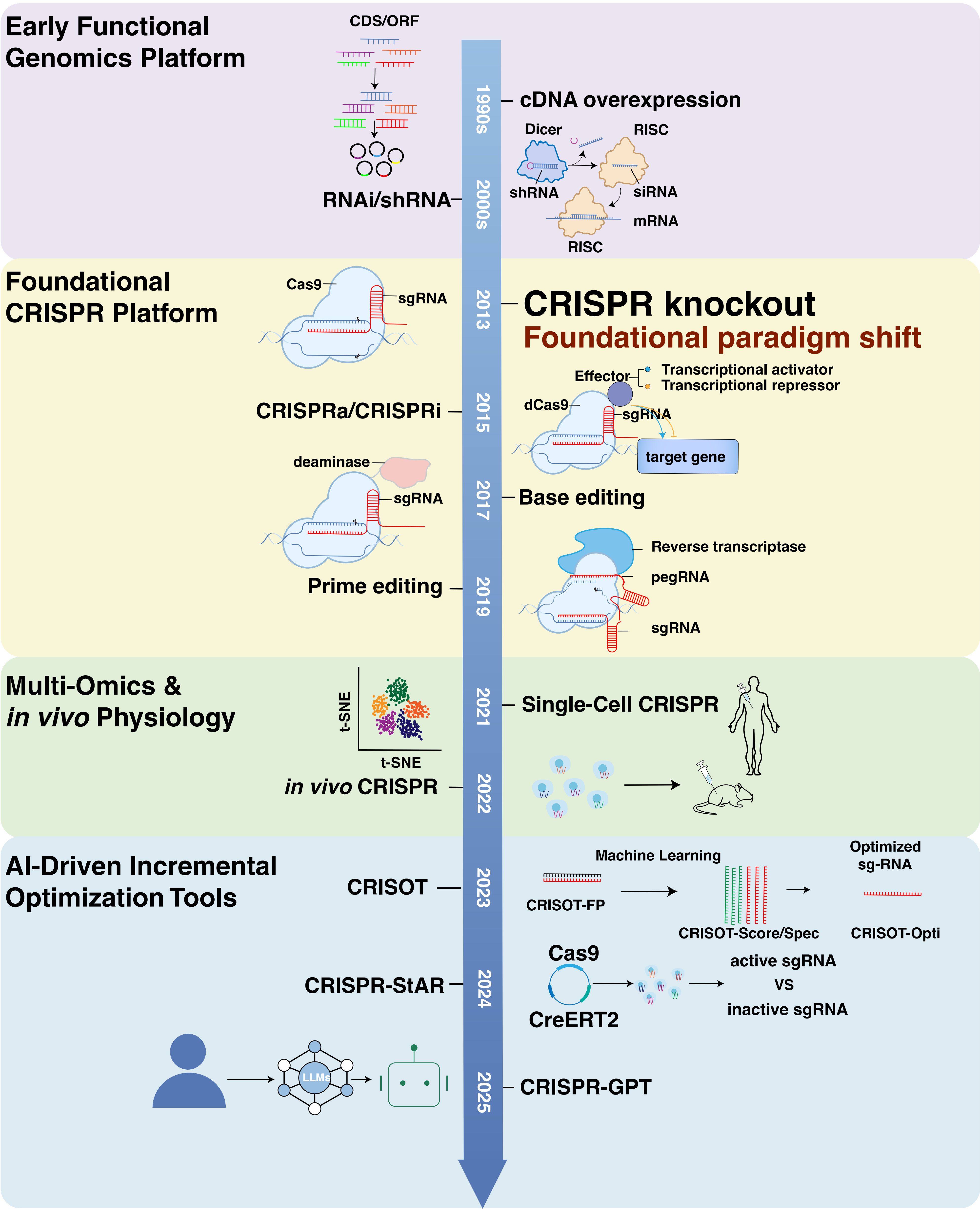
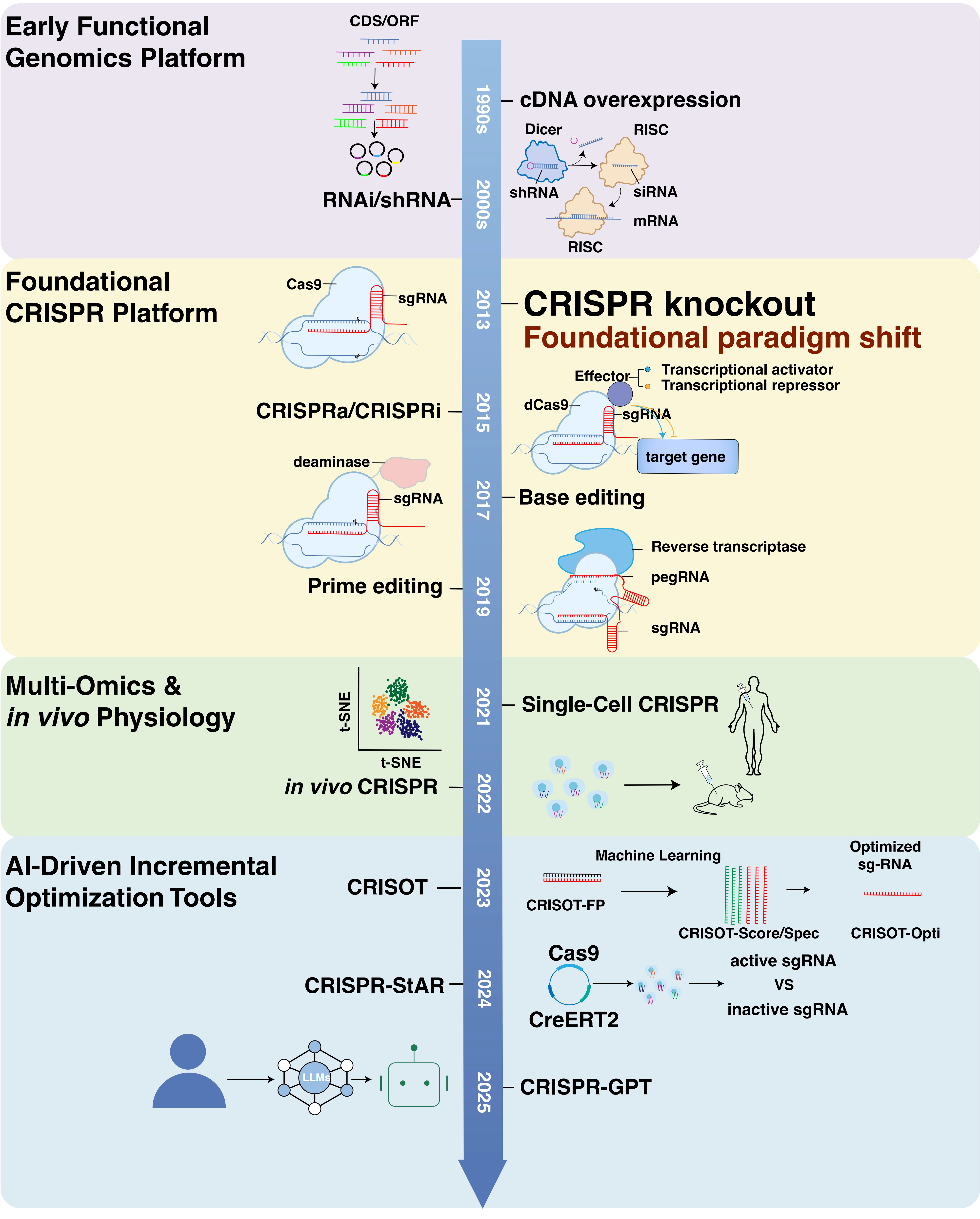
Figure 1. The evolution of functional library screening technologies. The field has progressed through four key developmental stages: (1) Early Functional Genomics Platform, characterized by cDNA overexpression and RNAi/shRNA; (2) Foundational CRISPR Platform, initiated by the introduction of Cas9/sgRNA knockout and expanded to include CRISPRa/i and base/prime editing, an event that laid the cornerstone for the functional CRISPR platform; (3) Multi-Omics & in vivo Physiology, which bridges genetic perturbation with single-cell transcriptomics and in vivo models to resolve cellular heterogeneity and physiological contexts; and (4) AI-Driven Incremental Optimization Tools, leveraging machine learning for automated design, off-target analysis, and tools like CRISPR-StAR to interrogate complex regulatory networks. Together, these stages represent a shift toward high-precision, high-throughput, and context-aware functional screening in diseases such as cancer and aging. CDS: coding sequence; ORF: open reading frame; RNAi: RNA interference; shRNA: short hairpin RNA; siRNA: small interfering RNA; RISC: RNA-induced silencing complex; CRISPR: clustered regularly interspaced short palindromic repeats; sgRNA: single guide RNA; pegRNA: prime editing guide RNA; CRISOT: CRISPR off-target screening and identification tool; CRISPR-StAR: CRISPR screening with temporal and regional control; CreERT2: Cre recombinase–estrogen receptor T2 (mutant); CRISPR-GPT: generative pre-trained transformer for CRISPR design; AI: artificial intelligence; CRISPRa: clustered regularly interspaced short palindromic repeats activation.
2.1 Early functional genomics via ectopic overexpression and RNA interference
Early functional genomics relied heavily on overexpression of complementary DNA (cDNA)[14-16] and RNAi-based libraries, including small interfering RNA (siRNA) and shRNA screens[17,18]. While foundational for early work in cell differentiation and signaling, this strategy faces critical hurdles in aging and cancer research. Constructing comprehensive cDNA libraries with adequate transcriptome coverage remains technically challenging[19,20]. cDNA screens driven by strong constitutive promoters often yield supraphysiological protein levels. Such non-physiological expression can artificially override the stringent cell cycle checkpoints governing senescence[21,22], potentially masking genuine drivers of senescence escape and generating confounding artifacts[20].
RNAi strategies present distinct challenges, primarily due to sequence-dependent toxicity. shRNA seed regions mimic endogenous microRNAs (miRNAs), inadvertently binding to the 3' untranslated regions of off-target transcripts and provoking widespread off-target effects[23,24]. High-level shRNA expression saturates the miRNA processing machinery by sequestering Exportin-5 and RNA-induced silencing complex (RISC)-Argonaute 2 (Ago2)[25]. The disruption of basal cellular homeostasis elicits non-specific stress responses. In aged cells, which already exhibit elevated proteotoxic and oxidative stress, this exogenous burden can induce senescence-like phenotypes or apoptosis independent of the target gene[25,26]. Distinguishing specific gene loss from general cellular collapse thus becomes extremely difficult, thereby restricting the utility of these tools for unbiased discovery in aged systems.
2.2 CRISPR-associated protein 9 (Cas9) knockout/knockin strategy
CRISPR-Cas9 knockout libraries transformed functional screening with superior signal-to-noise ratios[27-31]. With near-complete gene inactivation, researchers could systematically map tumor dependencies across diverse genetic backgrounds. This quickly and profoundly reoriented cancer research[27,28,32]. However, this application in aged organisms reveals a critical limitation. Cas9 works by inducing double-strand breaks (DSBs). Aging cells already carry a heavier DNA damage burden and heightened DNA damage response (DDR) sensitivity, so these exogenous breaks easily trigger p53-mediated senescence or exacerbate pre-existing senescence-associated phenotypes, thereby generating artifacts that obscure true genetic drivers in aged contexts[21,33]. Knockin strategies enable precise sequence insertion while preserving endogenous regulation, allowing modeling of specific mutations and in vivo tracking[34]. Yet they run into the same DSB-related constraints within fragile aged microenvironments. Thus, while essential for mapping tumor dependencies in young models, knockout approaches require rigorous controls or supplementation with non-destructive alternatives in aging research.
2.3 CRISPR interference (CRISPRi) and activation (CRISPRa)
To circumvent DSB-induced toxicity, CRISPRi and CRISPRa systems utilize catalytically inactive Cas9 (dCas9) fused to transcriptional modulators[35-38]. The advent of CRISPRi/a has proven particularly transformative for studying senescence, stem-cell fate transitions, and non-lethal phenotypes, as these biological processes are often highly sensitive to DNA damage and strong perturbations[39]. However, CRISPRi/a efficacy depends on chromatin accessibility. Aging is accompanied by profound heterochromatin formation and epigenetic silencing[6,40]. This compacted landscape in aged tissues can sterically hinder dCas9 binding, leading to reduced editing efficiency compared to young counterparts. Successful application in aging models therefore often demands careful gRNA design targeting accessible loci or the co-delivery of chromatin-modulating factors, a consideration less critical in young tumor models.
2.4 CRISPR base editing and prime editing
Base editing and prime editing enable precise substitutions without DSBs, addressing the safety concerns of genomic instability in aged organisms[41-48]. Prime editing has already demonstrated functional repair of disease-causing point mutations in patient-derived models[49], underscoring its utility for modeling age-associated mutations such as those driving clonal hematopoiesis of indeterminate potential (CHIP) and cancer initiation[50,51]. By avoiding DSBs that risk chromosomal rearrangements and p53 activation, these tools offer a theoretically safer profile for long-term studies in aged hosts[41-48].
Despite progress in coding regions, extending high-resolution CRISPR-based screening toolkits to noncoding regulatory elements remains challenging, especially in regions of structural variability and epigenetic silencing typical of aging genomes[6,52,53]. Moreover, phenotypic readouts in aged cells are highly sensitive to the altered cellular microenvironment[54,55], complicating the interpretation of subtle regulatory changes driven by single-nucleotide edits.
2.5 Single-cell CRISPR screening and in vivo screening
Traditional bulk screening averages out the cellular heterogeneity that is exacerbated by aging and cancer. Single-cell CRISPR screening, exemplified by Perturb-seq, links genetic perturbations to whole-transcriptome states within the same cell[11,56-59]. This method identifies regulators of cell fate decisions, state transitions, and lineage differentiation[60,61]. New tools such as CRISPR screening with temporal and regional control (CRISPR-STAR) enhance the resolution of in vivo screening through internal controls[62], while dedicated algorithms improve off-target prediction[63]. The generative pre-trained transformer for CRISPR design (CRISPR-GPT) system assists with experimental design to improve screening precision[64].
For instance, in vivo screening at single-cell resolution provides direct evidence of how genetic perturbations affect cell fate decisions in physiological contexts[65]. The aged tumor microenvironment specifically alters the impact of certain genetic perturbations on cancer cells[54,55]. Functional CRISPR screens have identified non-coding regulatory elements that modulate oncogene-induced senescence[66]. Independent studies have shown that aging reshapes the tumor immune microenvironment[55,67]. Organoid-based CRISPR screening provides a physiologically relevant platform to model cancer susceptibility[68,69]. Researchers are now combining functional screens with spatial omics and chromatin profiling. This integration is key to understanding genome regulation in specific tissue contexts.
2.6 Combinatorial screening: Progress and persistent challenges
Combinatorial screening has evolved from inefficient dual-shRNA methods to precise multiplex CRISPR systems. CRISPR-associated protein 12a (Cas12a) arrays can now autonomously process multiple CRISPR RNAs (crRNAs)[70]. Orthogonal Cas9/Cas12a platforms can achieve independent dual-gene editing, and the IN4MER platform offers superior sensitivity for detecting synthetic lethal interactions[71]. These advances have enabled the systematic screening of gene combinations, such as the synthetic lethal interaction between breast cancer susceptibility gene (BRCA) deficiency and poly (ADP-ribose) polymerase 1 (PARP1) inhibition. This therapeutic strategy, first established by killing BRCA2-deficient tumors and targeting DNA repair defects in BRCA-mutant cells, was subsequently validated in vivo in BRCA1-deficient mammary tumors responsive to AZD2281 alone or with platinum[72-74]. This strategy was further validated by specific BRCA1-PARP1 genetic interactions[75,76]. Some interactions translate to biomarkers of drug sensitivity, including frequent genetic alterations in the lysine demethylase 5C/6A (KDM5C/6A) histone demethylases, which sensitize cells to inhibition of poly (ADP-ribose) polymerase 7 (PARP7/TIPARP)[77]. These alterations were previously hidden by technical noise and poor co-delivery rates.
The combinatorial explosion problem still limits genome-wide mapping, as testing all human gene pairings far exceeds current experimental capabilities. The field has responded with a pragmatic strategy: integrate small, targeted libraries with large-scale multi-omics databases. Machine learning now predicts high-confidence interactions before wet-lab validation. CRISPR-GEM, for instance, uses neural networks to forecast editing effects, guiding experimental design[78]. More recent explainable AI models go further, integrating sequence features with epigenetic information to predict both gRNA efficiency and off-target risks. CCLMoff leverages RNA language models for versatile predictions across diverse next-generation sequencing (NGS) datasets[79]. This computationally guided, experimentally validated approach can efficiently navigate complex genetic interaction networks and accelerate the development of translational medicine breakthroughs, such as the use of Werner syndrome ATP-dependent helicase (WRN) inhibitors in microsatellite instability (MSI) cancers.
2.7 Platform selection, in vivo barriers, and future directions
Given the diverse array of CRISPR-based screening technologies now available, selecting the optimal modality requires aligning the specific biological question with the strengths of each platform (Figure 2 and Table 1). CRISPR knockout remains the gold standard for identifying essential genes in cancer dependency maps, whereas CRISPRi/a and base editing are increasingly preferred in aging research to model dosage effects and specific point mutations without triggering confounding DNA damage responses[80]. Emerging modalities such as spatial CRISPR screening[81,82] and RNA-targeting CRISPR[83,84] further expand the toolkit for aging and cancer models.
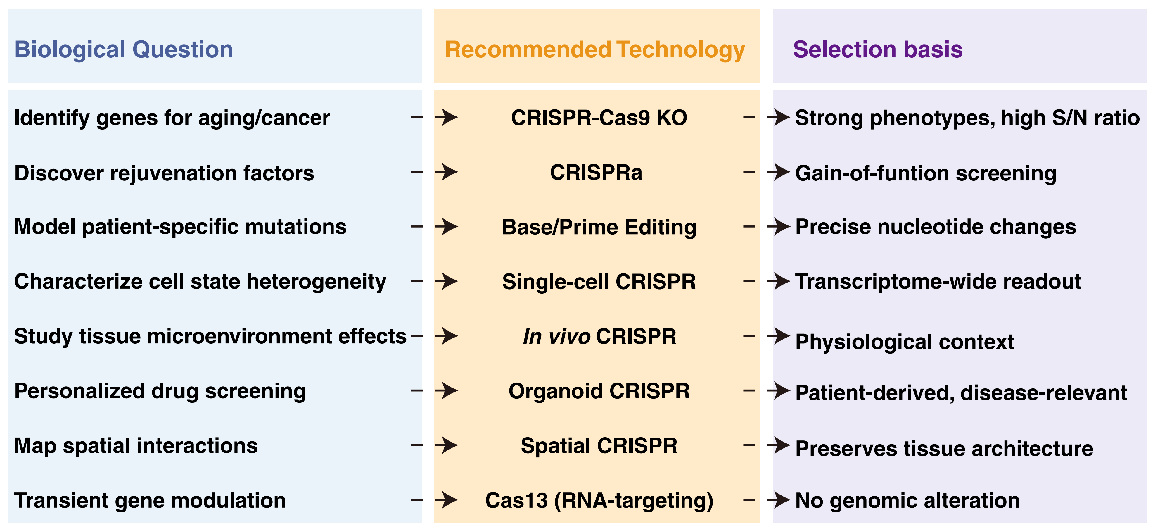
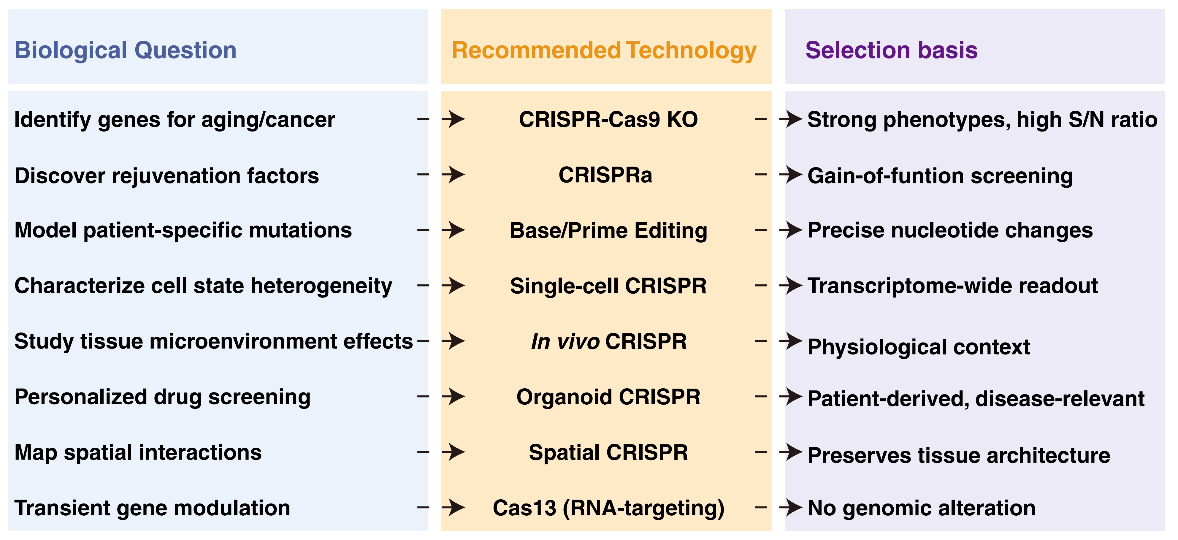
Figure 2. Functional screening technology selection guide. CRISPR: clustered regularly interspaced short palindromic repeats; Cas9: CRISPR-associated protein 9; Cas13: CRISPR-associated protein 13.
Table 1. Functional screening technology selection guide.
| Technology | Mechanism | Applications | Advantages | Limitations | Relevant to aging | Relevant to cancer | References |
| CRISPR-KO | Complete gene inactivation via DSBs | Genome-wide essential gene screening | Strong phenotype, high signal-to-noise | Lethal phenotypes | Identifying senescence genes | Cancer dependency maps | [27-30] |
| CRISPRi | Transcriptional repression via dCas9-KRAB | Non-lethal phenotypes | No DSB, reversible | Depends on chromatin accessibility | Senescence entry mechanisms | Non-oncogene addiction | [37,38] |
| CRISPRa | activation via dCas9-VP64/p300 | Gain-of-function | No DSB, reversible | Limited activation efficiency | Rejuvenation factors | Oncogene discovery | [39,241] |
| Base Editing | nCas9 fused to deaminases | Point mutation function | Single-nucleotide precision | Limited editing scope | Aging-related SNPs | Driver mutation modeling | [41,45] |
| Prime Editing | nCas9-RT utilizing pegRNA for editing. | Complex variants | Versatile editing, precise control | Lower efficiency | Progeria mutations | Fusion genes | [46-48] |
| Single-cell CRISPR | CRISPR with single-cell readout | Cell state transitions | High-dimensional readout, cell state heterogeneity | High cost, limited throughput | Aging subtypes | Cell plasticity | [11,56,94,232] |
| In vivo CRISPR | Perturbation in physiological context | Physiological context | Native microenvironment | Technical complexity | Tissue-specific aging | Metastasis, immunotherapy | [9,228,242,243] |
| Organoid CRISPR | CRISPR in 3D patient-derived organoid cultures | Imaging, viability, scRNA-seq, drug response | Physiological architecture, patient-derived, disease-relevant | Lower transduction efficiency, culture complexity, batch effects | Aging subtype classification, senescence heterogeneity | Drug screening, personalized medicine, resistance mechanisms | [68,69,227] |
| Spatial CRISPR(emerging) | CRISPR & spatial transcriptomics/proteomics | Spatial gene expression, cell-cell interactions | Preserves tissue architecture, microenvironment context | Very high cost, computational complexity, early-stage technology | Tissue aging gradients, niche interactions | Tumor microenvironment, immune cell exclusion | [81,82] |
| RNA-targeting CRISPR | Cas13-mediated RNA knockdown | Focused-Genome-wide | Targets RNA (no genomic alteration), multiplexing | Focused-Genome-wide | Transient senescence modulation, | Oncogene knockdown, alternative splicing | [83,84] |
CRISPR: clustered regularly interspaced short palindromic repeats; Cas9: CRISPR-associated protein 9; dCas9: catalytically dead Cas9 (D10A and H840A mutations); DSB: double-strand break; KO: knockout; CRISPRi: clustered regularly interspaced short palindromic repeats interference; CRISPRa: clustered regularly interspaced short palindromic repeats activation; pegRNA: prime editing guide RNA; Cas13: CRISPR-associated protein 13; KRAB: Krüppel-associated box; RT: reverse transcriptase; SNPs: single nucleotide polymorphisms; scRNA-seq: single-cell RNA sequencing.
Table 1 summarizes the core features and applications of ten major CRISPR screening platforms. The content covers the molecular mechanisms, typical applications, advantages, and limitations of each technology. It further details their specific utility in aging biology for identifying senescence-essential genes and rejuvenation factors, as well as in cancer research for driver gene screening and resistance mechanism analysis. This guide aims to assist researchers in selecting the optimal technical platform based on their specific biological questions.
In vivo translation of these systems remains fraught with age-specific hurdles. The 4.7 kb packaging limit of adeno-associated virus (AAV) vectors is compounded by serotype-dependent variations in tropism that shift with age. Transduction efficiency in aged individuals can decrease by around 40% compared to younger cohorts[85,86]. Editing outcomes display pronounced serotype- and tissue-specific heterogeneity[87,88], while certain serotypes, for example, AAV8/9, typically achieve robust hepatic transduction[89]. Others exhibit markedly diminished efficiency, particularly in the brain[87]. New strategies are emerging to navigate this landscape. CRISPR screening by AAV episome-sequencing (CrAAVe-seq) leverages PHP.eB capsids to enable scalable, cell-type-specific screening within the mouse brain[90], while in vivo screens in human T cells identified P2RY8-Gα13 as a negative regulator of tumor infiltration[91]. Safety is an even greater concern for aging studies due to risks like p53 activation and chromosomal shuffling in genomically unstable cells. That’s why many are switching to RNA-targeting CRISPR-associated protein 13 (Cas13). LwaCas13a, for example, hits > 90% knockdown with far fewer errors[92]. Meanwhile, lipid nanoparticles designed to cross the blood-brain barrier are opening new doors for glioblastoma treatment[93]. These barriers explain the field's shift toward targeted sub-libraries combined with computational prediction rather than genome-wide in vivo screens.
Looking forward, in vivo physiological models such as aged tissues and cancer, spatially resolved mapping, and AI-guided experimental design will become increasingly central[63,64,94]. These advances will provide powerful tools for dissecting the unique biology of the aging-cancer interface.
3. Applications of Library Screening in Aging Research
Cellular senescence is a multifaceted state driven by intrinsic and extrinsic stressors, including telomere attrition, DNA damage, mitochondrial dysfunction, and epigenetic drift. Characterized by irreversible cell-cycle arrest and the SASP, this state represents an active biological program rather than a process of passive decay.
3.1 Chromatin regulatory factors: Senescence as a stable epigenetic state
CRISPR screens have revealed that epigenetic regulators actively sustain senescence. In 2021, Wang and colleagues conducted a genome-wide CRISPR-Cas9 knockout screen in human mesenchymal progenitor cells (hMPCs) and identified the histone acetyltransferase KAT7 as a potent pro-senescence gene[95]. Genetic ablation of KAT7 markedly alleviated senescence phenotypes and restored proliferative capacity in vivo. A subsequent focused screen further identified H2AZ1 as a driver of senescence in human mesenchymal stem cells (hMSCs) by modulating enhancer chromatin state to repress key target genes[96]. RPL22 was found to destabilize heterochromatin through nucleolar disruption[97], and p300 activated senescence programs via de novo super-enhancers[98]. Conversely, a CRISPR activation screen identified SOX5 as a senescence suppressor that functions through HMGB2-mediated chromatin remodeling[99].
Collectively, these studies establish a conceptual framework where senescence is stabilized by an actively regulated epigenetic landscape, paralleling the role of chromatin dysregulation in cancer plasticity and therapeutic resistance[100-103].
3.2 Metabolic regulatory factors: The senescence-metabolism feedback loop
Far from being metabolically inert, senescent cells undergo extensive metabolic reprogramming. Disruptions in glucose utilization[104,105], mitochondrial homeostasis[106-109], redox balance[110-113], and proteostasis[114-117] can actively drive cellular senescence. This creates a self-reinforcing pathological cycle where metabolic imbalance perpetuates the senescent state[118-120].
Metabolic dysfunction acts as a critical upstream driver of the senescent state. In CRISPR-Cas9 screens of aged neural stem cells (NSCs), the glucose transporter GLUT4 emerged as a core regulator. Its deletion restricted glucose uptake sufficiently to reactivate dormant stem cells. This finding indicates that excessive metabolic input can precipitate senescence[13,121]. In parallel, genome-wide screens have identified neddylation networks and BRG1- or BRM-associated factor (BAF) complex components as modulators of protein synthesis fidelity in aging models[122,123]. These findings collectively underscore the interconnectedness of metabolism, proteostasis, and the aging phenotype.
Entry into senescence actively reshapes cellular and systemic metabolism by reconfiguring conserved growth and secretory pathways. As reviewed by Wiley and Campisi, the mechanistic target of rapamycin (mTOR) has emerged as a central metabolic hub that drives both entry into senescence and the production of the pro-tumorigenic SASP[124]. Such activity fundamentally alters the nutrient microenvironment. Likewise, whole-genome CRISPRi screens have pinpointed forkhead box O (FoxO) signaling and mitochondrial translation pathways as key downstream effectors[125]. These effectors, including the FOXO-regulated longevity factor OSER1[126], reinforce metabolic rewiring and drive systemic functional decline during the aging process[124]. Moreover, the dual role of FoxO in aging and cancer is highly context-dependent, with its functional outcomes differing across tissue types, disease stages, and genetic backgrounds[127-129].
3.3 SASP regulatory factors: Uncoupling arrest from secretion
A defining feature of senescence is the secretion of large amounts of pro-inflammatory cytokines, chemokines, growth factors, and proteases, collectively termed the SASP, which mediates communication between senescent cells and their microenvironment[3,130,131]. While transient SASP aids tissue repair, its persistence drives chronic inflammation and malignancy[22,132-134]. A CRISPR-Cas9 functional screen targeting 1,378 senescence-related genes in primary human dermal fibroblasts identified regulators that differentially control proliferative arrest and SASP expression[135]. Loss of CHEK2, HAS1, or MDK allowed partial bypass of senescence while maintaining or enhancing SASP, whereas depletion of MTOR, CRISPLD2, or MORF4L1 suppressed SASP output[135]. These results show that cell-cycle exit and secretory activity can be genetically uncoupled.
Distinct pathways can regulate SASP induction. For instance, CCAAT/enhancer-binding protein beta (CEBPB) promotes inflammation independently of the DNA damage response (DDR) in dyskeratosis congenita cells[136]. CRISPR-based screening approaches identified autophagy-related genes as critical regulators of senescence entry. Inhibition of macroautophagy via ULK1 depletion induces senescence in cancer cell lines, and combining ULK1 inhibition with the senolytic drug ABT-263 leads to apoptosis[137]. In addition, CRISPR screens targeting innate immune pathways have identified the cyclic GMP-AMP synthase (cGAS)–stimulator of interferon genes (STING) axis as a critical driver of SASP. Loss of function of key cytosolic DNA-sensing components suppresses inflammatory cytokine production without affecting proliferative arrest, demonstrating that innate immune signaling constitutes a dedicated regulatory module for SASP induction[138].
These findings position SASP as a modular, therapeutically targetable component, enabling strategies to suppress harmful inflammation while preserving the tumor-suppressive function of senescence-associated cell-cycle arrest.
3.4 Nucleocytoplasmic transport: A spatial control layer
Nucleocytoplasmic transport is emerging as a regulatory layer in senescence by governing the spatial organization of key regulatory factors[139,140]. By controlling the spatial distribution of transcription factors, RNA-binding proteins, ribosomal subunits, and stress-response regulators, this system operates at the interface of gene regulation and proteostasis.
Li et al. identified the export receptor XPO7 as a positive regulator of senescence in hMSCs[139]. Loss of XPO7 attenuated multiple senescence phenotypes and altered HDAC2 stability, suggesting that nuclear export influences senescence not merely through cargo transport, but by modulating the localization and turnover of key regulatory factors[139]. Genome-wide CRISPRi screening further revealed that RNA processing and nuclear export machinery selectively regulate SASP-associated gene expression without reversing proliferative arrest[125]. Single-cell platforms like Perturb-seq have reinforced the centrality of nuclear organization in senescence[141]. Given the established role of nuclear export in sustaining transcriptional plasticity in cancer, XPO7 may represent a point of convergence between senescence and tumorigenesis, with outcomes determined by context-specific transport dynamics.
3.5 From cellular senescence to organismal aging
Functional genomic screening is reshaping aging research by shifting the focus from single-gene models to integrated regulatory networks. Accumulating evidence suggests that cellular senescence is not a fixed endpoint defined by a single pathway, but a stable and actively maintained state shaped by interconnected layers, including chromatin organization[142], metabolic regulation[143,144], nucleocytoplasmic transport[140,145], and SASP-associated signaling[146].
While cell-based screens have largely focused on cellular senescence, emerging efforts now target organismal aging. Genome-wide RNAi screens in C. elegans have identified lifespan-extending genes, and recent mouse CRISPR screens incorporate in vivo cellular readouts linked to aging. For example, an in vivo screen in aged mice identified clusterin (Clu) as a driver of myeloid-biased differentiation, a hallmark of aging hematopoiesis[147]. Functional screening has also revealed systemic biomarkers that suppress immune function in aged hosts[54,55]. Epigenetic aging clocks are beginning to be functionally validated by complementary approaches like Mendelian randomization to distinguish causal drivers from mere correlates[148]. Meanwhile, transcriptomic and proteomic aging clocks are also emerging as complementary tools to distinguish causal drivers from mere correlates. Bridging cellular and organismal aging remains a critical challenge, calling for longitudinal in vivo Perturb-seq and cross-species comparative screening.
4. Applications of Library Screening in Cancer Research
From a functional genomics perspective, cancer is an adaptive state forged by multifactorial pressures[149,150], including genetic instability[151,152], metabolic stress[153,154], therapeutic intervention[155,156], and microenvironmental selection[157,158]. These pressures converge on cellular senescence, either inducing transient senescence to remodel the microenvironment or driving evasion through selective pressure. Cancer cells repurpose conserved stress, developmental, and homeostatic networks for survival. Functional library screening provides a systematic framework to resolve these rewired dependencies, revealing context-specific vulnerabilities[159]. CRISPR-based screens have been particularly instrumental in elucidating the cancer–senescence interface, covering state transitions, senescence bypass, senescence-mimetic microenvironment remodeling, and senescence-associated therapeutic vulnerabilities.
4.1 State transitions: Therapy-induced senescence and cellular plasticity
Therapeutic resistance is increasingly recognized as a dynamic process driven by cell-state reprogramming rather than solely by genetic selection. Therapeutic stress can induce a reversible therapy-induced senescence (TIS) state via cancer cell plasticity[160]. TIS cells can remodel the microenvironment through SASP or re-enter the cell cycle after stress removal, driving recurrence and metastasis. Studying TIS fate and plasticity can elucidate mechanisms of acquired resistance and define therapeutic windows.
A landmark study found that TIS mediates resistance to approximately half of FDA-approved anticancer drugs across diverse breast cancer models, including MCF7, T47D, MDA-MB-231, and Hs578T, despite their distinct mechanisms of action[161]. Integrated single-cell RNA sequencing and surface proteomics identified KRAS as a key driver of senescence escape in triple-negative breast cancer (TNBC)[161]. TNBC cells can escape senescence via multiple pathways. CD47-mediated immune evasion characterizes colorectal and MCF7 models[162], RSK3-driven invasive signaling[163], and non-coding RNA (ncRNA) regulation of p27-mediated senescence[164]. This underscores the widespread nature of senescence-escape regulatory networks.
CRISPR screens in lung cancer, melanoma models, and multiple other solid tumor models have systematically elucidated the molecular basis of senescence persistence versus entry. Ramponi et al. revealed that drug-tolerant persister cells and drug-tolerant expanding persister cells lack canonical SASP features but instead depend on one-carbon metabolism genes and H4K20me3 modification regulators[165]. Pharmacological inhibition of the responsible methyltransferases KMT5B and KMT5C restored inflammatory programs and induced persister cell death, uncovering a therapeutic vulnerability rooted in epigenetic regulation of senescence-associated chromatin states.
The development of inducible CRISPR screening platforms has further overcome technical barriers for non-proliferating cells, enabling gene editing after senescence establishment. A pioneering study employed genome-wide CRISPR/Cas9 screening in senescent lung cancer, liver cancer, and breast cancer cells to identify SLC25A23 as a universal vulnerability of senescent cancer cells[166]. Remarkably, the antibiotic salinomycin phenocopies SLC25A23 inhibition, and its combination with DR5 antibodies enhances immune-mediated clearance of senescent cancer cells via IL18-dependent activation of natural killer cells and CD8+ T cells[166]. Similarly, screens are now being extended to pancreatic cancer, glioblastoma, colorectal cancer, and prostate cancer. These efforts are expected to uncover additional vulnerabilities, including glutaminase, electron transport chain components, nucleotide synthesis pathways, and XPO1 dependency, further demonstrating that functional genomics is systematically mapping the targetable vulnerability landscape of senescent cells and providing new avenues for combination immunotherapy.
4.2 The aged host and tumor microenvironment remodeling
The tumor microenvironment (TME) shares hallmarks of aged tissues, including SASP-like secretion, stromal fibrosis, and impaired immune surveillance[22,167]. The aged host is not a passive backdrop but actively shapes tumor evolution through systemic metabolic changes and local microenvironment remodeling. Systemically, bone-marrow-derived taurine activates mTOR in leukemia stem cells via SLC6A6, while systemic lactate and methylmalonic acid suppress CD8+ T cell surveillance, creating a pro-tumorigenic niche[168-170]. The TME functions as a dynamic ecosystem of immune, stromal, and endothelial components that tumors co-opt for immune evasion.
In vivo screening has directly interrogated how aging alters microenvironment function and immunotherapy response. A CRISPRa screen identified immunostimulatory candidates, including CD80, TNFSF14, CXCL10, TNFSF18, and TNFSF9, with a three-gene combination achieving potent regression in metastatic models[171]. A Genome Biology Perturb-seq study in glioblastoma combined CRISPRi with single-cell RNA sequencing to perturb over 500 genes, identifying 49 radiotherapy-sensitivity genes and 230 growth-controlling genes while revealing that radiation rewires transcriptional responses to perturbations[172].
Beyond paracrine effects, functional screens have systematically mapped tumor-intrinsic regulators of immune evasion that operate through mechanisms analogous to age-related immune dysfunction. These findings underscore the necessity of age-stratified screening to uncover context-specific vulnerabilities and resistance mechanisms, bridging systemic host physiology with local TME remodeling to inform precision oncology in aging populations. Local tissue remodeling and systemic aging promote tumor initiation through CHIP. Functional screens reveal that age-related chronic inflammation creates a selective pressure favoring the expansion of CHIP mutant clones. These clones exhibit altered differentiation trajectories and enhanced resistance to apoptosis, lowering the threshold for malignant transformation. The aged microenvironment, rich in SASP factors, further accelerates this process by inducing DNA damage in neighboring cells and suppressing immune surveillance, creating a vicious cycle of clonal expansion and tissue dysfunction.
4.3 Senescence-associated vulnerabilities in regulatory networks
Cancer cell dependency on specific regulatory networks is not an intrinsic property of single mutations but an emergent vulnerability arising from rewired regulatory states, with senescence representing one such state defined by a unique dependency architecture.
Senolytic target discovery has accelerated through innovative screening approaches. The Death-seq platform revealed that SMAC mimetics synergize with ABT-199 to clear senescent cells while sparing human platelets, addressing the dose-limiting thrombocytopenia caused by ABT-263[173]. Genome-wide screening has identified a metabolic vulnerability in senescent cells characterized by ASNS downregulation, rendering them dependent on exogenous asparagine. Combining L-asparaginase with autophagy inhibitors depletes asparagine and effectively clears senescent cells in aged mice, representing a promising senolytic strategy[174].
The cFLIP and SLC25A23 have emerged as a recurring theme in senescence-specific vulnerabilities, with cFLIP inhibition sensitizing senescent cells to death receptor-mediated apoptosis[166,175]. Preclinical studies combining cyclin-dependent kinase 4 and cyclin-dependent kinase 6 (CDK4/6) inhibitors with DR5-targeting antibody–drug conjugates (ADCs) are exploiting the unique vulnerability of senescent cells to this specific therapeutic modality[176]. Functional screening thus provides a systematic framework for resolving state-specific vulnerabilities, enabling therapeutic strategies that either enforce senescence in intolerable cancers or selectively clear senescent cells that might otherwise drive relapse and microenvironmental remodeling.
4.4 Therapeutic outlook: From tumor eradication to state modulation
At the systems level informed by functional screening, cancer represents a dynamic adaptive reprogramming process in which hypoxia-driven genomic instability and other selective pressures shape tumor evolution[177]. More broadly, cancer cells must navigate multiple selective pressures, including therapeutic, immune, metabolic, and spatial constraints[149]. In response, cells progressively rewire their developmental programs, stress-response pathways, homeostatic networks, and evolutionarily conserved survival mechanisms[149]. Insights from senescence-focused screens are driving a paradigm shift from complete tumor eradication toward active modulation of cell states, targeting key regulatory pathways to either enforce senescence in cancers that cannot tolerate growth arrest or selectively eliminate senescent cells that would otherwise drive therapy resistance, metastasis, and immunosuppression[178].
The two-step senescence-then-senolysis model offers a promising preclinical framework, reconceptualizing senescence from a terminal endpoint to an exploitable intermediate state[179]. The core logic of this model lies in its functional temporal coupling. Genetic or pharmacological agents such as valosin-containing protein (VCP) inhibitors or CDK4/6 inhibitors are employed to drive tumor cells into a senescent state, thereby exposing selective vulnerabilities that remain hidden. Senolytic agents are introduced to target these newly acquired dependencies, which may involve autophagy, lysosomal function, or specific metabolic vulnerabilities such as GLS1[180], achieving precise elimination. This shape-then-clear strategy not only validates the feasibility of specific regimens, such as combining VCP inhibition with ABT-263 and conatumumab in cholangiocarcinoma as demonstrated by Yang and colleagues through genome-wide CRISPR-Cas9 screening, but also reveals that the choice of senescence inducer must be matched with the spectrum of vulnerabilities it creates[181]. Consequently, this approach offers a novel theoretical foundation for overcoming tumor drug resistance and recurrence.
Although this model demonstrates significant potential for clinical translation, particularly with combination regimens based on mature drugs like CDK4/6 inhibitors now entering clinical evaluation, its practical application faces several critical challenges[182]. The primary obstacle is the heterogeneity of the senescent state. This necessitates the development of molecular typing tools to predict specific sensitivities. Furthermore, the lack of robust technologies for monitoring senescence in vivo creates a bottleneck for precisely timing interventions and evaluating efficacy. Although emerging imaging approaches, including β-galactosidase-responsive magnetic resonance imaging (MRI) probes, near-infrared fluorescent probes, bioluminescent probes, and positron emission tomography (PET) radioligands such as [18F]FPyGal, show promise for in vivo senescence detection, reliable clinical-grade biomarkers are currently scarce. Additionally, therapeutic strategies must strictly maintain host homeostasis. The scope and intensity of senescence induction must be precisely controlled to steer tumor cells toward a state compatible with the host, avoiding accelerated tissue dysfunction or chronic inflammation caused by excessive systemic senescence induction. The senescence-associated secretory phenotype, while potentially beneficial in acute contexts, drives chronic inflammation, tissue degeneration, and organ dysfunction when senescent cells persist[183].
Looking ahead, innovations in functional library screening will be the key driver for deepening the application of this model. CRISPR-based functional genomic screening strategies explicitly designed to uncover senescence-state-specific dependencies offer a powerful roadmap for the rational development of next-generation senolytic therapies. By developing inducible Cas9 platforms to overcome the proliferation bias inherent in conventional screens, and utilizing in vivo Perturb-seq to simultaneously map genetic dependencies and transcriptional states under therapeutic pressure, researchers can more precisely decipher the logic of cell-fate decisions at the intersection of cancer and aging. While transcriptomic analyses have revealed distinct immune landscapes in aged versus young tumor microenvironments, large-scale in vivo functional screens directly comparing these age groups remain an urgent unmet need. Future research directions will focus on integrating multi-omics data to define senescence-associated dependency architectures, conducting age-stratified screening to identify vulnerabilities specific to aged versus young tumor microenvironments[184], and exploring the reprogramming effects on the immune microenvironment following senolytic clearance[185]. As cancer biology converges with geroscience, this functional screening-based strategy promises to transcend traditional cytotoxic paradigms, paving the way for precision therapies not only in oncology but also across a broad spectrum of non-tumor aging-related diseases.
5. Aging and cancer: A shared stress-response state transition system
From a systems biology perspective, aging and cancer represent two alternative stable states emerging from a shared stress-responsive regulatory network. Under persistent genotoxic, metabolic, and microenvironmental stress, cells undergo state transitions toward either a stable arrest program (senescence) or a maladaptive proliferative program (tumorigenesis)[186-188]. These outcomes are not dictated by single genetic lesions but by the dynamic reconfiguration of conserved regulatory modules governing genome integrity, epigenetic state, metabolism, and inflammatory signaling. The balance between damage sensing, state stability, and immune-mediated clearance determines the trajectory of this system (Figure 3).
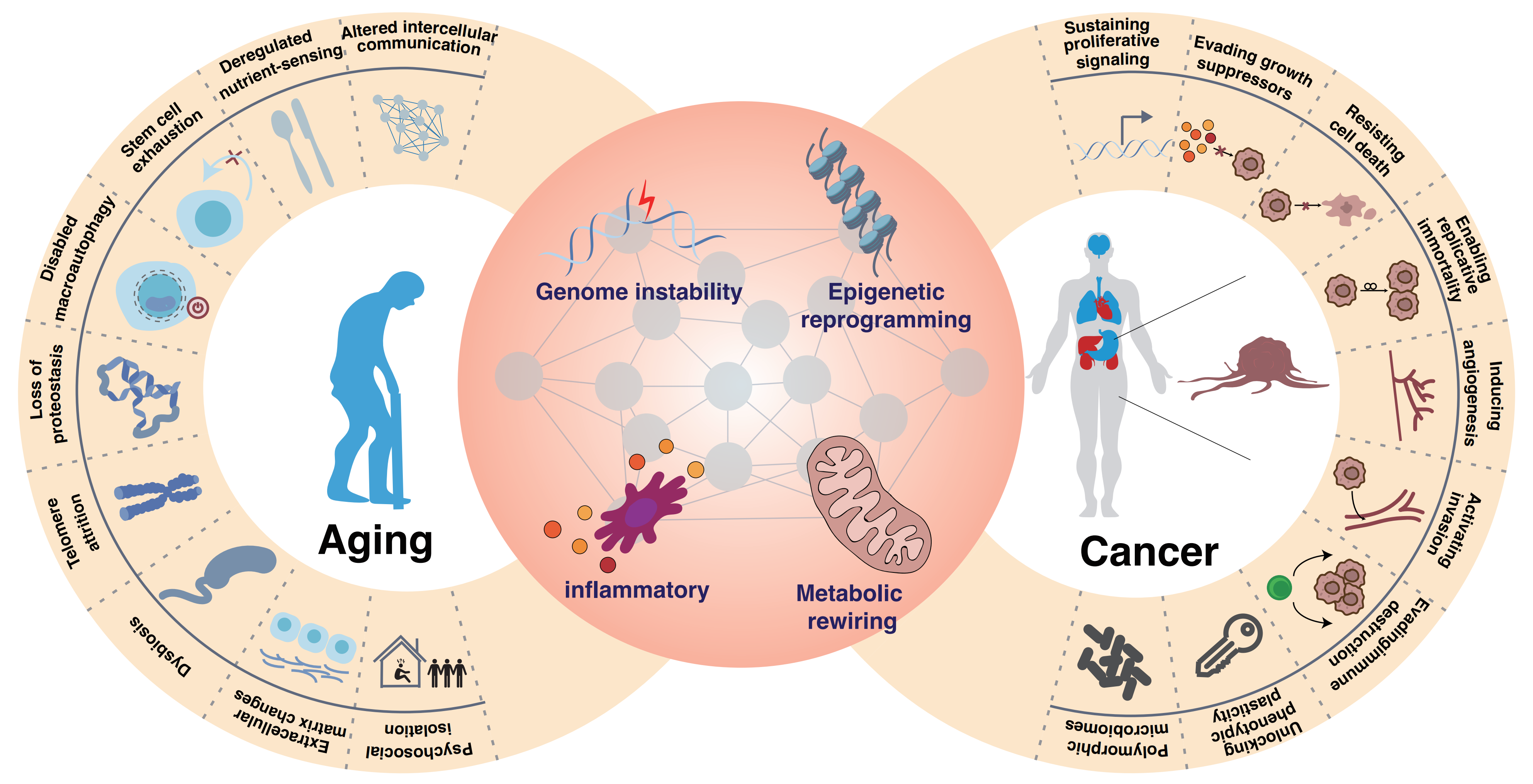
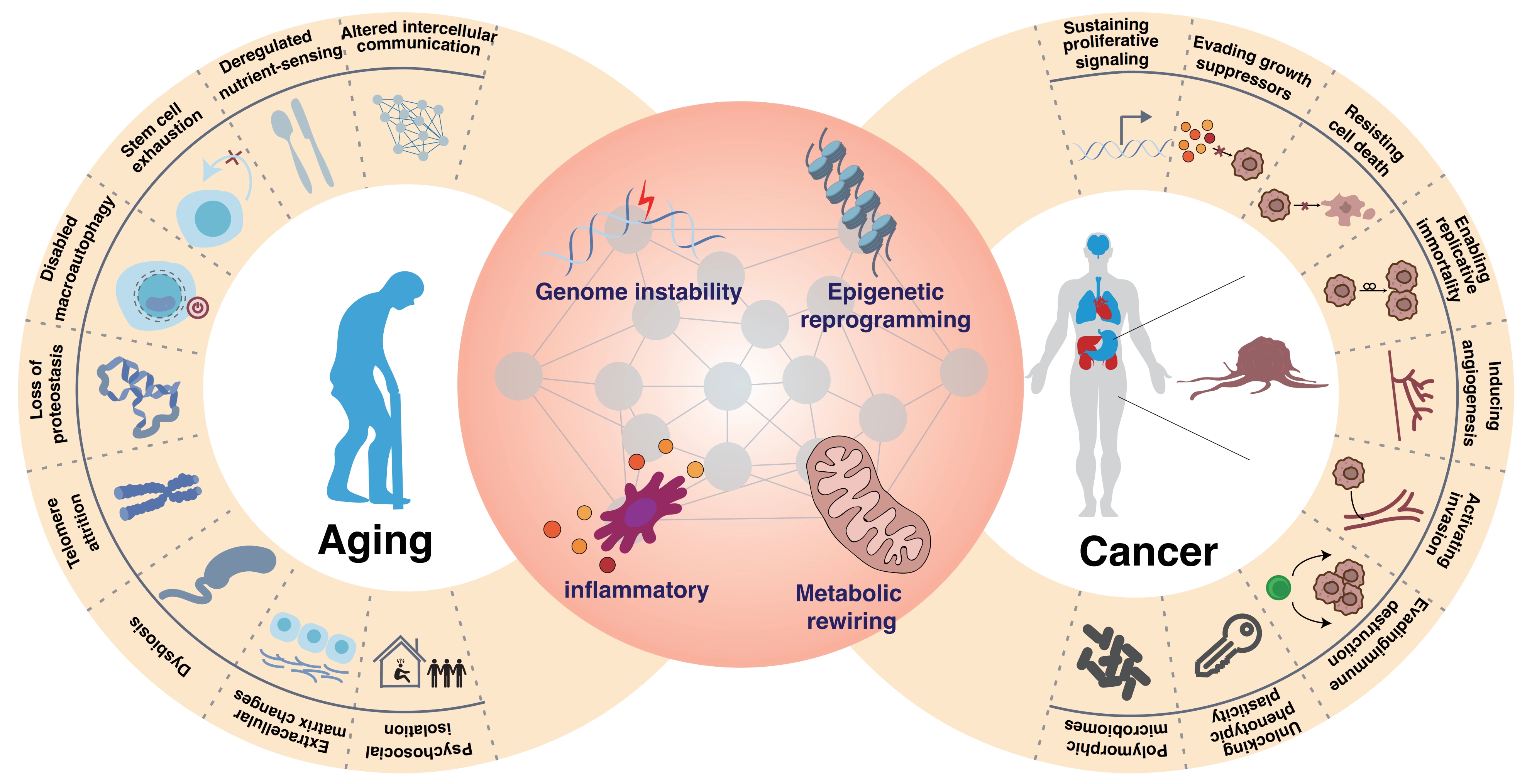
Figure 3. Interconnected hallmarks of aging and cancer. This figure illustrates the shared and distinct biological pathways linking aging and cancer. It highlights key mechanistic pillars, including genome instability, chronic inflammation, metabolic rewiring, and epigenetic reprogramming, that drive both processes. The diagram further clarifies the dual role of these mechanisms, acting as tumor-suppressive barriers in early aging while promoting tumorigenesis and tissue degeneration in late stages.
5.1 Genomic instability: Divergent fates under shared stress
Genomic instability serves as a primary upstream driver of both aging and cancer. In aging, accumulated DNA damage, such as telomere attrition and replication stress, activates the DNA Damage Response (DDR), enforcing stable cell-cycle arrest[189-193]. In cancer, defective repair coupled with selective pressure allows cells to bypass arrest, leading to mutation accumulation and clonal evolution[191,194,195].
The divergence is not deterministic but conditional on system-level clearance capacity. Chaib et al. demonstrated that chemotherapy-induced senescent cells within tumors can upregulate PD-L2 to evade immune clearance[196]. Such escaped senescent cells persist within tissues, acquiring pro-tumorigenic functions including immune modulation and niche remodeling[197]. Thus, genomic instability functions as a shared initiating stress, while downstream fate depends on whether checkpoint enforcement or escape-and-selection dominates. This axis defines genomic integrity control as a state bifurcation network linking aging and cancer outcomes.
5.2 Epigenetic reprogramming: A shared regulatory basis for state fate decisions
Epigenetic remodeling provides the structural basis for stabilizing or reversing cellular states under stress. Senescence is characterized by chromatin reorganization, including heterochromatin formation, DNA methylation drift, and transcriptional locking of proliferative programs[40,198,199]. In contrast, cancer cells exploit epigenetic plasticity to sustain a dedifferentiated state, lineage flexibility, and therapy adaptation, with these epigenetic alterations serving as emerging diagnostic biomarkers and therapeutic targets[200,201].
Screens targeting epigenetic regulators have identified critical genes and pathways governing these states. Studies on KAT7[95], H2AZ1[96], and RPL22[97] reveal that histone acetylation and nucleolar homeostasis dictate the entry and maintenance of senescence. Notably, KAT7 also promotes colorectal cancer progression[103], underscoring the dual roles of epigenetic regulators in aging and cancer. In cancer, regulators such as DNMT3A[202,203], TET2[204], and the Polycomb complex (PRC2/EZH2)[205] are pivotal for maintaining stem-like states and lineage plasticity. For instance, in acute myeloid leukemia (AML), alterations in DNMT3A and TET2 correlate with altered chromatin accessibility at differentiation loci, facilitating the reacquisition of stemness[206]. In solid tumors, EZH2-mediated H3K27 methylation drives dedifferentiation and metastasis, while enhancer reprogramming driven by transcription factors like GATA3 and JUN underpins endocrine resistance in breast cancer[207,208]. Thus, epigenetic machinery either locks the cell in a non-proliferative state or unlocks it for malignant progression.
5.3 Metabolic rewiring: A common mechanism for cellular adaptation and state maintenance
While both senescent and tumor cells exhibit metabolic rewiring, their objectives differ: senescent cells prioritize sustaining arrest and SASP production[209], whereas cancer cells prioritize biomass accumulation and environmental adaptation[209,210].
In aged neural stem cells (NSCs), elevated glucose uptake is associated with the senescent state, and genetic ablation of GLUT4 can restore activation, highlighting abnormal nutrient sensing as a key driver[13]. Conversely, cancer metabolism is often fueled by niche-derived resources. Sharma et al. integrated single-cell transcriptomics with in vivo screening to show that bone-marrow-derived taurine, transported via SLC6A6, activates mTOR signaling and glycolysis in leukemia stem cells, sustaining self-renewal and conferring venetoclax resistance[168]. This underscores that metabolic adaptation in cancer is not purely cell-intrinsic but relies on systemic metabolic cues, paralleling the metabolic shifts seen in aged tissues. Indeed, global metabolic deregulation in the aged host dynamically remodels the microenvironment. The accumulation of byproducts such as systemic lactate[211,212] and methylmalonic acid[170], coupled with CD38-mediated NAD+ depletion[213,214], actively contributes to CD8+ T cell dysfunction.
Thus, metabolic rewiring functions as an energetic constraint layer that biases system trajectories toward maintenance (senescence) or expansion (tumorigenesis).
5.4 Inflammatory signaling and SASP: The context-dependent bridge
The SASP functions as a dynamic signaling program linking intracellular states to tissue-level outcomes, with effects dictated by context, duration, and composition[215,216].
In acute or short-term conditions, such as embryonic development[217], wound healing, and early tumor suppression, the SASP serves a beneficial homeostatic role by reinforcing cell-cycle arrest via autocrine loops and recruiting immune effectors to clear senescent cells[4]. Conversely, when senescence becomes chronic due to unresolved DNA damage or persistent oncogenic stress, the SASP undergoes a malignant transformation driven by master regulators like nuclear factor kappa B (NF-κB) and mTOR[218]. This sustained secretion remodels the microenvironment into an immunosuppressive niche, recruiting myeloid-derived suppressor cells and inhibiting cytotoxic T cells, thereby accelerating tumor progression. Furthermore, therapy-induced senescence in normal tissues can promote adverse effects and relapse[197,219,220].
This continuum underscores that the tumor-suppressive versus tumor-promoting nature of senescence is not intrinsic but context-dependent. Recognizing this duality is critical for therapeutic design. Consequently, therapeutic strategies are evolving to target this spatiotemporal logic through two distinct approaches. Senolytics selectively eliminate senescent cells to extinguish the source of chronic SASP, while senomorphics modulate the SASP composition by inhibiting specific signaling pathways or neutralizing pro-tumor factors without inducing cell lysis. Deciphering the precise spatiotemporal rules governing SASP heterogeneity is therefore paramount for safely harnessing these strategies to disrupt the senescence–cancer axis[221-223].
5.5 Shared network, two fates: Identifying the determinant networks
Collectively, genomic instability, epigenetic regulation, metabolic rewiring, and inflammatory signaling constitute interconnected layers of a unified stress-response system[6,149,186]. Aging and cancer emerge as distinct stable endpoints of this system, defined by differential weighting of arrest, plasticity, and clearance mechanisms. The former tends toward cell-cycle arrest and sustained inflammatory signaling, while the latter tends toward proliferation, adaptation, and immune evasion[186,215] (Table 2). Therefore, understanding this relationship requires moving beyond simply listing shared genes to pinpointing the specific network nodes that dictate whether a cell becomes senescent or malignant (Table 3). However, the current predominance of cancer-focused studies limits our view of cancer-aging shared networks. Future studies must interpret existing data with caution and prioritize unified research that equally investigates both processes to overcome these biases.
Table 2. Convergent and divergent regulatory genes and pathways in aging and cancer uncovered by functional screens.
| Biological process | Genes/pathway | Screening Method | Role in Aging | Role in Cancer | Relationship | Therapeutic Strategy |
| Genomic Instability | WRN helicase | Synthetic lethality | Maintains stability | synthetic lethal | Shared | Inhibition |
| Telomere attrition | CRISPR | Induces senescence | Activation of telomerase | Divergent | Cancer: telomerase inhibitors | |
| Epigenetic Regulation | KAT7 | CRISPR-Cas9 KO (hMPCs) | Pro-senescencemaintains H3K14ac | Promotes CRC | Shared | Inhibition |
| H2A.Z.1 | CRISPR screen (hMSCs/RS/WS) | Drives senescence via enhancers | Promotes GBM, HCC, and lung | Shared | Inhibition | |
| RPL22 | CRISPR-Cas9 screen (hMSCs) | Destabilizes heterochromatin | Not reported | Aging-specific | Inhibition | |
| SOX5 | CRISPRa (hMSCs) | Senescence suppressor | Context-dependent | Aging-specific | Activation | |
| DNMT3A/TET2 | CRISPR (AML) | Clonal hematopoiesis | blocks differentiation | Shared | Prevent clonal expansion | |
| EZH2 | CRISPR KO | Not reported | Drives dedifferentiation | Cancer-specific | Inhibition | |
| ARID1A/B | Dependency maps | Not reported | mutant cancers | Cancer-specific | Synthetic lethality targeting ARID1B | |
| Metabolic Regulation | mTORC1 | CRISPRi/Cas9 (multiple) | Drives SASP | Promotes proliferation | Shared | Inhibition (rapamycin) extends lifespan & suppresses tumors |
| GLUT4 | CRISPR-Cas9 (aged NSCs) | Excessive uptake induces senescence | Not reported | Aging-specific | Inhibition | |
| FoxO signaling | CRISPRi | Mitochondrial translation | suppressor | Shared | Enhance in aging, context-specific in cancer | |
| SASP/Inflammation | NF-κBpathway | CRISPR-Cas9 (fibroblasts) | Master SASP regulator | Chronic inflammation | Shared | Inhibition |
| cGAS-STING axis | CRISPR screen | Induces SASP | Immune surveillance vs. escape | Shared | Aging: inhibition, Cancer: activate (context-dependent) | |
| IL-6/IL-8 | CRISPR screen | Core SASP | Promotes progression | Shared | Inhibition | |
| CEBPB | CRISPR screen | Promotes inflammation | NA | Aging-specific | Inhibition | |
| CHEK2/HAS1/MDK | CRISPR-Cas9 | Loss bypasses arrest | Not reported | Aging-specific | Inhibition | |
| MTOR/CRISPLD2/MORF4L1 | CRISPR (SASP regulators) | Required for SASP output | Not reported | Aging-specific | Inhibition | |
| Immune Evasion | PD-L2 | CRISPR | Evades immune clearance in TIS | Suppresses T cells | Shared | Inhibition |
WRN: werner syndrome ATP-dependent helicase; CRISPR: clustered regularly interspaced short palindromic repeats; CRISPR-Cas9: clustered regularly interspaced short palindromic repeats-CRISPR-associated protein 9; KO: knockout; hMPCs: human mesenchymal progenitor cells; hMSCs: human mesenchymal stem cells; AML: acute myeloid leukemia; CRISPRi: clustered regularly interspaced short palindromic repeats interference; SASP: senescence-associated secretory phenotype; NSCs: neural stem cells; FoxO: forkhead box O; NF-κB: nuclear factor kappa B; cGAS: cyclic GMP-AMP synthase; STING: stimulator of interferon genes; TIS: therapy-induced senescence; CRC: colorectal cancer; WS: Werner syndrome; GBM: glioblastoma; HCC: hepatocellular carcinoma; mTORC1: mechanistic target of rapamycin complex 1; IL-6: interleukin 6; IL-8: interleukin 8.
Table 3. Genes and pathways classification.
| Type | Definition&Criteria | Therapeutic Implication | Example | Supporting Evidence(references) |
| Shared (aligned) | Same direction of therapeutic benefit in both aging and cancer Intervention yields concordant positive outcomes. | Single intervention benefits both conditions, broadest therapeutic window | KAT7 | [95,103] |
| Shared (context-dependent) | Effect depends on tissue type, disease stage, or genetic background. Outcomes vary by context. | Requires careful patient stratification and monitoring | FoxO | [127-129] |
| Shared (therapeutic opportunity) | Exploitable vulnerability in cancer, protective in aging | Precision oncology approach, avoid systemic effects | DNMT3A/TET2 | [50,51,202,204] |
| Aging-specific | Primarily involved in aging/senescence; not reported in cancer | Rejuvenation targets, minimal cancer risk | RPL22 | [97] |
| Cancer-specific | Primarily involved in cancer progression/resistance | Oncology targets, may not affect aging | CD47 | [162] |
| Divergent | Opposing roles or therapeutic strategies required in aging vs. cancer | Cannot use same intervention, risk of adverse effects | p53-p21-Rb | [21,132,193,206,215,220,244] |
FoxO: forkhead box O.
6. Conclusions and Perspectives
6.1 Reframing the aging–cancer axis: From linear models to dynamic systems
The traditional linear view of the aging–cancer relationship is increasingly untenable[224]. Emerging evidence posits that both processes stem from a shared regulatory system that configures distinct outcomes based on context[186]. The central question thus shifts from identifying common genes to understanding how a single network reconfigures to drive cellular arrest, clearance, adaptation, or malignant progression[225].
Recent advances have shifted attention from individual genes to regulatory networks, enabled by functional genomic screening and single-cell multi-omics profiling. Genomic instability, epigenetic reprogramming, metabolic rewiring, and inflammatory signaling, often treated as separate mechanisms, may instead reflect interconnected layers of the same system responding to persistent stress. Addressing this complexity will require systematic perturbation of regulatory genes in a context-dependent manner. For example, coupling pooled CRISPR screens with single-cell transcriptomic and epigenomic readouts can reconstruct how network topology shifts across cell states[226].
6.2 Overcoming limitations: The next generation of functional genomics
Despite significant advances, critical challenges remain. First, the high context-dependency of aging and cancer necessitates more physiologically relevant models. Future efforts must prioritize primary cells, organoids, and in vivo platforms that preserve tissue architecture and immune interactions[227]. Second, temporal dynamics are often overlooked. Key processes, such as the evolution of the SASP or the long-term fate of therapy-induced senescent cells, unfold over extended periods[22]. Capturing these trajectories will require longitudinal strategies, such as time-resolved lineage tracing combined with inducible CRISPR systems. Third, resolving cell–cell interactions demands the integration of perturbation screens with spatial transcriptomics and multiplexed imaging.
To address these gaps, the field is advancing toward integrated multi-scale frameworks. In vivo CRISPR screening, empowered by engineered viral vectors and cell-type-specific promoters, enables systematic interrogation within native physiological contexts[228]. Genetic perturbations can now be coupled with information-rich readouts, including single-cell multi-omics and spatial transcriptomics[229,230]. This shifts screening from simple hit identification to building high-dimensional genotype–phenotype maps. Such strategies can directly resolve cell state transitions, including senescence emergence, maintenance, and reprogramming[231]. These strategies can also map how these trajectories interact with tumor evolution, immune responses, and therapies[232]. Crucially, expanding beyond transcriptional outputs to include protein-level and functional phenotypes will be essential for a holistic understanding of biological effects.
6.3 AI-driven discovery: From data integration to virtual cells
AI is transforming CRISPR screening from empirical discovery to predictive design through concrete applications. For off-target prediction, tools like CRISOT leverage RNA-DNA interaction fingerprints from molecular dynamics to predict genome-wide effects for Cas9 and base editors with superior accuracy[63]. In combinatorial design, NAIAD employs active learning to integrate single-gene data with adaptive gene embeddings. This approach identifies synergistic targets with a 40% performance gain across more than 350,000 interactions[233]. Deep learning models such as DeepCRISPR[234] and CRISPR-Net[235] now predict guide RNA specificity and quantify off-target activities caused by mismatches and indels, while transfer learning approaches like DeepCRISTL address cross-cell-type variability[236]. Causally informed frameworks like PDGrapher, a graph neural network incorporating causal structure, identified 13.37% more true therapeutic targets in chemical perturbation screens and successfully nominated KDR as a treatment target for non-small cell lung cancer, with recommended inhibitors including vandetanib and sorafenib[237]. It should be noted that the field is still largely at the preclinical stage, with most evidence coming from cell-line screens and computational modeling.
Despite these advances, critical bottlenecks constrain widespread adoption. The lack of high-quality standardized training data remains a primary limitation, as CRISPR screening datasets exhibit substantial heterogeneity due to differences in cell lines, screening protocols, and readout methods[238]. This variability directly contributes to poor cross-cell-type and cross-tissue generalizability, meaning models trained on cancer cell lines frequently fail to predict outcomes in primary cells or in vivo settings. The black box nature of deep learning hinders mechanistic insight, and integrating multi-omics readouts poses computational challenges. Addressing these issues requires coordinated efforts to generate standardized perturbation datasets across diverse biological contexts, develop transfer learning methods for improved cross-tissue prediction, and create interpretable AI frameworks that guide hypothesis-driven research rather than functioning as black boxes. The ultimate goal is to integrate large-scale perturbation data with computational modeling to build virtual cell frameworks capable of simulating cellular responses in silico before experimental validation, thereby reducing reliance on exhaustive empirical screening[239,240].
7. Outlook
Ultimately, the central shift in the field is not merely technological but conceptual. By framing aging and cancer as alternative stable states of a common regulatory network, we gain a more coherent basis for understanding their interplay. This perspective moves us beyond cataloging shared genes toward identifying the critical decision networks that dictate cellular fate. Leveraging the convergence of in vivo screening, spatial multi-omics, and AI-driven modeling will empower us to precisely steer these networks. Finally, suppressing malignant transformation while promoting healthy aging ushers in a new era of precision intervention.
Acknowledgements
The authors would like to thank all the researchers whose work is included in this review. We acknowledge the use of AI-assisted tools, including DeepSeek, for language editing and polishing of the manuscript. All authors have full control over the content of the manuscript, have thoroughly reviewed and revised all parts of the manuscript, and take full responsibility for all content of the manuscript.
Author contributions
Fan BT: Writing-original draft.
Wu AW: Investigation, supervision.
Pan X, Wang H: Conceptualization, writing-review & editing.
Conflicts of interest
Hu Wang is a Youth Editorial Board member of Ageing and Cancer Research & Treatment. The other authors declare no conflicts of interest.
Ethical approval
Not applicable.
Consent to participate
Not applicable.
Consent for publication
Not applicable.
Availability of data and materials
Not applicable.
Funding
This work was supported by the National Key R&D Program of China (2021YFA1102800 to H.W.) and by National Natural Science Foundation of China (82301746 to A.W.); “Pioneer” and “Leading Goose” R&D Program of Zhejiang (No.2026C02A1147); the Hangzhou Youth Innovation Team Project (No. TD2023020 to A.W.); and the HZNU scientific research and innovation team project (No. TD2025002 to H.W).
Copyright
© The Author(s) 2026.
References
-
1. Gorgoulis V, Adams PD, Alimonti A, Bennett DC, Bischof O, Bishop C, et al. Cellular senescence: Defining a path forward. Cell. 2019;179(4):813-827.[DOI]
-
2. Collado M, Serrano M. Senescence in tumours: Evidence from mice and humans. Nat Rev Cancer. 2010;10(1):51-57.[DOI]
-
5. Pérez-Mancera PA, Young ARJ, Narita M. Inside and out: The activities of senescence in cancer. Nat Rev Cancer. 2014;14(8):547-558.[DOI]
-
6. López-Otín C, Blasco MA, Partridge L, Serrano M, Kroemer G. Hallmarks of aging: An expanding universe. Cell. 2023;186(2):243-278.[DOI]
-
8. Tsherniak A, Vazquez F, Montgomery PG, Weir BA, Kryukov G, Cowley GS, et al. Defining a cancer dependency map. Cell. 2017;170(3):564-576.e16.[DOI]
-
9. Chen S, Sanjana NE, Zheng K, Shalem O, Lee K, Shi X, et al. Genome-wide CRISPR screen in a mouse model of tumor growth and metastasis. Cell. 2015;160(6):1246-1260.[DOI]
-
10. Chow RD, Chen S. Cancer CRISPR screens in vivo. Trends Cancer. 2018;4(5):349-358.[DOI]
-
12. Santinha AJ, Strano A, Platt RJ. Methods and applications of in vivo CRISPR screening. Nat Rev Genet. 2025;26(10):702-718.[DOI]
-
13. Ruetz TJ, Pogson AN, Kashiwagi CM, Gagnon SD, Morton B, Sun ED, et al. CRISPR–Cas9 screens reveal regulators of ageing in neural stem cells. Nature. 2024;634(8036):1150-1159.[DOI]
-
18. Cullen LM, Arndt GM. Genome-wide screening for gene function using RNAi in mammalian cells. Immunol Cell Biol. 2005;83(3):217-223.[DOI]
-
20. Temple G, Lamesch P, Milstein S, Hill DE, Wagner L, Moore T, et al. From genome to proteome: Developing expression clone resources for the human genome. Hum Mol Genet. 2006;15(suppl_1):R31-R43.[DOI]
-
23. Jackson AL, Bartz SR, Schelter J, Kobayashi SV, Burchard J, Mao M, et al. Expression profiling reveals off-target gene regulation by RNAi. Nat Biotechnol. 2003;21(6):635-637.[DOI]
-
26. Hipp MS, Kasturi P, Hartl FU. The proteostasis network and its decline in ageing. Nat Rev Mol Cell Biol. 2019;20(7):421-435.[DOI]
-
27. Wang T, Wei JJ, Sabatini DM, Lander ES. Genetic screens in human cells using the CRISPR-Cas9 system. Science. 2014;343(6166):80-84.[DOI]
-
28. Shalem O, Sanjana NE, Hartenian E, Shi X, Scott DA, Mikkelsen TS, et al. Genome-scale CRISPR-Cas9 knockout screening in human cells. Science. 2014;343(6166):84-87.[DOI]
-
30. Cong L, Ran FA, Cox D, Lin S, Barretto R, Habib N, et al. Multiplex genome engineering using CRISPR/Cas systems. Science. 2013;339(6121):819-823.[DOI]
-
31. Mali P, Yang L, Esvelt KM, Aach J, Guell M, DiCarlo JE, et al. RNA-guided human genome engineering via Cas9. Science. 2013;339(6121):823-826.[DOI]
-
32. Wang T, Birsoy K, Hughes NW, Krupczak KM, Post Y, Wei JJ, et al. Identification and characterization of essential genes in the human genome. Science. 2015;350(6264):1096-1101.[DOI]
-
33. Pickar-Oliver A, Gersbach CA. The next generation of CRISPR–Cas technologies and applications. Nat Rev Mol Cell Biol. 2019;20(8):490-507.[DOI]
-
34. Hsu PD, Lander ES, Zhang F. Development and applications of CRISPR-Cas9 for genome engineering. Cell. 2014;157(6):1262-1278.[DOI]
-
35. Sander JD, Joung JK. CRISPR-Cas systems for editing, regulating and targeting genomes. Nat Biotechnol. 2014;32(4):347-355.[DOI]
-
37. Qi LS, Larson MH, Gilbert LA, Doudna JA, Weissman JS, Arkin AP, et al. Repurposing CRISPR as an RNA-guided platform for sequence-specific control of gene expression. Cell. 2013;152(5):1173-1183.[DOI]
-
38. Fulco CP, Munschauer M, Anyoha R, Munson G, Grossman SR, Perez EM, et al. Systematic mapping of functional enhancer—promoter connections with CRISPR interference. Science. 2016;354(6313):769-773.[DOI]
-
39. Kampmann M. CRISPRi and CRISPRa Screens in mammalian cells for precision biology and medicine. ACS Chem Biol. 2018;13(2):406-416.[DOI]
-
40. Zhang W, Qu J, Liu GH, Belmonte JCI. The ageing epigenome and its rejuvenation. Nat Rev Mol Cell Biol. 2020;21(3):137-150.[DOI]
-
41. Zhang X, Zhu B, Chen L, Xie L, Yu W, Wang Y, et al. Dual base editor catalyzes both cytosine and adenine base conversions in human cells. Nat Biotechnol. 2020;38(7):856-860.[DOI]
-
43. Sakata RC, Ishiguro S, Mori H, Tanaka M, Tatsuno K, Ueda H, et al. Base editors for simultaneous introduction of C-to-T and A-to-G mutations. Nat Biotechnol. 2020;38(7):865-869.[DOI]
-
44. Kurt IC, Zhou R, Iyer S, Garcia SP, Miller BR, Langner LM, et al. CRISPR C-to-G base editors for inducing targeted DNA transversions in human cells. Nat Biotechnol. 2021;39(1):41-46.[DOI]
-
45. Komor AC, Kim YB, Packer MS, Zuris JA Liu DR. Programmable editing of a target base in genomic DNA without double-stranded DNA cleavage. Nature. 2016;533(7603):420-424.[DOI]
-
46. Anzalone AV, Randolph PB, Davis JR, Sousa AA, Koblan LW, Levy JM, et al. Search-and-replace genome editing without double-strand breaks or donor DNA. Nature. 2019;576(7785):149-157.[DOI]
-
47. Chen PJ, Hussmann JA, Yan J, Knipping F, Ravisankar P, Chen PF, et al. Enhanced prime editing systems by manipulating cellular determinants of editing outcomes. Cell. 2021;184(22):5635-5652.e29.[DOI]
-
48. Anzalone AV, Gao XD, Podracky CJ, Nelson AT, Koblan LW, Raguram A, et al. Programmable deletion, replacement, integration and inversion of large DNA sequences with twin prime editing. Nat Biotechnol. 2022;40:731-740.[DOI]
-
49. Schene IF, Joore IP, Oka R, Mokry M, van Vugt AHM, van Boxtel R, et al. Prime editing for functional repair in patient-derived disease models. Nat Commun. 2020;11(1):5352.[DOI]
-
53. Shalem O, Sanjana NE, Zhang F. High-throughput functional genomics using CRISPR-Cas9. Nat Rev Genet. 2015;16(5):299-311.[DOI]
-
54. Chen ACY, Jaiswal S, Martinez D, Yerinde C, Ji K, Miranda V, et al. The aged tumor microenvironment limits T cell control of cancer. Nat Immunol. 2024;25(6):1033-1045.[DOI]
-
55. Duan R, Jiang L, Wang T, Li Z, Yu X, Gao Y, et al. Aging-induced immune microenvironment remodeling fosters melanoma in male mice via γδ17-Neutrophil-CD8 axis. Nat Commun. 2024;15(1):10860.[DOI]
-
58. Datlinger P, Rendeiro AF, Schmidl C, Krausgruber T, Traxler P, Klughammer J, et al. Pooled CRISPR screening with single-cell transcriptome readout. Nat Methods. 2017;14(3):297-301.[DOI]
-
66. Han R, Li L, Ugalde AP, Tal A, Manber Z, Barbera EP, et al. Functional CRISPR screen identifies AP1-associated enhancer regulating FOXF1 to modulate oncogene-induced senescence. Genome Biol. 2018;19(1):118.[DOI]
-
67. Zhang C, Lei L, Yang X, Ma K, Zheng H, Su Y, et al. Single-cell sequencing reveals antitumor characteristics of intratumoral immune cells in old mice. J Immunother Cancer. 2021;9(10):e002809.[DOI]
-
68. He J, Papa G, Azizi F, Kirsche L, Artola-Boran M, Cassio RF, et al. Gastric organoid-based ectopic and orthotopic in vivo CRISPR screening for tumor suppressors in gastric cancer. Gastroenterology. 2026;170(3):495510.[DOI]
-
73. Rottenberg S, Jaspers JE, Kersbergen A, Van Der Burg E, Nygren AO, Zander SA, et al. High sensitivity of BRCA1-deficient mammary tumors to the PARP inhibitor AZD2281 alone and in combination with platinum drugs. Proc Natl Acad Sci U S A. 2008;105(44):17079-17084.[DOI]
-
74. Farmer H, McCabe N, Lord C J, Tutt AN, Johnson DA, Richardson TB, et al. Targeting the DNA repair defect in BRCA mutant cells as a therapeutic strategy. Nature. 2005;434(7035):917-921.[DOI]
-
77. Fong SH, Kuenzi BM, Mattson NM, Lee J, Sanchez K, Bojorquez-Gomez A, et al. A multilineage screen identifies actionable synthetic lethal interactions in human cancers. Nat Genet. 2025;57(1):154-164.[DOI]
-
80. Przybyla L, Gilbert LA. A new era in functional genomics screens. Nat Rev Genet. 2022;23(2):89-103.[DOI]
-
81. Dhainaut M, Rose SA, Akturk G, Wroblewska A, Nielsen SR, Park ES, et al. Spatial CRISPR genomics identifies regulators of the tumor microenvironment. Cell. 2022;185(7):1223-1239.e20.[DOI]
-
82. Binan L, Jiang A, Danquah SA, Valakh V, Simonton B, Bezney J, et al. Simultaneous CRISPR screening and spatial transcriptomics reveal intracellular, intercellular, and functional transcriptional circuits. Cell. 2025;188(8):2141-2158.e18.[DOI]
-
83. Hart SK, Müller S, Wessels HH, Méndez-Mancilla A, Drabavicius G, Choi O, et al. Precise RNA targeting with CRISPR-Cas13d. Nat Biotechnol. 2026;44(1):64-69.[DOI]
-
91. Knudsen NH, Escobar G, Korell F, Kienka T, Nobrega C, Anderson S, et al. In vivo CRISPR screens identify modifiers of CAR T cell function in myeloma. Nature. 2025;646(8086):953-962.[DOI]
-
94. Bock C, Datlinger P, Chardon F, Coelho MA, Dong MB, Lawson KA, et al. High-content CRISPR screening. Nat Rev Methods Primers. 2022;2:8.[DOI]
-
96. Li MH, Jiang X, Jing Y, Yan K, Bi SJ, Wang S, et al. CRISPR-based screening pinpoints H2AZ1 as a driver of senescence in human mesenchymal stem cells. Protein Cell. 2025;16(4):293-299.[DOI]
-
104. Kitada K, Nakano D, Ohsaki H, Hitomi H, Minamino T, Yatabe J, et al. Hyperglycemia causes cellular senescence via a SGLT2- and p21-dependent pathway in proximal tubules in the early stage of diabetic nephropathy. J Diabetes Complications. 2014;28(5):604-611.[DOI]
-
107. Wiley CD, Velarde MC, Lecot P, Liu S, Sarnoski EA, Freund A, et al. Mitochondrial dysfunction induces senescence with a distinct secretory phenotype. Cell Metab. 2016;23(2):303-314.[DOI]
-
108. Kujoth GC, Leeuwenburgh C, Prolla TA. Mitochondrial DNA mutations and apoptosis in mammalian aging. Cancer Res. 2006;66(15):7386-7389.[DOI]
-
109. Trifunovic A, Wredenberg A, Falkenberg M, Spelbrink JN, Rovio AT, Bruder CE, et al. Premature ageing in mice expressing defective mitochondrial DNA polymerase. Nature. 2004;429(6990):417-423.[DOI]
-
110. Van Vliet T, Casciaro F, Demaria M. To breathe or not to breathe: Understanding how oxygen sensing contributes to age-related phenotypes. Ageing Res Rev. 2021;67:101267.[DOI]
-
113. Nakazawa MS, Keith B, Simon MC. Oxygen availability and metabolic adaptations. Nat Rev Cancer. 2016;16(10):663-673.[DOI]
-
115. Morimoto RI, Cuervo AM. Proteostasis and the aging proteome in health and disease. J Gerontol A Biol Sci Med Sci. 2014;69(Suppl 1):S33-S38.[DOI]
-
116. Kaushik S, Cuervo AM. Proteostasis and aging. Nat Med. 2015;21(12):1406-1415.[DOI]
-
118. Hartl FU. Cellular homeostasis and aging. Annu Rev Biochem. 2016;85(1):1-4.[DOI]
-
119. Moldakozhayev A, Gladyshev VN. Metabolism, homeostasis, and aging. Trends Endocrinol Metab. 2023;34(3):158-169.[DOI]
-
121. Leto D, Saltiel AR. Regulation of glucose transport by insulin: Traffic control of GLUT4. Nat Rev Mol Cell Biol. 2012;13(6):383-396.[DOI]
-
122. Breusegem SY, Houghton J, Romero-Bueno R, Fragoso-Luna A, Kentistou KA, Ong KK, et al. A multiparametric anti-aging CRISPR screen uncovers a role for BAF in protein synthesis regulation. Nat Commun. 2025;16:1681.[DOI]
-
123. Saurat N, Minotti AP, Rahman MT, Sikder T, Zhang C, Cornacchia D, et al. Genome-wide CRISPR screen identifies neddylation as a regulator of neuronal aging and AD neurodegeneration. Cell Stem Cell. 2024;31(8):1162-1174.e9.[DOI]
-
126. Song J, Li Z, Zhou L, Chen X, Sew WQ, Herranz H, et al. FOXO-regulated OSER1 reduces oxidative stress and extends lifespan in multiple species. Nat Commun. 2024;15(1):7144.[DOI]
-
127. Calissi G, Lam EW, Link W. Therapeutic strategies targeting FOXO transcription factors. Nat Rev Drug Discov. 2021;20(1):21-38.[DOI]
-
131. Young ARJ, Narita M. SASP reflects senescence. EMBO Rep. 2009;10(3):228-230.[DOI]
-
133. Acosta JC, Banito A, Wuestefeld T, Georgilis A, Janich P, Morton JP, et al. A complex secretory program orchestrated by the inflammasome controls paracrine senescence. Nat Cell Biol. 2013;15(8):978-990.[DOI]
-
134. Orjalo AV, Bhaumik D, Gengler BK, Scott GK, Campisi J. Cell surface-bound IL-1α is an upstream regulator of the senescence-associated IL-6/IL-8 cytokine network. Proc Natl Acad Sci U S A. 2009;106(40):17031-17036.[DOI]
-
142. Ma F, Sen R. Physiological aging in three dimensions. Trends Cell Biol. 2026;36(3):230-245.[DOI]
-
143. Chini CCS, Cordeiro HS, Le Kim Tran N, Chini EN. NAD metabolism: Role in senescence regulation and aging. Aging Cell. 2024;23:e13920.[DOI]
-
144. Palmer AK, Jensen MD. Metabolic changes in aging humans: Current evidence and therapeutic strategies. J Clin Investig. 2022;132(16):e158451.[DOI]
-
146. Giroud J, Bouriez I, Paulus H, Pourtier A, Debacq-Chainiaux F, Pluquet O. Exploring the communication of the SASP: Dynamic, interactive, and adaptive effects on the microenvironment. Int J Mol Sci. 2023;24(13):10788.[DOI]
-
147. Sun N, Lin CH, Li MY, Wang Y, Chen D, Ren X, et al. Clusterin drives myeloid bias in aged hematopoietic stem cells by regulating mitochondrial function. Nat Aging. 2025;5(8):1510-1527.[DOI]
-
150. Flavahan WA, Gaskell E, Bernstein BE. Epigenetic plasticity and the hallmarks of cancer. Science. 2017;357(6348):eaal2380.[DOI]
-
151. Lengauer C, Kinzler KW, Vogelstein B. Genetic instabilities in human cancers. Nature. 1998;396(6712):643-649.[DOI]
-
153. Jin S, White E. Role of autophagy in cancer: Management of metabolic stress. Autophagy. 2007;3(1):28-31.[DOI]
-
154. Munir R, Lisec J, Swinnen JV, Zaidi N. Lipid metabolism in cancer cells under metabolic stress. Br J Cancer. 2019;120(12):1090-1098.[DOI]
-
155. Sawyers C. Rational therapeutic intervention in cancer: Kinases as drug targets. Curr Opin Genet Dev. 2002;12(1):111-115.[DOI]
-
156. Whiteside T. Immune suppression in cancer: Effects on immune cells, mechanisms and future therapeutic intervention. Semin Cancer Biol. 2006;16(1):3-15.[DOI]
-
157. Gatenby RA, Gillies RJ. A microenvironmental model of carcinogenesis. Nat Rev Cancer. 2008;8(1):56-61.[DOI]
-
159. Arafeh R, Shibue T, Dempster JM, Hahn WC, Vazquez F. The present and future of the cancer dependency map. Nat Rev Cancer. 2025;25(1):59-73.[DOI]
-
165. Ramponi V, Richart L, Kovatcheva M, Stephan-Otto Attolini C, Capellades J, Lord AE, et al. H4K20me3-mediated repression of inflammatory genes is a characteristic and targetable vulnerability of persister cancer cells. Cancer Res. 2025;85(1):32-51.[DOI]
-
175. Wang L, Jin H, Jochems F, Wang S, Lieftink C, Martinez IM, et al. cFLIP suppression and DR5 activation sensitize senescent cancer cells to senolysis. Nat Cancer. 2022;3(11):1284-1299.[DOI]
-
177. Suvac A, Ashton J, Bristow RG. Tumour hypoxia in driving genomic instability and tumour evolution. Nat Rev Cancer. 2025;25(3):1667-1688.[DOI]
-
178. Feng T, Xie F, Lee LMY, Lin Z, Tu Y, Lyu Y, et al. Cellular senescence in cancer: From mechanism paradoxes to precision therapeutics. Mol Cancer. 2025;24(1):213.[DOI]
-
179. Bousset L, Gil J. Targeting senescence as an anticancer therapy. Mol Oncol. 2022;16(21):3855-3880.[DOI]
-
187. Loaiza N, Demaria M. Cellular senescence and tumor promotion: Is aging the key? Biochim Biophys Acta Rev Cancer. 2016;1865(2):155-167.[DOI]
-
188. Lecot P, Alimirah F, Desprez PY, Campisi J, Wiley C. Context-dependent effects of cellular senescence in cancer development. Br J Cancer. 2016;114(11):1180-1184.[DOI]
-
189. Hayflick L, Moorhead PS. The serial cultivation of human diploid cell strains. Exp Cell Res. 1961;25(3):585-621.[DOI]
-
191. Deng Y, Chang S. Role of telomeres and telomerase in genomic instability, senescence and cancer. Lab Invest. 2007;87(11):1071-1076.[DOI]
-
194. Lee S, Schmitt CA. The dynamic nature of senescence in cancer. Nat Cell Biol. 2019;21(1):94-101.[DOI]
-
195. Di Mitri D, Alimonti A. Non-cell-autonomous regulation of cellular senescence in cancer. Trends Cell Biol. 2016;26(3):215-226.[DOI]
-
196. Chaib S, López-Domínguez JA, Lalinde-Gutiérrez M, Prats N, Marin I, Boix O, et al. The efficacy of chemotherapy is limited by intratumoral senescent cells expressing PD-L2. Nat Cancer. 2024;5(3):448-462.[DOI]
-
198. Rando TA, Chang HY. Aging, rejuvenation, and epigenetic reprogramming: Resetting the aging clock. Cell. 2012;148(1):46-57.[DOI]
-
199. Kane AE, Sinclair DA. Epigenetic changes during aging and their reprogramming potential. Crit Rev Biochem Mol Biol. 2019;54(1):61-83.[DOI]
-
200. Suvà ML, Riggi N, Bernstein BE. Epigenetic reprogramming in cancer. Science. 2013;339(6127):1567-1570.[DOI]
-
201. da Silva Costa PM, Sales SLA, Pinheiro DP, Pontes LQ, Maranhão SS, do Ó Pessoa C, et al. Epigenetic reprogramming in cancer: From diagnosis to treatment. Front Cell Dev Biol. 2023;11:1116805.[DOI]
-
202. Shah MY, Licht JD. DNMT3A mutations in acute myeloid leukemia. Nat Genet. 2011;43(4):289-290.[DOI]
-
204. Weissmann S, Alpermann T, Grossmann V, Kowarsch A, Nadarajah N, Eder C, et al. Landscape of TET2 mutations in acute myeloid leukemia. Leukemia. 2012;26(5):934-942.[DOI]
-
205. Blackledge NP, Klose RJ. The molecular principles of gene regulation by Polycomb repressive complexes. Nat Rev Mol Cell Biol. 2021;22(12):815-833.[DOI]
-
208. Kim E, Kim M, Woo DH, Shin Y, Shin J, Chang N, et al. Phosphorylation of EZH2 activates STAT3 signaling via STAT3 methylation and promotes tumorigenicity of glioblastoma stem-like cells. Cancer Cell. 2013;23(6):839-852.[DOI]
-
210. Wei Q, Qian Y, Yu J, Wong CC. Metabolic rewiring in the promotion of cancer metastasis: Mechanisms and therapeutic implications. Oncogene. 2020;39(39):6139-6156.[DOI]
-
212. Ippolito L, Morandi A, Giannoni E, Chiarugi P. Lactate: A metabolic driver in the tumour landscape. Trends Biochem Sci. 2019;44(2):153-166.[DOI]
-
215. Campisi J. Aging, cellular senescence, and cancer. Annu Rev Physiol. 2013;75:685-705.[DOI]
-
216. Tchkonia T, Zhu Y, van Deursen J, Campisi J, Kirkland JL. Cellular senescence and the senescent secretory phenotype: Therapeutic opportunities. J Clin Invest. 2013;123(3):966-972.[DOI]
-
217. Storer M, Mas A, Robert-Moreno A, Pecoraro M, Ortells MC, Di Giacomo V, et al. Senescence is a developmental mechanism that contributes to embryonic growth and patterning. Cell. 2013;155(5):1119-1130.[DOI]
-
218. Laberge RM, Sun Y, Orjalo AV, Patil CK, Freund A, Zhou L, et al. MTOR regulates the pro-tumorigenic senescence-associated secretory phenotype by promoting IL1A translation. Nat Cell Biol. 2015;17(8):1049-1061.[DOI]
-
220. Demaria M, O’Leary MN, Chang J, Shao L, Liu S, Alimirah F, et al. Cellular senescence promotes adverse effects of chemotherapy and cancer relapse. Cancer Discov. 2017;7(2):165-176.[DOI]
-
222. Kirkland JL, Tchkonia T. Cellular senescence: A translational perspective. EBioMedicine. 2017;21:21-28.[DOI]
-
224. Muñoz-Espín D, Serrano M. Cellular senescence: From physiology to pathology. Nat Rev Mol Cell Biol. 2014;15(7):482-496.[DOI]
-
225. Vidal M, Cusick ME, Barabási AL. Interactome networks and human disease. Cell. 2011;144(6):986-998.[DOI]
-
227. Lancaster MA, Knoblich JA. Organogenesis in a dish: Modeling development and disease using organoid technologies. Science. 2014;345(6194):1247125.[DOI]
-
229. Baysoy A, Bai Z, Satija R, Fan R. The technological landscape and applications of single-cell multi-omics. Nat Rev Mol Cell Biol. 2023;24(10):695-713.[DOI]
-
230. Tian L, Chen F, Macosko EZ. The expanding vistas of spatial transcriptomics. Nat Biotechnol. 2023;41(6):773-782.[DOI]
-
232. Le J, Dian Y, Zhao D, Guo Z, Luo Z, Chen X, et al. Single-cell multi-omics in cancer immunotherapy: From tumor heterogeneity to personalized precision treatment. Mol Cancer. 2025;24(1):221.[DOI]
-
233. Qin J, Wessels HH, Fernandez-Granda C, Hao Y. Active learning for efficient discovery of optimal combinatorial perturbations. In: Singh A, Fazel M, Hsu D, Lacoste-Julien S, Berkenkamp F, Maharaj T, Wagstaff K, Zhu J, editors. Proceedings of the 42nd International Conference on Machine Learning; 2026 Jan 7-2025 Jul 19; Vancouver, Canada. Brookline: PMLR; 2025. p. 50363-50383. Available from: https://openreview.net/pdf?id=fGbGF0kl5L
-
235. Lin J, Zhang Z, Zhang S, Chen J, Wong KC. CRISPR‐net: A recurrent convolutional network quantifies CRISPR off‐target activities with mismatches and indels. Adv. Sci. 2020;7(13):1903562.[DOI]
-
238. Vinceti A, Iannuzzi RM, Boyle I, Trastulla L, Campbell CD, Vazquez F, et al. A benchmark of computational methods for correcting biases of established and unknown origin in CRISPR-Cas9 screening data. Genome Biol. 2024;25(1):192.[DOI]
-
242. Dervovic D, Malik AA, Chen ELY, Narimatsu M, Adler N, Afiuni-Zadeh S, et al. In vivo CRISPR screens reveal Serpinb9 and Adam2 as regulators of immune therapy response in lung cancer. Nat Commun. 2023;14:3150.[DOI]
-
243. Su P, Liu Y, Chen T, Xue Y, Zeng Y, Zhu G, et al. In vivo CRISPR screens identify a dual function of MEN1 in regulating tumor-microenvironment interactions. Nat Genet. 2024;56(9):1890-1902.[DOI]
-
244. Chang BD, Xuan Y, Broude EV, Zhu H, Schott B, Fang J, et al. Role of p53 and p21waf1/cip1 in senescence-like terminal proliferation arrest induced in human tumor cells by chemotherapeutic drugs. Oncogene. 1999;18(34):4808-4818.[DOI]
Copyright
© The Author(s) 2026. This is an Open Access article licensed under a Creative Commons Attribution 4.0 International License (https://creativecommons.org/licenses/by/4.0/), which permits unrestricted use, sharing, adaptation, distribution and reproduction in any medium or format, for any purpose, even commercially, as long as you give appropriate credit to the original author(s) and the source, provide a link to the Creative Commons license, and indicate if changes were made.
Publisher’s Note
Science Exploration remains a neutral stance on jurisdictional claims in published
maps
and institutional affiliations. The views expressed in this article are solely those
of
the author(s) and do not reflect the opinions of the Editors or the publisher.
Share And Cite


Science Exploration Style
Fan B, Wu A, Pan X, Wang H. From RNAi to single-cell CRISPR: Evolving functional screening in aging and cancer. Ageing Cancer Res Treat. 2026;3:202621. https://doi.org/10.70401/acrt.2026.0031
Tips
Copy completed.
Submit a Manuscript
Author Instructions
Cite this Article
Article Metrics
0
View
0
Download
Cited
Article Updates

- Abstract
- Keywords
- 1. Introduction
- 2. Technological Advances for Aging-Cancer Research
- 3. Applications of Library Screening in Aging Research
- 4. Applications of Library Screening in Cancer Research
- 5. Aging and cancer: A shared stress-response state transition system
- 6. Conclusions and Perspectives
- 7. Outlook
- Acknowledgements
- Author contributions
- Conflicts of interest
- Ethical approval
- Consent to participate
- Consent for publication
- Availability of data and materials
- Funding
- References
- Copyright
 Get citation
Science Exploration Style

Fan B, Wu A, Pan X, Wang H. From RNAi to single-cell CRISPR: Evolving functional screening in aging and cancer. Ageing Cancer Res Treat. 2026;3:202621. https://doi.org/10.70401/acrt.2026.0031
copy
Share Link
copy