Interplay between hypoxia and inflammation in cancer progression, therapy resistance, and natural cancer resistance

Yandong Xu
1,2

,
Yang Zhao
1,2,*
*Correspondence to:
Yang Zhao, Department of Physiology and Department of Hepatobiliary and Pancreatic Surgery, The First Affiliated Hospital, Zhejiang University School of Medicine, Hangzhou 301158, Zhejiang, China.
E-mail: yang.zhao@zju.edu.cn
Ageing Cancer Res Treat. 2026;3:202612. 10.70401/acrt.2026.0032
Received: March 23, 2026Accepted: July 17, 2026Published: July 17, 2026
Abstract
The microenvironment of solid tumors is characterized by dynamic availability of oxygen and persistent, complex inflammatory signaling. Accumulating evidence suggests that hypoxia and inflammation actively influence tumor progression and therapeutic response. In physiological conditions, hypoxic and inflammatory responses function as evolutionarily conserved protective mechanisms. Acute hypoxia promotes temporary metabolic adaptation to preserve tissue viability, while acute inflammation facilitates clearance of damaged or abnormal cells and restores homeostasis. In tumors, by contrast, hypoxia and inflammation become chronic and unresolved. Persistent activation of these protective signals gradually renders the microenvironment permissive to the sustained adaptation of abnormal cells rather than eliminating them. This transition from clearance to tolerance reshapes cellular metabolism, immune cell behavior, and tissue organization within the tumor microenvironment. As a result, tumor cells increasingly adapt to metabolic stress and evade effective immune surveillance, contributing to malignancy and therapy resistance. Interestingly, some subterranean species, including the naked mole rat and blind mole rat, show natural cancer resistance. They either evolved adaptations counteracting inflammation, or take the immune response to their advantage to prevent cancer. These anti-cancer mechanisms are a result of adaptation to their hypoxic subterranean habitats. In this review, we examine the fundamental features of hypoxia and inflammation in tumors, explore their functional interplay, and discuss how these mechanisms are evaded or harnessed by cancer resistant subterranean mammals. We further propose a simple clearance to resistance framework that links acute protective stress responses, chronic tumor-promoting adaptation, and naturally evolved cancer resistance.
Keywords
Cancer resistance, hypoxia, inflammation, subterranean species
1. Introduction
Hypoxia and Inflammation are major hallmarks of the cancer microenvironment. While both have evolved as protective responses to stresses, functioning to preserve tissue integrity and homeostasis, their persistence in tumors leads to fundamental reprogramming of cell behavior and immune functions. Chronic hypoxia establishes adaptive transcriptional and epigenetic modulations of cells that provide metabolic flexibility and immune evasion[1,2]. Unresolved inflammation shifts the immune landscape from active clearance toward sustained tolerance[3-6]. Hypoxia and inflammation are mutually regulated. Hypoxia activates nuclear factor-κB (NF-κB), promoting the transcription of downstream, pro-inflammatory factors[7]; meanwhile, it represses the transcription and translation of some inflammatory genes directly. Together, these processes establish a microenvironment in tumors that favors the survival of cancer cells with resistance to therapy. Intriguingly, some subterranean species such as the naked mole rat (NMR, Heterocephalus glaber) and the blind mole rat (BMR, Spalax), adapted to hypoxic habitats, exhibit strong resistance to tumorigenesis[8,9]. They prevent cancer through mechanisms modulating the inflammatory response, reflecting the interplay between hypoxia and inflammation on an evolutionary timescale. In this review, we examine the molecular foundations of cells responding to hypoxia and inflammation in cancer, explore their interplay in driving tumor persistence, and discuss how cancer resistant species circumvent these pathological responses for tumor suppression. In this framework, tumor progression is viewed as a staged transition from acute clearance to chronic tolerance, and finally to stabilized resistance; cancer-resistant hypoxia-adapted species are discussed as natural examples in which this maladaptive loop is interrupted.
2. Cellular Response to Hypoxia: From Adaptive Survival to Tumor Persistence
Hypoxia is a hallmark of solid tumors and arises when the demand for molecular oxygen required by the cells exceeds the capacity of the vascular supply, a condition commonly caused by rapid cellular proliferation and structural abnormality in tumor vasculature[10]. Abnormal tumor vasculature, characterized by structural disorganization and poor perfusion, results in regions with fluctuating or sustained oxygen deprivation[11]. In the bone marrow, lymphoid tissue, placenta and intestinal mucosa, physiologically low oxygen functions as a spatially and temporally regulated signal that contribute to tissue homeostasis and immune-cell adaptation[12]. Central to this response is the stabilization of hypoxia-inducible factor α (HIF-α) subunits. HIF is regulated by prolyl hydroxylase domain proteins (PHDs) and factor inhibiting HIF (FIH, encoded by HIF1AN), an oxygen- and 2-oxoglutarate-dependent dioxygenase, which control HIF-α protein stability and transcriptional activity, seperatelly[13]. Under normoxic conditions, PHDs hydroxylate HIF-α subunits, enabling their recognition by the von Hippel-Lindau (VHL) E3 ubiquitin ligase complex and subsequent proteasomal degradation. Under normoxic conditions, hydroxylation by FIH hinders the HIF α-subunits from binding to their transcriptional co-activators, the histone acetyl transferases CREB-binding protein (CBP) and p300, preventing the formation of a functional transcriptional complex and thereby repressing HIF activity in an oxygen-dependent manner[14]. When oxygen levels decline, the enzymatic activity of the PHD and FIH are reduced. Consequently, HIF-α escapes VHL-mediated degradation, accumulates, and translocates to the nucleus, where it dimerizes with the constitutively expressed HIF-β subunit. Reduced FIH activity also permits more efficient recruitment of CBP/p300, enabling the HIF complex to activate the transcription of genes involved in angiogenesis, glucose metabolism, erythropoiesis, and cellular stress adaptation[13].
Through these transcriptional regulations, acute hypoxia promotes metabolic flexibility and supports temporary cell survival (Figure 1).
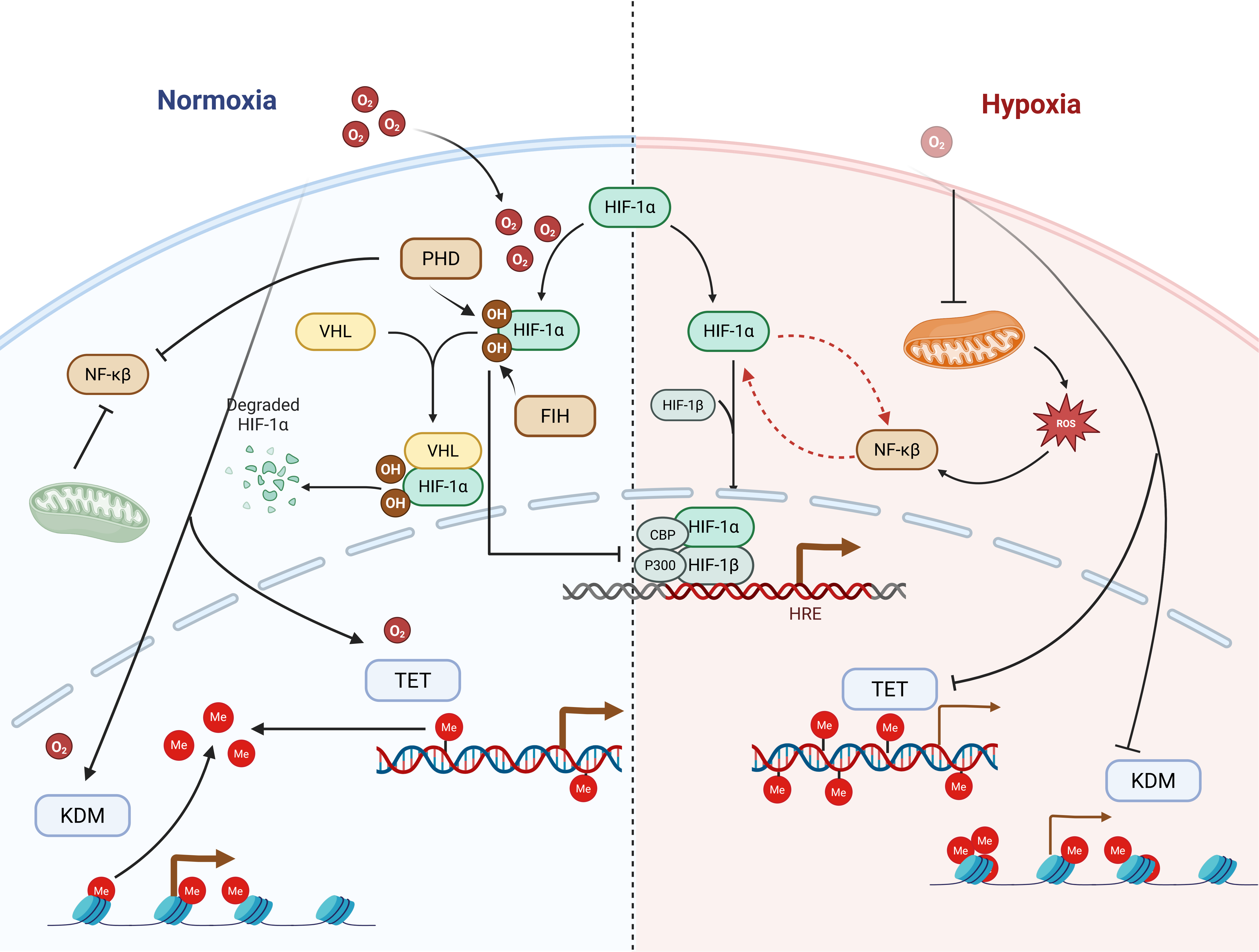
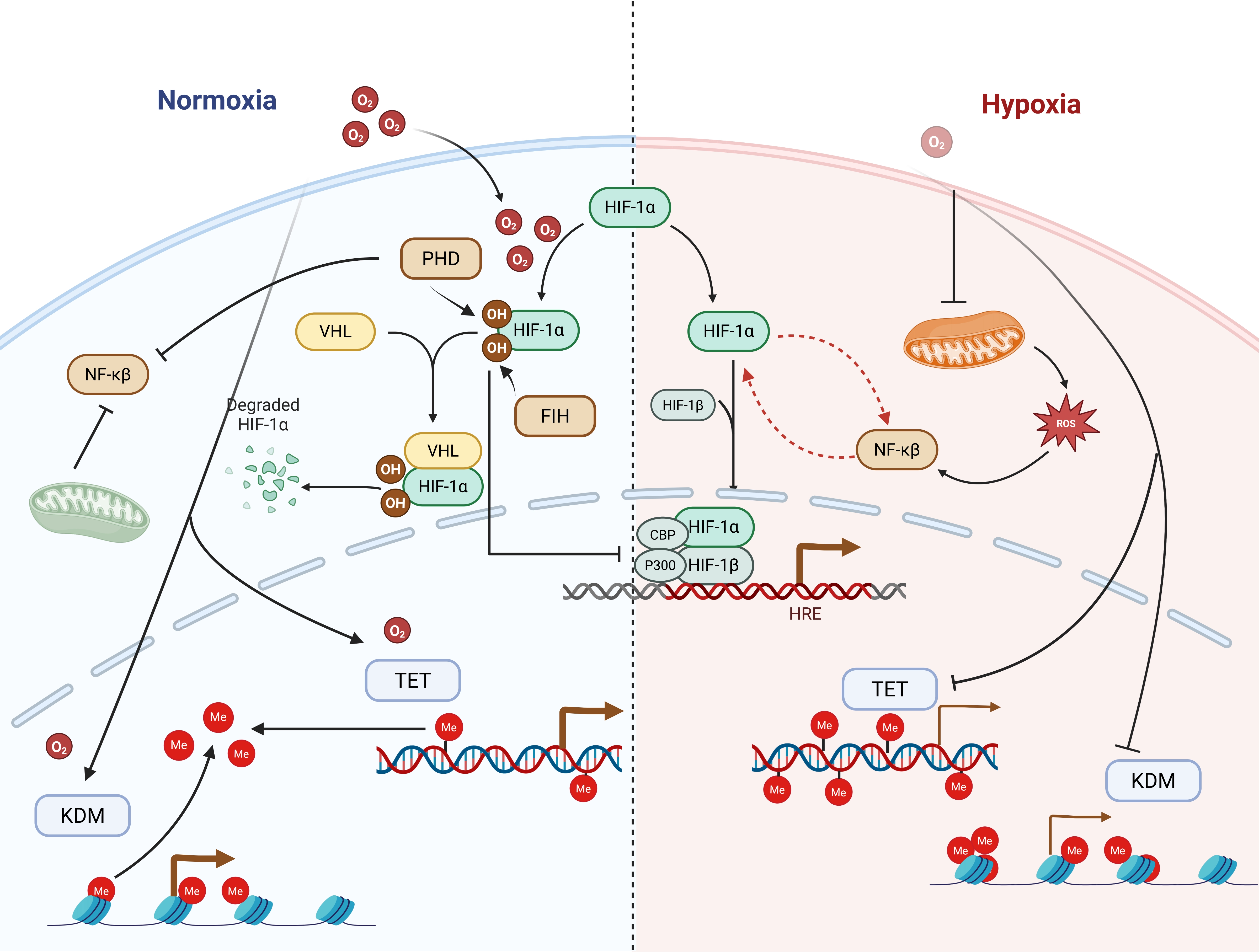
Figure 1. Multilayered oxygen-sensing system and its crosstalk with inflammatory signaling in cancer. Under normoxic conditions, PHDs hydroxylate HIF-1α, promoting its recognition by the VHL E3 ubiquitin ligase complex and subsequent proteasomal degradation. FIH further represses HIF transcriptional activity by blocking the recruitment of the transcriptional co-activators CBP/p300. Oxygen-dependent epigenetic regulators, including JmjC KDMs and TET DNA dioxygenases, remain active under sufficient oxygen availability and regulate histone and DNA methylation states. Under hypoxia, reduced oxygen availability inhibits PHD and FIH activity, leading to HIF-1α stabilization, dimerization with HIF-1β, recruitment of CBP/p300, and activation of HRE-containing target genes. Hypoxia also impairs oxygen-dependent KDM and TET activity, resulting in altered chromatin and DNA methylation landscapes. In parallel, mitochondrial ROS increase under hypoxic stress and act as secondary messengers that reinforce HIF-1α stabilization and activate NF-κB signaling. NF-κB can further enhance HIF-1α expression, forming a reciprocal hypoxia-inflammation regulatory loop. Together, these oxygen-sensitive pathways coordinate transcriptional, epigenetic, metabolic, and inflammatory responses that contribute to tumor adaptation, immune modulation, and cancer progression. Created in BioRender.com. Xu, Y. (2026) https://BioRender.com/jdbnyey. PHDs: prolyl hydroxylase domain proteins; HIF: hypoxia-inducible factor; VHL: von Hippel-Lindau; FIH: factor inhibiting hypoxia-inducible factor; CBP: CREB-binding protein; TET: ten-eleven translocation; KDMs: domain-containing histone demethylases; HRE: hypoxia-responsive element; ROS: reactive oxygen species; NF-κB: nuclear factor-κB; JmjC: Jumonji C.
Beyond the canonical, HIF-dependent pathway, mammalian cells possess a broader network of oxygen-dependent enzymes that participate in oxygen sensing and cellular adaptation. A major group of these enzymes belongs to the 2-oxoglutarate-dependent dioxygenases (2-OGDDs)[15], which utilize molecular oxygen, ferrous iron, and 2-oxoglutarate as cofactors to catalyze oxidative reactions. 2-OGDDs were first identified in studies on collagen biosynthesis, where they were found to catalyze the hydroxylation of prolyl and lysyl residues[16,17]. Interestingly, several classes of these enzymes play important roles in epigenetic regulation. Jumonji C domain-containing histone demethylases (JmjC-KDMs) regulate histone methylation states[15]. JmjC-domain containing KDMs are oxygen-dependent enzymes, but their oxygen sensitivities and transcriptional consequences differ among family members and cellular contexts. Hypoxia can inhibit selected KDMs and TET enzymes, although compensatory expression, metabolite availability, and locus-specific recruitment may result in divergent epigenetic outcomes[18-21]. Under hypoxia, these KDMs fail at demethylating specific histone marks, influencing transcriptional programs controlling cell differentiation, proliferation, and stress responses[17,19,22]. Similarly, ten-eleven translocation (TET) DNA dioxygenases, which catalyze DNA demethylation, are also oxygen-dependent enzymes whose activity can be reduced under hypoxic conditions, leading to global changes in DNA methylation patterns[23,24]. Through these mechanisms, hypoxia can influence gene expression not only through HIF-mediated transcription but also through epigenetic remodeling (Figure 1).
At the chromatin level, the consequence of KDM inhibition is context dependent. Hypoxia-induced inhibition of KDM5 can increase H3K4 methylation at promoters and enhancers, whereas inhibition of KDM6A/B can maintain H3K27me3 and thereby restrain differentiation or immune related transcriptional programs. Therefore, oxygen dependent epigenetic regulation should be interpreted together with tumor lineage, oxygen tension, and the basal activity of individual KDMs rather than as a uniform consequence of hypoxia[15,19].
In the physiological context, hypoxia represents a reversible and tightly regulated adaptation. However, in tumors, hypoxia is rarely transient. Persistent oxygen deprivation leads to chronic activation of hypoxia-responsive pathways. Instead of resolving, these adaptive responses become stabilized, resulting in long-term metabolic reprogramming[1]. Tumor cells increasingly rely on glycolysis and undergo extensive remodeling of mitochondrial metabolism[25], activate antioxidant programs through hypoxia response pathways[26], thereby improving their ability to tolerate oxidative and therapeutic stress[27]. Cancer cells can hijack this metabolic plasticity, contributing to survival under hypoxia stress[2].
Chronic hypoxia also reshapes the tumor microenvironment[28]. It promotes angiogenic factor secretion, alters extracellular matrix remodeling, and influences immune cell recruitment and function. Hypoxia reshape the tumor microenvironment in a context dependent manner. While prolonged and severe hypoxia stress can impair cytotoxic T cell function, promote T cell exhaustion and facilitate regulatory T cell accumulation transient or moderate HIF activation may support certain aspects of CD8+ tumor infiltrating lymphocyte (TIL) function and adaptation. These divergent effect depends on immune cell type, hypoxia intensity, and duration of exposure. Therefore, hypoxia and HIF signaling (including HIF1α and HIF2α) can either promote or restrain antitumor immunity depending on the specific tumor context, contributing to the complex regulation of tumor immune escape[29,30]. KDMs are aberrantly overexpressed in different types of cancers and are associated with the growth, invasion, and metastasis of cancer cells[31]. Thus, hypoxia becomes a structural force that stabilizes a stress-adapted ecosystem. Importantly, this transition marks a shift from adaptive survival to tumor persistence. Chronic hypoxia reinforces a state in which tumor cells are better equipped to endure hostile conditions, including chemotherapy, radiotherapy, and immune attack[32,33]. In this way, hypoxia contributes directly to the development of cancer resistance by converting protective stress signaling into a sustained survival advantage, driving tissue dysfunction and disease development[30,34]. Reactive oxygen species (ROS) also play an important complementary role in cellular responses to hypoxia[35]. Under hypoxic conditions, mitochondrial electron transport chain activity becomes altered, particularly at complexes I and III, leading to increased electron leakage and enhanced production of ROS[36]. Rather than acting as primary oxygen sensors, ROS function as secondary messengers that integrate changes in oxygen availability with intracellular signaling pathways.
ROS also connect hypoxia to inflammation by tuning redox sensitive transcription factors, including NF-κB, activator protein 1 (AP-1) and nuclear factor erythroid 2–related factor 2 (NRF2); depending on intensity and duration, this signaling can either support inflammatory clearance or reinforce antioxidant and pro-survival programs in cancer cells[26,37].
3. Inflammation as a Mediator between Immune Clearance and Immune Tolerance
Inflammation is a fundamental biological response evolved to eliminate harmful stimuli and restore tissue integrity[38]. Acute inflammation is tightly regulated and self-limiting[39]. In response to infection, tissue injury, or the emergence of abnormal cells, innate immune cells such as macrophages and natural killer cells are rapidly recruited. This initial response promotes the production of pro-inflammatory cytokines, enhances antigen presentation, and facilitates cytotoxic T cell activation, ultimately leading to the clearance of damaged or infected cells. Once the threat is eliminated, inflammatory signaling subsides and tissue homeostasis is restored[40].
During tumorigenesis, however, inflammatory responses often do not resolve. Inflammatory responses play decisive roles at different stages of tumor development, including initiation, promotion, malignant conversion, invasion, and metastasis[3]. Early transformed cells can trigger immune recognition and elimination, a process often described as immune surveillance[41]. Type I interferon (IFN) is a major mediator of tumor immune surveillance, which is activated by cytoplasmic nucleic acid sensing pathways including retinoic acid-inducible gene I (RIG-I) and melanoma differentiation-associated protein 5 (MDA5) pathways that sense double-stranded RNA (dsRNA) or the cyclic GMP-AMP synthase (cGAS)-stimulator of interferon genes (STING) pathway that senses dsDNA[42]. This mechanism, first evolved as a defense, has been co-opted to eliminate abnormal cells such as cancer cells, which is termed viral mimicry[43-46]. Yet when abnormal cells persist and tissue damage signals remain active, inflammation becomes chronic. Continuous exposure to cytokines, chemokines, and danger-associated molecular patterns sustains immune activation but gradually alters immune cell function[47]. Cytotoxic T cells may enter a state of exhaustion characterized by reduced effector function and sustained expression of inhibitory receptors. Macrophages shift from pro-inflammatory phenotypes toward immunoregulatory or tissue-remodeling states. Regulatory T cells accumulate, further suppressing anti-tumor immunity[6]. Rather than promoting elimination, the inflammatory milieu begins to favor tissue maintenance and damage limitation.
Mechanistically, chronic activation of IFN promotes survival of cancer cells through activating a distinct set of downstream genes. Acute IFN response results in the formation of an IFN-stimulated gene factor 3 (ISGF3), composed of IFN response factor 9 (IRF9) and tyrosine-phosphorylated signal transducer and activator of transcription 1 (STAT1) and STAT2. ISGF3, acting as a transcription factor, then activates a spectrum of anti-viral genes collectively called interferon-stimulated genes (ISGs). However, when IFN is sustained, the accumulation of IRF9, STAT1, and STAT2 will form a new complex lacking the tyrosine phosphorylation of STAT1 and STAT2. This new complex, termed unphosphorylated interferon-stimulated gene factor 3 (U-ISGF3), activates the expression of a group of genes, namely the IFN-related DNA damage resistance signature (IRDS) that promotes the survival of malignant cells[48]. Beyond cytotoxic injury, persistent IFN/STAT signaling can select for IRDS-high cells that tolerate DNA damage and therapy, and may cooperate with checkpoint programs rather than simply reflecting effective anti-viral immunity. Thus, the same IFN module may have opposite outputs in acute and chronic settings[48].
This shift represents a transition from immune-mediated elimination to a regulated tolerance state within the microenvironment, enabling cancer cells to evade immune surveillance and gain survival advantages[49]. Importantly, tolerance to inflammation does not imply immune inactivity; instead, it reflects a regulated state in which the immune system limits excessive cytotoxicity to prevent collateral tissue injury under persistent stress. Within tumors, this protective adaptation becomes maladaptive. The sustained inflammatory environment no longer eliminates transformed cells but instead stabilizes a niche in which tumor cells can persist, evade immune attack, and resist therapy[50]. Thus, chronic inflammation in cancer does not simply reflect ongoing immune failure; it represents a reprogrammed immune state shaped by prolonged stress exposure. By converting a transient protective response into a sustained tolerance program, inflammation becomes a central contributor to cancer persistence.
4. Coordinated Stress Adaptation in Cancer by Interplay between Hypoxia and Inflammation
Hypoxia and inflammation do not operate as independent pathological processes in tumors[28]. Instead, they form an integrated stress sensing and response network mediated in part by the reciprocal regulation between HIFs and NF-κB. Under physiological conditions, oxygen-dependent PHDs restrain HIF signaling by promoting HIF-α hydroxylation and VHL-mediated degradation. When oxygen levels decline, reduced PHD activity allows HIF-α to accumulate and activate transcription. In parallel, hypoxia can engage IKK–NF-κB signaling and induce an NF-κB-dependent transcriptional program[51,52]. The resulting activation of inflammatory gene expression links hypoxic stress directly to inflammatory signaling. While the HIF pathway mainly responds to hypoxic conditions, its expression can also be elevated by various non-hypoxic stimuli, particularly immune signals such as bacterial lipopolysaccharide (LPS), tumor necrosis factor-α (TNF-α), reactive oxygen species, hepatocyte growth factor, and interleukin-18 (IL-18), largely through crosstalk with the NF-κB signaling pathway[53,54]. HIF and NF-κB pathways reinforce each other at multiple levels. NF-κB enhances HIF-1α transcription[55] whereas HIF signaling can amplify inflammatory responsiveness[56], establishing a crosstalk that sustains stress signaling even in response to modest perturbations. Beyond HIF regulation, hypoxia induced ROS accumulation can activate NF-κB signaling through redox-sensitive mechanisms, thereby linking oxygen deprivation to inflammatory responses that contribute to tumor progression[57]. We therefore define the hypoxia-inflammation axis as a three-phase process: early clearance, chronic tolerance, and stabilized resistance.
This coordinated activation drives stage-dependent reprogramming of the immune microenvironment. In early phases, the cooperation between HIF and NF-κB supports innate immune function, promoting neutrophil recruitment, survival, and effector activity to eliminate abnormal cells. However, intra-tumoral persistent hypoxic and inflammatory stimulation progressively shifts immune behavior. Intra-tumoral hypoxia promotes CD8+ T cell dysfunction via chronic activation of the integrated stress response transcription factor activating transcription factor 4 (ATF4)[58]. Hypoxic niches in glioblastoma attract and sequester tumor-associated macrophages and cytotoxic T cells, reprogramming them for immunosuppressive function[59]. Regulatory T cells expand, myeloid-derived suppressor cells accumulate, and adenosine-mediated signaling dampens cytotoxic T cell responses. A recent study shows that tumoral hypoxia also directly down-regulates the transcription and translation of genes from the IFN pathway in multiple cancer types[60]. What initially serves as a protective immune response transitions into a regulated tolerance state aimed at limiting tissue damage under chronic stress, and hypoxia is playing a significant role. These effects are context dependent across tumor types. In immunologically “hot” tumors, hypoxia may blunt pre-existing T cell activity and contribute to acquired resistance to immune checkpoint inhibitors, whereas in “cold” tumors it may reinforce immune exclusion, poor antigen presentation, and myeloid/Treg-dominant inflammation. Hypoxia can also induce immune checkpoint or “don’t-eat-me” signals such as programmed death ligand 1 (PD-L1) and CD47, and promote extracellular vesicles/exosome-mediated communication that transfers immunosuppressive cargo to immune and stromal cells[61-63].
In addition to directly regulating immune response pathways, tumoral hypoxia driven vascular and metabolic changes further stabilize this tolerant microenvironment. HIF-dependent signaling stimulates angiogenic factor production, yet tumor-associated blood vessels are typically structurally abnormal and inefficient, resulting in heterogeneous perfusion and persistent oxygen deprivation. This abnormal vasculature prolongs hypoxic stress and reinforces inflammatory activation, forming a self-sustaining feedback loop. Concurrently, HIF-mediated metabolic reprogramming promotes glycolytic metabolism and alters nutrient availability within the tumor microenvironment. The accumulation of acidic metabolic byproducts and competition for nutrients further suppress immune cell activity while favoring tumor cells capable of adapting to metabolic stress. Together, these vascular and metabolic alterations confer a stabilized microenvironment characterized by chronic hypoxia, immune suppression, and sustained inflammatory signaling.
Taken together, hypoxia and inflammation form a coordinated adaptive system that evolves through a functional complex: from immune clearance, to regulated tolerance, and ultimately to microenvironmental stabilization. In the stabilization phase, aberrant vasculature, metabolic rewiring, and immunosuppression establish the features of the tumor ecosystem. Resilience of cancer therefore emerges not from a single molecular pathway, but from the chronic consolidation of this stress-adapted state.
4.1 Therapeutic opportunities of the hypoxia-inflammation axis
Clinically, the most actionable nodes are those that are both measurable and druggable, including HIF2α, PHD/HIF signaling, JmjC-KDMs, and cytokine pathways such as IL-1β or Janus kinase (JAK)/signal transducer and activator of transcription (STAT). Among these approaches, HIF-2α inhibition has achieved the strongest clinical validation. These efficacy of HIF-2α inhibitor belzutifan in VHL-associated renal cell carcinoma and previously treated advanced clear-cell renal cell carcinoma provides that hypoxia biology can be therapeutically targeted in specific clinical settings[64]. However, the current application of this strategy remains limited to selected tumor types and molecular contexts. Other intervention targeting hypoxia associated pathways are at different stages of clinical evaluation. VEGF blockade has an established role in cancer therapy by altering tumor angiogenesis, although adaptive resistance and persistent hypoxia frequently limited its efficacy. Hypoxia activated prodrugs (HAPs) are designed to selectively eliminate hypoxic tumor cell and has shown encouraging activity in preclinical studies, but their clinical translation has been constrained by intertumoral heterogeneity and variable drug activation. Approaches targeting inflammatory pathways, including IL-1β inhibition, immune checkpoint blockade, and epigenetic modulation through DNMT1 inhibition, may influence hypoxia associated immune suppression and tumor adaptation. However, their therapeutic effects are likely dependent on tumor type, molecular features, and appropriate combination strategies. Given the physiological importance of hypoxia signaling, systemic inhibition of this pathway may cause toxicity and interfere with normal adaptive responses. Therefore, identifying biomarkers that predict treatment response will be essential. Potential biomarkers include hypoxia signatures, HIF target expression, IFN/IRDS activity, immune infiltration status, and extracellular vesicle (EV)-associated immunosuppressive cargo[63].
4.2 Cancer resistance of subterranean animals through immune modulation
The following species are discussed not as an exhaustive catalogue of long-lived animals, but as natural experiments in which chronic or intermittent hypoxia has been decoupled from cancer-promoting inflammation. This rationale connects NMR, BMR, and bowhead whale adaptations back to the clearance-to-tolerance framework.
Although tumoral hypoxia triggers tumor immune escape, some hypoxia-FFtolerant species have evolved natural longevity and cancer resistance. The NMR, which inhabits subterranean burrows in East Africa, is the longest-lived rodent species with striking cancer resistance[9]. With a maximum lifespan exceeding 40 years, NMRs rarely get cancer, and their cells are very resistant to oncogene-induced transformation[65]. Multiple mechanisms have been reported implicating cancer resistance and longevity in the NMR, including cGAS-mediated DNA repair[66], resistance to induced pluripotent stem cell (iPSC) reprogramming by OSKM factors (Oct4, Sox2, Klf4, and Myc)[67-69], and early contact inhibition (ECI) through high-molecular-mass hyaluronan (HMM-HA) and an additional INK4a/INK4b hybrid locus (pALTINK4a/b)[70,71]. The NMR epigenome is very stable, exhibiting increased enrichment of repressive H3K27 methylation marks. H3K27 methylation is controlled by oxygen-sensing enzymes of the KDM6 family[72]. It remains unclear whether the enrichment of repressive H3K27 methylation in hypoxia-adapted NMR results from diminished KDM6 activity. Hyaluronan is a linear polysaccharide and a major component of the extracellular matrix. While short to medium sized HA has pro-survival and pro-inflammatory functions, HMM-HA exerts antiproliferative and anti-inflammatory effects[73-75]. NMR HMM-HA plays important roles in inhibiting cell proliferation, suppressing tumorigenesis[70], and also protects cells from oxidative stress[76,77]. HMM-HA is enriched in a wide range of subterranean mammalian species, but not in phylogenetically related aboveground species[78], suggesting that its accumulation may be associated with adaptation to subterranean environments. However, whether HMM-HA directly contributes to hypoxia adaptation or cancer resistance remains incompletely understood. Current evidence indicates that HMM-HA can restrict cell proliferation, maintain tissue integrity, and modulate inflammatory responses, which may provide a favorable physiological context for long-term survival under environmental stress. In the naked mole rat (NMR), HMM-HA has been proposed to contribute to cancer resistance through mechanisms including early contact inhibition (ECI), genomic stability, and regulation of inflammatory signaling. One possibility is that HMM-HA helps prevent excessive inflammatory activation in chronically hypoxic environments, thereby limiting conditions that favor tumor progression. However, this model remains a hypothesis and requires further experimental validation.
Another remarkable long-lived, cancer-resistant subterranean species is the BMR[9]. BMRs spend their entire lives in subterranean burrows and thus have been extensively studied as models of hypoxia tolerance. Their remarkable resistance to cancer was identified subsequently. BMR cells exhibit an anti-tumor phenotype called concerted cell death (CCD), which is mediated by interferon β (IFN-β)[79-81]. Cells cultured in vitro and continuously passaged to 10-15 population doublings enter a growth arrest. Within a few days, the cells undergo necrotic cell death[79].Due to naturally reduced DNA methyltransferase 1 (DNMT1) expression in BMR tissues and cells, the spontaneous activation of IFN upon cell over-proliferation is caused by the derepression of retrotransposable elements, which triggers the cGAS-STING pathway[81]. This mechanism differs from that observed in NMRs and suggests that BMRs may utilize innate immune activation as an alternative strategy to eliminate potentially transformed cells. BMR p53 contains two naturally occurring mutations that reduce its transcriptional activity toward pro-apoptotic target genes[82]. In contrast to NMRs, BMRs appear to rely more heavily on immune surveillance mechanisms, in which retrotransposon activation generates endogenous danger signals that contribute to IFN-mediated antitumor responses (Figure 2). Similar mechanisms have been observed in other contexts, where reduced DNMT1 activity promotes retrotransposon derepression and induces viral mimicry responses through innate immune activation[83]. Although reduced p53 activity could potentially increase cancer susceptibility, the coexistence of attenuated p53 function and low DNMT1 expression has been proposed as an evolutionary adaptation that balances cell survival under hypoxic conditions with enhanced immune surveillance. However, whether weak p53 and low DNMT1 represent a co-evolved compensatory anticancer strategy remains speculative and requires further investigation.
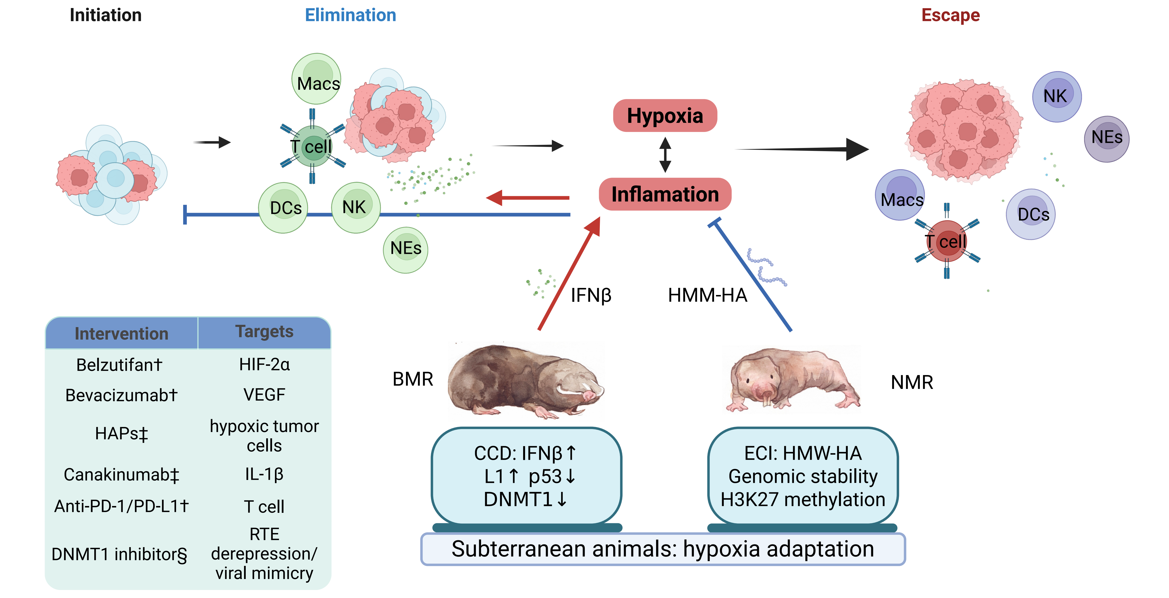
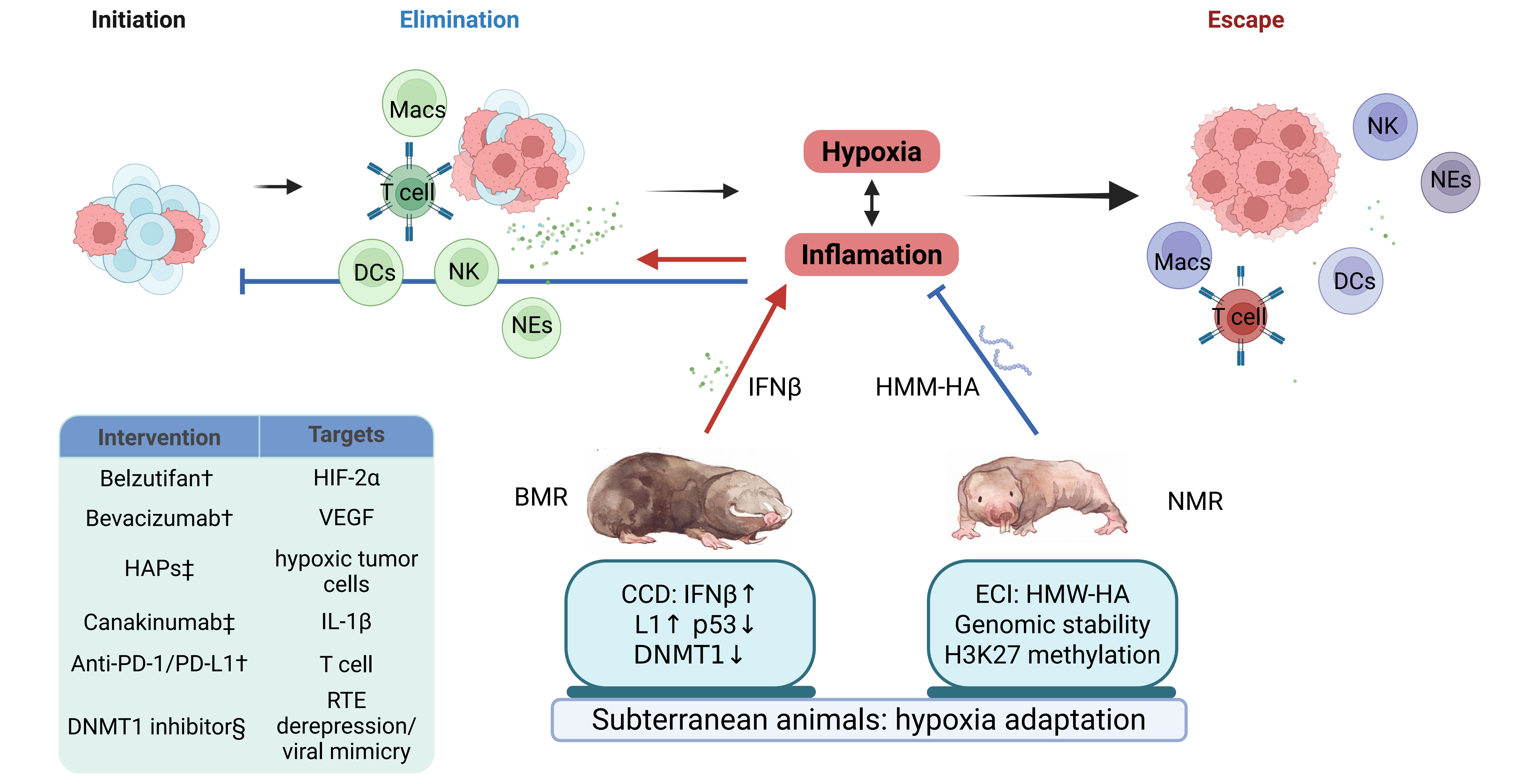
Figure 2. Subterranean Species Modulate the Interplay between Hypoxia and Inflammation for Tumor Suppression and provide potential therapeutic implications. During tumorigenesis, abnormal cells may initially be recognized and eliminated by immune cells, including T cells, Macs, DCs, NK cells, and NEs. However, persistent hypoxia and inflammation progressively reshape the tumor microenvironment and promote immune escape, characterized by impaired immune elimination and tumor persistence. Subterranean mammals have evolved distinct adaptive mechanisms to cope with chronic hypoxic habitats, which may also contribute to their cancer resistance. In the BMR, reduced DNMT1 expression is associated with LINE-1 (L1) derepression and activation of an IFNβ-mediated CCD response, thereby enhancing immune-mediated elimination of abnormal cells. In the NMR, HMM-HA contributes to ECI, genomic stability, and H3K27 methylation-associated epigenetic stability, which may suppress excessive inflammation and prevent malignant progression. These divergent strategies suggest that subterranean animals can either reinforce immune clearance or limit chronic inflammation, thereby interrupting the maladaptive hypoxia–inflammation loop that supports tumor escape. Potential therapeutic interventions targeting this axis include HIF-2α inhibition by belzutifan, VEGF blockade by bevacizumab, HAPs targeting hypoxic tumor cells, IL-1β blockade by canakinumab, immune checkpoint blockade targeting PD-1/PD-L1, and DNMT1 inhibition to induce transposable element-mediated viral mimicry. Created in BioRender.com. Guo, K. (2026) https://BioRender.com/meq9qag. †: Clinically validated or approved in selected cancer settings; ‡: Under clinical investigation or supported mainly by preclinical evidence; §: Currently supported primarily by preclinical studies; Macs: macrophages; DCs: dendritic cells; NK: natural killer; NEs: neutrophils; BMR: blind mole rat; DNMT1: DNA methyltransferase 1; IFN: interferon; CCD: concerted cell death; NMR: naked mole rat; HMM-HA: high-molecular-mass hyaluronan; ECI: early contact inhibition; HIF: hypoxia-inducible factor; HAPs: hypoxia-activated prodrugs; LINE-1: long interspersed nuclear element 1; H3K27: histone H3 lysine 27; VEGF: vascular endothelial growth factor; IL-1β: interleukin 1-beta; PD-1: programmed cell death protein 1; PD-L1: programmed death-ligand 1.
Another long-lived species that has adapted to intermittent hypoxia is the bowhead whale, which has been investigated as a model of longevity and stress adaptation[84,85]. Unlike subterranean mammals that experience chronic hypoxia, bowhead whales encounter repeated hypoxic episodes during prolonged diving, providing a distinct physiological context for studying oxygen stress responses. Several studies have identified enhanced genome maintenance mechanisms in bowhead whales. For,example, Cold-inducible RNA-binding protein (CIRBP) is highly expressed in bowhead fibroblasts and tissues and has been associated with improved DNA repair capacity, including enhanced non-homologous end joining and homologous recombination repair[86]. In addition, increased expression of DNA repair-related pathways may contribute to genome stability and reduced accumulation of cellular damage during aging. Although bowhead whales and subterranean mammals share adaptations to oxygen limitation and exceptional longevity, the mechanisms underlying their cancer resistance appear to involve distinct biological strategies. Therefore, in this review, the bowhead whale is considered a brief example of intermittent hypoxia adaptation rather than a central model of hypoxia–inflammation-mediated cancer resistance, as direct links between its hypoxia responses and inflammatory regulation remain insufficiently defined.
5. Conclusion
Hypoxia and inflammation are fundamental stress responses that evolved to preserve tissue integrity under adverse conditions. In physiological conditions, both processes are transient and tightly regulated, supporting adaptation, immune-mediated clearance, and restoration of homeostasis. However, within the tumor microenvironment, these responses become chronic and unresolved. Persistent oxygen deprivation and sustained inflammatory signaling progressively reprogram cellular and immune behavior, shifting the tissue state from active clearance toward regulated tolerance.
Rather than acting independently, hypoxia and inflammation form an interconnected network that stabilizes a microenvironment adapted to metabolic, oxidative, and immune pressure. Tumor cells exploit this chronic stress landscape to enhance survival, evade immune elimination, and resist therapeutic intervention. In this sense, resistance of cancer cells to therapy and immune surveillance does not arise solely from intrinsic genetic alterations, but also from the prolonged activation of protective signaling that becomes maladaptive when sustained. Understanding how transient adaptive responses transition into chronic tolerance states may provide new insight into therapeutic strategies. Interventions that restore effective clearance mechanisms or disrupt the stabilization of stress-adapted microenvironments could help overcome resistance and improve treatment outcomes.
Naturally cancer-resistant subterranean species provide unique and valuable models to understand this transition. Long adapted to hypoxia, these species have reshaped their immune systems in favor of cancer resistance, either through anti-inflammatory modulation of HMM-HA in NMRs or by enhanced innate immune surveillance in BMRs. Together, these divergent strategies ultimately converge on cancer resistance, highlighting the deep evolutionary interplay between hypoxia and inflammation. From a translational perspective, these species suggest two principles: dampening maladaptive chronic inflammation without abolishing clearance, or forcing stressed premalignant cells back into immune elimination.
Acknowledgements
We thank Ms. Ke Guo for drawing the animals of Figure 2.
Authors contribution
Zhao Y: Conceptualization, project administration, writing-original draft, writing-review & editing.
Xu Y: Visualization, project administration, writing-original draft, writing-review & editing.
Conflicts of interest
The authors declare no competing interest.
Ethical approval
Not applicable.
Consent to participate
Not applicable.
Consent for publication
Not applicable.
Availability of data and materials
Not applicable.
Funding
This work was supported by General Program of National Natural Science Foundation of China (Grant no. 32371236) and Zhejiang Provincial Natural Science Foundation of China (Grant no. LZ23C110002) to Y.Z.
Copyright
© The Author(s) 2026.
References
-
2. Pavlova NN, Thompson CB. The emerging hallmarks of cancer metabolism. Cell Metab. 2016;23(1):27-47.[DOI]
-
3. Kumar KH, Venakteshaiah SU. Inflammation and cancer. Endocr Metab Immune Disord Drug Targets. 2021;21(2):193-194.[DOI]
-
6. Gallimore AM, Simon AK. Positive and negative influences of regulatory T cells on tumour immunity. Oncogene. 2008;27(45):5886-5893.[DOI]
-
7. Pham K, Parikh K, Heinrich EC. Hypoxia and inflammation: Insights from high-altitude physiology. Front Physiol. 2021;12:676782.[DOI]
-
10. Bosco MC, D’Orazi G, Del Bufalo D. Targeting hypoxia in tumor: A new promising therapeutic strategy. J Exp Clin Cancer Res. 2020;39(1):8.[DOI]
-
11. Varsha VK, Savita JK, Girish HC, Shyamala K. Role of hypoxia and epithelial-mesenchymal transition in the formation and maintenance of oral cancer stem cells in oral squamous cell carcinomas and metastatic lymph node: An immunohistochemical analysis. J Oral Maxillofac Pathol. 2023;27(2):307-314.
-
13. Taylor CT, Scholz CC. The effect of HIF on metabolism and immunity. Nat Rev Nephrol. 2022;18(9):573-587.[DOI]
-
23. McDonough MA, Loenarz C, Chowdhury R, Clifton IJ, Schofield CJ. Structural studies on human 2-oxoglutarate dependent oxygenases. Curr Opin Struct Biol. 2010;20(6):659-672.[DOI]
-
24. Rose NR, McDonough MA, King ONF, Kawamura A, Schofield CJ. Inhibition of 2-oxoglutarate dependent oxygenases. Chem Soc Rev. 2011;40(8):4364.[DOI]
-
26. Bae T, Hallis SP, Kwak MK. Hypoxia, oxidative stress, and the interplay of HIFs and NRF2 signaling in cancer. Exp Mol Med. 2024;56:501-514.[DOI]
-
28. Wu Q, You L, Nepovimova E, Heger Z, Wu W, Kuca K, et al. Hypoxia-inducible factors: Master regulators of hypoxic tumor immune escape. J Hematol Oncol. 2022;15(1):77.[DOI]
-
31. He Y, Korboukh I, Jin J, Huang J. Targeting protein lysine methylation and demethylation in cancers. Acta Biochim Biophys Sin. 2012;44(1):70-79.[DOI]
-
33. Martinez-Bosch N, Vinaixa J, Navarro P. Immune evasion in pancreatic cancer: From mechanisms to therapy. Cancers. 2018;10(1):6.[DOI]
-
35. Ortega-Sáenz P, López-Barneo J. Physiology of the carotid body: From molecules to disease. Annu Rev Physiol. 2020;82:127-149.[DOI]
-
36. Cummins EP, Strowitzki MJ, Taylor CT. Mechanisms and consequences of oxygen and carbon dioxide sensing in mammals. Physiol Rev. 2020;100(1):463-488.[DOI]
-
38. Medzhitov R. Origin and physiological roles of inflammation. Nature. 2008;454(7203):428-435.[DOI]
-
39. Barton GM. A calculated response: Control of inflammation by the innate immune system. J Clin Invest. 2008;118(2):413-420.[DOI]
-
41. Dunn GP, Bruce AT, Ikeda H, Old LJ, Schreiber RD. Cancer immunoediting: From immunosurveillance to tumor escape. Nat Immunol. 2002;3(11):991-998.[DOI]
-
46. Zhao Y, Simon M, Seluanov A, Gorbunova V. DNA damage and repair in age-related inflammation. Nat Rev Immunol. 2023;23(2):75-89.[DOI]
-
47. Fernandes Q, Inchakalody VP, Bedhiafi T, Mestiri S, Taib N, Uddin S, et al. Chronic inflammation and cancer; the two sides of a coin. Life Sci. 2024;338:122390.[DOI]
-
48. Cheon H, Wang Y, Wightman SM, Jackson MW, Stark GR. How cancer cells make and respond to interferon-I. Trends Cancer. 2023;9(1):83-92.[DOI]
-
49. Hanahan D, Michielin O, Pittet MJ. Convergent inducers and effectors of T cell paralysis in the tumour microenvironment. Nat Rev Cancer. 2025;25:41-58.
-
52. Perkins ND. Post-translational modifications regulating the activity and function of the nuclear factor kappa B pathway. Oncogene. 2006;25(51):6717-6730.[DOI]
-
55. Belaiba RS, Bonello S, Zähringer C, Schmidt S, Hess J, Kietzmann T, et al. Hypoxia up-regulates hypoxia-inducible factor-1alpha transcription by involving phosphatidylinositol 3-kinase and nuclear factor kappaB in pulmonary artery smooth muscle cells. Mol Biol Cell. 2007;18(12):4691-4697.
-
57. Ciepła J, Smolarczyk R. Tumor hypoxia unveiled: insights into microenvironment, detection tools and emerging therapies. Clin Exp Med. 2024;24:235.[DOI]
-
61. Noman MZ, Desantis G, Janji B, Hasmim M, Karray S, Dessen P, et al. PD-L1 is a novel direct target of HIF-1α, and its blockade under hypoxia enhanced MDSC-mediated T cell activation. J Exp Med. 2014;211(5):781-790.[DOI]
-
62. Zhang H, Lu H, Xiang L, Bullen JW, Zhang C, Samanta D, et al. HIF-1 regulates CD47 expression in breast cancer cells to promote evasion of phagocytosis and maintenance of cancer stem cells. Proc Natl Acad Sci U S A. 2015;112(45):E6215-E6223.[DOI]
-
64. Choueiri TK, Powles T, Peltola K, de Velasco G, Burotto M, Suarez C, et al. Belzutifan versus everolimus for advanced renal-cell carcinoma. N Engl J Med. 2024;391(8):710-721.[DOI]
-
65. Zhao J, Tian X, Zhu Y, Zhang Z, Rydkina E, Yuan Y, et al. Reply to: Transformation of naked mole-rat cells. Nature. 2020;583(7814):E8-E13.[DOI]
-
71. Tian X, Azpurua J, Ke Z, Augereau A, Zhang ZD, Vijg J, et al. INK4locus of the tumor-resistant rodent, the naked mole rat, expresses a functional p15/p16 hybrid isoform. Proc Natl Acad Sci U S A. 2015;112(4):1053-1058.[DOI]
-
79. Gorbunova V, Hine C, Tian X, Ablaeva J, Gudkov AV, Nevo E, et al. Cancer resistance in the blind mole rat is mediated by concerted necrotic cell death mechanism. Proc Natl Acad Sci U S A. 2012;109(47):19392-19396.[DOI]
-
81. Zhao Y, Oreskovic E, Zhang Q, Lu Q, Gilman A, Lin YS, et al. Transposon-triggered innate immune response confers cancer resistance to the blind mole rat. Nat Immunol. 2021;22(10):1219-1230.[DOI]
-
82. Ashur-Fabian O, Avivi A, Trakhtenbrot L, Adamsky K, Cohen M, Kajakaro G, et al. Evolution of p53 in hypoxia-stressed Spalax mimics human tumor mutation. Proc Natl Acad Sci U S A. 2004;101(33):12236-12241.[DOI]
-
83. Leonova KI, Brodsky L, Lipchick B, Pal M, Novototskaya L, Chenchik AA, et al. p53 cooperates with DNA methylation and a suicidal interferon response to maintain epigenetic silencing of repeats and noncoding RNAs. Proc Natl Acad Sci U S A. 2013;110(1):E89-E98.[DOI]
-
84. Tyshkovskiy A, Ma S, Shindyapina AV, Tikhonov S, Lee SG, Bozaykut P, et al. Distinct longevity mechanisms across and within species and their association with aging. Cell. 2023;186(13):2929-2949.e20.[DOI]
Copyright
© The Author(s) 2026. This is an Open Access article licensed under a Creative Commons Attribution 4.0 International License (https://creativecommons.org/licenses/by/4.0/), which permits unrestricted use, sharing, adaptation, distribution and reproduction in any medium or format, for any purpose, even commercially, as long as you give appropriate credit to the original author(s) and the source, provide a link to the Creative Commons license, and indicate if changes were made.
Publisher’s Note
Science Exploration remains a neutral stance on jurisdictional claims in published
maps
and institutional affiliations. The views expressed in this article are solely those
of
the author(s) and do not reflect the opinions of the Editors or the publisher.
Share And Cite


Science Exploration Style
Xu Y, Zhao Y. Interplay between hypoxia and inflammation in cancer progression, therapy resistance, and natural cancer resistance. Ageing Cancer Res Treat. 2026;3:202612. https://doi.org/10.70401/acrt.2026.0032
Tips
Copy completed.
Submit a Manuscript
Author Instructions
Cite this Article
Article Metrics
0
View
0
Download
Cited
Article Updates

- Abstract
- Keywords
- 1. Introduction
- 2. Cellular Response to Hypoxia: From Adaptive Survival to Tumor Persistence
- 3. Inflammation as a Mediator between Immune Clearance and Immune Tolerance
- 4. Coordinated Stress Adaptation in Cancer by Interplay between Hypoxia and Inflammation
- 5. Conclusion
- Acknowledgements
- Authors contribution
- Conflicts of interest
- Ethical approval
- Consent to participate
- Consent for publication
- Availability of data and materials
- Funding
- References
- Copyright
 Get citation
Science Exploration Style

Xu Y, Zhao Y. Interplay between hypoxia and inflammation in cancer progression, therapy resistance, and natural cancer resistance. Ageing Cancer Res Treat. 2026;3:202612. https://doi.org/10.70401/acrt.2026.0032
copy
Share Link
copy