Organoid and organ-on-a-chip models for the tumor immune microenvironment: A call to incorporate immunosenescence

Yining Feng
1,#
,
Luqi Wang
1,#
,
Dameng Li
1,2,3,#
,
Chenglong Mu
1,2,3,#
,
Liang Wei
1,2,3,*

,
Bo Ma
1,2,3,*
*Correspondence to:
Bo Ma, Cellular Therapeutics School of Medicine, Xuzhou Medical University, Xuzhou 221004, Jiangsu, China; Center of Clinical Oncology, The Affiliated Hospital of Xuzhou Medical University, Xuzhou 221002, Jiangsu, China; Jiangsu Center for the Collaboration and Innovation of Cancer Biotherapy, Xuzhou Medical University, Xuzhou 221004, Jiangsu, China.
E-mail: boma@xzhmu.edu.cn
Liang Wei, Cellular Therapeutics School of Medicine, Xuzhou Medical University, Xuzhou 221004, Jiangsu, China; Center of Clinical Oncology, The Affiliated Hospital of Xuzhou Medical University, Xuzhou 221002, Jiangsu, China; Jiangsu Center for the Collaboration and Innovation of Cancer Biotherapy, Xuzhou Medical University, Xuzhou 221004, Jiangsu, China. E-mail: weiliang@xzhmu.edu.cn
Liang Wei, Cellular Therapeutics School of Medicine, Xuzhou Medical University, Xuzhou 221004, Jiangsu, China; Center of Clinical Oncology, The Affiliated Hospital of Xuzhou Medical University, Xuzhou 221002, Jiangsu, China; Jiangsu Center for the Collaboration and Innovation of Cancer Biotherapy, Xuzhou Medical University, Xuzhou 221004, Jiangsu, China. E-mail: weiliang@xzhmu.edu.cn
Ageing Cancer Res Treat. 2027;4:202625. 10.70401/acrt.2026.0039
Received: May 26, 2026Accepted: September 02, 2026Published: September 03, 2026
This article belongs to the Special lssue Immunosenescence and Cancer: From Mechanisms to Therapy
Abstract
The tumor immune microenvironment (TIME) is a central determinant of cancer progression and therapeutic response. Although organoid and organ-on-a-chip technologies have substantially advanced the modeling of tumor-immune interactions, most current platforms overlook a critical dimension of clinical reality: immunosenescence. This process profoundly reshapes immune cell composition, functional capacity, and tumor-immune crosstalk, yet it remains largely absent from existing in vitro systems. In this review, we provide a conceptual and methodological overview of organoid- and microfluidic-based models for reconstructing the TIME. We highlight their respective strengths in capturing tumor heterogeneity and dynamic immune processes, while critically evaluating their limitations in modeling long-term immune evolution and age-associated dysfunction. Importantly, we propose immunosenescence as a missing but essential modeling axis and argue that it should be treated as a controllable experimental variable rather than a background condition. By integrating aging-associated immune features into advanced 3D and microfluidic platforms, next-generation models are poised to better recapitulate patient heterogeneity and improve the prediction of immunotherapy outcomes. This perspective underscores a shift toward age-aware, physiologically relevant tumor models and provides a framework for advancing precision immuno-oncology in an aging population.
Keywords
Tumor immune microenvironment, immunosenescence, organoid, organ-on-a-chip
1. Introduction
The tumor immune microenvironment (TIME) has emerged as a central determinant of cancer initiation, progression, metastasis, and therapeutic response[1]. Rather than behaving as isolated entities, tumor cells are embedded within an intricate and evolving ecosystem composed of immune cells, stromal components, extracellular matrix (ECM), soluble mediators, and diverse biophysical cues. Continuous reciprocal interactions among these components shape immune surveillance, immune escape, and treatment outcomes. Consequently, the TIME is now recognized not only as a diagnostic hallmark but also as a critical target for therapeutic intervention[2].
Over the past decade, immunotherapy, encompassing immune checkpoint inhibitors, adoptive cell therapies, bispecific antibodies, and cancer vaccines, has transformed the treatment landscape of multiple malignancies[3]. Durable responses in a subset of patients have validated the immune system as a powerful therapeutic lever. However, despite these breakthroughs, clinical efficacy remains highly heterogeneous and often difficult to predict.
This variability is particularly conspicuous in elderly patients, who constitute the majority of cancer cases[4,5]. Aging is accompanied by profound remodeling of the immune system, a phenomenon broadly referred to as immune aging[6]. This umbrella term encompasses two interconnected yet mechanistically distinct dimensions: immunosenescence, which denotes the progressive functional decline of the immune system, manifested as thymic involution, contraction of the naïve lymphocyte pools, accumulation of exhausted and senescent effector cells, and shrinkage of the immune repertoire; and inflammaging, which describes the chronic, low-grade systemic inflammation driven largely by the senescence-associated secretory phenotype (SASP) and characterized by sustained elevation of pro-inflammatory cytokines such as interleukin (IL)-6, tumor necrosis factor (TNF-α), and IL-8. Throughout this Review, we distinguish immunosenescence from cellular senescence and T cell exhaustion. Immunosenescence refers to system-level immune aging, whereas cellular senescence denotes cell-intrinsic growth arrest, and T cell exhaustion describes antigen-driven functional decline. These three states may overlap but are not interchangeable. These two processes operate in a self-reinforcing loop and collectively reshape the TIME, compromising antitumor immunity and therapeutic responsiveness. Given that functional deterioration of the immune system represents the most clinically consequential aspect of this remodeling, in this Review we primarily employ the term immunosenescence to describe the age-associated immune defects that undermine tumor control and immunotherapy efficacy, while using inflammaging specifically in contexts related to chronic inflammation and SASP-driven immune suppression[7].
Yet, a critical limitation of current preclinical models is the prevailing assumption of a “young” immune system, which fails to reflect the aged immune landscape observed in clinical settings[8]. Traditional experimental models are insufficient to address this gap. Two-dimensional (2D) cell culture systems, although experimentally convenient, lack tissue-level architecture and spatial organization[9]. Animal models have provided indispensable insights into cancer biology and immunology, yet species-specific differences in immune regulation, tumor evolution, and lifespan limit their translational relevance[10,11]. In particular, human age-related immune remodeling is poorly captured in commonly used murine models. These shortcomings have driven intense interest in human-relevant in vitro platforms capable of reconstructing the TIME with higher physiological fidelity.
Tumor organoids and organ-on-a-chip technologies have emerged as leading next-generation modeling platforms[12,13]. While organoids preserve tumor-intrinsic genetic and phenotypic heterogeneity, organ-on-a-chip systems, which leverage bioengineered microfluidic platforms, introduce dynamic perfusion, vascular interfaces, and precise spatiotemporal control. Together, they offer complementary advantages for modeling the complexity of tumor-immune interactions[14-16].
In this review, we provide a methodological perspective on these emerging platforms and critically examine their capabilities in reconstructing the TIME. Importantly, we position immunosenescence as a missing but essential modeling dimension and propose a framework for integrating immune aging into next-generation in vitro systems. By doing so, we aim to bridge the gap between experimental models and clinical reality, ultimately fostering more effective immunotherapy strategies for aging populations.
2. Cellular, Stromal, and Biophysical Components of the TIME
Accurate reconstruction of the TIME requires integrating diverse cellular, stromal, and biophysical components within a dynamically evolving system. Rather than a static assembly of cell types, the TIME represents a highly coordinated and context-dependent network in which immune, stromal, and tumor cells continuously interact under biochemical and mechanical constraints. Importantly, these interactions are not only spatially organized but also temporally regulated, with aging serving as a primary driver of temporal remodeling.
2.1 Immune cells
The immune compartment of the TIME encompasses a broad spectrum of innate and adaptive immune cells, including cytotoxic CD8+ T cells, CD4+ helper T cells, regulatory T cells (Tregs), B cells, natural killer (NK) cells, dendritic cells (DCs), tumor-associated macrophages (TAMs), neutrophils, and myeloid-derived suppressor cells (MDSCs). These populations display substantial phenotypic and functional heterogeneity. Depending on contextual cues, immune cells may exert potent antitumor activity or be co-opted into immunosuppressive and tumor-promoting roles[17,18].
Functional plasticity is a defining feature of immune cells within the TIME[19-21]. Signals such as tumor-derived cytokines, chronic antigen exposure, hypoxia, and metabolic stress can drive transitions between activation, exhaustion, and cellular senescence[22]. For example, CD8+ T cells may shift from highly cytotoxic effectors to dysfunctional exhausted cells, while macrophages can polarize along a spectrum from pro-inflammatory (M1-like) to immunosuppressive (M2-like) phenotypes[23-26].
Importantly, immune aging introduces an additional layer of complexity to immune cell states. Immunosenescence leads to a contraction of the naive T cell pool, accumulation of senescent and exhausted lymphocytes, impaired antigen presentation, and expansion of immunosuppressive myeloid populations[27-30]. These age-associated alterations fundamentally reshape antitumor immunity and contribute to therapy resistance. Therefore, capturing both functional plasticity and age-associated immune remodeling is essential for physiologically relevant TIME modeling (Figure 1).
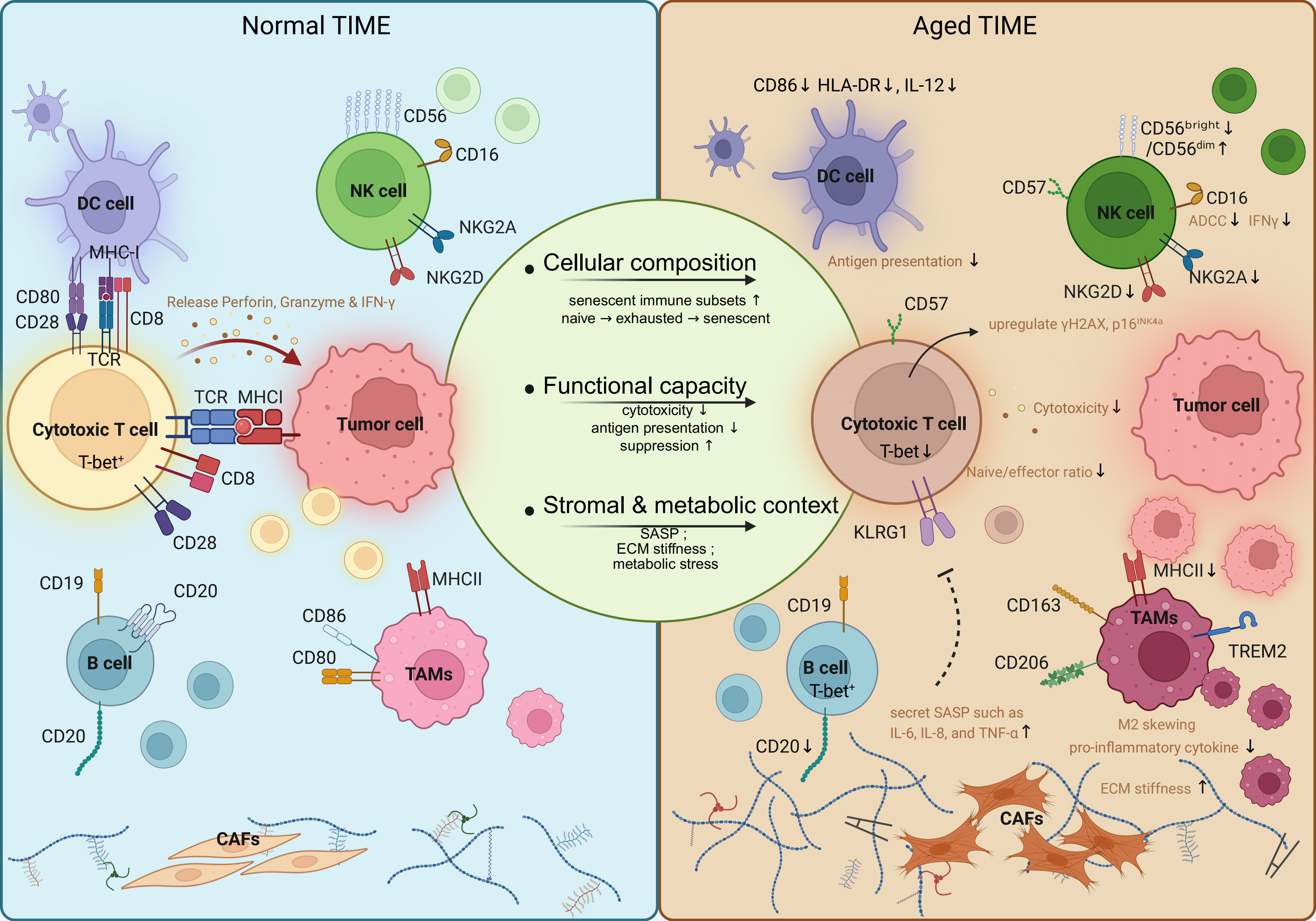
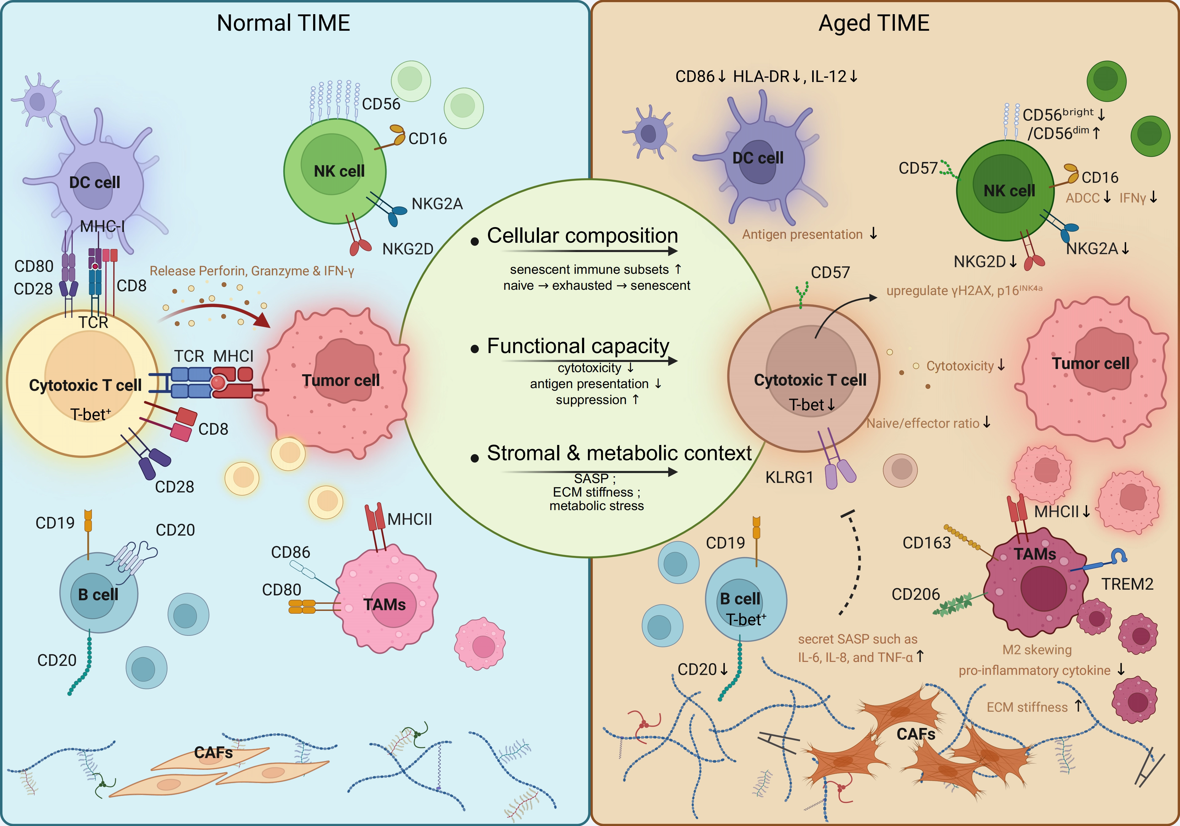
Figure 1. Aging reshapes the TIME. Schematic comparison of a normal (left) versus aged (right) TIME. Aging reshapes the TIME across three interconnected axes: cellular composition (altered proportions of lymphocyte and myeloid subsets), functional capacity (diminished effector functions and enhanced suppression), and stromal & metabolic context (SASP, ECM stiffness, hypoxia, and metabolic stress). Key immune cell types depicted include CD8+ T cells, NK cells, dendritic cells, tumor-associated macrophages, and B cells. Created in BioRender. Feng, Y. (2026) https://BioRender.com/tlspmpr. TIME: tumor immune microenvironment; SASP: senescence-associated secretory phenotype; ECM: extracellular matrix; DCs: dendritic cells; CAFs: cancer-associated fibroblasts; TAMs: tumor-associated macrophages; MHC: major histocompatibility complex; NK: natural killer.
Central to the interplay between immunosenescence and inflammaging is the SASP, a hallmark feature in which senescent cells secrete a plethora of pro-inflammatory cytokines, chemokines, growth factors, and matrix-remodeling enzymes. In the TIME, the SASP is produced by multiple cellular sources, including senescent CD8+ T cells, NK cells, tumor-associated macrophages, cancer-associated fibroblasts, and senescent epithelial cells[31,32]. Once secreted, SASP factors such as IL-6, IL-8, and TNF-α exert potent paracrine effects that propagate senescence to neighboring cells, a process termed bystander senescence, while simultaneously recruiting and polarizing MDSCs and TAMs toward immunosuppressive phenotypes[31,33,34]. This SASP-driven inflammatory milieu not only sustains chronic low-grade inflammation but also establishes a feed-forward loop that reinforces immune dysfunction and fosters a tumor-permissive microenvironment. Therefore, accurate modeling of the aged TIME necessitates not only the inclusion of senescent immune cells but also the reconstitution of SASP-mediated intercellular signaling and its microenvironmental consequences[35].
Mechanistically, the integration of immunosenescence into these models can be achieved through three primary methodological routes: (1) Primary Cell Sourcing: utilizing autologous immune cells directly isolated from elderly donors or cancer patients; (2) In Vitro Induction: driving T cell senescence through chronic T-cell receptor (TCR) stimulation, replicative aging, or exposure to “inflammaging” mediators such as IL-6 and TNF-α; and (3) Microenvironmental Stress: recapitulating age-associated dysfunction by precisely controlling oxygen tension (hypoxia), nutrient deprivation, and metabolic waste accumulation (e.g., lactate) within the microfluidic device (Table 1).
Table 1. Key characteristics of TIME immune cells: Normal vs. immunosenescent statesa,b.
| Immune Cell Type | Molecular Markers | Key Features & Impact on TIME | Key Parameters for In Vitro Modeling | Citation |
| CD8+ T cells | CD3+, CD8+, CD28+,CD62L+CD45RO- (naive), CD62L-CD45RO+ (memory), T-bet+, Eomes+ | Cytotoxic effector; Perforin/granzyme B-mediated killing; IFN-γ production; Antigen-specific tumor cell elimination | • Microfluidic infiltration assay (3D collagen, physiological shear stress) • Tumor organoid-T cell co-culture (optimized ratio, e.g., 1:1-1:5) • Peptide/MHC-multimer tracking (37 ℃) • 3D bioprinted ECM-rich tumor model | [36-69] |
| Aged CD8+ T cells | CD3+, CD8+, CD28-, CD27-, CD57+, KLRG1+, p16INK4a ↑, γH2AX foci ↑, naive/effector ratio ↓, TCF-1 ↓ | Proliferation ↓, Cytotoxicity ↓; Oligoclonal expansion; Impaired TCR signaling; Impaired neoantigen responsiveness; Reduced response to ICB | • Chronic antigen exposure in a microfluidic channel (e.g., OVA peptide, extended culture) • Hypoxia (pathophysiological O2 tension, e.g., 1% O2) + ROS induction (e.g., H2O2) • Co-culture with SASP+ senescent fibroblasts • Use donors ≥ 65 years vs. 25-35 years as a normal control | [31,40-44] |
| CD4+ T cells | CD3+, CD4+, CD28+,CD62L+CD45RO- (naive), CD62L-CD45RO+ (memory) | Helper functions; Supports CD8+ and B cells; Orchestrates adaptive immunity | • Autologous DC-tumor organoid-T cell tri-culture • Tunable 3D hydrogel for lymphoid mechanics (stiffness range: 0.5-5 kPa) • Antigen-specific priming assessment (tetramer) | [45-47] |
| Aged CD4+ T cells | CD28-, CD57+, KLRG1+, PD-1 ↑, p16INK4a ↑, γH2AX foci ↑ | Helper function ↓; Repertoire diversity ↓; Th1/Th2/Th17 skewing (Th1: IFN-γ ↓, Th2: IL-4 ↑, Th17: IL-17 dysregulated); Impaired CD8+ priming | • Aged donor CD4+ T cells (≥ 65 years) • Suboptimal TCR stimulation (low-dose anti-CD3 or weak agonist peptide) • Co-culture with aged autologous DCs • TCR-seq repertoire analysis | [48] |
| Treg cells | CD4+, CD25+, FOXP3+; CD39+, CD73+ (activated subset), CTLA-4+, PD-1+, GITR+ | Suppresses effector T-cell responses; IL-2 consumption; Secretes inhibitory cytokines (TGF-β, IL-10, IL-35); Maintains immune homeostasis | • iTreg induction with TGF-β + IL-2 ± ATRA in 3D hydrogel • Foxp3-reporter tracking (GFP or RFP) • CFSE-based suppression assay in microfluidic co-culture | [49-51] |
| Aged Treg cells | FOXP3+, CD28-, CTLA-4 ↑, PD-1 ↑ | Suppressive function ↑; Accumulation in tumor and lymphoid tissues; Impairs antitumor immunity | • Stiff ECM hydrogel (e.g., > 10 kPa) + lactate-rich microenvironment • Compare CTLA-4/PD-1 expression and suppressive capacity vs. normal Tregs | [52-54] |
| B lymphocytes | CD19+, CD20+, CD27+ (memory), CD138+ (plasma cells) | Antibody production; Antigen presentation; TLS formation; Antitumor via ADCC and phagocytosis | • 3D engineered TLS with FDC networks (e.g., CD21L+ fibroblasts) • BCR stimulation in 3D collagen (e.g., anti-IgM) • CD40L + IL-21 + tumor antigen | [55-57] |
| Aged B lymphocytes | CD19+CD27-IgD+ (naive B ↓), switched memory B ↓, CD19+CD21-CD11c+T-bet+ (ABCs ↑) | High-affinity antibody ↓, BCR diversity ↓, BCR clonality ↑; CSR ↓, SHM ↓, TNF-α/IL-6 ↑, Antigen presentation ↓; Pro-tumor inflammation | • TLR7 agonist (e.g., R848) + IL-21 + IFN-γ to induce ABCs (CD11c+T-bet+) • Co-culture with aged Tfh cells in TLS models • Assess CSR/SHM (e.g., IgH isotype sequencing) and TNF-α/IL-6 secretion | [58,59] |
| TAMs | CD68+, CD163low/-, CD86+ (M1-like), HLA-DR+ | High plasticity; M1 antitumor (phagocytosis, antigen presentation, IL-12/TNF-α); M2 pro-tumor (immunosuppressive, tissue repair, angiogenesis) | • M-CSF for M0 differentiation (5-7 days) • M1 polarization: LPS (100 ng/mL) + IFN-γ (20 ng/mL) in 3D matrix (48 h) • M2 polarization: IL-4/IL-13 (20 ng/mL each) in 3D matrix (48 h) • CCL2-gradient microfluidic recruitment assay | [60-62] |
| Aged TAMs | CD163+, CD206+ (M2-like ↑), HLA-DR ↓, TREM2 ↑, LILRB4 ↑ | M2 skewing ↑; Pro-inflammatory cytokine ↓, phagocytosis ↓; Immunosuppressive, tumor-promoting environment | • M2 skewing on stiff substrate (e.g., > 10 kPa) + IL-4/IL-13 • Co-culture with senescent tumor cells or TIS (tumor-induced senescent fibroblasts) • Evaluate CD163/CD206/TREM2/LILRB4 and phagocytic index | [63,64] |
| MDSCs | CD11b+, CD33+, HLA-DRlow/-,CD14-CD15+ (G-MDSC); CD14+CD15- (M-MDSC) | Immunosuppression in pathological conditions; Minimal in normal TIME | • PBMC induction with GM-CSF + IL-6 + TCM in 3D bone marrow niche(5-7 days) • Hypoxic gradient chamber (e.g., 5%→1% O2) • PDO spatial co-culture | [65-67] |
| Aged MDSCs | CD11b+, CD33+, HLA-DRlow/-,PD-L1↑, ARG1 ↑, ROS ↑, LOX-1+, S100A8/A9 ↑(G-MDSC); iNOS ↑, NO ↑, IDO1 ↑, ARG1 ↑, PD-L1 ↑, IL-10 ↑, TGF-β ↑ (M-MDSC) | Expansion ↑, ARG1 ↑, ROS ↑, NO ↑, T cell suppression ↑; Amplified immunosuppressive network; Resistance to immunotherapy | • Aged donor MDSCs (≥ 65 years) or normal PBMC aged with lipid-rich microenvironment • Assess suppression of T cell proliferation (CFSE) and ROS/NO production • Compare PD-L1, ARG1, S100A8/A9 by flow cytometry | [68,69] |
| DC cells | HLA-DR+, CD11c+, CD86+, CD80+, CD1c+ (cDC2), CD141+ (cDC1) | Antigen uptake, processing, and presentation; T cell priming; Type I IFN production; Central for adaptive antitumor immunity | • Mo-DC differentiation: GM-CSF (50 ng/mL) + IL-4 (20 ng/mL) for 5-7 days • Maturation cocktail: TNF-α, IL-1β, PGE2, LPS (24-48 h) • 3D migration assay through lymphatic endothelial microfluidic chip | [70-72] |
| Aged DC cells | CD86 ↓, HLA-DR ↓, IL-12 ↓, pDC:IDO ↑ | Antigen presentation ↓, cross-presentation ↓; Type I IFN ↓; Regulatory DC expansion ↑; Reduced T cell priming; Immune tolerance | • Aged donor Mo-DCs (≥ 65 years) or normal Mo-DCs pre-treated with chronic low-dose TNF-α (1-5 ng/mL, 7-14 days) • Assess CD86/HLA-DR downregulation and cross-presentation defect (e.g., SIINFEKL-OVA model) | [73,74] |
| NK cells | CD56+, CD16+, NKG2D+, NKp30+, NKp46+ | Innate Cytotoxicity; ADCC via CD16; IFN-γ secretion; Early antitumor surveillance; Effective against MHC-low tumors | • Live-imaging tumor spheroid killing (optimized E: T ratio) • Shear stress platform (0.5-4 dyn/cm2) for ADCC with therapeutic antibodies | [75,76] |
| Aged NK cells | CD56dim ↑, CD57↑, KIR↑; CD56bright ↓, NKG2A↓, NKG2D↓, NKp30↓ | Perforin/granzyme B ↓, ADCC ↓, IFN-γ ↓; Diminished innate surveillance; Impaired ADCC | • Aged donor NK expansion (≥ 65 years, IL-2 100-500 U/mL, 7-14 days) • Chronic IL-12 + IL-15 stimulation (7 days) → NKG2D ↓ • Assess killing under metabolic stress (low glucose, 10-20 mM lactate) • Measure CD56dimCD57+KIR+ subset by flow cytometry | [77,78] |
| Neutrophils | CD11b+, CD15+, CD16+, CD62L+, CXCR2+ | Phagocytosis; NETosis; cytokine secretion; N1 antitumor (CD86+ICAM1+TNF-α+) vs. N2 pro-tumor (PD-L1+VEGF+MMP-9+) plasticity | • Vascular-perfusable organ-on-a-chip for rolling/adhesion/TEM • Short-term culture (≤ 4 h; or HL-60 differentiation for longer studies) • Chemotaxis to fMLP (10-7-10-6 M) under flow | [79-81] |
| Aged Neutrophils | CD62L ↓, CXCR2 ↓, CD101+, PD-L1 ↑ | Chemotaxis ↓; NETosis dysregulated; N2-like polarization ↑; Impaired inflammation; Pro-tumorigenic functions | • Aged donor neutrophils (≥ 65 years, use within 2-4 h of isolation) • Exposure to aged plasma or SASP-conditioned medium • Stiff hydrogel (e.g., > 10 kPa) under flow • Measure CD62L/CXCR2 ↓ and PD-L1 ↑; assess NETosis | [82,83] |
| a: Cells with gray backgrounds indicate immunosenescent populations.b: Immunosenescent populations in this table exhibit cell type-specific changes. In addition, the following canonical senescence markers may be considered as reference indicators: (i) p16INK4a/p21/p53 upregulation; (ii) SA-β-gal activity; (iii) SASP factors (IL-6, IL-8, TNF-α); (iv) telomere attrition; and (v) mitochondrial dysfunction. Notably, these markers may be variably expressed and are not necessarily present in all immunosenescent cell types. | [84,85] | |||
3D: three-dimensional; ECM: extracellular matrix; TCR: T-Cell receptor; OVA: ovalbumin; ROS: reactive oxygen species; SASP: senescence-associated secretory phenotype; DC: dendritic cell; CTLA-4: cytotoxic T-lymphocyte-associated protein 4; GITR: glucocorticoid-induced TNFR-related protein; PD-1: programmed cell death protein 1; TGF: transforming growth factor; IL: interleukin; ATRA: all-trans retinoic acid; GFP: green fluorescent protein; RFP: red fluorescent protein; CFSE: carboxyfluorescein succinimidyl ester; FDC: follicular dendritic cell; ADCC: antibody-dependent cellular cytotoxicity; TNF: tumor necrosis factor; TLR: toll-like receptor; IFN: interferon; TAM: tumor-associated macrophage; LPS: lipopolysaccharide; MDSC: myeloid-derived suppressor cell; GM-CSF: granulocyte-macrophage colony-stimulating factor; TCM: tumor-conditioned medium; PDO: patient-derived organoid; TIME: tumor immune microenvironment; ARG1: arginase 1; LOX-1: lectin-type oxidized LDL receptor 1; NKG2D: natural killer group 2 member D; NK: natural killer; KIR: killer-cell immunoglobulin-like receptor; NETosis: neutrophil extracellular trap formation; VEGF: vascular endothelial growth factor.
2.2 Stromal components and extracellular matrix
Beyond immune cells, the TIME is structurally and functionally defined by cancer-associated fibroblasts, endothelial cells, pericytes, and other stromal elements. These cells actively participate in immune regulation by secreting immunomodulatory cytokines and chemokines, remodeling the ECM, and forming physical barriers that impede immune infiltration[86,87]. The ECM itself provides more than just structural support; it transmits biochemical and mechanical signals that orchestrate immune cell migration, activation, and survival[88]. In the context of aging, alterations in stromal cell secretory profiles (e.g., SASP) and increased ECM cross-linking further complicate these interactions[89]. Among stromal cells, cancer-associated fibroblasts (CAFs) are particularly influential in shaping the aged TIME, as they not only contribute to ECM remodeling but also actively modulate immune cell function through both contact-dependent and paracrine mechanisms. In hepatocellular carcinoma, CD90+LAMA4+ extracellular matrix cancer-associated fibroblasts (eCAFs) induce CD8+ T cell senescence through LAMA4 binding to its receptor ITGA6 and activating ATM/CHK2/H2AX axis-mediated DNA damage signaling, as evidenced by upregulation of p53, p16, p21, β-galactosidase, reactive oxygen species (ROS), and RAS[90]. Assouline et al. reported that in pancreatic ductal adenocarcinoma, senescent cancer-associated fibroblasts restrict CD8+ T cell activation through their senescence-associated secretory phenotype and upregulation of immune regulatory genes, and that senolytic elimination of these cells with agents such as ABT-199 restores T cell activation and potentiates immunotherapy[91].
2.3 Biophysical and metabolic determinants
Biophysical parameters such as tissue stiffness, interstitial flow, fluid shear stress, and oxygen tension profoundly shape immune behavior[92-94]. In the normal TIME, these cues are tightly regulated to maintain immune surveillance. However, in the context of aging and tumor progression, they undergo pathological alterations[95]. Aberrant tumor vasculature disrupts interstitial flow, impedes immune cell infiltration, and generates unstable shear stress that impairs lymphocyte activation and migration[96]. Zhang et al. reported that biomechanical stress drives CD8+ T cell exhaustion through the Piezo1/Osr2 signaling axis, wherein mechanical signals sensed by Piezo1 activate a transcriptional program centered on Osr2, ultimately promoting terminal exhaustion and undermining antitumor immunity[97]. Meanwhile, chronic hypoxia, driven by rapid tumor proliferation and dysfunctional blood supply, suppresses T cell infiltration and drives exhaustion through mitochondrial dysfunction and HIF-1α-mediated transcriptional reprogramming[98]. Scharping et al. further reported a strong correlation between severe hypoxia and exhausted intratumoral T cells, noting that hypoxia drives T cell dysfunction through mitochondrial dysfunction and ROS formation[94].
Metabolic constraints similarly shape immune behavior in the TIME. Under normal conditions, balanced glycolysis and oxidative phosphorylation support T cell proliferation and cytokine production[99]. In tumors, hypoxia and nutrient deprivation impair immune effector function and promote exhaustion[94]. In aging, metabolic dysregulation becomes more pronounced. Aged immune cells exhibit enhanced glycolysis, mitochondrial dysfunction, and elevated ROS production[100]. Liu et al. reported that metabolic reprogramming drives T cell senescence through enhanced glycolysis and mitochondrial dysfunction, reinforcing epigenetic alterations that compromise antitumor immunity[77]. van Beek et al. further showed that in aged macrophages, mitochondrial dysfunction and altered lipid metabolism directly fuel inflammaging and reinforce the immunosuppressive TIME[63]. Additionally, the tumor microenvironment imposes further barriers through nutrient competition and lactate accumulation. Ringel et al. demonstrated that obesity reshapes tumor metabolism to suppress antitumor immunity via these mechanisms[101]. These constraints exacerbate age-related immune decline, as aged T cells exhibit compromised metabolic flexibility and reduced adaptability to microenvironmental stress, as demonstrated by Park et al. Spatiotemporal gradients of glucose, lactate, amino acids, and lipids further modulate immune cell fate[102]. Incorporating these dynamic metabolic factors remains a major challenge for in vitro modeling but is essential for achieving high physiological relevance, particularly when modeling the aged TIME.
In summary, an ideal model of the TIME must integrate diverse immune cell populations, stromal components with immunomodulatory functions, and precisely controllable biophysical and metabolic cues. However, conventional 2D cultures and static 3D models struggle to simultaneously fulfill these complex requirements. In the following sections, we will review two cutting-edge technologies, namely tumor organoids (Section 3) and organ-on-a-chip platforms (Section 4), to explore their respective technical advantages and current limitations in addressing the aforementioned modeling dimensions.
3. Organoid-Based Models for Tumor-Immune Interactions
Tumor organoids are 3D structures derived from patient tissues or genetically engineered cells that self-organize to recapitulate key features of tumor architecture and cellular heterogeneity (Figure 2). As highly physiological platforms, organoid technology has rapidly gained traction for cancer modeling, drug screening, and precision oncology[103].
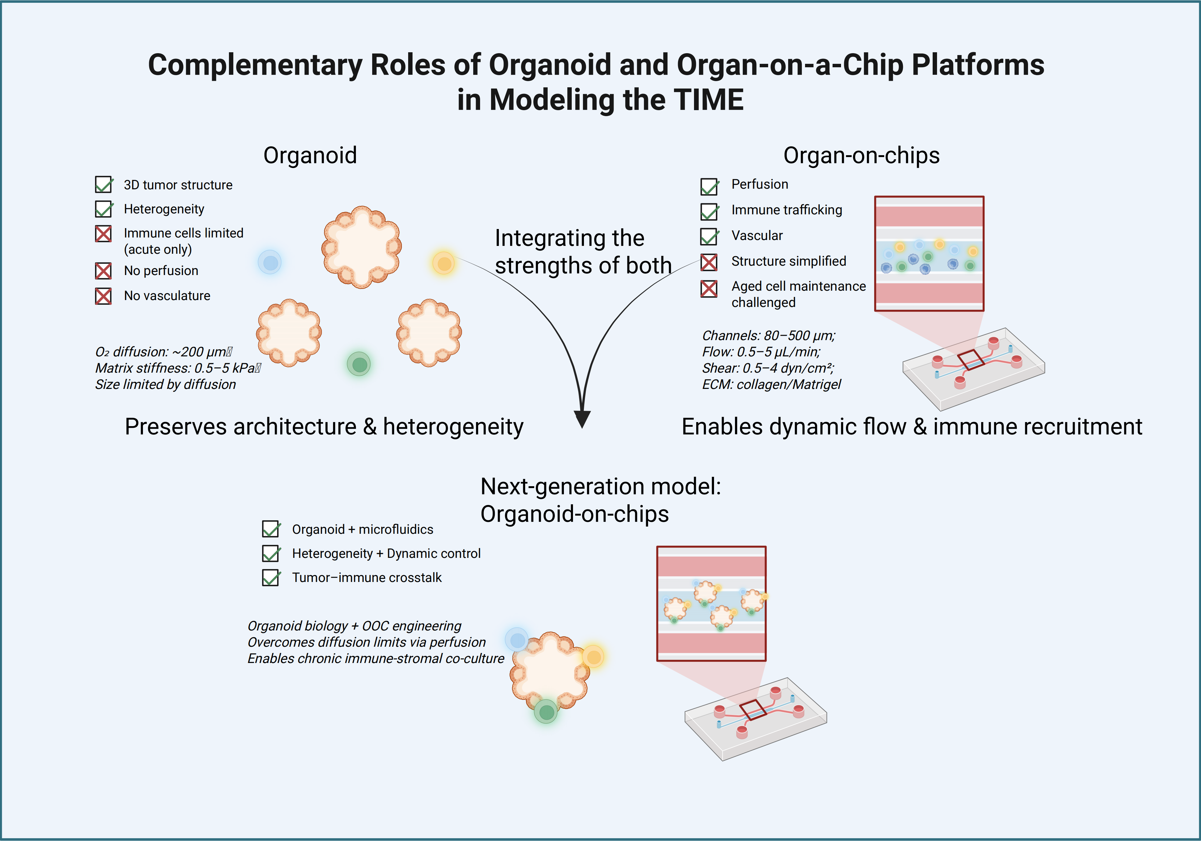
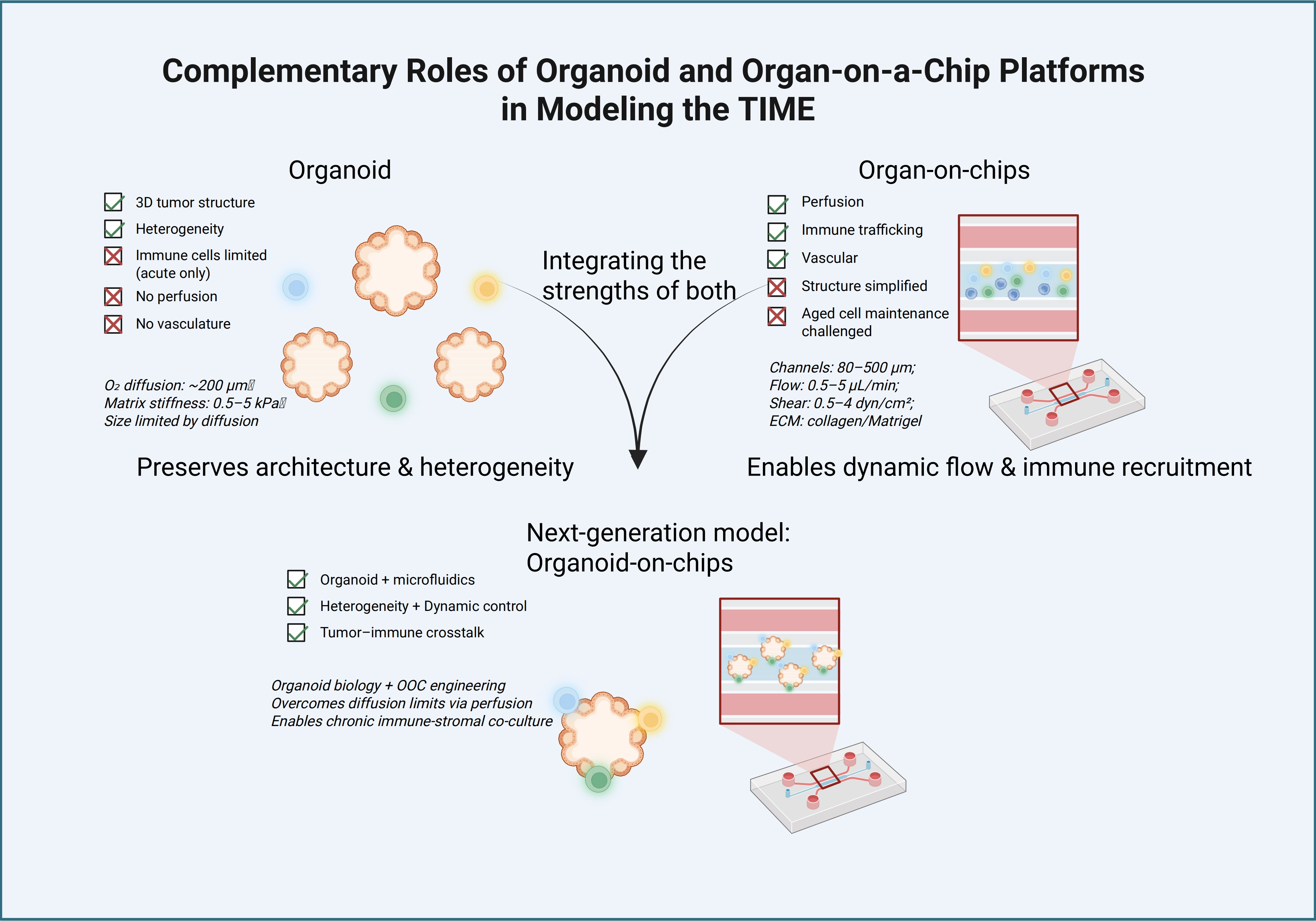
Figure 2. Complementary roles of organoid and organ-on-a-chip platforms in modeling the tumor immune microenvironment. Comparison of organoid, organ-on-a-chip, and hybrid platforms for modeling the tumor immune microenvironment. Organoids preserve tumor architecture and cellular heterogeneity but face intrinsic biological limitations, including restricted oxygen diffusion (~ 200 μm), matrix stiffness in the range of 0.5-5 kPa, and size constraints imposed by diffusion, which collectively limit their capacity for sustained immune-tumor co-culture. Organ-on-a-chip platforms enable controlled fluid flow, vascular interactions, and immune cell dynamics, with key design parameters annotated: channel dimensions (80-500 μm), flow rate (0.5-5 μL/min), shear stress (0.5-4 dyn/cm2), and ECM composition (collagen I/Matrigel). Hybrid systems integrate organoid biology with microfluidic engineering control, overcoming diffusion limits via perfusion and enabling chronic immune-stromal co-culture. The bottom panel illustrates the hybrid concept as a next-generation model that combines structural fidelity with dynamic control for investigating tumor-immune crosstalk. Created in BioRender. Feng, Y. (2026) https://BioRender.com/f848qrb. TIME: tumor immune microenvironment; 3D: three-dimensional; OOC: organ-on-a-chip.
3.1 Generation and characteristics of tumor organoids
Tumor organoids preserve the genetic alterations, epigenetic landscapes, and lineage hierarchies of their parental tumors. They can be expanded long-term, cryopreserved, and subjected to genetic manipulation, enabling systematic interrogation of tumor-intrinsic mechanisms. Importantly, organoids maintain the intra-tumoral heterogeneity that is often lost in conventional cell lines.
Recent multi-omic analyses have provided compelling evidence for this fidelity. As noted by Alison P. McGuigan, technologies such as single-cell RNA sequencing (scRNA-seq), single-cell assay for transposase-accessible chromatin with sequencing (scATAC-seq), and single-cell whole-genome sequencing (scWGS-seq) have been validated across three dimensions, namely genetics, epigenetics, and transcriptomic lineages, showing that tumor organoids faithfully preserve the essential features of their parental tissues (Table 2). These tools not only validate the reliability of patient-derived organoids (PDOs) for precision oncology but also offer powerful means to explore tumor heterogeneity evolution, microenvironmental adaptation, and mechanisms of drug resistance[113]. However, it is worth noting that these single-cell technologies provide high-resolution snapshots at discrete time points, offering limited capacity to track the continuous, long-term evolution of cell populations, a critical limitation when modeling time-dependent processes such as immunosenescence. Kumar et al. systematically compared PDOs with primary tumors through a single-cell atlas of gastric cancer, integrated with spatial transcriptomics, independent cohort validation, and functional assays. Their results revealed that organoid epithelial cells exhibited significant enrichment of epithelial-mesenchymal transition (EMT), cell motility, and oncogenic gene modules, and faithfully retained three sublineages, namely EpiC (chief cell-like), EpiInt (intestinal-type), and EpiPit (pit cell-like), with the EpiInt1 subset showing high expression of key gastric cancer oncogenes and serving as a critical population in malignant transformation. These findings highlight the high fidelity of organoids in preserving tumor epithelial lineage features, providing a robust cellular foundation for reconstructing the complex interactions within the TIME[114].
Table 2. Single-cell technologies validate that tumor organoids highly retain key features of the parental tumor.
| Technical Dimension | Technology | Year | Types of cancers | Citation |
| Genetic | WGS-seqscWGS-seq | 2019 | Ovarian cancer | [104] |
| Genetic | scWGS-seq+ scRNA-seq | 2022 | Glioblastoma | [105] |
| Epigenetic | scGET-seq | 2022 | Colorectal cancer | [106] |
| Transcriptomic Lineage + Epigenetic | scRNA-seq + EPIC array | 2025 | Glioblastoma | [107] |
| Transcriptomic Lineage | scRNA-seq+ scTCR-seq | 2018 | Colon cancer, pancreatic cancer, lung cancer, bile duct ampullary adenocarcinoma, brain schwannoma and salivary gland pleomorphic adenoma | [36] |
| Transcriptomic Lineage | scRNA-seq | 2020 | Glioblastoma | [108] |
| Transcriptomic Lineage | scRNA-seq | 2021 | Pancreatic ductal adenocarcinoma | [109] |
| Transcriptomic Lineage | scRNA-seq | 2020 | Colorectal and gastroesophageal cancer | [110] |
| Transcriptomic Lineage | scRNA-seq | 2026 | Ovarian cancer | [111] |
| Transcriptomic Lineage | scRNA-seq | 2022 | Prostate cancer | [112] |
EPIC: Infinium MethylationEPIC BeadChip (array); scWGS-seq: single-cell whole-genome sequencing; scRNA-seq: single-cell RNA sequencing; scGET-seq: single-cell genome and epigenome by transposase sequencing; scTCR-seq: single-cell T-cell receptor sequencing.
3.2 Immune co-culture strategies
Several strategies have been developed to incorporate immune components into tumor organoid systems. Co-culture with peripheral blood mononuclear cells (PBMCs), tumor-infiltrating lymphocytes (TILs), or engineered immune cells [e.g., chimeric antigen receptor T-cells (CAR-T cells)] allows investigation of immune infiltration, cytotoxicity, and immune escape[115-117].
First, “antigen-agnostic” expansion platforms facilitate the study of personalized responses. Cattaneo et al. addressed the challenge of expanding tumor-reactive T cells by co-culturing peripheral blood lymphocytes with autologous tumor organoids. This approach requires no neoantigen prediction and yields enriched CD8+ T cells within two weeks, with success rates of 33%-50% in non-small cell lung cancer and microsatellite instable colorectal cancer. After co-culture, T cell purity exceeds 90%, expansion ranges from 3- to 20-fold, and tumor organoid killing rates reach 20%-80%, providing an efficient platform for personalized immunotherapy[118].
Alternatively, “native” tumor-immune architectures can be preserved through specialized culture methods. Zhao et al. preserved tumor-infiltrating immune cells in ovarian cancer organoids by directly culturing patient tumor fragments (~ 1 mm) without enzymatic dissociation or red blood cell removal, demonstrating high responsiveness to chemotherapy and maintaining a diverse immune landscape including T cells, B cells, and macrophages[119]. Similarly, air-liquid interface (ALI) models offer short-term access to native interactions. Neal et al. utilized an ALI method to retain diverse endogenous immune cells within patient-derived organoids (PDOs), achieving high concordance (R2 = 0.719) in T cell receptor repertoires between organoids and original tumors[36].
To further mimic stromal and spatial complexities, newer interface models and bioengineering approaches have emerged. Li et al. established a gel-liquid interface (GLI) co-culture model, enabling efficient interaction between lung cancer organoids and autologous PBMCs by providing a liquid interface that facilitates free immune cell migration and infiltration[120]. Furthermore, induced pluripotent stem cell (iPSC)-derived organoids and hybrid systems have expanded the scope of modeling for immune-privileged sites. Peng et al. generated individualized patient tumor models by implanting tumor fragments into iPSC-derived brain organoids, successfully preserving tumor heterogeneity, resident immune cells, and even rudimentary vascular structures[107].
3.3 Methodological strengths and limitations
Despite their structural fidelity, tumor organoids exhibit a significant “immune void”. As underscored by Kumar et al., while organoids preserve epithelial lineage features with high fidelity, they suffer from a marked depletion of resident immune populations, particularly plasma cells and mature myeloid subsets[114]. This immune deficiency is compounded by the absence of functional vasculature and stromal components, which precludes modeling of immune cell trafficking, extravasation, and chronic stromal-immune crosstalk[121,122]. Moreover, the functional persistence of exogenous immune cells in co-culture is largely confined to acute timeframes, falling short of the sustained surveillance required to capture the progressive functional decline inherent to immunosenescence[123]. Even the long-term stability of organoids themselves is not assured; serial passaging may gradually erode the very heterogeneity that constitutes their primary advantage[104]. This inherent limitation suggests that relying solely on organoids may yield an incomplete and potentially overoptimistic assessment of the tumor immune landscape. Consequently, the integration of exogenous immune components under dynamic flow, as facilitated by microfluidic technologies, is not merely an enhancement but a functional necessity for faithfully modeling the TIME.
4. Organ-on-a-Chip Platforms for Modeling the TIME
While organoids excel at preserving intrinsic tumor heterogeneity, they inherently lack vasculature, perfusion, and the dynamic spatiotemporal cues essential for immune cell behavior. Organ-on-a-chip (OOC) platforms address these specific dimensions. Rather than being substitutes for one another, the two approaches together form the foundation for integrating next-generation tumor immune microenvironment models. Organ-on-a-chip technology integrates microfabrication, microfluidics, and tissue engineering to recreate tissue-level organization and function under dynamic conditions; tumor-on-a-chip platforms have become increasingly sophisticated, enabling incorporation of multiple cell types, real-time monitoring, and precise control of microenvironmental parameters.
4.1 Microfluidic control and dynamic perfusion
The primary advantage of microfluidic perfusion is the continuous delivery of nutrients and oxygen alongside the efficient removal of metabolic waste, which supports long-term culture and the generation of stable chemical gradients. This dynamic environment is critical for modeling of immune cell trafficking, extravasation, and infiltration, dynamic processes that are difficult to recapitulate in static systems[124]. For instance, Zou et al. developed a hepatocellular carcinoma (HCC) organoid-on-a-chip featuring vertically crossed microchannels that enable dynamic drug delivery and uniform culture, significantly enhancing the precision of immunotherapy response predictions[125]. Similarly, to overcome the low success rates and long culture durations of conventional PDOs, Ding et al. utilized droplet microfluidics to rapidly generate micro-organospheres. This chip-based approach successfully captured endogenous T cells and myeloid cells, allowing for the clinical assessment of programmed cell death protein 1 (PD-1) blockade and bispecific antibodies within a physiologically active immune microenvironment[126].
4.2 Vascularized tumor-on-a-chip models
Vascularized tumor chips incorporate endothelial-lined channels that mimic blood vessels. These platforms allow immune cells to circulate, adhere to the endothelium, and transmigrate into the tumor and stromal compartments[127]. Such models have provided profound mechanistic insights into immune surveillance, immune exclusion, and therapy-induced vascular remodeling[128,129]. Shirure et al. systematically delineated the design strategies, namely self-assembly and patterning, for vascularized organoid-on-a-chip platforms. They outlined the critical roles of integrating endothelial cells, stromal cells, and organ-specific cells to enable the in vitro recapitulation of tumor angiogenesis, transendothelial migration of immune cells, and hematogenous dissemination of tumor cells, thereby establishing a key framework for modeling inter-organ immune interactions and evaluating immunotherapeutic outcomes[122]. Quintard et al. engineered a microfluidic platform employing a serpentine microchannel architecture integrated with a U-shaped trapping mechanism, enabling the stable immobilization of organoids and the self-assembly of surrounding endothelial networks. This system successfully achieved functional anastomosis and perfusion between the endogenous vasculature of blood vessel organoids and the externally formed endothelial beds, thereby offering a scalable in vitro platform for investigating hematogenous tumor metastasis, immune cell-endothelial crosstalk, and interconnected multi-organ immune responses[130].
4.3 Multi-compartment and multi-organ integration: A holistic perspective
Beyond local tumor-immune interactions, the efficacy of immunotherapy is often dictated by systemic factors. Advanced chip designs can interconnect multiple tissue compartments to study complex processes such as immune cell education and metabolic coupling. Kawakita et al. leveraged microfluidic organ-on-a-chip technology combined with induced pluripotent stem cell-derived cells to establish a blood-brain barrier model that recapitulates physiologically relevant barrier functions and multicellular composition. They further noted that physically interconnecting such chip modules with tumor, immune, or metabolic organs, such as the liver, intestine, and bone marrow, enables the study of systemic interactions across interconnected tissue compartments. This interconnected approach allows for the investigation of systemic cytokine signaling and tumor-immune interactions within a more holistic physiological context[131]. Crucially, such multi-organ integration provides the technical infrastructure required to incorporate systemic variables, most notably immunosenescence, by simulating the communication between the aged bone marrow, the immune source, and the distal tumor site.
However, applying OOC platforms to aged immunity confronts two critical technical gaps: the poor survival and adhesion of aged primary immune cells under prolonged perfusion, and the oversimplification of aged vascular biology in current endothelial models, together with limited throughput and inter-chip variability that hinder standardization[132].
5. Advanced Modeling Dimensions of the TIME
Despite significant advances, current organoid and organ-on-a-chip models remain incomplete representations of the TIME. Most notably, they often fail to capture long-term immune evolution and age-associated remodeling.
First, immune cell dynamics in many systems are short-lived, limiting the ability to model chronic processes such as exhaustion and senescence. Second, the majority of studies rely on immune cells derived from young, healthy donors, which do not reflect the aged immune landscape of cancer patients. Third, systemic factors associated with aging, including chronic inflammation (inflammaging), metabolic alterations, and endocrine signaling, are largely absent from current in vitro platforms. As a result, these models may overestimate immune functionality and therapeutic efficacy, thereby limiting their translational predictive power. Addressing these gaps requires a conceptual shift from static reconstruction toward dynamic, age-aware modeling of the TIME.
5.1 Spatial organization, mechanical cues, and metabolic gradients
Next-generation TIME models increasingly incorporate spatial patterning, tunable matrix stiffness, and controlled metabolic gradients[122]. These biophysical features are not merely background settings; they directly influence immune cell recruitment, activation, and exhaustion. Microengineering approaches now allow for the systematic interrogation of how physical and metabolic constraints regulate immune function and shape therapeutic responses[133,134].
5.2 Modeling immunosenescence: From biological phenomenon to experimental variable
While recent advances have substantially improved reconstruction of the TIME, most existing models implicitly assume a “young” or immunologically intact immune system. This assumption contrasts sharply with clinical reality, where cancer incidence and immunotherapy are concentrated in elderly populations.
Immunosenescence should not be viewed merely as a biological phenomenon but rather as a critical and controllable modeling parameter in TIME reconstruction. From a methodological perspective, it encompasses multiple dimensions, including altered immune cell composition, reduced functional capacity, chronic inflammatory signaling, and impaired intercellular communication[77]. Specifically, immune cell composition shifts toward a lower naive-to-memory T cell ratio, contraction of the naive B cell pool, and expansion of Tregs, MDSCs, and age-associated B cells[135-140]. Functional capacity decline is manifested as a reduction in the overall number of CD8+ T cells, particularly the loss of the NKG2C+GZMB- cytotoxic memory subset, along with an approximately 2- to 3-fold increase in IL-4 levels upon stimulation[44]. Chronic inflammatory signaling is driven by the SASP, with IL-6, IL-8, and TNF-α elevated in aged microenvironments[31]. Aging is associated with thymic involution, contraction of the naive T cell pool, accumulation of exhausted and senescent T cells, impaired antigen presentation, and myeloid skewing toward immunosuppressive phenotypes. These changes synergize with tumor-derived signals to establish a permissive microenvironment for tumor progression and immune escape[141,142].
Several strategies can be employed to incorporate immunosenescence into in vitro systems. These include the use of immune cells derived from elderly donors, in vitro induction of senescence through chronic stimulation or replicative aging, and exposure to inflammatory or metabolic stress conditions that mimic aging-associated environments. Direct use of aged donor cells (≥ 65 years) provides the highest clinical relevance but is limited by donor availability and inter-individual variability[143]. As an alternative, in vitro induction through chronic antigen stimulation can be used. For example, daily peptide stimulation for 5 days induces the expression of multiple inhibitory receptors, including PD-1, CD244, CD160, and LAG-3, on CD8+ T cells[144]. Oxidative stress using sublethal H2O2 or chronic hypoxia (1%-2% O2) upregulates p16INK4a and γH2AX foci[42,145,146], while co-culture with SASP+ senescent fibroblasts induces bystander senescence via paracrine signaling[147]. A tiered approach is proposed: in vitro induction for early mechanistic studies requiring reproducibility; aged donor cells for confirmatory translational validation. In microfluidic platforms, parameters such as cytokine gradients, oxygen tension, and nutrient availability can be precisely controlled to recapitulate features of inflammaging.
Organ-on-a-chip platforms offer unique opportunities to incorporate immunosenescence into TIME models. Immune cells derived from elderly donors, in vitro aged immune cells, or immune cells subjected to chronic inflammatory and metabolic stress can be introduced into microfluidic systems under controlled conditions[143,148]. Dynamic perfusion supports sustained immune-tumor interactions, while regulation of oxygen tension, cytokine gradients, and matrix stiffness enables modeling of inflammaging-associated features. Specifically, oxygen tension can be modulated to recapitulate tumor-relevant gradients, enabling the monitoring of intracellular mitochondrial superoxide, the on-membrane T cell exhaustion marker PD-1, and secreted extracellular H2O2[149,150]. In organ-on-a-chip platforms, fluid shear stress can be precisely controlled as a biomechanical parameter. By recapitulating the altered flow patterns and elevated shear forces characteristic of aged or tumor-stiffened tissues, such platforms enable the modeling of progressive immune dysfunction and exhaustion associated with immunosenescence[132]. Immune cell ratios should be deliberately skewed to mirror the aged TIME. For instance, adjusting the balance of immune cell populations by enriching the proportion of Tregs relative to CD8+ effector T cells can model the immunosuppressive skew observed in the aged TIME[36]. Substrate stiffness can regulate macrophage polarization, with lower stiffness, such as 11 kPa, promoting M2-like phenotypes and higher stiffness favoring pro-inflammatory phenotypes[64]. In addition, O’Connor et al. found that substrate stiffness regulates T cell activation by influencing proximal T cell receptor signaling, with T cell proliferation markedly impaired on extremely stiff substrates exceeding 2.3 MPa[151].
Hybrid organoid-chip systems further bridge structural fidelity and immune aging. Co-culture of tumor organoids with senescent immune populations enables systematic investigation of age-dependent immune infiltration, cytotoxicity, immune checkpoint expression, and therapeutic responsiveness. To ensure that immunosenescence has been meaningfully incorporated, models should satisfy both phenotypic and functional criteria. Phenotypically, introduced immune populations must express expected aging hallmarks, including p16INK4a upregulation, loss of CD28/CD27, and acquisition of CD57/KLRG1. Functionally, three quantitative benchmarks are proposed: (i) a clear differential in tumor organoid killing by aged versus young CD8+ T cells, measured by live-cell imaging or caspase-3/7 assays; (ii) diminished responsiveness to anti-PD-1/PD-L1, mirroring the lower objective response rates observed clinically in patients aged ≥ 65 years[152,153]; and (iii) significantly elevated SASP factors (IL-6, TNF-α) in culture supernatants versus young controls, quantified by multiplex cytokine assays[154]. Such integrated platforms are particularly valuable for deconstructing the mechanisms behind the increased toxicity and diminished efficacy of immunotherapies observed in older patients, ultimately fostering the development of age-tailored therapeutic interventions (Figure 3).
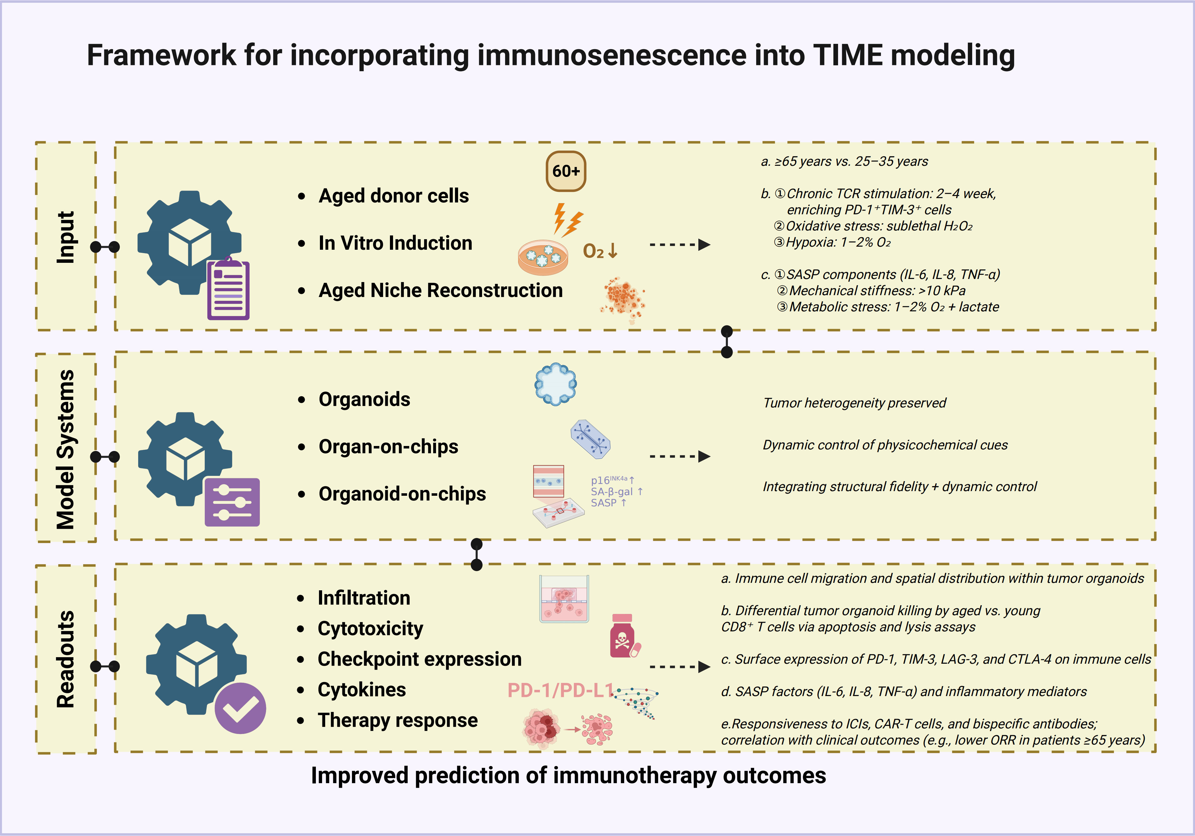
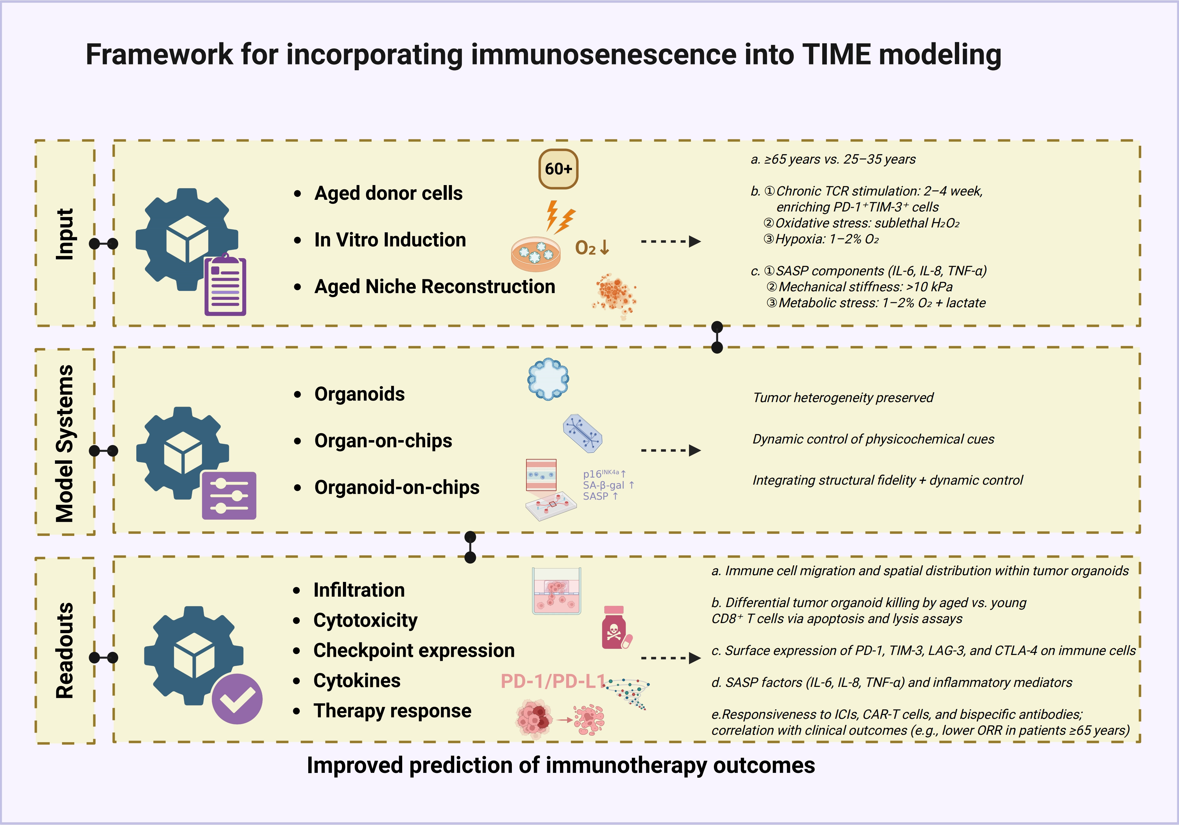
Figure 3. Framework for incorporating immunosenescence into TIME models. Three interconnected parts guide the design, execution, and validation of age-aware TIME models. Part I presents three strategies: aged donor cells, in vitro senescence induction, and aged niche reconstruction. Part II shows their implementation across organoid, organ-on-a-chip, and hybrid platforms. Part III lists five functional readouts (infiltration, cytotoxicity, checkpoint expression, cytokines, and therapy response) to confirm successful integration. Together, these components provide a systematic framework to improve immunotherapy prediction for aging populations. Created in BioRender. Feng, Y. (2026) https://BioRender.com/zn90ecx. TIME: tumor immune microenvironment; TCR: T-cell receptor; PD-1: programmed cell death protein 1; TIM-3: T-cell immunoglobulin and mucin-domain containing-3; SASP: senescence-associated secretory phenotype; IL: interleukin; TNF: tumor necrosis factor; LAG-3: lymphocyte-activation gene 3; CTLA-4: cytotoxic T-lymphocyte-associated protein 4; ICI: immune checkpoint inhibitor; CAR-T: chimeric antigen receptor T-cell.
6. Applications in Immunotherapy Evaluation and Drug Development
Emerging evidence has demonstrated the translational potential of organoid- and chip-based platforms for evaluating immunotherapies across multiple modalities. For immune checkpoint inhibitors (ICIs), Meißner et al. established an autologous patient-derived organoid-PBMC co-culture platform that captures patient-specific tumor-immune interactions and predicts ICI responses in head and neck squamous cell carcinoma[155]. Wang et al. developed a microfluidic patient-derived organotypic tumor spheroid (PDOTS) model for hepatocellular carcinoma that preserves the native tumor-immune microenvironment and enables ex vivo evaluation of immunotherapeutic responses[156]. Esposito et al. constructed a colorectal cancer immunity-organoid interaction platform to model ICI response and identified cancer-specific tissue markers associated with immunotherapy resistance. A gut-on-a-chip platform incorporating patient fecal microbiome has further been applied to predict ICI responses in melanoma[157].
For CAR-T cell therapy, Logun et al. reported a unique phase 1 clinical trial design in which patient-derived glioblastoma organoids (GBOs) were treated concurrently with the same autologous CAR-T cell products as the patients, enabling real-time assessment of CAR-T cell bioactivity[158]. Ma et al. engineered a bioengineered immunocompetent “preclinical trial-on-chip” platform for leukemia that enables real-time spatiotemporal monitoring of CAR-T cell functions and models heterogeneous clinical responses including remission, resistance, and relapse[159]. Wu et al. comprehensively reviewed recent advances in organoid- and organ-on-a-chip-based platforms for the preclinical validation of CAR-T therapies, highlighting that models of solid tumors such as pancreatic ductal adenocarcinoma and glioblastoma show high predictive value for CAR-T clinical outcomes[160].
For bispecific antibodies, patient-derived intestinal organoids and tumouroids supplemented with immune cells have been used to study the on-target, off-tumour toxicities of T-cell-engaging bispecific antibodies, capturing clinical toxicities not predicted by conventional models as well as inter-patient variability in TCB responses[161-163].
For cancer vaccines, Ferreira N et al. developed an integrated organoid-immune co-culture pipeline using pancreatic ductal adenocarcinoma patient-derived organoids and matched HLA immune cells for functional testing of cancer nanovaccines[164]. Organoid platforms have also been applied to investigate tumor-immune cell interactions, aiding in the design and testing of immune-based therapies and vaccines[165].
Despite these promising advances, direct evidence linking immunosenescence-integrated organoid and organ-on-a-chip models to clinical outcomes in elderly populations remains limited. Nevertheless, these platforms offer unique advantages that position them to address this critical gap (Table 3). First, they enable the assessment of immune-related adverse events (irAEs) within a senescent inflammatory background, offering mechanistic insights into why older patients may exhibit distinct toxicity profiles. Second, they facilitate the discovery of novel biomarkers associated with age-related immune exhaustion, supporting the rational design of therapies tailored to the specific immunological context of aging. Third, these platforms provide an unprecedented opportunity for preclinical testing of rejuvenation strategies, such as senolytics or epigenetic reprogramming, aimed at restoring immune potency or bypassing age-associated barriers to infiltration. Collectively, shifting toward immunosenescence-integrated modeling will be instrumental in bridging the translational gap, moving the field closer to truly personalized and age-optimized immunotherapy.
Table 3. Comparison of current TIME modeling platforms and their ability to capture immunosenescence.
| Feature | 2D culture | Organoids | Chips (Organ-on-chips) | Hybrid (Organoid-on-chips) |
| Tumor heterogeneity | - | ++ | ++ | +++ |
| Immune cell integration | + | ++ | +++ | +++ |
| Perfusion/Flow | - | - | ++ | +++ |
| Vascularization | - | + | ++ | ++ |
| ECM/Stroma | - | ++ | + | ++ |
| Aging modeling | - | + | ++ | +++ |
| Long-term culture | - | + | ++ | ++ |
| Throughput | +++ | ++ | + | + |
| Physiological relevance | + | ++ | +++ | ++++ |
Symbols: -: absent/poor; +: limited/basic; ++: moderate/good; +++: strong/very good; ++++: excellent. TIME: tumor immune microenvironment; 2D: two-dimensional; ECM: extracellular matrix.
7. Challenges, Standardization, and Future Perspectives
Despite the conceptual and technical advances outlined above, the integration of immunosenescence into organoid and organ-on-a-chip platforms faces a series of interconnected challenges that extend beyond mere technical optimization. Foremost among these is a fundamental temporal-scale mismatch. Immunosenescence, in physiological reality, unfolds over years to decades, progressively reshaping immune repertoire, functional competence, and intercellular communication networks. Yet the typical culture windows of organoids and OOC platforms, ranging from days to several weeks, fall orders of magnitude short of capturing this chronic trajectory. Although in vitro induction of senescence via chronic TCR stimulation, for example over a period of two to four weeks, or exposure to oxidative stress can accelerate the appearance of senescence-associated phenotypes, these accelerated models remain compressed surrogates rather than faithful recapitulations of the aging process in vivo. The use of aged donor-derived cells provides a cross-sectional snapshot of the endpoint state, but offers no insight into the dynamic path by which that state was reached. This temporal constraint is not a peripheral limitation but a structural bottleneck that challenges the very premise of modeling a chronic, progressive process within inherently acute experimental systems.
This temporal bottleneck manifests in several concrete technical obstacles that current platforms have yet to overcome. First, the functional persistence of aged primary immune cells, particularly T cells and NK cells, under prolonged perfusion is markedly compromised, as these cells exhibit reduced adhesion, survival, and responsiveness to microenvironmental cues. This is not merely a technical inconvenience but reflects the intrinsic biological frailty of senescent cells, which are poorly adapted to the mechanical and metabolic demands of ex vivo maintenance. Second, current vascularized chip models, while sophisticated in their microengineering, remain gross simplifications of the aged vascular niche. They lack the endothelial senescence, glycocalyx degradation, pericyte dysfunction, and chronic low-grade inflammatory tone that collectively define the aging vasculature and profoundly influence immune cell trafficking and effector function. Third, the field faces a persistent standardization dilemma. The same biological complexity that makes immunosenescence a compelling modeling target, including donor heterogeneity, differential aging trajectories across individuals, and context-dependent SASP profiles, also renders cross-study comparison and systematic benchmarking extraordinarily difficult. Unlike oncogenic driver mutations, which can be defined by discrete genetic events, immunosenescence lacks a universally accepted molecular signature. This absence of a common reference point impedes the validation and intercomparison of different model systems.
Addressing these obstacles requires a shift from the pursuit of a single definitive model toward a framework of transparent, stratified reporting and validation. We propose that any claim of age-aware modeling should be accompanied by a minimum dataset that includes donor chronological age and, where feasible, immune age metrics such as the naïve-to-memory T-cell ratio. Quantitative senescence markers, for example SA-β-gal activity and p16INK4a expression levels, should be reported alongside core SASP factors in culture supernatants such as IL-6, IL-8, and TNF-α, as well as telomere length or attrition rate. These parameters should be presented together with the specific culture conditions under which they were measured, such as perfusion rate, oxygen tension, matrix stiffness, and cytokine supplementation. These variables profoundly influence senescence phenotypes and can confound cross-laboratory comparisons if left unreported. We emphasize that these reporting criteria are not intended as a rigid checklist. They are proposed as a means of enabling meaningful comparison across models, recognizing that different experimental questions may legitimately demand different degrees and modes of senescence integration. Beyond chronological age, emerging multi-omics immune aging clocks offer orthogonal metrics to benchmark the senescence burden of in vitro models, thereby providing a quantitative framework for cross-experimental comparison and strengthening the translational relevance of age-aware TIME modeling[166-168].
Looking beyond these hurdles, three interrelated trajectories will define the path forward. First, computational approaches integrating multi-omics data can predict immune aging trajectories and guide experimental design. Second, hybrid organoid-chip platforms offer a practical framework for validating these predictions under controlled biophysical and biochemical conditions. Third, rigorous clinical correlation, including directly comparing platform-based predictions with outcomes in elderly patient cohorts, will be essential to establish translational value.
8. Conclusions
As organoid and organ-on-a-chip technologies continue to mature, the incorporation of immunosenescence is no longer an optional enhancement but a functional necessity. By treating immune aging as a controllable experimental variable, we can bridge the long-standing gap between preclinical models and the clinical reality of cancer patients. This paradigm shift will be instrumental in the next era of precision oncology, enabling the rational design of effective and safe immunotherapies for an aging global population.
Acknowledgements
During the preparation of this manuscript, the authors used DeepSeek-V3 tools solely for language editing and proofreading (grammar, spelling, and readability improvement). No AI tools were used for data analysis, figure generation, or conceptual design. The authors reviewed and edited all AI-assisted outputs and take full responsibility for the content of this publication.
Authors contribution
Feng Y, Wang L: Conceptualization, writing-original draft, visualization.
Li D, Mu C: Writing-review & editing.
Wei L, Ma B: Conceptualization, supervision, funding acquisition, writing-review & editing.
Conflicts of interest
The authors declare no conflicts of interest.
Ethical approval
Not applicable.
Consent to participate
Not applicable.
Consent for publication
Not applicable.
Availability of data and materials
Not applicable.
Funding
This study was supported by the Major Program of the Natural Science Foundation of the Jiangsu Higher Education Institutions of China (Grant No. 24KJA310009).
Copyright
© The Author(s) 2026.
References
-
14. Ronaldson-Bouchard K, Teles D, Yeager K, Tavakol DN, Zhao Y, Chramiec A, et al. A multi-organ chip with matured tissue niches linked by vascular flow. Nat Biomed Eng. 2022;6(4):351-371.[DOI]
-
16. Perucca A, Llonín AG, Benach OM, Hallopeau C, Rivas EI, Linares J, et al. Micro Immune Response On-chip (MIRO) models the tumour-stroma interface for immunotherapy testing. Nat Commun. 2025;16:1279.[DOI]
-
17. Yang M, Ma B, Shao H, Clark AM, Wells A. Macrophage phenotypic subtypes diametrically regulate epithelial-mesenchymal plasticity in breast cancer cells. BMC Cancer. 2016;16(1):419.[DOI]
-
26. Kim DM, Lee JH, Pan Q, Han HW, Shen Z, Eshghjoo S, et al. Nutrient-sensing growth hormone secretagogue receptor in macrophage programming and meta-inflammation. Mol Metab. 2024;79:101852.[DOI]
-
32. Ajoolabady A, Pratico D, Tang D, Zhou S, Franceschi C, Ren J. Immunosenescence and inflammaging: Mechanisms and role in diseases. Ageing Res Rev. 2024;101:102540.[DOI]
-
36. Neal JT, Li X, Zhu J, Giangarra V, Grzeskowiak CL, Ju J, et al. Organoid modeling of the tumor immune microenvironment. Cell. 2018;175(7):1972-1988.e16.[DOI]
-
39. Camacho-Gomez D, Movilla N, Borau C, Martin A, Oñate Salafranca C, Pardo J, et al. An agent-based method to estimate 3D cell migration trajectories from 2D measurements: Quantifying and comparing T vs CAR-T 3D cell migration. Comput Methods Programs Biomed. 2024;255:108331.[DOI]
-
41. Jung JH, Yang Y, Kim Y. cPLA2α-driven Cox-2/PGE2 axis promotes hypoxia-induced senescence in Jurkat T cells. Biochem Biophys Res Commun. 2025;790:152894.[DOI]
-
43. Ferrara R, Naigeon M, Auclin E, Duchemann B, Cassard L, Jouniaux JM, et al. Circulating T-cell immunosenescence in patients with advanced non-small cell lung cancer treated with single-agent PD-1/PD-L1 inhibitors or platinum-based chemotherapy. Clin Cancer Res. 2021;27(2):492-503.
-
44. Terekhova M, Swain A, Bohacova P, Aladyeva E, Arthur L, Laha A, et al. Single-cell atlas of healthy human blood unveils age-related loss of NKG2C+GZMB-CD8+ memory T cells and accumulation of type 2 memory T cells. Immunity. 2023;56(12):2836-2854.e9.[DOI]
-
51. Collison LW, Vignali DAA. In vitro Treg suppression assays. In: Kassiotis G, Liston A, editors. Regulatory T cells. Totowa (NJ): Humana Press; 2011. p. 21-37.[DOI]
-
58. Frasca D, Diaz A, Romero M, Blomberg BB. Leptin induces immunosenescence in human B cells. Cell Immunol. 2020;348:103994.[DOI]
-
64. Sridharan R, Cavanagh B, Cameron AR, Kelly DJ, O’Brien FJ. Material stiffness influences the polarization state, function and migration mode of macrophages. Acta Biomater. 2019;89:47-59.[DOI]
-
66. Marigo I, Bosio E, Solito S, Mesa C, Fernandez A, Dolcetti L, et al. Tumor-induced tolerance and immune suppression depend on the C/EBPβ transcription factor. Immunity. 2010;32(6):790-802.[DOI]
-
78. Pereira BI, Devine OP, Vukmanovic-Stejic M, Chambers ES, Subramanian P, Patel N, et al. Senescent cells evade immune clearance via HLA-E-mediated NK and CD8+ T cell inhibition. Nat Commun. 2019;10:2387.[DOI]
-
82. Zhang D, Chen G, Manwani D, Mortha A, Xu C, Faith JJ, et al. Neutrophil ageing is regulated by the microbiome. Nature. 2015;525(7570):528-532.[DOI]
-
84. Gorgoulis V, Adams PD, Alimonti A, Bennett DC, Bischof O, Bishop C, et al. Cellular senescence: Defining a path forward. Cell. 2019;179(4):813-827.[DOI]
-
91. Assouline B, Kahn R, Hodali L, Condiotti R, Engel Y, Elyada E, et al. Senescent cancer-associated fibroblasts in pancreatic adenocarcinoma restrict CD8+ T cell activation and limit responsiveness to immunotherapy in mice. Nat Commun. 2024;15:6162.[DOI]
-
97. Zhang J, Li J, Hou Y, Lin Y, Zhao H, Shi Y, et al. Osr2 functions as a biomechanical checkpoint to aggravate CD8+ T cell exhaustion in tumor. Cell. 2024;187(13):3409-3426.e24.[DOI]
-
103. Yan HHN, Chan AS, Lai FPL, Leung SY. Organoid cultures for cancer modeling. Cell Stem Cell. 2023;30(7):917-937.[DOI]
-
109. Peng T, Ma X, Hua W, Wang C, Chu Y, Sun M, et al. Individualized patient tumor organoids faithfully preserve human brain tumor ecosystems and predict patient response to therapy. Cell Stem Cell. 2025;32(4):652-669.e11.[DOI]
-
113. Pieraccioli M, Ciucci A, Corti C, Mastrantonio R, Scarpone EK, Cesari E, et al. Single-cell transcriptome analysis of patient-derived organoids captures inter- and intratumor heterogeneity and uncovers targetable pathways in high grade serous ovarian cancer. Drug Resist Updat. 2026;85:101354.[DOI]
-
114. Song H, Weinstein HNW, Allegakoen P, Wadsworth MH, Xie J, Yang H, et al. Single-cell analysis of human primary prostate cancer reveals the heterogeneity of tumor-associated epithelial cell states. Nat Commun. 2022;13:141.[DOI]
-
116. Qin Z, Zhang H, Li Y, Yang J, Liu H, Guan Z, et al. Transcriptome profiling of tumor-infiltrating lymphocyte-mediated cytotoxicity against patient-derived lung cancer organoids. Commun Biol. 2025;8:1835.[DOI]
-
117. Zhao Z, Zhang S, Jiang N, Zhu W, Song D, Liu S, et al. Patient-derived immunocompetent tumor organoids: A platform for chemotherapy evaluation in the context of T-cell recognition. Angew Chem Int Ed. 2024;63(9):e202317613.[DOI]
-
120. Li K, Liu C, Sui X, Li C, Zhang T, Zhao T, et al. An organoid co-culture model for probing systemic anti-tumor immunity in lung cancer. Cell Stem Cell. 2025;32(8):1218-1234.e7.[DOI]
-
121. Rashidan K, Ayadilord M, Hazrati A, Nazerian A, Shafiee A, Hashemi SM. Immune and immune-integrated organoids as NextGeneration platforms for disease modeling. MedComm. 2025;6(12):e70531.[DOI]
-
123. Wang Q, Yuan F, Zuo X, Li M. Breakthroughs and challenges of organoid models for assessing cancer immunotherapy: A cutting-edge tool for advancing personalised treatments. Cell Death Discov. 2025;11:222.[DOI]
-
127. Gao Q, Wang J, Zhang H, Wang J, Jing Y, Su J. Organoid vascularization: Strategies and applications. Adv Healthc Mater. 2025;14(20):2500301.[DOI]
-
128. Maiullari F, Ceraolo MG, Presutti D, Fratini N, Galbiati M, Fasciani A, et al. Modeling breast cancer dynamics through modulable small Vessel Environment Bioreactor (sVEB). Biomaterials. 2025;323:123441.[DOI]
-
129. Du Y, Wang YR, Bao QY, Xu XX, Xu C, Wang S, et al. Personalized vascularized tumor organoid-on-a-chip for tumor metastasis and therapeutic targeting assessment. Adv Mater. 2025;37(6):2412815.[DOI]
-
130. Quintard C, Tubbs E, Jonsson G, Jiao J, Wang J, Werschler N, et al. A microfluidic platform integrating functional vascularized organoids-on-chip. Nat Commun. 2024;15:1452.[DOI]
-
134. Zhao L, Zhao G, Fu J, Liu Y, Feng J, Zhang G, et al. Programmed cell death protein 1 engagement impairs cytoskeletal forces and nuclear mechanotransduction in T cells. ACS Nano. 2025;19(46):39667-39681.[DOI]
-
135. Thyagarajan B, Faul J, Vivek S, Kim JK, Nikolich-Žugich J, Weir D, et al. Age-related differences in T-cell subsets in a nationally representative sample of people older than age 55: Findings from the health and retirement study. J Gerontol A Biol Sci Med Sci. 2022;77(5):927-933.
-
145. Duan J, Duan J, Zhang Z, Tong T. Irreversible cellular senescence induced by prolonged exposure to H2O2 involves DNA-damage-and-repair genes and telomere shortening. Int J Biochem Cell Biol. 2005;37(7):1407-1420.[DOI]
-
146. Bannoud N, Dalotto-Moreno T, Kindgard L, García PA, Blidner AG, Mariño KV, et al. Hypoxia supports differentiation of terminally exhausted CD8 T cells. Front Immunol. 2021;12:660944.[DOI]
-
147. Tchkonia T, Yi Z, van Deursen J, Campisi J, Kirkland JL. Cellular senescence and the senescent secretory phenotype: Therapeutic opportunities. J Clin Invest. 2013;123(3):966-972.[DOI]
-
153. Yao J, Li S, Bai L, Chen J, Ren C, Liu T, et al. Efficacy and safety of immune checkpoint inhibitors in elderly patients with advanced non-small cell lung cancer: A systematic review and meta-analysis. eClinicalMedicine. 2025;81:103081.[DOI]
-
157. Esposito A, Agostini A, Quero G, Piro G, Priori L, Caggiano A, et al. Colorectal cancer patients-derived immunity-organoid platform unveils cancer-specific tissue markers associated with immunotherapy resistance. Cell Death Dis. 2024;15:878.[DOI]
-
160. Wu T, Wu D, Zeng H. Insights into the efficacy and safety of CAR-T therapies using organoids and organs-on-chips. TrAC Trends Anal Chem. 2026;200:118827.[DOI]
-
163. Liu Y, Wang C, Chen G, Chen J, Chen W, Lei K, et al. Patient derived cancer organoids model the response to HER2-CD3 bispecific antibody (BsAbHER2) generated from hydroxyapatite gene delivery system. Cancer Lett. 2024;597:217043.[DOI]
-
164. Ferreira N, Agorku D, Rosa A, Roosz J, Christ L, Anderle N, et al. Advanced human immune cell-organoid co-cultures for functional testing of cancer nanovaccines. Adv Sci. 2026;13(10):e15199.[DOI]
Copyright
© The Author(s) 2027. This is an Open Access article licensed under a Creative Commons Attribution 4.0 International License (https://creativecommons.org/licenses/by/4.0/), which permits unrestricted use, sharing, adaptation, distribution and reproduction in any medium or format, for any purpose, even commercially, as long as you give appropriate credit to the original author(s) and the source, provide a link to the Creative Commons license, and indicate if changes were made.
Publisher’s Note
Science Exploration remains a neutral stance on jurisdictional claims in published
maps
and institutional affiliations. The views expressed in this article are solely those
of
the author(s) and do not reflect the opinions of the Editors or the publisher.
Share And Cite


Science Exploration Style
Feng Y, Wang L, Li D, Mu C, Wei L, Ma B. Organoid and organ-on-a-chip models for the tumor immune microenvironment: A call to incorporate immunosenescence. Ageing Cancer Res Treat. 2027;4:202625. https://doi.org/10.70401/acrt.2026.0039
Tips
Copy completed.
Submit a Manuscript
Author Instructions
Cite this Article
Article Metrics
0
View
0
Download
Cited
Article Updates

- Abstract
- Keywords
- 1. Introduction
- 2. Cellular, Stromal, and Biophysical Components of the TIME
- 3. Organoid-Based Models for Tumor-Immune Interactions
- 4. Organ-on-a-Chip Platforms for Modeling the TIME
- 5. Advanced Modeling Dimensions of the TIME
- 6. Applications in Immunotherapy Evaluation and Drug Development
- 7. Challenges, Standardization, and Future Perspectives
- 8. Conclusions
- Acknowledgements
- Authors contribution
- Conflicts of interest
- Ethical approval
- Consent to participate
- Consent for publication
- Availability of data and materials
- Funding
- References
- Copyright
 Get citation
Science Exploration Style

Feng Y, Wang L, Li D, Mu C, Wei L, Ma B. Organoid and organ-on-a-chip models for the tumor immune microenvironment: A call to incorporate immunosenescence. Ageing Cancer Res Treat. 2027;4:202625. https://doi.org/10.70401/acrt.2026.0039
copy
Share Link
copy