Radical-mediated C7 cholesterol oxidation during erastin-induced ferroptosis

Yuta Akiba
1,#
,
Kotoko Arisawa
1,#

,
Takayuki Kaneko
1
,
Kei Ishida
1
,
Emiko Sato
2
,
Takashi Toyama
1
,
Yoshiro Saito
1,*
*Correspondence to:
Yoshiro Saito, Laboratory of Molecular Biology and Metabolism, Graduate School of Pharmaceutical Sciences, Tohoku University, Sendai, Miyagi 980-8578, Japan.
E-mail: Yoshiro.saito.a8@tohoku.ac.jp
EXO. 2026;1:202629. 10.70401/EXO.2026.0022
Received: August 17, 2026Accepted: October 28, 2026Published: October 28, 2026
Abstract
Aims: Ferroptosis is driven by iron-dependent lipid peroxidation, but whether cholesterol undergoes radical-mediated oxidation during this process remains incompletely understood. We examined the temporal relationship between lipid peroxidation, cell death, and C7 cholesterol oxidation during erastin-induced ferroptosis.
Methods: Human SH-SY5Y cells were treated with erastin. Cell viability, lipid hydroperoxides, and lipid radicals were assessed using Alamar Blue, Liperfluo, and LipiRADICAL, respectively. Total 7α-hydroxycholesterol (t7α-OHCh), total 7β-hydroxycholesterol (t7β-OHCh), and 7-ketocholesterol (7-ketoCh) were quantified by gas chromatography–mass spectrometry (GC–MS) after triphenylphosphine reduction and saponification.
Results: Erastin-induced cell death was suppressed by deferoxamine, ferrostatin-1, and α-tocopherol. Liperfluo fluorescence increased before detectable LipiRADICAL fluorescence and loss of cell viability. t7α-OHCh increased during the early phase, whereas t7β-OHCh increased later; no statistically significant change in 7-ketoCh was detected. Because t7α-OHCh and t7β-OHCh represent composite pools of pre-existing free and esterified hydroxycholesterols and hydroperoxide-derived products, the early t7α-OHCh signal cannot distinguish enzymatic hydroxylation from a 7α-hydroperoxycholesterol (7α-OOHCh)-derived product. The increase in t7β-OHCh together with the overall C7 stereochemical profile is consistent with radical-mediated C7 cholesterol oxidation.
Conclusion: These findings support radical-mediated C7 cholesterol oxidation as an early component of erastin-induced ferroptotic lipid damage. The biological activity and extracellular transfer of these products remain to be established.
Keywords
Ferroptosis, cholesterol oxidation, lipid hydroperoxide, 7-hydroxycholesterol, oxysterol
1. Introduction
Ferroptosis is an iron-dependent form of regulated cell death driven by the uncontrolled accumulation of lipid hydroperoxides in cellular membranes[1,2]. Erastin induces ferroptosis by inhibiting the cystine/glutamate antiporter system, thereby limiting glutathione synthesis and impairing the lipid hydroperoxide-reducing activity of glutathione peroxidase 4 (GPX4)[3,4]. GPX4 is a selenium-containing enzyme that directly reduces membrane lipid hydroperoxides, and the incorporation of selenocysteine into GPX4 is critical for protection against hydroperoxide-induced ferroptosis[3-5]. Selenium-dependent antioxidant systems therefore provide an important link between the control of lipid hydroperoxides and ferroptotic cell death.
Most mechanistic studies of ferroptosis have focused on the oxidation of polyunsaturated fatty acid-containing phospholipids[6,7]. However, cholesterol (Ch) is another major membrane lipid and is also susceptible to free-radical oxidation[8,9]. We previously demonstrated enhanced lipid peroxidation in oxidatively stressed cells, including selenium-deficient Jurkat cells and glutamate-treated neuronal cells[10,11]. In addition, C7 Ch oxidation products, including products derived from hydroperoxycholesterol (Ch-OOH), accumulated in selenium-deficient and radical initiator-treated cells[10,12], suggesting that these products can serve as sensitive indicators of cellular lipid peroxidation. Ch abundance and cholesterol-dependent remodeling of mevalonate-pathway flux have been shown to influence cellular susceptibility to ferroptosis[13], and 7-ketocholesterol (7-ketoCh) has been detected in ferroptotic cells[14]. Nevertheless, the temporal and stereochemical profiles of Ch oxidation during ferroptosis remain insufficiently characterized, and it remains unclear whether these products could be used as in vivo indicators of ferroptosis-associated lipid oxidation. Ferroptotic lipid peroxidation is spatially organized across intracellular membranes, and the relative contributions of individual organelles may vary depending on the cellular and experimental context[15,16]. Because Ch is enriched in the plasma membrane but is continuously trafficked through endosomal and lysosomal compartments, the subcellular localization of early lipid hydroperoxide accumulation may be relevant to understanding which membrane lipid pools are susceptible to oxidation[17,18].
Because Ch hydroperoxides are chemically labile and difficult to quantify directly, they were reduced during sample preparation and analyzed as their more stable hydroxy derivatives. Following reduction and saponification, the measured total 7α- and 7β-hydroxycholesterol (t7α- and t7β-OHCh) include free and esterified OHChs together with the corresponding 7-hydroperoxide forms (7-OOHCh)[8,9,19]. Thus, this approach does not directly distinguish the initial hydroperoxides from pre-existing OHChs but permits inference of possible oxidation pathways from the stereochemical C7 product profile. Non-enzymatic free-radical oxidation of Ch yields both 7α- and 7β-products[20], whereas CYP7A1 catalyzes the regio- and stereoselective formation of 7α-OHCh[21]. Accordingly, concomitant increases in the two epimers are consistent with radical-mediated Ch oxidation, whereas preferential or earlier accumulation of the 7α-isomer may suggest an additional enzymatic contribution. The product profile alone, however, cannot establish the involvement of a specific enzyme.
Ferroptosis is increasingly recognized as a non-cell-autonomous process. Cadherin-mediated cell–cell adhesion can regulate ferroptosis sensitivity through NF2–YAP signaling, while lipid peroxidation can propagate between closely apposed plasma membranes of neighboring cells[22,23]. Whether cholesterol oxidation products participate in such intercellular processes remains unknown.
In the present study, we characterized the temporal progression of lipid peroxidation during erastin-induced ferroptosis in SH-SY5Y cells by monitoring cell viability, lipid hydroperoxides, and lipid radicals. We further examined the intracellular lipid hydroperoxides and quantified C7 Ch oxidation products by gas chromatography–mass spectrometry (GC–MS) after reduction and saponification, using their temporal and stereochemical profiles to infer possible oxidation pathways.
2. Methods
2.1 Cell culture and treatments
Human SH-SY5Y neuroblastoma cells were cultured in high-glucose Dulbecco’s modified Eagle’s medium (DMEM; Nacalai Tesque) supplemented with 10% heat-inactivated fetal bovine serum (Sigma-Aldrich) and 1% penicillin-streptomycin at 37 °C in a humidified atmosphere containing 5% CO2. Cells were seeded 24 h before treatment. Ferroptosis was induced using erastin (10 μM; Sigma-Aldrich). Apoptosis was induced using staurosporine (100 nM, FUJIFILM Wako Pure Chemical), whereas necroptosis was induced using a combination of tumor necrosis factor-α, cycloheximide, and Z-VAD-FMK (TCZ). For TCZ treatment, cells were pretreated with cycloheximide (10 μg/mL) and Z-VAD-FMK (25 μM) for 30 min, followed by tumor necrosis factor-α (50 ng/mL). Where indicated, cells were treated with deferoxamine, necrostatin-1, Z-VAD-FMK, ferrostatin-1, or α-tocopherol.
2.2 Cell viability assay
Cells were seeded in 96-well plates at 2.0 × 10⁴ cells in 100 μL per well. At the indicated times after treatment, the medium was replaced with DMEM containing 10% Alamar Blue Cell Viability Reagent (Invitrogen) and 10% fetal bovine serum. After incubation for 60 min at 37 °C, fluorescence was measured at excitation and emission wavelengths of 544 and 585 nm, respectively, using a SpectraMax iD5 microplate reader (Molecular Devices). Cell viability was expressed relative to that of the corresponding untreated control cells.
2.3 Measurement of lipid hydroperoxides and lipid radicals
Intracellular lipid hydroperoxides were measured using Liperfluo (Dojindo Laboratories). Cells were seeded in 96-well plates at 2.0 × 10⁴ cells per well and treated as indicated. At each measurement time, the medium was replaced with DMEM containing 20 μM Liperfluo and 10% fetal bovine serum. After incubation for 30 min at 37 °C, cells were washed twice with phosphate-buffered saline (PBS), and fluorescence (Ex 480 nm, Em 580 nm) was measured using a SpectraMax iD5 microplate reader. Nine points were measured per well using bottom-read mode.
Lipid radicals were detected using LipiRADICAL (Funakoshi). Cells were incubated with 1 μM LipiRADICAL for 20 min before treatment with erastin. At the indicated times, cells were washed twice with PBS, and fluorescence was measured at excitation and emission wavelengths of 470 and 540 nm, respectively, using the microplate reader.
2.4 Fluorescence microscopy
For visualization of intracellular lipid hydroperoxides, SH-SY5Y cells were seeded at 5 × 10⁴ cells per well on poly-D-lysine-coated coverslips in 12-well plates. At the indicated times after treatment with erastin (10 μM), cells were incubated with Liperfluo (20 μM) for 30 min at 37 °C, washed twice with PBS, and fixed with 4% paraformaldehyde for 10 min at room temperature. Cells were mounted using a 4',6-diamidino-2-phenylindole (DAPI)-containing mounting medium and observed using an FV1000 confocal laser-scanning microscope (Olympus).
2.5 Extraction and GC-MS analysis of C7 cholesterol oxidation products
Ch oxidation products were measured as described previously[19]. SH-SY5Y cells were seeded in six-well plates and treated with erastin (10 μM) for the indicated periods. Cells were washed twice with PBS, collected using a cell scraper, and divided into aliquots for lipid extraction and protein quantification. Lipids were extracted overnight at -30 °C under nitrogen using methanol containing butylated hydroxytoluene (50 mg/L) and chloroform containing triphenylphosphine (5 g/L). These low-temperature, antioxidant-containing, and nitrogen-protected conditions were used to minimize artifactual cholesterol oxidation during sample preparation. A 10-ng aliquot of 19-OHCh was added as an internal standard. Following addition of 0.9% KCl, the lower organic phase was collected and evaporated under nitrogen. Samples were saponified in methanolic KOH for 30 min at 37 °C and extracted with chloroform/ethyl acetate (4:1, v/v) after addition of 10% acetic acid. Oxysterols were purified using silica solid-phase extraction. The cartridges were washed with 0.5% 2-propanol in hexane, and oxysterols were eluted with 30% 2-propanol in hexane. The eluates were evaporated under nitrogen and derivatized with 100 μL of N-methyl-N-trimethylsilyl trifluoroacetamide for 30 min at 37 °C. Oxysterols were analyzed using a GCMS-QP2010 Plus gas chromatograph-mass spectrometer (Shimadzu) equipped with a BPX5 column (Trajan Scientific and Medical). Helium was used as the carrier gas at a flow rate of 1.14 mL/min. The oven temperature was maintained at 60 °C for 2 min, increased to 330 °C at 15 °C/min, and maintained at 330 °C for 5 min. The ion-source and interface temperatures were 200 °C and 280 °C, respectively. Electron-impact ionization and selected-ion monitoring were used for detection. Total cellular 7α-OHCh, 7β-OHCh, and 7-ketoCh were quantified using the internal-standard method and normalized to cellular protein content. Hereafter, t7α-OHCh and t7β-OHCh denote composite pools comprising pre-existing free and esterified 7-OHCh together with the corresponding 7-OOHCh-derived products generated by triphenylphosphine reduction. The reported 7-ketoCh value includes free and esterified 7-ketoCh recovered after saponification but does not include a hydroperoxide-derived component.
2.6 Statistical analysis
Statistical analyses were performed using GraphPad Prism version 10. Data are presented as the mean ± standard deviation from three experiments unless otherwise stated. Comparisons with the corresponding control or 0-h group were performed using one-way analysis of variance followed by Dunnett’s multiple-comparisons test, as specified in the figure legends. A value of P < 0.05 was considered statistically significant. Unless otherwise indicated, all experiments were performed independently three times, and n = 3 represents three independent biological experiments.
3. Results
3.1 Early accumulation of lipid hydroperoxides precedes detectable lipid radical accumulation during erastin-induced ferroptosis
To determine whether lipid hydroperoxide accumulation was selectively associated with ferroptosis rather than being a general consequence of cell death, SH-SY5Y cells were exposed to different regulated cell death stimuli under conditions producing a partial loss of cell viability over 24 h. Staurosporine, TCZ, and erastin were used to induce apoptosis, necroptosis, and ferroptosis, respectively. Cell viability remained largely unchanged in untreated control cells, whereas each treatment caused a time-dependent decrease in viability, with TCZ producing the most pronounced reduction at 24 h (Figure 1a). The erastin-induced decrease in cell viability was suppressed by deferoxamine, ferrostatin-1, and α-tocopherol, but not by Z-VAD-FMK or necrostatin-1, supporting the involvement of ferroptotic cell death (Figures S1-S3). α-Tocopherol did not significantly affect the Alamar Blue signal in cells not treated with erastin at any concentration tested, although small, nonsignificant elevations in the mean values were observed.
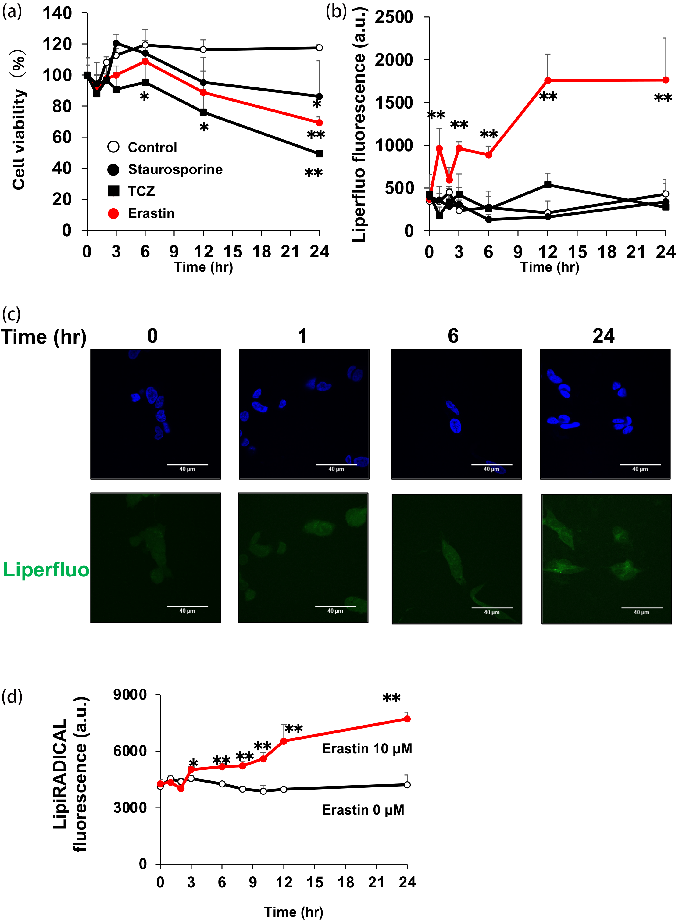
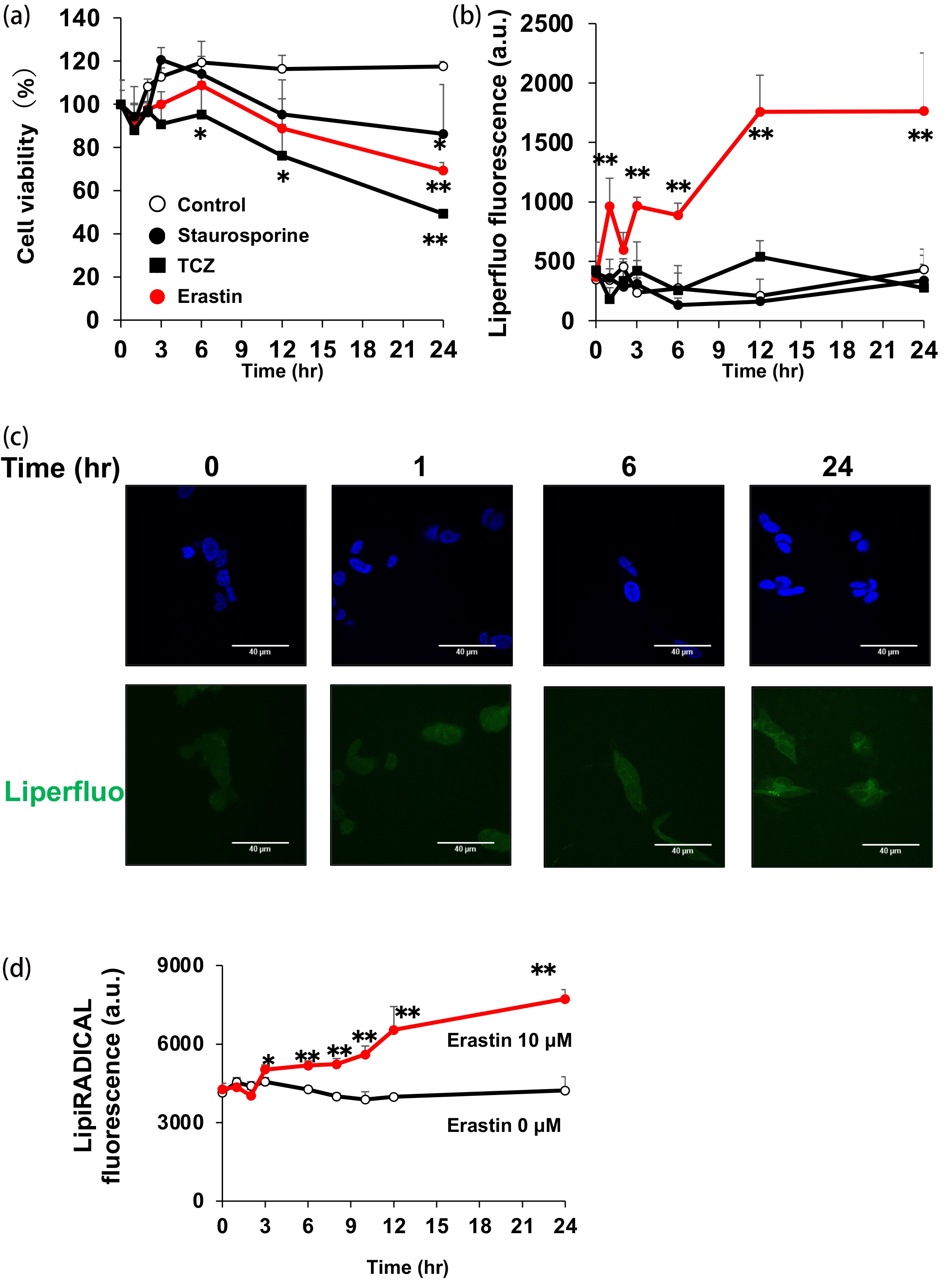
Figure 1. Selective early accumulation of lipid hydroperoxides precedes detectable lipid radical accumulation during erastin-induced ferroptosis. (a, b) SH-SY5Y cells were treated with staurosporine (100 nM), TNF-α (50 ng/mL)/cycloheximide (10 μg/mL)/Z-VAD-FMK (25 μM) (TCZ), or erastin (10 μM). For TCZ treatment, cells were pretreated with cycloheximide and Z-VAD-FMK for 30 min before the addition of TNF-α. Cell viability (a) and lipid hydroperoxide accumulation detected using Liperfluo (b) were measured at the indicated times; (c) Representative images of DAPI (blue) and Liperfluo (green) staining after erastin treatment. Scale bars, 40 μm; (d) LipiRADICAL fluorescence in cells treated with vehicle or erastin (10 μM). Data are presented as the mean ± SD (n = 3 independent biological experiments). *P < 0.05 and **P < 0.01 versus 0 h by one-way ANOVA followed by Dunnett’s test. TNF-α: tumor necrosis factor alpha; DAPI: 4',6-diamidino-2-phenylindole.
We next measured intracellular lipid hydroperoxide accumulation using Liperfluo. Despite the induction of cell death by staurosporine and TCZ, neither treatment caused a marked increase in Liperfluo fluorescence (Figure 1b). In contrast, erastin induced a rapid and sustained increase in Liperfluo fluorescence, which was detectable as early as 1 h after treatment, when cell viability was still preserved (Figure 1b). Notably, TCZ caused a greater loss of cell viability than erastin at 24 h without inducing a comparable increase in Liperfluo fluorescence. These findings indicate that lipid hydroperoxide accumulation was not simply a secondary consequence of reduced cell viability but was selectively associated with erastin-induced ferroptosis. Fluorescence microscopy further confirmed the time-dependent increase in intracellular Liperfluo fluorescence following erastin treatment (Figure 1c).
We subsequently monitored lipid radical accumulation using LipiRADICAL. Whereas Liperfluo fluorescence increased within 1 h, the LipiRADICAL signal remained near its basal level during the earliest phase and began to increase after approximately 3 h, followed by a progressive increase up to 24 h (Figure 1d). Thus, under our experimental conditions, the detectable accumulation of lipid hydroperoxides preceded both the increase in the lipid radical signal and the loss of cell viability. These results identify lipid hydroperoxide accumulation as an early and selective oxidative event during erastin-induced ferroptosis.
3.2 Radical-mediated C7 cholesterol oxidation occurs during erastin-induced ferroptosis
Liperfluo detects lipid hydroperoxides but does not identify the oxidized lipid species. We therefore examined the time-dependent accumulation of the C7 cholesterol oxidation products targeted by our analytical method after erastin treatment. Radical-mediated Ch oxidation can generate both 7α- and 7β-OHCh, as well as 7-ketoCh, whereas CYP7A1 catalyzes the regioselective formation of 7α-OHCh (Figure 2a).
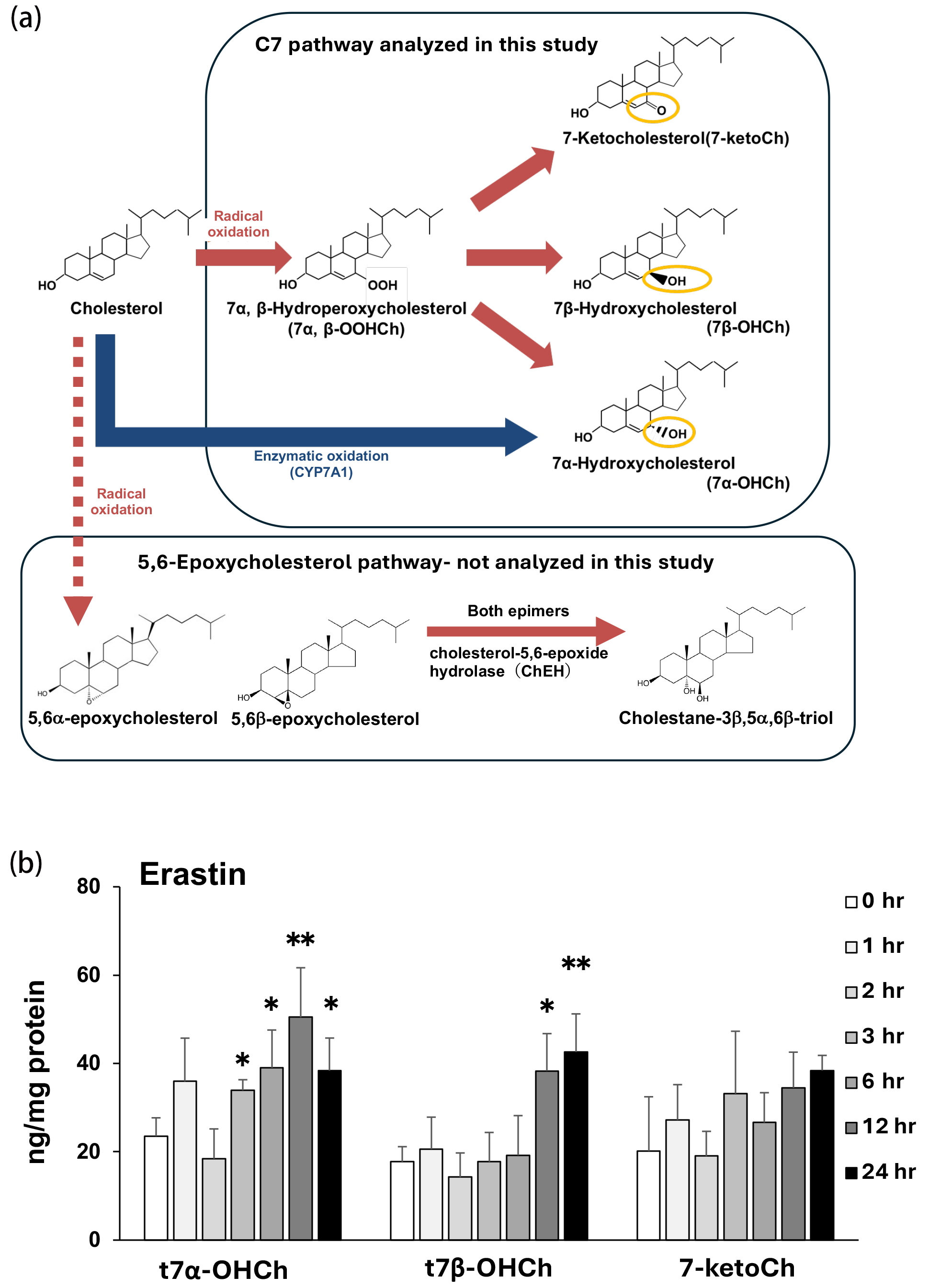
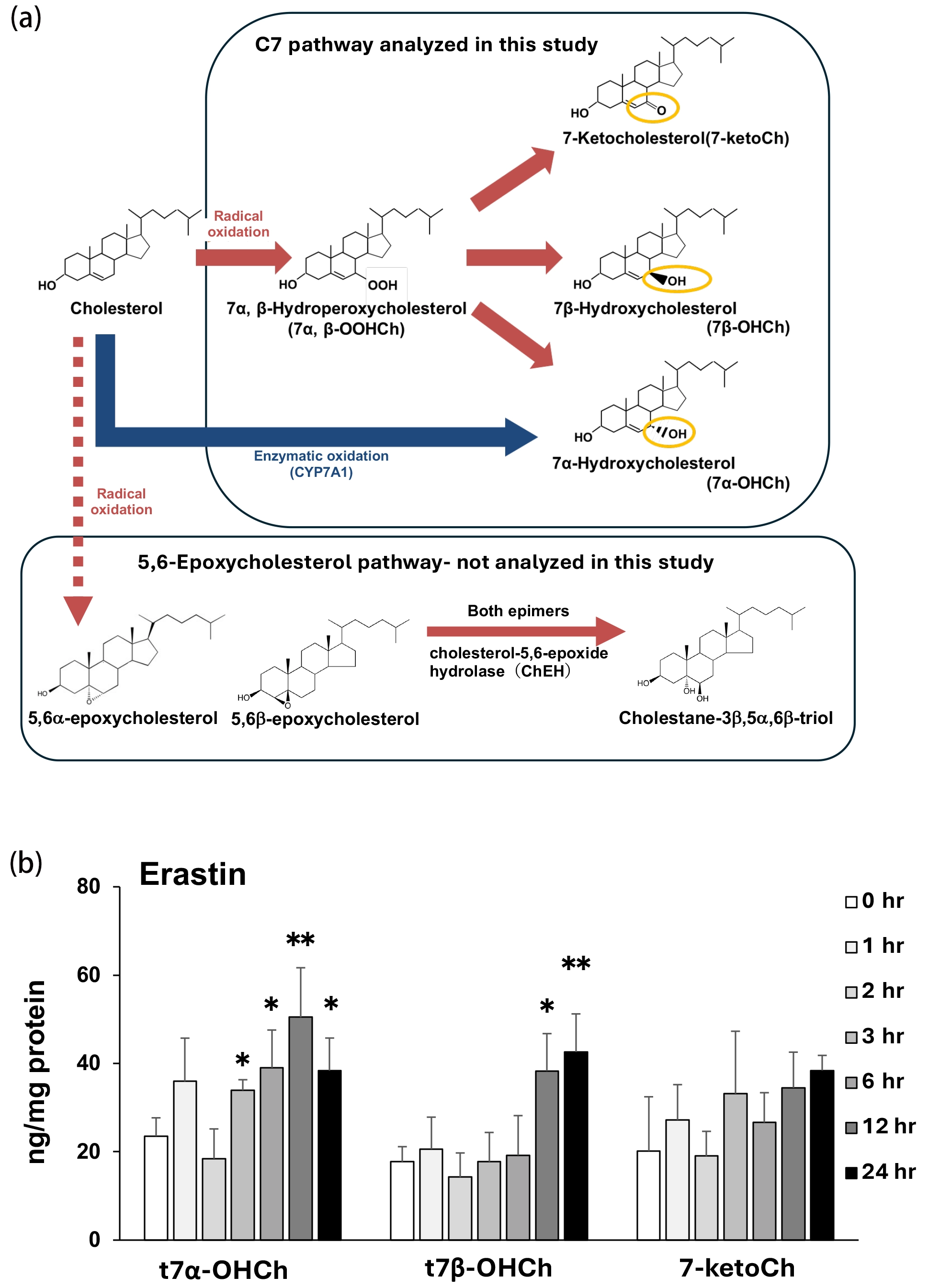
Figure 2. Radical-mediated C7 cholesterol oxidation during erastin-induced ferroptosis. (a) Simplified scheme of the C7 cholesterol oxidation pathway analyzed in this study and the 5,6-epoxycholesterol pathway not examined in the present study. Upper box indicates the C7 products quantified by GC–MS. Radical oxidation generates C7 cholesterol hydroperoxides and both 7α-OHCh and 7β-OHCh, whereas CYP7A1-mediated oxidation selectively generates 7α-OHCh. The conversion of 5,6α- and 5,6β-epoxycholesterol to cholestane-3β,5α,6β-triol by ChEH is shown for context but was not analyzed. This simplified scheme is not intended to provide a comprehensive representation of cholesterol oxidation; (b) Cellular levels of total 7α-OHCh, total 7β-OHCh, and 7-ketoCh after treatment with erastin (10 μM) for the indicated periods were quantified by GC–MS and normalized to cellular protein content. t7α-OHCh and t7β-OHCh represent composite pools measured after triphenylphosphine reduction and saponification. Data are presented as the mean ± SD (n = 3 independent biological experiments). *P < 0.05 and **P < 0.01 versus 0 h; Dunnett’s test. GC–MS: gas chromatography–mass spectrometry; 7α-OHCh: 7α-hydroxycholesterol; 7β-OHCh: 7β-hydroxycholesterol; t7α-OHCh: total 7α-hydroxycholesterol; t7β-OHCh: total 7β-hydroxycholesterol; ChEH: cholesterol-5,6-epoxide hydrolase.
GC–MS analysis with reduction and saponification revealed time-dependent changes in cellular Ch oxidation products after erastin treatment (Figure 2b). The level of t7α-OHCh increased during the early phase and was significantly elevated at several time points from 3 h onward. In contrast, t7β-OHCh increased during the later phase, with significant elevations observed at 12 and 24 h. No statistically significant change in 7-ketoCh was detected (Figure 2b). Because t7α-OHCh may include an enzymatically generated component, its increase alone cannot be attributed exclusively to radical-mediated oxidation. Instead, the increase in t7β-OHCh, together with the overall C7 stereochemical profile, provides the principal evidence for radical-mediated C7 cholesterol oxidation during erastin-induced ferroptosis. Although t7α-OHCh reached statistical significance at earlier time points than t7β-OHCh, their rates of accumulation were not directly compared statistically. This temporal pattern should therefore be interpreted descriptively and does not establish different kinetic rates between the two epimers.
4. Discussion
In the present study, we characterized the temporal features of lipid oxidation during erastin-induced ferroptosis in SH-SY5Y cells, with particular emphasis on Ch oxidation. Erastin-induced cell death was inhibited by deferoxamine, ferrostatin-1, and α-tocopherol, but not by inhibitors of apoptosis or necroptosis, demonstrating the involvement of iron-dependent lipid peroxidation. Lipid hydroperoxides accumulated before a decrease in cell viability and before lipid radicals became detectable. Furthermore, GC–MS analysis demonstrated the time-dependent accumulation of both t7α-OHCh and t7β-OHCh. This C7 stereochemical profile supports radical-mediated Ch oxidation as a component of the lipid damage accompanying erastin-induced ferroptosis.
Liperfluo fluorescence increased from the early phase of erastin treatment, whereas an increase in LipiRADICAL fluorescence became detectable later. This temporal difference should not be interpreted as indicating that hydroperoxides are chemically formed before radicals, because radical formation is required for initiation and propagation of lipid peroxidation. Rather, short-lived lipid radicals may remain below the detection limit during the earliest phase, whereas the relatively stable lipid hydroperoxides generated through chain oxidation can accumulate to detectable levels. Differences in probe sensitivity, selectivity, and reaction kinetics may also contribute to the observed separation. The present fluorescence microscopy included no organelle-specific markers or quantitative colocalization analysis. Therefore, the observed intracellular Liperfluo fluorescence does not establish the subcellular localization of lipid hydroperoxide accumulation or cholesterol oxidation, and possible involvement of endoplasmic reticulum (ER) or endolysosomal membranes remains hypothetical. Nevertheless, the results indicate that the accumulation of lipid hydroperoxides is an early measurable event that precedes overt loss of cell viability and may therefore provide a sensitive index of the developing ferroptotic response.
Lipid peroxidation during ferroptosis is spatially organized across intracellular membranes, and its predominant site may depend on the cell type and mode of ferroptosis induction[15,16]. The endoplasmic reticulum has been identified as an important early site of ferroptotic lipid peroxidation, while more recent studies have also implicated lysosomal lipid peroxidation and lysosomal membrane permeabilization in the propagation of ferroptotic damage. Ch is enriched in the plasma membrane but is continuously transported through endosomal and lysosomal compartments[17,18]. These membrane pools may therefore be exposed to distinct local oxidative environments.
An important feature of the present analytical procedure is that unstable OOHChs were reduced with triphenylphosphine before analysis, followed by saponification to release esterified products[19,20,24]. Consequently, the measured t7α-OHCh and t7β-OHCh values include free and esterified OHChs together with the corresponding OOHChs converted to their stable hydroxyl derivatives during sample preparation. The concurrent accumulation of the α- and β-epimers is consistent with the stereochemical product pattern expected from radical-mediated oxidation at C7 of Ch. The later increase in t7β-OHCh, together with the increase in detectable lipid radical fluorescence, further supports the occurrence of radical Ch oxidation during ferroptosis. The absence of a significant increase in 7-ketoCh does not exclude this mechanism, because reduction of the primary hydroperoxides during sample preparation may favor their recovery as OHChs before further conversion to secondary oxidation products. It should also be noted that the use of three independent biological experiments may have limited the statistical power to detect a modest change in 7-ketoCh. Previous studies have also linked Ch autoxidation and 7-ketoCh formation to ferroptotic conditions[14]. The present results extend these observations by providing a time-resolved C7 epimer profile during erastin-induced ferroptosis. Cell–cell interactions can influence ferroptosis in two complementary ways. Cadherin-mediated adhesion regulates cellular ferroptosis sensitivity through NF2–YAP signaling, whereas lipid peroxidation and ferroptotic death can propagate between closely apposed plasma membranes of neighboring cells[22,23]. In this context, the accumulation of t7α- and t7β-OHCh observed here raises the possibility that Ch oxidation may contribute to intercellular lipid damage.
The early increase in t7α-OHCh cannot be attributed exclusively to radical-mediated oxidation. CYP7A1 catalyzes the regio- and stereoselective conversion of cholesterol to 7α-OHCh[21], whereas radical-mediated cholesterol autoxidation generates 7α-OOHCh, which is reduced to 7α-OHCh during sample preparation. Because t7α-OHCh includes products from both routes, the present method cannot resolve their relative contributions. Thus, the earlier increase in t7α-OHCh relative to t7β-OHCh is compatible with, but does not establish, an additional enzymatic contribution. Accordingly, our inference of radical-mediated C7 cholesterol oxidation is based primarily on the increase in t7β-OHCh together with the overall C7 stereochemical profile. The potential contribution of CYP7A1 remains to be investigated. Although butylated hydroxytoluene, low-temperature extraction, and a nitrogen atmosphere were used to limit ex vivo oxidation, the method did not include a dedicated isotope-labeled cholesterol artifact monitor. Therefore, a minor contribution from oxidation during sample preparation cannot be completely excluded.
Beyond direct membrane contact, C7 Ch oxidation products may travel between cells through extracellular lipid carriers. The primary radical oxidation products, 7α/7β-OOHCh, are relatively unstable precursors of 7α/7β-OHCh and 7-ketoCh. Nevertheless, cholesterol hydroperoxides can spontaneously transfer from cell membranes to LDL[25,26]. The more stable C7 oxidation products, including 7α/7β-OHCh and 7-ketoCh, can be exported from cells to high density lipoprotein (HDL), with ABCG1 contributing to this efflux[27,28]. HDL-associated oxysterols can subsequently be taken up by recipient cells[29]. These findings suggest that 7-OOHCh generated during ferroptosis could either transfer directly to extracellular lipoproteins or be converted to 7-OHCh or 7-ketoCh before efflux. Passive release of these products or oxidized membrane fragments following ferroptotic membrane rupture is also conceivable. However, extracellular release or intercellular transfer of C7 Ch oxidation products from ferroptotic cells has not been established. Because the present study measured only cell-associated oxysterols, paired analysis of cellular and extracellular fractions together with recipient-cell assays will be required to address this possibility.
Cholesterol oxidation is not restricted to the C7 branch examined in the present study. Free-radical- and peroxide-dependent cholesterol oxidation can also generate 5,6α-epoxycholesterol and 5,6β-epoxycholesterol (Figure 2a)[30]. Both epimers are substrates of cholesterol-5,6-epoxide hydrolase (ChEH) and are converted to cholestane-3β,5α,6β-triol[31]. This triol can undergo further enzymatic metabolism to biologically active sterol products, illustrating that primary cholesterol oxidation products may serve as metabolic precursors rather than solely as indices of oxidative damage[32]. However, our targeted GC-MS analysis quantified only total C7 cholesterol oxidation products and did not measure 5,6-epoxycholesterols, cholestane-3β,5α,6β-triol, or their downstream metabolites. Accordingly, the present findings characterize the C7 branch of cholesterol oxidation but do not define cholesterol oxidation globally. Whether the 5,6-epoxycholesterol/ChEH pathway is activated during ferroptosis remains to be determined. The present study demonstrates the accumulation of C7 cholesterol oxidation products as indicators of oxidative damage during erastin-induced ferroptosis. However, it does not establish that these products function as biologically active metabolites. Although oxysterols can exert signaling or cytotoxic effects in other biological contexts, whether the C7 products generated during ferroptosis affect the producing cells or neighboring cells remains unknown. Their potential biological and intercellular effects should therefore be regarded as hypotheses requiring direct functional investigation.
Oxidized Ch products can be quantified in cells, tissues, and biological fluids, providing a potential advantage over highly reactive and short-lived lipid radicals. The total C7-OHCh profile may therefore complement existing indices of lipid peroxidation in future in vivo studies. Nevertheless, these products are not specific to ferroptosis because they can arise during other forms of oxidative stress, and total 7α-OHCh may contain both enzymatically and nonenzymatically generated components. Their potential use as ferroptosis-associated biomarkers will consequently require validation together with functional evidence of iron-dependent cell death. Whether these C7 cholesterol oxidation products exert biological activity or contribute to intercellular propagation remains to be determined. Additional limitations of the present study include the use of a single cell line and ferroptosis inducer, indirect fluorescence-based detection of early oxidative events, the absence of organelle-specific oxysterol measurements, and the lack of in vivo validation.
5. Conclusion
In conclusion, our findings support radical-mediated oxidation within the C7 branch of cholesterol oxidation during erastin-induced ferroptosis and suggest that time-resolved analysis of C7 Ch oxidation products may complement existing measures of lipid peroxidation. In addition, whether these C7 cholesterol oxidation products are released extracellularly, exert biological activity, or contribute to intercellular propagation remains to be determined.
Supplementary materials
The supplementary material for this article is available at: Supplementary materials.
Acknowledgements
This manuscript incorporates and extends preliminary findings previously presented as a Japanese Society of Toxicology 2024 conference abstract (P-6S): https://doi.org/10.14869/toxpt.51.1.0_P-6S. More detailed experiments, analyses, and discussion are presented in the manuscript. During the preparation of this manuscript, the authors used ChatGPT to assist with English-language editing of the text. The authors reviewed and edited the output and take full responsibility for the content.
Authors contribution
Akiba Y, Arisawa K: Conceptualization, methodology, formal analysis, visualization, writing-review & editing.
Kaneko T: Methodology.
Ishida K, Toyama T: Conceptualization, writing-review & editing.
Sato E: Formal analysis.
Saito Y: Conceptualization, methodology, visualization, supervision, funding acquisition, writing-original draft, writing-review & editing.
All authors have read and approved the final version of the manuscript.
Conflicts of interest
The authors declare no conflicts of interest.
Ethical approval
Not applicable.
Consent to participate
Not applicable.
Consent for publication
Not applicable.
Availability of data and materials
The data generated and/or analyzed during the current study are available from the corresponding author on reasonable request.
Funding
This work was supported in part by JSPS KAKENHI (Grant Numbers 21H05270, 21K19321, 24H00590, and 26K23303 for Y.S.) and Japan Agency for Medical Research and Development (AMED, Grant Number JP20ek0210144 and BINDS for Y.S.).
Copyright
© The Author(s) 2026.
References
-
1. Berndt C, Alborzinia H, Amen VS, Ayton S, Barayeu U, Bartelt A, et al. Ferroptosis in health and disease. Redox Biol. 2024;75:103211.[DOI]
-
2. Dixon SJ, Olzmann JA. The cell biology of ferroptosis. Nat Rev Mol Cell Biol. 2024;25(6):424-442.[DOI]
-
9. Saito Y, Noguchi N, Niki E. Cholesterol is more readily oxidized than phospholipid linoleates in cell membranes to produce cholesterol hydroperoxides. Free Radic Biol Med. 2024;211:89-95.[DOI]
-
10. Saito Y, Yoshida Y, Akazawa T, Takahashi K, Niki E. Cell death caused by selenium deficiency and protective effect of antioxidants. J Biol Chem. 2003;278(41):39428-39434.[DOI]
-
11. Saito Y, Nishio K, Akazawa YO, Yamanaka K, Miyama A, Yoshida Y, et al. Cytoprotective effects of vitamin E homologues against glutamate-induced cell death in immature primary cortical neuron cultures: Tocopherols and tocotrienols exert similar effects by antioxidant function. Free Radic Biol Med. 2010;49(10):1542-1549.
-
16. Saimoto Y, Kusakabe D, Morimoto K, Matsuoka Y, Kozakura E, Kato N, et al. Lysosomal lipid peroxidation contributes to ferroptosis induction via lysosomal membrane permeabilization. Nat Commun. 2025;16(1):3554.[DOI]
-
18. McCauliff LA, Langan A, Li R, Ilnytska O, Bose D, Waghalter M, et al. Intracellular cholesterol trafficking is dependent upon NPC2 interaction with lysobisphosphatidic acid. eLife. 2019;8:e50832.[DOI]
-
19. Saito Y, Shichiri M, Hamajima T, Ishida N, Mita Y, Nakao S, et al. Enhancement of lipid peroxidation and its amelioration by vitamin E in a subject with mutations in the SBP2 gene. J Lipid Res. 2015;56(11):2172-2182.[DOI]
-
20. Yoshida Y, Umeno A, Shichiri M. Lipid peroxidation biomarkers for evaluating oxidative stress and assessing antioxidant capacity in vivo. J Clin Biochem Nutr. 2013;52(1):9-16.[DOI]
-
24. Saito Y. Selenium metabolism, lipid peroxidation, and ferroptosis sensitivity: Lessons from selenium deficiency. Ferroptosis Oxid Stress. 2026;2(4):202627.[DOI]
-
25. Brown AJ, Leong SL, Dean RT, Jessup W. 7-Hydroperoxycholesterol and its products in oxidized low density lipoprotein and human atherosclerotic plaque. J Lipid Res. 1997;38(9):1730-1745.
-
27. Chen Y, Arnal-Levron M, Lagarde M, Moulin P, Luquain-Costaz C, Delton I. THP1 macrophages oxidized cholesterol, generating 7-derivative oxysterols specifically released by HDL. Steroids. 2015;99:212-218.[DOI]
-
28. Terasaka N, Nan W, Yvan-Charvet L, Tall AR. High-density lipoprotein protects macrophages from oxidized low-density lipoprotein-induced apoptosis by promoting efflux of 7-ketocholesterol via ABCG1. Proc Natl Acad Sci U S A. 2007;104(38):15093-15098.[DOI]
-
29. Xu M, Zhou H, Tan KCB, Guo R, Shiu SWM, Wong Y. ABCG1 mediated oxidized LDL-derived oxysterol efflux from macrophages. Biochem Biophys Res Commun. 2009;390(4):1349-1354.[DOI]
-
30. Sevanian A, McLeod LL. Cholesterol autoxidation in phospholipid membrane bilayers. Lipids. 1987;22(9):627-636.[DOI]
Copyright
© The Author(s) 2026. This is an Open Access article licensed under a Creative Commons Attribution 4.0 International License (https://creativecommons.org/licenses/by/4.0/), which permits unrestricted use, sharing, adaptation, distribution and reproduction in any medium or format, for any purpose, even commercially, as long as you give appropriate credit to the original author(s) and the source, provide a link to the Creative Commons license, and indicate if changes were made.
Publisher’s Note
Science Exploration remains a neutral stance on jurisdictional claims in published
maps
and institutional affiliations. The views expressed in this article are solely those
of
the author(s) and do not reflect the opinions of the Editors or the publisher.
Share And Cite


Science Exploration Style
Akiba Y, Arisawa K, Kaneko T, Ishida K, Sato E, Toyama T, et al. Radical-mediated C7 cholesterol oxidation during erastin-induced ferroptosis. EXO. 2026;1:202629. https://doi.org/10.70401/EXO.2026.0022
Tips
Copy completed.
Submit a Manuscript
Author Instructions
Cite this Article
Article Metrics
0
View
0
Download
Cited
Article Updates

 Get citation
Science Exploration Style

Akiba Y, Arisawa K, Kaneko T, Ishida K, Sato E, Toyama T, et al. Radical-mediated C7 cholesterol oxidation during erastin-induced ferroptosis. EXO. 2026;1:202629. https://doi.org/10.70401/EXO.2026.0022
copy
Share Link
copy