Metabolic regulations by p53 in tumour suppression: From cell-intrinsic homeostasis to microenvironmental control

Stephanie Gurres
1
,
Yuhui Lu
1
,
Marcel Leist
2
,
Ivano Amelio
1,*

*Correspondence to:
Ivano Amelio, Chair of Systemic Toxicology, Department of Biology, University of Konstanz, Konstanz 78464, Germany.
E-mail: Ivano.amelio@uni-konstanz.de
EXO. 2026;1:202624. 10.70401/EXO.2026.0023
Received: June 30, 2026Accepted: October 28, 2026Published: October 28, 2026
Abstract
The transcription factor p53, widely known as the “guardian of the genome”, is a central tumour suppressor that coordinates cellular responses to stress. Upon DNA damage and other perturbations, p53 activates transcriptional programs that promote cell-cycle arrest, senescence, and apoptosis. However, it has become increasingly clear that these canonical pathways alone do not fully account for the tumour-suppressive functions of p53. A growing body of evidence instead highlights metabolic regulation as a major component of the p53 network. p53 coordinates cellular metabolism to preserve genomic and redox homeostasis, regulate adaptation to metabolic stress, and influence cell-fate decisions through multiple forms of regulated cell death. Beyond cell-intrinsic effects, p53-dependent metabolic programs also shape the tumour microenvironment by modulating nutrient availability, immune-cell function, and intercellular signalling. In this review, we discuss how p53 regulates metabolism at both cell-intrinsic and cell-extrinsic levels and how these functions contribute to tumour suppression. We further examine how distinct p53 mutations reprogram metabolic pathways and alter the tumour-suppressive activities of wild-type p53, emphasizing the context dependence and functional heterogeneity of mutant p53. Together, these findings position metabolic regulation as a central and multifaceted component of p53 biology with important implications for understanding tumour development and therapeutic vulnerability.
Keywords
p53, metabolism regulation, cell death, genomic integrity, tumour suppressor
1. Introduction
The transcription factor p53, encoded by the TP53 gene, is widely known as the “guardian of the genome”[1]. Under homeostatic conditions, p53 is usually kept at low levels primarily through MDM2-mediated ubiquitination and proteasomal degradation[2,3]. p53 is activated in response to multiple cellular stressors, such as DNA damage, oncogene activation, ribosomal stress, nutrient deprivation, UV radiation and hypoxia, and subsequently induces diverse downstream programs, including cell cycle arrest (e.g. through p21), senescence, and apoptosis (e.g. through PUMA, NOXA), which contribute to preserving genomic integrity and prevent oncogenesis[4,5]. However, p53-mediated tumour suppression cannot always be explained by its canonical effectors. For example, triple-knockout of p21, Puma and Noxa in mice fails to reproduce the tumour-prone phenotype of p53-null mice[6,7], highlighting the importance of identifying additional effectors within the p53 network and elucidating the mechanisms through which they contribute to tumour suppression. In this context, ZMAT3, an RNA-binding protein involved in the regulation of RNA homeostasis, has emerged as an important component of the p53-mediated tumour suppression network[8]. Puma/p21/Zmat3 triple-knockout mice developed tumours at a significantly higher frequency than wild-type mice, or the Puma/Zmat3 or p21/Zmat3 double-knockout mice. Notably, both the triple-knockout and Puma/Zmat3 double-knockout animals predominantly succumbed to lymphoma, whereas p21/Zmat3 double-knockout mice developed mainly solid tumours[9]. Thus, while ZMAT3 appears to play a critical role in the pathway, disruption of distinct p53-regulated targets can influence both the efficiency of tumour suppression and the spectrum of malignancies that arise.
Accumulating evidence indicates that p53-mediated metabolic regulation constitutes a relevant mechanistic layer of tumour suppression[10], helps maintain genomic and cellular homeostasis, constrains metabolic adaptation to stress, and influences cell-fate decisions. p53-mediated metabolic regulation can also extend beyond the tumour cell itself by shaping the extracellular microenvironment and thereby influencing tumour cells[11]. In addition, mutant p53 can reprogram cellular metabolism to support tumour growth and adaptive survival. Understanding how different p53 mutations alter metabolic networks and disrupt the tumour-suppressive functions of wild-type p53 may reveal metabolic vulnerabilities.
In this review, we summarize the regulatory mechanisms and biological consequences of p53-mediated metabolic regulation in both wild-type and mutant p53 settings. We focus on how these metabolic regulations contribute to tumour suppression through cell-intrinsic and extrinsic mechanisms. We also discuss how mutant p53 rewires metabolism and alters these tumour-suppressive networks.
2. Cell-Intrinsic Metabolic Mechanisms of p53-Mediated Tumour Suppression
2.1 Metabolic control of genomic integrity by p53
During genome replication and maintenance, cells are continuously challenged by DNA damage, mutations, telomere shortening, aberrant epigenomic states, and chromatin abnormalities[12]. Failure to adequately resolve these stresses can lead to genomic instability and ultimately promote tumorigenesis[13]. p53-mediated metabolic regulation links cellular homeostasis to genomic integrity through multiple mechanisms.
Nucleotide metabolic homeostasis is crucial for faithful DNA replication, DNA repair and transcription, thereby limiting replication stress and the subsequent development of genomic instability[14]. p53 maintains adequate nucleotide availability by restraining hypertranscription and regulating the expression of specific genes involved in deoxyribonucleoside triphosphate (dNTP) synthesis[15]. For example, p53 inactivation decreases the expression of the ribonucleotide reductase subunit p53R2 (RRM2B), leading to dNTP pool imbalance and compromising the fidelity of nucleotide incorporation during DNA replication[14,16,17]. Beyond dNTP supply itself, p53/p53R2 loss has also been found to enhance pentose phosphate pathway (PPP) activity and subsequent substrates for NAD+ salvage, thereby promoting persistent PARP1 activation and replication-fork vulnerability[18]. Similarly, the urea cycle enzyme argininosuccinate synthase (ASS1), a transcriptional target of p53, has been reported to support the balance of the purine and pyrimidine nucleotide pool, and consequentially, DNA repair. Loss of ASS1 enhances DNA damage and exacerbates genomic instability[19].
Consistent with these findings, p53-deficient cells exhibit a strong reliance on adenosine-associated metabolic pathways. Accordingly, pharmacological inhibition or genetic depletion of the purine biosynthetic enzyme phosphoribosylformylglycinamidine synthase (PFAS) preferentially suppresses the growth of p53-deficient tumours in vivo, highlighting their dependence on nucleotide availability for tumour growth[20].
p53 shapes the availability of metabolic substrates and cofactors that influence the epigenomic landscape and chromatin remodelling. p53 maintains sufficient levels of S-adenosylmethionine (SAM), a universal methyl donor, through transcriptional regulation of one-carbon metabolism and methionine uptake via SLC43A2[21]. In the absence of p53, limited SAM availability results in heterochromatin derepression and hypertranscription of satellite RNAs, with consequent R-loop associated replication stress and chromosomal aberrations[21]. Overall, coordination of metabolism appears to be an important aspect of the p53-mediated safeguard of genomic stability: loss of p53 disrupts interconnected processes, such as epigenetic control, transcription and DNA replication, leading to replication stress, chromosomal instability, and metabolic dependencies.
Redox metabolism also contributes to the preservation of genomic integrity by limiting oxidative stress. p53 plays a central role in coordinating cellular antioxidant defences, in part by directly and indirectly regulating glutathione (GSH) availability. This occurs through transcriptional control of glutaminase 2 (GLS2) and through modulation of cellular dependence on serine and glycine biosynthetic pathways[22-24]. Together, these metabolic programs enhance cellular protection against reactive oxygen species (ROS)-induced damage, including oxidative lesions to DNA. Importantly, the redox-regulatory functions of p53 are context dependent and vary with the intensity and duration of stress. Under basal or mild stress, p53 limits ROS accumulation and oxidative DNA damage, whereas severe or prolonged stress can shift p53 toward promoting cell death to eliminate damaged cells. However, as discussed below, in some contexts p53-mediated metabolic regulation may promote oxidative forms of cell death, including ferroptosis.
Collectively, p53 coordinates an interconnected metabolic network that sustains metabolic homeostasis, facilitates adaptation to cellular stress, and preserves genomic integrity (Figure 1).
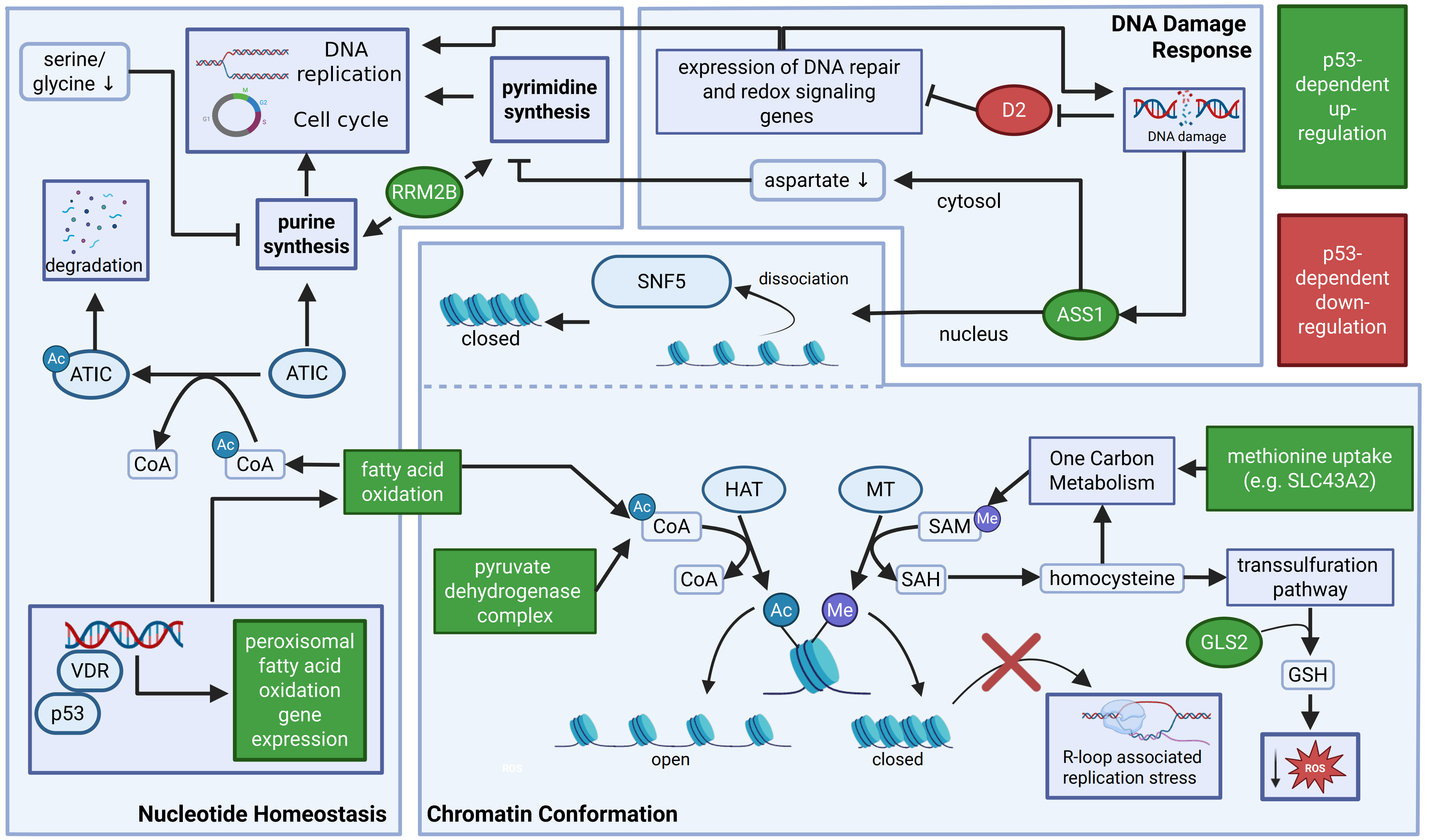
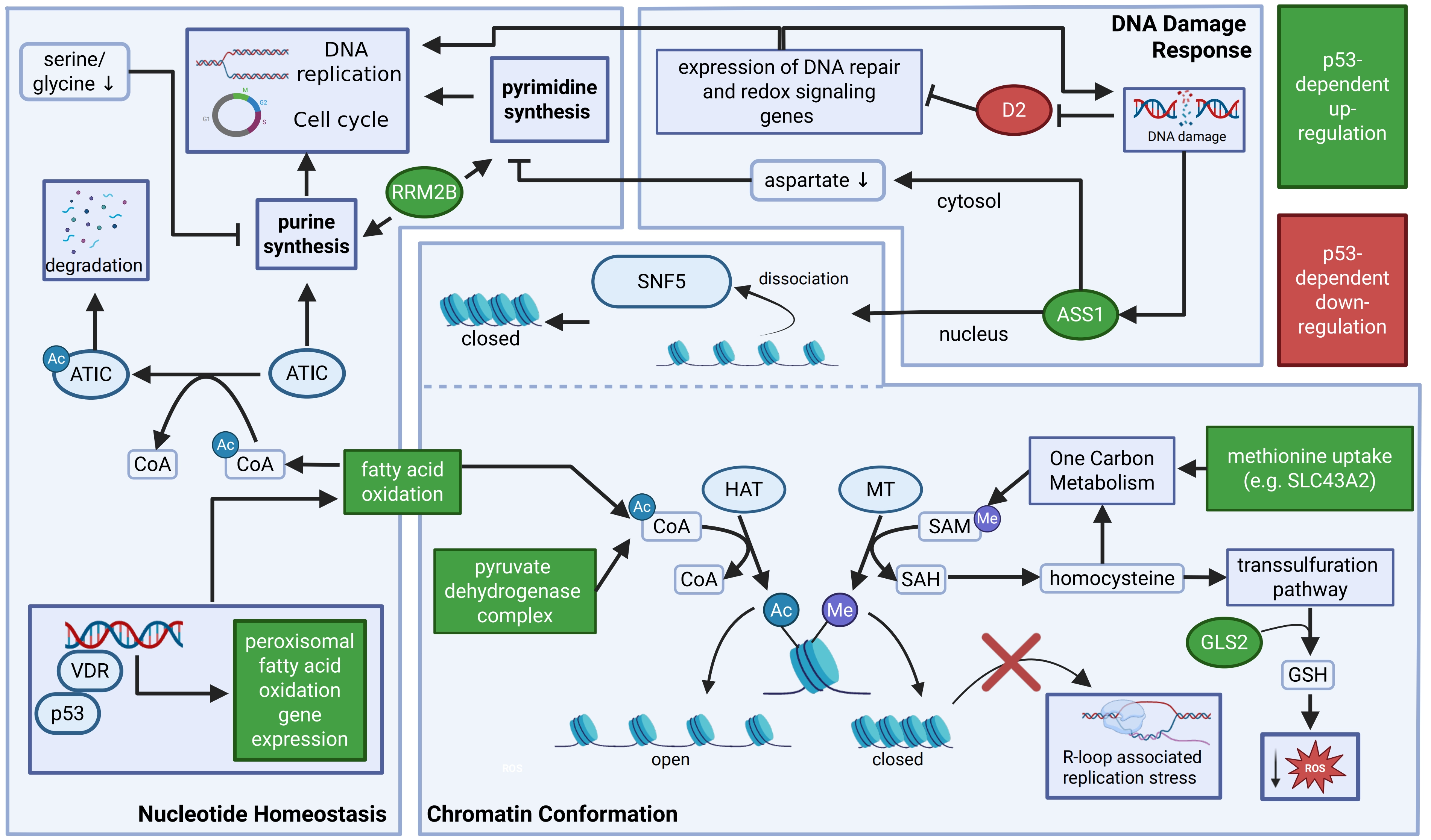
Figure 1. Metabolic regulation by p53 supports genomic integrity. Selected mechanisms linking p53-dependent metabolic regulation to nucleotide homeostasis, chromatin organisation and the DNA damage response. The ribonucleotide reductase subunit RRM2B supports deoxyribonucleotide availability for DNA replication and repair, whereas changes in serine, glycine and aspartate availability influence nucleotide biosynthesis, consequent DNA replication and cell-cycle progression. Cooperation between p53 and the VDR promotes peroxisomal fatty-acid oxidation, increasing acetyl-CoA availability for acetylation and degradation of the purine biosynthetic enzyme ATIC. Following DNA damage, ASS1 regulates cytosolic aspartate availability and contributes to nuclear chromatin remodelling through fumarate-dependent modification of the SWI/SNF complex. p53-dependent repression of type 2 deiodinase (D2) provides an additional connection between metabolic signalling and the expression of DNA repair and redox-regulatory genes. Metabolic control of acetyl-CoA and SAM availability supports histone acetylation and methylation, respectively. In particular, regulation of methionine uptake through SLC43A2 and one-carbon metabolism sustains SAM availability, helping preserve heterochromatin and limit aberrant transcription and R-loop-associated replication stress. GLS2 and transsulfuration provide complementary inputs supporting GSH synthesis and antioxidant defence. Green and red elements indicate reported p53-dependent upregulation and downregulation, respectively, including direct and indirect effects. The schematic integrates mechanisms identified in distinct experimental contexts. Created in BioRender. Amelio, I. (2026) https://BioRender.com/uer2dvd. Ac: acetyl group; ATIC: bifunctional AICAR transformylase/IMP cyclohydrolase; HAT: histone acetyltransferase; Me: methyl group; MT: methyltransferase; ROS: reactive oxygen species; SAH: S-adenosylhomocysteine; ASS1: argininosuccinate synthase 1; SAM: S-adenosylmethionine; GLS2: glutaminase 2; GSH: glutathione; RRM2B: ribonucleotide reductase subunit p53R2; VDR: vitamin D receptor; SWI/SNF: SWItch/sucrose non-fermentable.
2.2 Metabolic regulation of cell death by p53
Cell death represents an important terminal mechanism of tumour suppression by eliminating severely stressed or compromised cells. Notably, cell-fate decisions are highly dependent on cellular context and stress intensity, which can influence both the extent and mode of cell death. Increasing evidence indicates that p53-mediated metabolic regulation contributes to diverse forms of regulated cell death associated with metabolic stress.
2.2.1 Context-dependent regulation of ferroptosis by p53
Ferroptosis is an iron- and ROS-dependent form of regulated cell death characterized by excessive lipid peroxidation and subsequent loss of membrane integrity[25]. Cellular protection against ferroptosis can be broadly categorized into four mechanisms. (i) Direct detoxification of phospholipid hydroperoxides. The System Xc-–GSH–glutathione peroxidase 4 (GPX4) axis represents a canonical pathway for inhibiting phospholipid peroxidation. GPX4 serves as a central defence against ferroptosis by using GSH to reduce phospholipid hydroperoxides (PLOOHs) to phosphatidyl alcohols (PLOHs), thereby preserving membrane integrity. System Xc-, a cystine–glutamate antiporter composed of SLC7A11 and SLC3A2, imports cystine into cells, which is subsequently reduced to cysteine, an essential precursor for GSH synthesis[26]. GPX1 has recently been identified as an additional ferroptosis suppressor that, together with OSBPL8, is recruited to the endoplasmic reticulum, where it reduces oxidized phosphatidic acid[27]; (ii) NADPH-dependent antioxidant defence. The FSP1–ubiquinone/Vitamin K system[26,28,29] and the GCH1–BH4–DHFR[30,31] axis both provide GPX4-independent protection against ferroptosis. Ubiquinone, vitamin K, and BH4 function as radical-trapping antioxidants that limit ROS-driven lipid peroxidation; (iii) Iron homeostasis. Aberrant accumulation of Fe2+ resulting from dysregulated iron metabolism promotes the generation of hydroxyl radicals through the Fenton reaction, thereby facilitating lipid peroxidation[32,33]; (iv) Lipid metabolism and phospholipid peroxidation. The ACSL4–LPCAT3–LOXs[34-37] pathway promotes the incorporation and peroxidation of polyunsaturated fatty acids (PUFAs) in membrane phospholipids, thereby driving ferroptosis. Overall, ferroptosis induction generally results from the failure of one or more of the cellular defence mechanisms rather than from an actively initiated process.
p53 has been shown to promote ferroptosis by modulating multiple metabolic defence mechanisms. In 2015, p53 was first reported to transcriptionally repress SLC7A11 and thereby alter the sensitivity of mouse embryonic fibroblasts (MEFs) to erastin-induced ferroptosis[38]. p533KR, an acetylation-defective mutant that is unable to induce cell-cycle arrest, retains the capacity to repress SLC7A11 transcription. Overexpression of SLC7A11 or treatment with the ferroptosis inhibitor ferrostatin-1 rescued erastin-induced ferroptosis in cells expressing p533KR, supporting a direct role for SLC7A11 repression in p53-mediated ferroptotic sensitivity. In p53-null H1299 human non-small cell lung cancer cells, ectopic p53 expression was also shown to promote nuclear translocation of the deubiquitinase USP7, resulting in reduced monoubiquitination of histone H2B at the SLC7A11 locus and further suppression of SLC7A11 expression[39]. In addition, p53 transcriptionally represses OTUD5, a deubiquitinase that interacts with and stabilizes GPX4. p53-mediated repression of OTUD5 increases lipid peroxidation and sensitizes human gastric cancer cell lines (AGS and HGC-27) as well as mouse forestomach carcinoma (MFC) cells to ferroptosis[40]. Together, these findings broaden the mechanistic framework through which p53 regulates the GPX4-centered ferroptosis defence system.
p53 also regulates GPX4-independent mechanisms of ferroptosis suppression. Vitamin K epoxide reductase complex subunit 1-like 1 (VKORC1L1) has been identified as a direct transcriptional target of p53 and as a potential ferroptosis suppressor. Repression of VKORC1L1 following p53 activation, or pharmacological inhibition of VKORC1L1, reduces the generation of reduced vitamin K and thereby increases ferroptosis sensitivity across several cancer cell models, including p53-mutant pancreatic cancer cells[41]. In parallel, p53-mediated inhibition of the PPP may further weaken nicotinamide adenine dinucleotide phosphate (NADPH)-dependent antioxidant defences[42]. NADPH is required for the regeneration of reduced GSH from oxidized glutathione (GSSG) and supports NADPH-dependent ferroptosis-suppressive pathways, including the FSP1–ubiquinone/vitamin K system. Thus, by limiting NADPH availability, p53 in this context may further compromise cellular antioxidant capacity and increase susceptibility to pro-ferroptotic stressors.
Lipid composition represents another important determinant of ferroptosis sensitivity. Monounsaturated fatty acids (MUFAs) are less susceptible to lipid peroxidation than PUFAs and can therefore limit the propagation of lipid-derived free radicals[26]. In human hepatoma cells, stearoyl-CoA desaturase 1 (SCD1), a key enzyme in MUFA synthesis (Figure 2), has been shown to confer resistance to ferroptosis, whereas p53 counteracts this protective effect through transcriptional repression of SCD1[43]. In addition, p53 disrupts the interaction between SREBP1 and its co-factor DEPDC1, thereby impairing formation of a transcriptional activator complex that promotes SCD1 expression through binding to its promoter[44]. More recently, the p53–Parkin–ACSL4 axis has been identified as another mechanism through which p53 promotes ferroptosis in rat cardiac myoblasts and in mouse models. p53 represses Parkin transcription, reducing Parkin-mediated ubiquitination of ACSL4 and thereby promoting ACSL4 accumulation. Increased ACSL4 activity facilitates the incorporation of PUFAs, particularly arachidonic acid, into membrane phospholipids[45]. Through these mechanisms, p53 may increase the abundance of oxidation-prone PUFA-containing phospholipids and thereby enhance cellular susceptibility to ferroptotic stress.
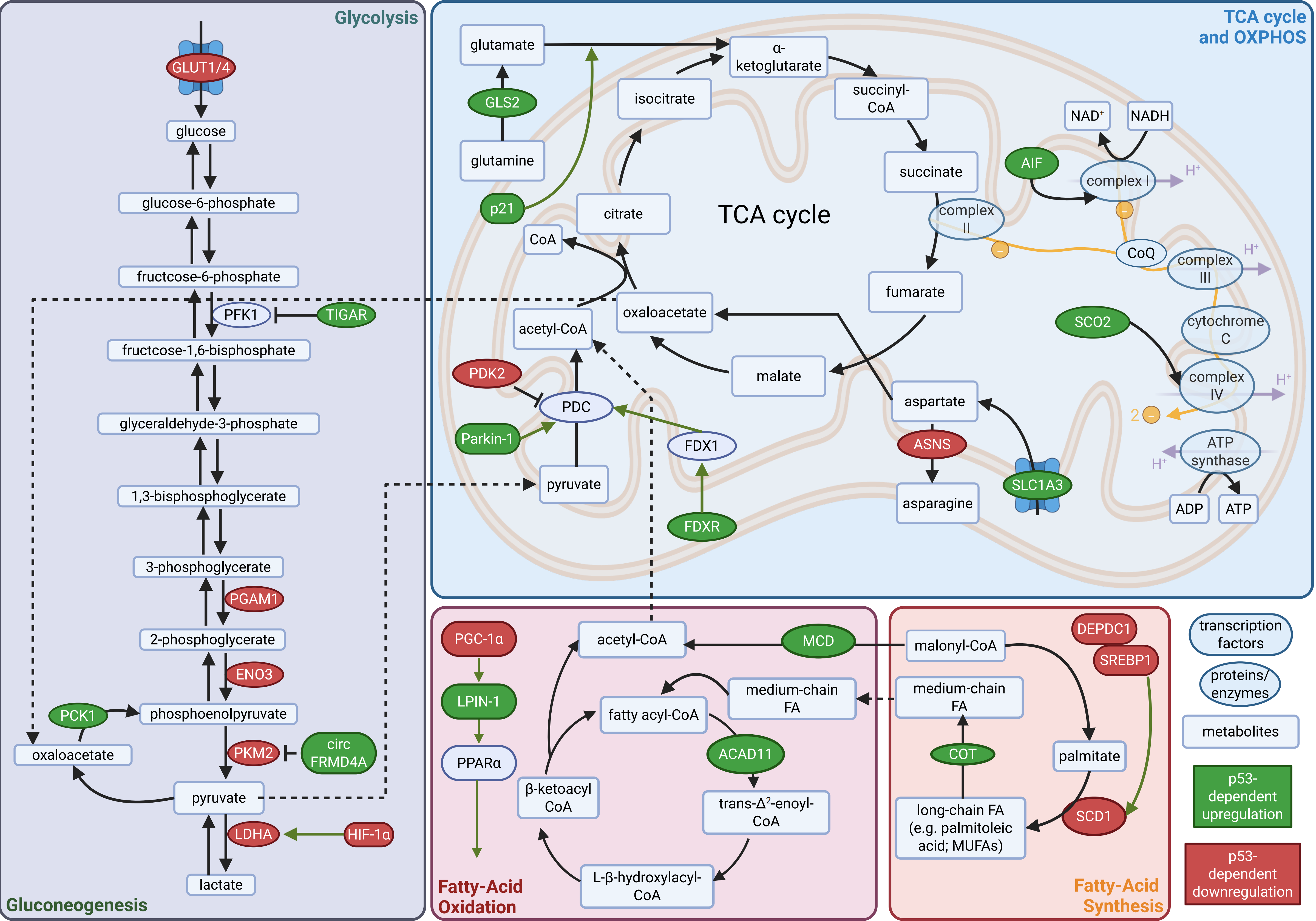
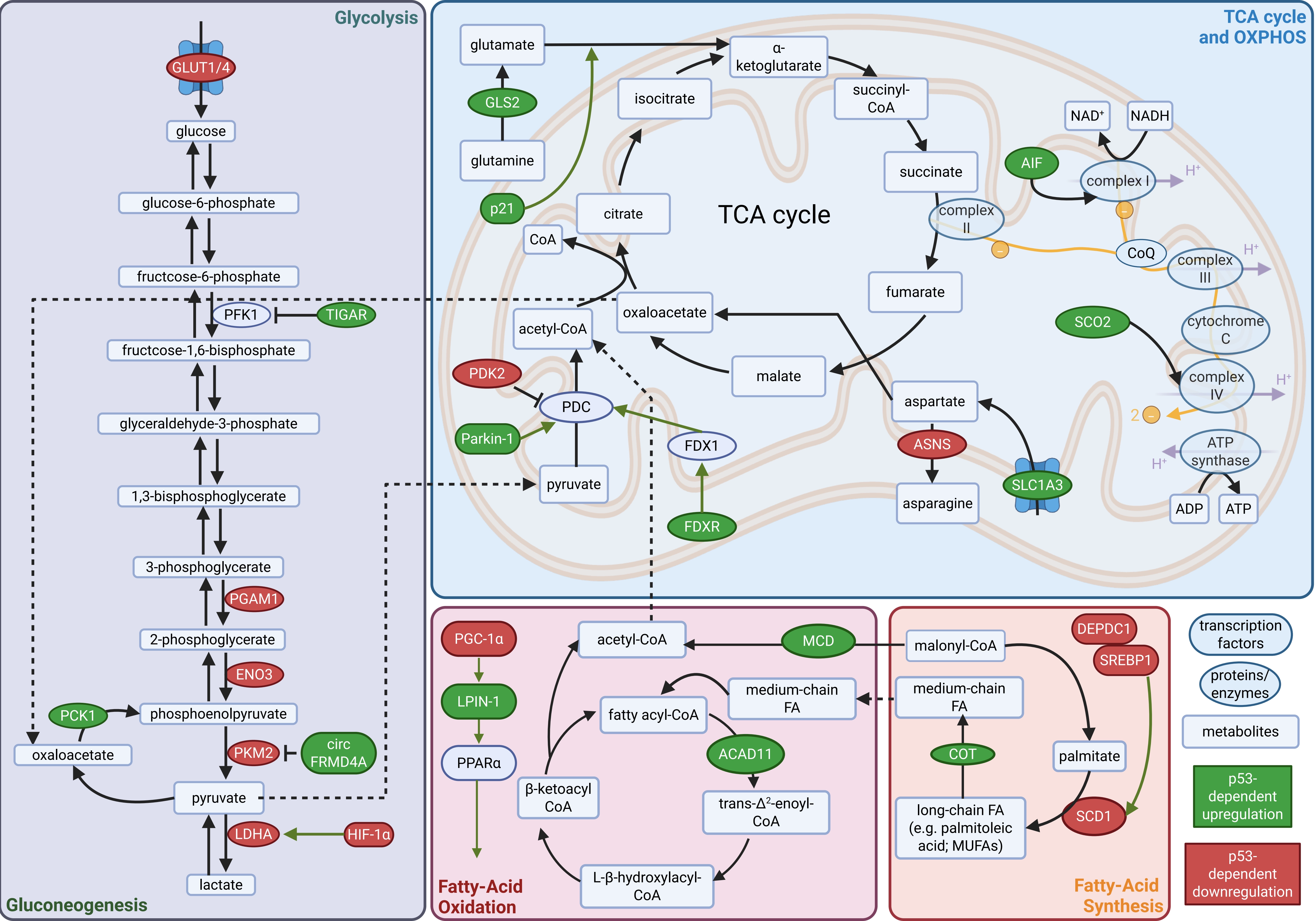
Figure 2. p53-dependent regulation of energy metabolism. Schematic overview of selected mechanisms through which wild-type p53 regulates glucose metabolism, mitochondrial respiration and fatty-acid metabolism. p53 modulates glucose uptake and glycolytic activity while supporting mitochondrial pyruvate utilisation through regulation of PDK2 and Parkin. Regulation of GLS2, p21, the aspartate transporter SLC1A3 and ASNS further links amino-acid metabolism to the availability of TCA cycle intermediates. FDXR and FDX1 connect p53-regulated mitochondrial functions to protein lipoylation, whereas AIF and SCO2 support respiratory-chain function. p53 also regulates fatty-acid utilisation through LPIN1, PPARα, ACAD11, MCD and COT, and modulates monounsaturated fatty-acid synthesis through the DEPDC1–SREBP1–SCD1 pathway. Together, these metabolic activities can influence cellular adaptation to nutrient stress and susceptibility to metabolically regulated cell death. Green and red nodes indicate reported p53-dependent increases and decreases, respectively, in expression or activity; these effects may be direct or indirect. The diagram integrates findings from different experimental systems and does not imply that all regulatory relationships occur simultaneously or uniformly across tissues and stress conditions. Created in BioRender. Amelio, I. (2026) https://BioRender.com/4ydphoj. OXPHOS: oxidative phosphorylation; PDC: pyruvate dehydrogenase complex; CoQ: coenzyme Q; FA: fatty acid; MUFAs: monounsaturated fatty acids; PDK2: pyruvate dehydrogenase kinase 2; GLS2: glutaminase 2; ASNS: asparagine synthetase; TCA: tricarboxylic acid; FDXR: ferredoxin reductase; FDX1: ferredoxin 1; AIF: apoptosis-inducing factor; SCO2: synthesis of cytochrome c oxidase 2; LPIN1: lipin 1; PPARα: peroxisome proliferator-activated receptor α; ACAD11: acyl-CoA dehydrogenase family member 11; MCD: malonyl-CoA decarboxylase; COT: carnitine O-octanoyltransferase.
An additional pathway through which p53 could promote ferroptosis involves the regulation of iron metabolism. Under certain conditions (deficiency of the ER-resident disulfide isomerase AGR2), p53 represses the expression of the iron exporter ferroportin 1 (FPN1), thereby inducing intracellular iron overload and promoting ferroptosis[46]. Conversely, ferroptotic triggers induce AGR2 expression, which restrains p53 activation and thereby limits ferroptosis in pancreatic cancer models.
The effect of p53 on ferroptosis can also shift toward a protective role, with distinct outcomes observed across cancer types and cellular states. For example, Tang et al. reported that p53 suppresses erastin-induced ferroptosis in HCT116 colorectal cancer (CRC) cells by promoting SLC7A11 expression and limiting dipeptidyl peptidase-4 (DPP4) activity[47]. Consistent with this observation, another study showed that p53 can inhibit ferroptosis in CRC cells by reducing PUFA abundance through the autophagy-dependent FBXO2–FABP5 axis[48]. As discussed above, a substantial component of the antioxidant activity of p53 is also mediated through the maintenance of GSH availability, suggesting that p53-dependent control of GSH metabolism may further shape ferroptotic susceptibility in specific contexts.
The cellular consequences of p53 presence or absence may therefore vary across tumour types and metabolic status, suggesting the importance of considering p53 status in ferroptosis and tumour suppression. Importantly, the apparent dual role of p53 in either promoting or restraining ferroptosis should be interpreted in light of the intrinsic ferroptotic sensitivity of the experimental model, as well as the specificity and potential off-target effects of the ferroptosis inducers employed. These variables may substantially influence the observed phenotype and should therefore be carefully considered when reconciling apparently conflicting findings across studies (Table 1).
Table 1. p53-dependent metabolic regulation and its crosstalk with cell death modalities.
| Connection to cell death | Pathway | Regulated genes | Species | Cell line | Effects |
| Ferroptosis | GSH synthesis | SLC7A11[38,39] ↓; H2Bub1[39] ↓ | Homo sapiens; Mus musculus | MEFs[38]; Human non-small cell lung cancer H1299 cell[39] | promote |
| GPX4 | OTUD5[40] ↓ | Homo sapiens; Mus musculus | Human gastric cancer AGS and HGC-27 cells; MFC cell | promote | |
| Vitamin K System | VKORC1L1[41] ↓ | Homo sapiens; Mus musculus | Human fibrosarcoma HT1080 cell; Human melanoma A375 cell; Xenograft model in mice | promote | |
| Monounsaturated fatty acid synthesis | SCD1[43] ↓ | Homo sapiens; Mus musculus | Human hepatocellular Carcinoma JHH-1 and HepG2 cells; Xenograft model in mice; HCC patient derived organoids; Clinical data | promote | |
| Phospholipids formation | Parkin[45] ↓; ACSL4[45] ↑ | Rattus norvegicus; Mus musculus | Rat embryonic heart myoblast H9c2 cell; C57BL/6J mice | promote | |
| Iron metabolism | FPN1[46] ↓; | Homo sapiens; Mus musculus | Human pancreatic cancer Capan2 and HPAC cells; C57BL/6 and BALB/c nude mice; Clinical data | promote | |
| Lipid peroxidation | DPP4 activity[47] ↓; SLC7A11[47] ↑ | Homo sapiens; Mus musculus | Human colorectal cancer SW48 and HCT116 cells; Xenograft model in nude mice | suppress | |
| PUFA metabolism | FBXO2[48] ↑; FABP5[48] ↓ | Homo sapiens; Mus musculus | Human colorectal cancer RKO and HCT116 cells; BALB/c nude mice;Clinical data | suppress | |
| Cuproptosis | Drive glycolytic flux toward the TCA cycle | circFRMD4A[49] ↑; PKM2[49] ↓ | Homo sapiens; Mus musculus | Human breast cancer CAL-51 cell; Human colorectal cancer HCT116 cell; Xenograft model in mice | promote |
| Upregulation of FDX1 | FDXR[50] ↑; FDX1[50] ↑ | Homo sapiens; Mus musculus | Human hepatocellular Carcinoma Hep3B2.1-7, PLC/PRF/5 and HepG2 cells; Xenograft model in mice | promote | |
| Copper accumulation and oxidation stress | COX17[51] ↑ | Homo sapiens; Mus musculus | Human gastric cancer AGS cell; HUVECs; Xenograft model in mice | promote | |
| Apoptosis and Necroptosis | Glycolytic activity suppression | HK[52] ↓; PDH[52] ↓; PFKP[52] ↓ | Homo sapiens | Human mesothelioma MSTO-211HAcT and H2452AcT cells | promote |
Overview over metabolic pathways that are regulated in a p53-dependent manner, the genes used for this regulation and the connected cell death pathways. ↓ indicates down-; ↑ indicates up-regulation of pathway/gene expression/cell death pathway; GSH: glutathione; MEFs: mouse embryonic fibroblasts; GPX4: glutathione peroxidase 4; VKORC1L1: vitamin K epoxide reductase complex subunit 1-like 1; SCD1: stearoyl-CoA desaturase 1; FPN1: ferroportin 1; DPP4: dipeptidyl peptidase-4; PUFA: polyunsaturated fatty acid; PKM2: pyruvate kinase M2; FDXR: ferredoxin reductase; HUVECs: human umbilical vein endothelial cells; FDX1: ferredoxin 1; H2Bub1: monoubiquitinated histone H2B; SLC7A11: solute carrier family 7 member 11; HK: hexokinase; PDH: pyruvate dehydrogenase; PFKP: phosphofructokinase, platelet; HCC: hepatocellular carcinoma; MFC: mouse forestomach carcinoma.
2.2.2 Beyond ferroptosis: p53-dependent metabolic control of other cell-death modalities
Beyond its roles in ferroptosis sensitivity, p53 can shape cellular susceptibility to diverse forms of regulated cell death through its extensive control of metabolism (Table 1). By modulating mitochondrial activity, nutrient utilization, redox balance, and metabolite availability, p53 can create metabolic states that either promote or restrain cell death in a context-dependent manner.
In 2022, Tsvetkov et al. postulated a distinct copper-dependent form of regulated cell death, named cuproptosis, that is closely linked to mitochondrial metabolism and proteotoxic stress[53]. Mechanistically, ferredoxin 1 (FDX1) promotes the reduction of Cu2+ to Cu+ and acts as an upstream regulator of mitochondrial protein lipoylation. Copper directly binds to lipoylated components of the tricarboxylic acid (TCA) cycle, promoting their aggregation and the subsequent loss of iron–sulfur (Fe–S) cluster-containing proteins, ultimately triggering proteotoxic stress and cell death[53,54]. These observations suggest that metabolic interventions that increase mitochondrial and TCA-cycle dependence could potentially enhance cuproptosis sensitivity in tumour cells.
p53 activity has also been associated with cuproptosis sensitivity. In human colorectal and breast carcinoma models, pharmacological activation of p53 has been reported to synergize with the cuproptosis inducer elesclomol, thereby enhancing cuproptosis sensitivity. Mechanistically, p53 induces the transcription of circFRMD4A, which inhibits pyruvate kinase M2 (PKM2), thereby redirecting glycolytic flux toward the TCA cycle[49]. This enhancement of mitochondrial metabolism may contribute to a pro-cuproptotic effect of p53 activation, as observed in mouse xenograft models in which p53 activation suppresses tumour growth.
Ferredoxin reductase (FDXR) may function as an upstream enabler of cuproptosis by donating reducing equivalents to FDX1, a key regulator of mitochondrial copper metabolism that has been implicated in the reduction of Cu2+ to Cu+. FDXR has been shown to be transcriptionally induced by p53 in hepatocellular carcinoma, providing an additional potential link between p53 activation and cuproptosis[50,55]. Moreover, p53 contributes to the maintenance of Fe–S cluster biosynthesis by regulating the expression of the Fe–S cluster assembly enzyme (ISCU) and mitochondrial frataxin[56-59], thereby potentially modulating cellular susceptibility to cuproptosis.
p53 may further influence cuproptosis by regulating cellular copper and redox homeostasis. In gastric cancer cells, the cytochrome c oxidase copper chaperone COX17 has been identified as a p53 transcriptional target and a downstream effector associated with cuproptosis[51]. COX17 upregulation or overexpression synergizes with elesclomol to increase intracellular Cu2+ accumulation and inhibit human umbilical vein endothelial cell (HUVEC) tube formation and proliferation. Combined elesclomol treatment and COX17 overexpression also reduce GSH and superoxide dismutase (SOD) levels, indicating disruption of cellular redox homeostasis. Given that GSH can function as a copper chelator[60], p53-dependent suppression of GSH synthesis could further promote the accumulation of labile copper required for cuproptosis initiation.
p53 supports mitochondrial metabolism by promoting acetyl-CoA production, fatty acid oxidation (FAO), TCA cycle anaplerosis, and oxidative phosphorylation. It enhances pyruvate entry into the TCA cycle by inhibiting pyruvate dehydrogenase kinase-2 (PDK2)[61] and inducing Parkin[62], while also stimulating FAO through carnitine O-octanoyltransferase (COT)[63], LPIN1/PPARα[64], acyl-CoA dehydrogenase family member 11 (ACAD11)[65], and malonyl-CoA decarboxylase (MCD)[66]. During nutrient stress, p53 maintains TCA cycle intermediates by regulating GLS2[23], p21[67], SLC1A3[68], and asparagine synthetase (ASNS)[69] (Figure 2). In parallel, p53 promotes oxidative phosphorylation by transcriptionally activating synthesis of cytochrome c oxidase 2 (SCO2)[70] and apoptosis-inducing factor (AIF)[71,72], thereby supporting respiratory complexes IV and I and maintaining mitochondrial energy production. Collectively, these findings suggest that p53 may enhance cellular sensitivity to cuproptosis through indirect mechanisms, including regulation of mitochondrial metabolism, Fe–S cluster homeostasis, copper handling, and redox balance, rather than by acting exclusively on the control of the core machinery postulated to control cuproptosis execution.
The impact of p53-mediated metabolic regulation on cell death extends beyond ferroptosis and cuproptosis and can also indirectly influence apoptosis, necrosis, and necroptosis. In cells that are highly dependent on glycolysis, a p53-driven shift toward oxidative metabolism may induce an energetic crisis, thereby suppressing tumour growth and ultimately promoting distinct forms of cell death depending on the cellular context. Consistent with this concept, p53-dependent suppression of aerobic glycolysis, or the Warburg effect, has been shown to trigger the concurrent activation of apoptosis and necroptosis in mesothelioma cells[52]. Conversely, p53 can also employ metabolic regulation to promote cell survival and limit stress-induced damage. For example, under metabolic stress conditions such as serine and glycine deprivation, p53 can protect cells from ROS-mediated damage by remodelling GSH metabolism[24]. Finally, p53 also exerts also an important crosstalk with the mammalian target of rapamycin (mTOR). The p53–mTOR crosstalk is highly reciprocal and strongly dependent on cellular context and stress conditions and plays a critical role in enabling cells and organisms to maintain homeostasis. p53 generally suppresses mTORC1 activity through transcriptional induction of negative regulators, including REDD1, LKB1, AMPKβ, TSC2, PTEN, and Sestrins, thereby limiting growth and promoting stress adaptation[73]. Conversely, mTOR signalling regulates p53 abundance and activity by controlling MDM2, AKT-dependent phosphorylation, protein translation and post-translational modification. Together, these bidirectional interactions coordinate growth, metabolism, survival and stress responses in a cell type- and context-dependent manner[73].
These findings underscore the context-dependent duality of p53-mediated metabolic control. On the one hand, p53 can enhance cellular stress tolerance by limiting oxidative damage and supporting metabolic adaptation. On the other hand, it can reprogram metabolic pathways in ways that facilitate the induction of multiple forms of cell death. This flexibility enables p53 to fine-tune cell fate according to the nature and severity of the stress and may also provide alternative routes to cell death when canonical p53-dependent death pathways are impaired.
3. Cell-Extrinsic Metabolic Functions of p53 in the Tumour Microenvironment
As tumorigenesis proceeds, tumour cells progressively remodel the surrounding microenvironment to create a parasitic system that sustains their growth and survival. This system comprises tumour cells, stroma and tissue-resident cells, blood vessels, various immune cells (T-cells, macrophages, etc.), and non-cellular components of the extracellular matrix like collagens, growth factors and cytokines[74,75]. Neighbouring non-malignant cells may initially function as a local barrier, sensing tumour-derived danger signals and alterations in the extracellular environment. This barrier, however, is dynamic and complex, as metabolic signals are exchanged among diverse cell populations to establish a bidirectional network. Within this network, p53 in stromal and immune cells can be either activated or suppressed by cell-cell communication, environmental stress, and metabolic perturbations, thereby mounting anti- or pro-tumour responses within the local tumour microenvironment (TME).
Expression and activation of p53 in cancer cells can influence the TME at multiple levels, including the composition and functional state of tumour-infiltrating immune cells, whereas loss of p53 can promote an immunosuppressive and immune-evasive TME in a context-dependent manner. In autochthonous lung and pancreatic tumour models, tumour-specific p53 loss has been associated with increased recruitment of suppressive myeloid populations and regulatory T cells, thereby enhancing protection of tumour cells from immune-mediated elimination[76]. Conversely, pharmacological activation of p53 has been shown to increase T-cell infiltration and enhance the efficacy of immune checkpoint blockade in implanted B16 melanoma models[77]. p53 can also promote innate immune signalling through the cyclic GMP–AMP synthase (cGAS)-stimulator of interferon genes (STING) pathway. Mechanistically, wild-type p53 induces TRIM24-dependent degradation of the cytosolic DNA exonuclease TREX1, resulting in cytosolic DNA accumulation, cGAS-STING activation and increased type-I interferon signalling, thereby contributing to both cell-intrinsic and cell-extrinsic tumour-suppressive responses[78]. In parallel, p53 activation can induce cellular senescence, and the resulting senescence-associated secretory phenotype (SASP) exerts highly context-dependent effects on neighbouring cells[79]. Transient SASP-associated cytokine and chemokine secretion can facilitate immune-cell recruitment and clearance of senescent tumour cells, whereas persistent SASP signalling can promote chronic inflammation, immunosuppression and tumour progression[80].
Tumour-associated macrophages (TAMs) represent one of the most abundant immune populations within the TME and exhibit substantial plasticity and heterogeneity, contributing to tumour progression, invasion, metastasis, and therapeutic resistance. In several cancer models, p53 activation has been shown to promote TAM polarization toward an anti-tumour, classically activated M1-like state characterized by the production of pro-inflammatory cytokines and chemokines that support tumour cell elimination[81,82]. Conversely, loss of p53 activity can favour pro-tumour macrophage states through metabolic reprogramming. p53 inactivation also derepresses IL-34, which induces expression of the fatty acid transporter CD36 and promotes FAO, thereby driving M2-like polarization in bone marrow-derived macrophages and xenograft models[83]. Consistently, p53 sequestration has been reported to derepress xCT and Arg1 transcription, enhancing glutathione synthesis and arginine metabolism to support M2-like polarization and ferroptosis resistance in esophageal squamous cell carcinoma[84]. Together, these findings indicate that the tumour-suppressive effects of p53 within the TME are highly context dependent and can shape the surrounding microenvironmental cues.
Metabolic competition represents an additional layer of regulation within the TME. Many cancer cells exhibit a strong dependence on glycolysis, a hallmark of the Warburg effect[85,86], which contributes to lactate accumulation, extracellular acidification, and hypoxia, thereby promoting tumour invasion and metastasis[87]. As discussed above, p53 can restrict glucose uptake and glycolytic flux in tumour cells, thereby reducing tumour-cell glucose consumption and preserving extracellular glucose availability. This metabolic rewiring may lessen competition for glucose between tumour cells and neighbouring immune populations, potentially explaining the p53-mediated modulation of immune-cell function[88].
4. Metabolic Regulations by Mutant p53
p53 is mutated in approximately half of all human cancers. Notably, the most frequent alterations are missense mutations within the DNA-binding domain (DBD), resulting in the expression of mutant p53 (mutp53) proteins. These mutations can be broadly classified as contact or conformational mutants and may exert diverse cellular effects through loss of wild-type function, dominant-negative activity, and gain-of-function (GOF) properties[89,90]. Increasing evidence indicates that mutp53 proteins can also reprogram cancer cell metabolism. Given the substantial functional heterogeneity among individual p53 mutants, however, it is important to distinguish their specific metabolic consequences rather than considering mutp53 as a uniform entity.
The human p53R175H mutant, corresponding to murine p53R172H, is a conformational mutant in which impaired zinc binding destabilizes the DBD and promotes misfolding. Ectopic expression of p53R172H or p53R175H in H1299 cells or MEFs enhances the Warburg phenotype by promoting GLUT1 translocation to the plasma membrane, an effect also reported for the R248Q and R273H mutants[91]. This metabolic output is, however, highly context dependent. Lonetto et al. showed that mesenchymal stem cells (MSCs) harbouring p53R172H progress toward a more aggressive tumour-derived state (MSC-TLs). Compared with wild-type p53 MSCs, p53R172H MSCs display increased glycolytic activity, whereas p53R172H MSC-TLs exhibit enhanced mitochondrial oxidative metabolism accompanied by changes in mitochondrial morphology, mass, and function[92]. Depletion of mutp53 reverses these phenotypes, supporting their dependence on p53R172H itself. In addition, in a comparative study of murine pancreatic ductal adenocarcinoma (PDAC) KPCR270H and KPCR172H cells, both carrying KrasG12D, Caporali et al. found that p53R172H was associated with an unexpected attenuation of urea-cycle metabolism and alterations in the related metabolic enzymes Arg2 and CkB, producing a phenotype that may contribute to tumour suppression[93].
The human p53R273H mutant, corresponding to murine p53R270H, is a hotspot missense mutation and a representative contact mutant that has been associated with increased invasive capacity in lung, breast, and colorectal cancer models[94-97]. Among several mutants profiled in p53-null H1299 cells, p53R273H was the only variant that significantly increased both glycolytic rate and maximal glycolytic capacity. It also enhanced mitochondrial oxidation, although this effect was evident only following inducible expression in H1299 and HCT116 cells and was not observed in stable cell lines, suggesting that compensatory adaptation may obscure the phenotype during prolonged expression[98]. In human PDAC, p53R273H has also been reported to increase mitochondrial respiration through upregulation of myosin heavy chain 14 (MYH14) and to promote mitochondrial fragmentation[99]. Consistent with these findings, the comparative KPC study described above showed that p53R270H sustains TCA-cycle activity and glutathione metabolism, with Assay for Transposase-Accessible Chromatin with high-throughput sequencing (ATAC-seq) revealing variant-specific differences in chromatin accessibility[93]. Importantly, however, these mutant-specific metabolic changes did not confer a measurable selective advantage or alter cellular fitness in that experimental setting. Beyond these metabolic effects, p53R273H has been shown to impair antioxidant capacity in HCT116 and H1299 cells through repression of NRF2 expression[100], while also increasing ROS production and pro-inflammatory cytokine release and activating mTOR signalling to suppress autophagy[101].
The p53R280K mutant has likewise been implicated in metabolic reprogramming that supports tumour progression. In human breast cancer cells, p53R280K promotes serine and glycine biosynthesis, including through upregulation of phosphoserine aminotransferase 1 (PSAT1), and enhances the expression of amino-acid transport systems such as the L-type amino acid transporter 1 (LAT1)/CD98 heavy-chain heterodimer. These changes are particularly evident under amino-acid-deprived conditions, enabling tumour cells to maintain proliferation despite reduced nutrient availability[102]. In the same context, p53R280K also co-opts the lipogenic transcription factor SREBP1 to form a transcriptional co-activation complex that induces fatty acid synthase (FASN) expression and promotes lipid accumulation[103].
Additional mutp53 variants have also been linked to metabolic regulation. For example, murine p53R178C has been shown to increase lipolysis by binding to and transactivating the beta-3 adrenergic receptor (ADRB3), further illustrating the capacity of specific p53 mutants to influence lipid metabolism[104].
Given the large and sometimes contradictory body of literature on mutp53-dependent metabolic regulation, careful consideration of experimental context is essential[89,90]. Differences between in vitro and in vivo systems, as well as microenvironmental conditions such as oxygen tension, nutrient availability, and stromal composition, may substantially influence the phenotypes attributed to individual mutants. This is exemplified by mutp53-dependent transcriptional regulation of extracellular matrix (ECM) components through cooperation with hypoxia-inducible factor 1 (HIF-1), an effect that becomes apparent specifically under hypoxic conditions[105] (Figure 3). Beyond direct effects on the ECM, including reprogramming of the tumour-cell secretome, mutp53 has also been reported to modulate cancer-associated fibroblasts and tumour–immune crosstalk, frequently contributing to the establishment of a tumour-supportive microenvironment[106].
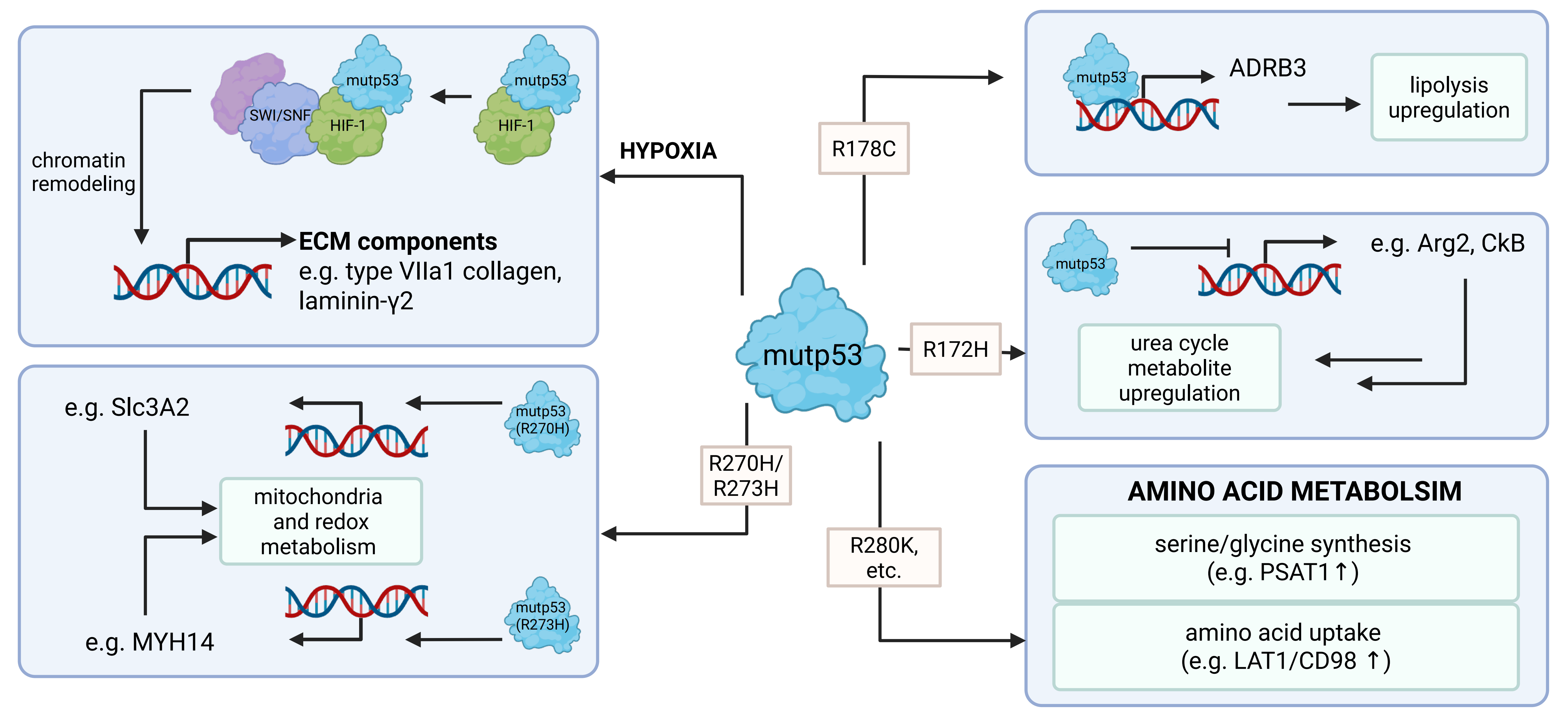
Figure 3. Variant- and context-dependent metabolic regulation by mutant p53. Selected examples of how distinct mutant p53 proteins influence cellular metabolism and extracellular matrix organisation. Under hypoxia, mutant p53 cooperates with HIF-1 and the SWI/SNF chromatin-remodelling complex to promote expression of ECM components, including collagen VII α1 and laminin γ2. Murine p53 R270H and its human counterpart R273H influence mitochondrial and redox metabolism in pancreatic cancer models, with reported changes involving SLC3A2 and MYH14, respectively. Murine p53 R178C, corresponding to human R181C, promotes expression of the ADRB3 and enhances lipolysis. In murine pancreatic cancer models, p53 R172H, corresponding to human R175H, restrains Arg2 and CkB expression and attenuates urea-cycle-associated metabolism; depletion of this mutant increases the abundance of associated metabolites. In breast cancer models, R280K and additional tested variants support serine and glycine biosynthesis and amino-acid uptake through regulation of enzymes and transporters, including PSAT1 and the LAT1/CD98 transporter complex. These effects become particularly relevant under limited amino-acid availability. The depicted relationships encompass direct and indirect mechanisms established in different experimental systems and should not be interpreted as a shared programme of all p53 mutants. Their consequences depend on tissue, nutrient availability, oxygen tension and tumour state, and metabolic alterations do not invariably confer increased cellular fitness. Created in BioRender. Amelio, I. (2026) https://BioRender.com/k2f3jvs. ARG2: arginase 2; CkB: creatine kinase B; LAT1: L-type amino-acid transporter 1; mutp53: mutant p53; HIF-1: hypoxia-inducible factor 1; SWI/SNF: SWItch/sucrose non-fermentable; ECM: extracellular matrix; MYH14: myosin heavy chain 14; ADRB3: β3-adrenergic receptor; PSAT1: phosphoserine aminotransferase 1.
Together, these studies emphasize the importance of considering both mutp53 variant identity and microenvironmental context when evaluating metabolic GOF phenotypes. Whether such metabolic alterations directly contribute to tumour-cell proliferation, metastasis, or survival remains under debate, particularly in light of broad clustered regularly interspaced short palindromic repeats (CRISPR)/Cas9-based analyses such as that of Wang et al., which found no consistent requirement for mutp53 in the tested cancer cell lines[107]. One possibility is that mutp53-dependent metabolic reprogramming may be more relevant during earlier stages of tumour evolution, where it could shape the metabolic landscape and improve access to energy and nutrients, rather than being universally required for maintenance of established tumours.
5. Conclusions and Future Perspectives
p53-mediated metabolic regulation is emerging as an important component of tumour suppression that extends well beyond the classical induction of cell-cycle arrest and apoptosis. By coordinating nucleotide availability, redox balance, mitochondrial metabolism and nutrient utilisation, p53 can preserve genomic integrity while simultaneously determining whether stressed cells adapt or undergo metabolic forms of cell death. These functions further extend beyond tumour cells, where p53-dependent metabolic changes can reshape immune-cell behaviour and nutrient competition within the tumour microenvironment. However, the increasingly complex literature also argues against a universal metabolic programme downstream of p53. Ferroptosis provides a particularly clear example, as p53 can either enhance or restrain ferroptotic sensitivity depending on cellular metabolism, tissue context and experimental conditions. Likewise, metabolic phenotypes attributed to mutant p53 differ markedly between individual variants and do not invariably translate into increased cellular fitness. Future studies should therefore move beyond isolated metabolic endpoints and establish whether specific metabolic alterations are functionally required for tumour initiation, maintenance or progression. Genetically defined models, physiological nutrient and oxygen conditions, spatial and isotope-resolved metabolic approaches, and direct comparison of individual TP53 variants will be particularly important. Ultimately, defining the contexts in which p53-dependent metabolic states represent genuine tumour dependencies may reveal vulnerabilities that can be exploited therapeutically.
Acknowledgements
The authors thank all the members of the Amelio laboratory for their constructive discussions. The authors apologize for any relevant articles that could not be included or discussed because of space limitations.
Authors contribution
Gurres S: Conceptualization, writing-original draft.
Lu Y: Writing-original draft.
Leist M: Writing-review & editing.
Amelio I: Conceptualization, supervision, writing-review & editing.
Conflicts of interest
The authors declare no conflicts of interest.
Ethical approval
Not applicable.
Consent to participate
Not applicable.
Consent for publication
Not applicable.
Availability of data and materials
Not applicable.
Funding
The work of the Amelio Lab has been supported by the DFG, (under the TRR353 “Death Decision” projects A05), the Carl Zeiss Stiftung (Endowed Professorship, #15972218, 2022-2027), the Excellence Strategy of the German Federation and States via the Blue Sky Grant (#81967325), and the Deutsche Krebshilfe (#70117491).
Copyright
© The Author(s) 2026.
References
-
3. Kubbutat MHG, Jones SN, Vousden KH. Regulation of p53 stability by Mdm2. Nature. 1997;387(6630):299-303.[DOI]
-
8. Boutelle AM, Mabene AR, Yao D, Xu H, Wang M, Tang YJ, et al. Integrative multiomic approaches reveal ZMAT3 and p21 as conserved hubs in the p53 tumor suppression network. Cell Death Differ. 2025;32(11):1954-1969.[DOI]
-
9. Brennan MS, Brinkmann K, Romero Sola G, Healey G, Gibson L, Gangoda L, et al. Combined absence of TRP53 target genes ZMAT3, PUMA and p21 cause a high incidence of cancer in mice. Cell Death Differ. 2024;31(2):159-169.[DOI]
-
12. Langie SAS, Koppen G, Desaulniers D, Al-Mulla F, Al-Temaimi R, Amedei A, et al. Causes of genome instability: The effect of low dose chemical exposures in modern society. Carcinogenesis. 2015;36(Suppl 1):S61-S88.[DOI]
-
13. Yao Y, Dai W. Genomic instability and cancer. J Carcinog Mutagen. 2014;5:1000165.[DOI]
-
16. Lebedeva MA, Eaton JS, Shadel GS. Loss of p53 causes mitochondrial DNA depletion and altered mitochondrial reactive oxygen species homeostasis. Biochim Biophys Acta BBA Bioenerg. 2009;1787(5):328-334.[DOI]
-
20. Urgel-Solas J, Benito-Ferrer A, Ortet L, Kourtis S, Abad E, Coll Manzano A, et al. Nuclear adenosine metabolism defines a metabolic vulnerability unmasked by TP53 loss. BioRxiv [Preprint]. 2026.[DOI]
-
21. Panatta E, Butera A, Mammarella E, Pitolli C, Mauriello A, Leist M, et al. Metabolic regulation by p53 prevents R-loop-associated genomic instability. Cell Rep. 2022;41(5):111568.[DOI]
-
42. Jiang P, Du W, Wang X, Mancuso A, Gao X, Wu M, et al. p53 regulates biosynthesis through direct inactivation of glucose-6-phosphate dehydrogenase. Nat Cell Biol. 2011;13(3):310-316.[DOI]
-
49. Liao Q, Deng J, Tong J, Gan Y, Hong W, Dong H, et al. p53 induces circFRMD4A to suppress cancer development through glycolytic reprogramming and cuproptosis. Mol Cell. 2025;85(1):132-149.e7.[DOI]
-
54. Qin W, Sheng H, Hu X, Burden RE, Martin SL, Wu H. Cuproptosis in cancer: Emerging mechanism and therapeutic opportunities. Trends Pharmacol Sci. 2026;47(4):386-402.[DOI]
-
63. Sanford JD, Franklin D, Grois GA, Jin A, Zhang Y. Carnitine o-octanoyltransferase is a p53 target that promotes oxidative metabolism and cell survival following nutrient starvation. J Biol Chem. 2023;299(7):104908.[DOI]
-
65. Jiang D, LaGory EL, Kenzelmann Brož D, Bieging KT, Brady CA, Link N, et al. Analysis of p53 transactivation domain mutants reveals Acad11 as a metabolic target important for p53 pro-survival function. Cell Rep. 2015;10(7):1096-1109.[DOI]
-
69. Koo KY, Moon K, Song HS, Lee MS. Metabolic regulation by p53: Implications for cancer therapy. Mol Cells. 2025;48(4):100198.[DOI]
-
70. Matoba S, Kang JG, Patino WD, Wragg A, Boehm M, Gavrilova O, et al. p53 regulates mitochondrial respiration. Science. 2006;312(5780):1650-1653.[DOI]
-
75. Whiteside TL. The tumor microenvironment and its role in promoting tumor growth. Oncogene. 2008;27(45):5904-5912.[DOI]
-
76. Blagih J, Zani F, Chakravarty P, Hennequart M, Pilley S, Hobor S, et al. Cancer-specific loss of p53 leads to a modulation of myeloid and T cell responses. Cell Rep. 2020;30(2):481-496.e6.[DOI]
-
80. Feng T, Xie F, Lee LMY, Lin Z, Tu Y, Lyu Y, et al. Cellular senescence in cancer: From mechanism paradoxes to precision therapeutics. Mol Cancer. 2025;24(1):213.[DOI]
-
83. Nian Z, Dou Y, Shen Y, Liu J, Du X, Jiang Y, et al. Interleukin-34-orchestrated tumor-associated macrophage reprogramming is required for tumor immune escape driven by p53 inactivation. Immunity. 2024;57(10):2344-2361.e7.[DOI]
-
85. Warburg O. On the origin of cancer cells. Science. 1956;123(3191):309-314.[DOI]
-
86. Barba I, Carrillo-Bosch L, Seoane J. Targeting the Warburg effect in cancer: Where do we stand? Int J Mol Sci. 2024;25(6):3142.[DOI]
-
87. Zhang D, Zhao Y, Guo D. Application and potential of the tumor microenvironment, ferroptosis, cuproptosis, and disulfidptosis in cancer treatment and monitoring. Crit Rev Oncol Hematol. 2026;218:105066.[DOI]
-
90. Butera A, Amelio I. Deciphering the significance of p53 mutant proteins. Trends Cell Biol. 2025;35(3):258-268.[DOI]
-
91. Zhang C, Liu J, Liang Y, Wu R, Zhao Y, Hong X, et al. Tumour-associated mutant p53 drives the Warburg effect. Nat Commun. 2013;4:2935.[DOI]
-
96. Zhao Y, Li Y, Sheng J, Wu F, Li K, Huang R, et al. P53-R273H mutation enhances colorectal cancer stemness through regulating specific lncRNAs. J Exp Clin Cancer Res. 2019;38:379.[DOI]
-
98. Eriksson M, Ambroise G, Ouchida AT, Lima Queiroz A, Smith D, Gimenez-Cassina A, et al. Effect of mutant p53 proteins on glycolysis and mitochondrial metabolism. Mol Cell Biol. 2017;37(24):e00328-17.[DOI]
-
101. Romeo MA, Gilardini Montani MS, Benedetti R, Arena A, D’Orazi G, Cirone M. p53-R273H sustains ROS, pro-inflammatory cytokine release and mTOR activation while reducing autophagy, mitophagy and UCP2 expression, effects prevented by wtp53. Biomolecules. 2021;11(3):344.[DOI]
Copyright
© The Author(s) 2026. This is an Open Access article licensed under a Creative Commons Attribution 4.0 International License (https://creativecommons.org/licenses/by/4.0/), which permits unrestricted use, sharing, adaptation, distribution and reproduction in any medium or format, for any purpose, even commercially, as long as you give appropriate credit to the original author(s) and the source, provide a link to the Creative Commons license, and indicate if changes were made.
Publisher’s Note
Science Exploration remains a neutral stance on jurisdictional claims in published
maps
and institutional affiliations. The views expressed in this article are solely those
of
the author(s) and do not reflect the opinions of the Editors or the publisher.
Share And Cite


Science Exploration Style
Gurres S, Lu Y, Leist M, Amelio I. Metabolic regulations by p53 in tumour suppression: From cell-intrinsic homeostasis to microenvironmental control. EXO. 2026;1:202624. https://doi.org/10.70401/EXO.2026.0023
Tips
Copy completed.
Submit a Manuscript
Author Instructions
Cite this Article
Article Metrics
0
View
0
Download
Cited
Article Updates

- Abstract
- Keywords
- 1. Introduction
- 2. Cell-Intrinsic Metabolic Mechanisms of p53-Mediated Tumour Suppression
- 3. Cell-Extrinsic Metabolic Functions of p53 in the Tumour Microenvironment
- 4. Metabolic Regulations by Mutant p53
- 5. Conclusions and Future Perspectives
- Acknowledgements
- Authors contribution
- Conflicts of interest
- Ethical approval
- Consent to participate
- Consent for publication
- Availability of data and materials
- Funding
- References
- Copyright
 Get citation
Science Exploration Style

Gurres S, Lu Y, Leist M, Amelio I. Metabolic regulations by p53 in tumour suppression: From cell-intrinsic homeostasis to microenvironmental control. EXO. 2026;1:202624. https://doi.org/10.70401/EXO.2026.0023
copy
Share Link
copy