Smart device-mediated joint therapy: From biochemical microenvironment modulation to macro-structural regeneration

*Correspondence to:
Yue Hou, School of Integrated Circuits, Wuhan University, Wuhan 430072, Hubei, China.
E-mail: yuehou@whu.edu.cn
Xinxin Yan, Department of Orthopedics, Renmin Hospital of Wuhan University, Wuhan 430060, Hubei, China. E-mail: xinxinyan@whu.edu.cn
Xinxin Yan, Department of Orthopedics, Renmin Hospital of Wuhan University, Wuhan 430060, Hubei, China. E-mail: xinxinyan@whu.edu.cn
Smart Mater Devices. 2026;2:202647. 10.70401/smd.2026.0045
Received: August 01, 2026Accepted: September 08, 2026Published: September 08, 2026
This article belongs to the Special lssue 3D Printing of Smart Materials and Structures: From Synthesis to Function
Abstract
Joint disorders, such as osteoarthritis (OA) and rheumatoid arthritis (RA), significantly impair patients’ quality of life and pose persistent challenges for current clinical management. The articular cavity presents a uniquely challenging therapeutic environment characterized by dense avascular cartilage, stringent synovial barriers, rapid synovial fluid clearance, and complex dynamic mechanical loading. Conventional therapeutic strategies, ranging from systemic pharmacological administration and intra-articular (IA) injections to invasive surgical approaches, are severely constrained by poor local bioavailability, rapid drug clearance, off-target toxicity, and the inherent passivity of static scaffold designs. Consequently, there is an urgent need for intelligent platforms capable of responsively adapting to pathological microenvironments to orchestrate concurrent biochemical regulation and tissue reconstruction. This review summarizes recent advances in smart devices for joint tissue intervention and repair, focusing on multi-scale therapeutic strategies specifically for the management of arthritis and related joint disorders, encompassing microneedle (MN)-based systems and 3D-printed structural scaffolds. Finally, the primary translational bottlenecks and future perspectives of these technologies are outlined to guide next-generation joint therapeutics.
Keywords
Osteoarthritis, rheumatoid arthritis, microneedles, 3D printing, joint repair, transdermal delivery
1. Introduction
As the core architecture supporting bodily locomotion and maintaining joint homeostasis, the musculoskeletal system is chronically subjected to high-load physiological environments. Pathological degeneration within this system is typically driven by the aberrant cross-talk between mechanical microenvironment deterioration and biochemical signaling dysregulation. Whether triggered by age-related senescence, chronic mechanical stress, or acute trauma, the progression of joint lesions beyond the critical threshold of endogenous self-repair precipitates a vicious cycle of self-amplifying inflammatory cascades and extracellular matrix (ECM) degradation. The resulting heterogeneous disease spectrum, including osteoarthritis (OA), rheumatoid arthritis (RA), gouty arthritis, and meniscus tears, represents a leading cause of disability worldwide[1-4]. Crucially, in the absence of timely and stage-matched tissue-repair interventions, both acute structural defects and age-associated metabolic dysregulation can trigger a cascading deterioration of local mechanical and biochemical niches, thereby driving progression toward end-stage OA and culminating in intractable pain and substantial socioeconomic burdens[5,6]. However, conventional clinical strategies remain largely restricted to superficial symptom alleviation and passive physical replacement. Active interventions capable of traversing the “acute injury-chronic degeneration” continuum to exert disease-modifying effects remain profoundly lacking[7].
Joint tissue repair is severely challenged by formidable anatomical barriers and complex microenvironmental niches. Intrinsically, articular cartilage is a hypocellular, avascular, and aneural tissue with an exceptionally sluggish anabolic rate, which fundamentally deprives it of endogenous self-healing capacity[8,9]. Structurally, the osteochondral interface exhibits a continuous gradient ranging from superficial parallel fibers to deep vertical mineralization, imposing stringent demands on the architectural configuration and mechanical matching of biomimetic scaffolds[10,11]. Complicating matters further, trauma- or degeneration-induced low-grade chronic inflammation frequently evolves into a self-perpetuating detrimental feedback loop of microenvironmental dysregulation, matrix degradation, and synovitis[12,13]. Against this background of disrupted homeostasis, conventional static materials and supportive strategies struggle to simultaneously achieve structural reconstruction and long-term mechanical homeostasis restoration, and thus fail to accommodate multiaxial, complex physiological loading profiles[14]. Despite the abundance of currently available modalities, critical bottlenecks persist across micro- and macro-scale joint repair paradigms. Systemic drug delivery is substantially hampered by low localized bioavailability and pronounced systemic off-target toxicities. Meanwhile, therapeutics administered via intra-articular (IA) injection exhibit an extremely short half-life owing to synovial fluid dilution and rapid lymphatic clearance[15]. This approach necessitates frequent invasive punctures that carry inherent infection risks, yet it still fails to provide controllable, on-demand release capable of adapting to dynamic pathological heterogeneity. Similarly, surgical and tissue engineering modalities face formidable barriers. Allografting is severely constrained by donor scarcity and long-term mechanical mismatch, whereas most contemporary scaffolds rely on static designs. These static systems lack the capacity for responsiveness and feedback regulation to localized microenvironmental cues and dynamic mechanical signals, ultimately compromising the quality of regenerated tissues[9,10].
In this context, smart devices integrating micro/nanotechnology, smart materials, bioelectronics, and additive manufacturing (AM) represent a promising strategy for joint repair. Unlike conventional passive drug delivery or static support, these devices achieve adaptive regulation of localized pathological fluctuations through a synergistic orchestration at the material, structural, and systemic levels, thereby offering more targeted solutions for joint regeneration[16,17]. Addressing joint degeneration fundamentally requires a cross-scale therapeutic paradigm bridging external wearable bio-interfaces and internal implantable architectures. At the micro-scale, wearable and minimally invasive platforms, particularly microneedles (MNs), serve as a pivotal platform to traverse the cutaneous barrier in a minimally invasive manner. Instead of directly puncturing the deep fibrous joint capsule as with conventional intra-articular injections, MNs bypass stratum corneum resistance to establish localized intradermal or peri-articular depots. These platforms enable the targeted transdermal delivery of drugs, bioactive factors, or physical stimuli (electrical, thermal, or mechanical signals) to adjacent lesion sites via local diffusion and microvascular circulation, achieving responsive controlled release and active intervention to reverse the inflammatory microenvironment and protect chondrocytes[14,18-20]. At the macro-scale, implantable 3D-printed structured scaffolds form the internal physical framework. By leveraging AM to construct multi-material modulus gradients, anisotropic fiber networks, and spatially compartmentalized bioactive cues, these implantable scaffolds provide essential mechanical buffering and topographical guidance for chondrocytes and stem cells, effectively orchestrating zone-specific matrix deposition and osteochondral or meniscal tissue reconstruction[21-23].
While existing literature predominantly centers on standalone domains, such as MNs for RA therapy, stimulus-responsive intra-articular delivery for OA, or isolated 3D-printed cartilage and osteochondral scaffolds, these strategies are rarely evaluated within a cohesive pathophysiological continuum. In reality, joint degeneration spans a coupled multi-scale spectrum where micro-scale inflammatory storms destroy the extracellular matrix, while macro-scale biomechanical destabilization perpetuates chronic synovitis. Distinct from single-topic overviews, herein, this review establishes an integrated cross-scale framework bridging external wearable interfaces and internal implantable architectures for joint repair, with the overall conceptual design illustrated in Figure 1. We systematically delineate the design logic and implementation pathways spanning from micro-scale pathological signaling regulation to macro-scale structural reconstruction across diverse stimulus modalities, delivery strategies, and regenerative functions. Furthermore, fundamental translational bottlenecks, cross-scale mechanical adaptations, and prospective clinical integration across both platforms are critically evaluated, offering forward-looking insights for the development of next-generation joint therapeutics.
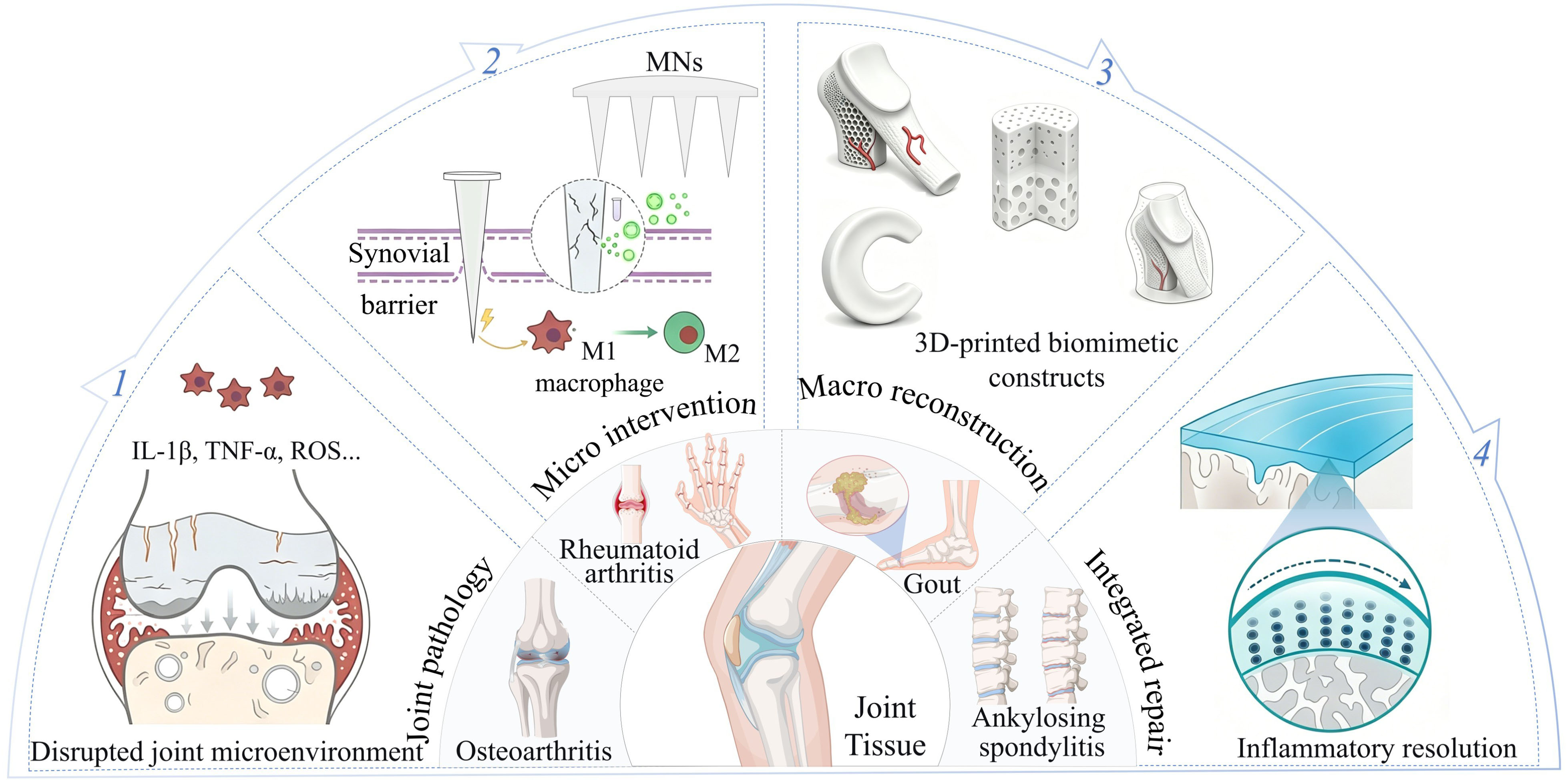
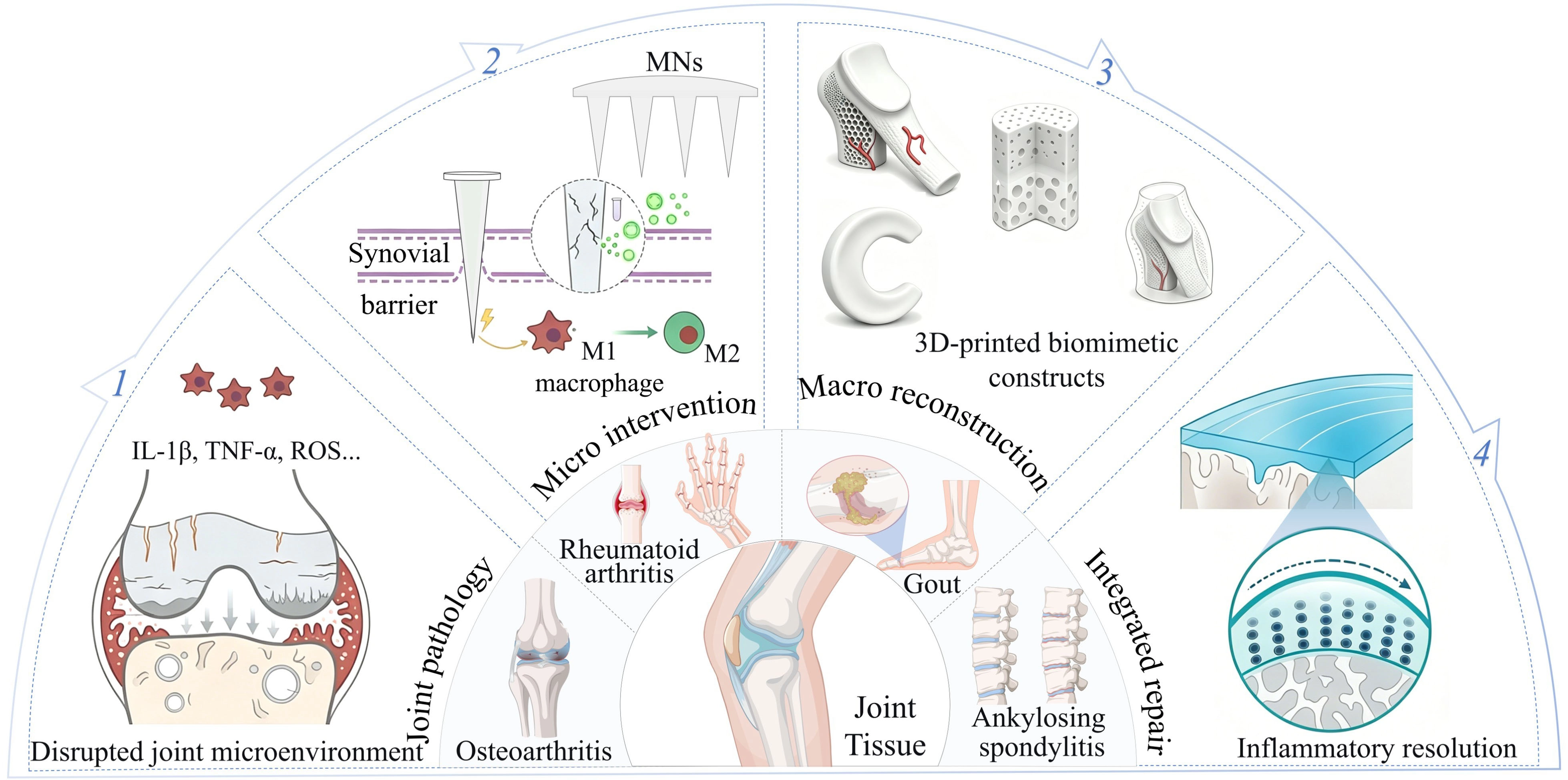
Figure 1. Schematic overview of the micro- and macro-scale joint repair paradigm. MNs: microneedles. IL-1β: interleukin-1β; TNF-α: tumor necrosis factor-alpha; ROS: reactive oxygen species.
2. Micro-Scale Smart Biointerfaces for Local Joint Modulation
2.1 MNs for controlled biochemical factor release
Joint diseases, such as OA and RA, are characterized by multi-scale and multi-cascade pathological features. Conventional systemic administration or IA injections are heavily constrained by the physical barriers of cartilage and synovium, alongside the rapid clearance of synovial fluid, which impairs the achievement of long-term disease-modifying therapy. Against this background, biochemical factor-controlled-release MNs, leveraging material chemistry design and microenvironmental sensing mechanisms, bypass the limitations of passive administration to serve as controlled-release vehicles that actively intervene in pathological progression. These platforms recognize joint pathological cues, such as mechanical strain, oxidative stress, metabolic surges, and cytokine cascades, to trigger on-demand drug release or structural adaptation. Therefore, they not only achieve the spatio-temporally controlled delivery of small-molecule anti-inflammatory drugs, but also effectively expand the delivery of biomacromolecules, such as polysaccharides, exosomes, genes, and peptides, thereby demonstrating distinct advantages in analgesia, cartilage regeneration, and immunoregulation.
Frequent mechanical stretching and inflammatory swelling within the joint environment often precipitate the detachment of conventional MNs or cause disordered drug diffusion. Consequently, establishing stable and burst-release-resistant delivery pathways is fundamental for long-term therapeutic interventions. As illustrated in Figure 2a, inspired by the adhesion mechanisms of octopus suckers and mussel byssus[24], Zhang et al. developed biomimetic adhesive MNs integrating micro-cavities and a polydopamine hydrogel substrate. The base utilizes Schiff base/Michael addition and catechol interactions for tissue adhesion, while suction-cup microchambers generate physical anchoring under wet, dynamic motion. This design enabled robust mechanical anchoring under dynamic joint conditions, and upon loading with glucocorticoids, effectively promoted cartilage repair in OA models. To enhance penetration capability, Cai et al. designed thumbtack-shaped titanium MNs with in situ grown titanium dioxide (TiO2) nanotubes on the surface[25]. By functionalizing the nanotubes with a crosslinked poly(vinyl alcohol) (PVA)-octadecyltrimethoxysilane (PVA-OTS) sol-gel coating TiO2 nanotubes as a diffusional barrier, this platform sustained diclofenac diethylamine release for over 8 h, which, combined with continuous physical needling at peri-articular acupoints (ST35, ST36, GB34), effectively alleviated OA inflammation, pain, and secondary cartilage erosion (Figure 2b). Addressing the toxicity risks associated with local burst release, Pham et al. fabricated the microneedle-based integrated release with advanced composite layered efficacy three-layer core-shell MN system[26]. The intermediate hydrophobic poly(lactic-co-glycolic) acid (PLGA) subshell minimizes contact between the drug core and surrounding aqueous fluids, suppressing hydrolytic burst degradation and achieving a sustained release profile with significantly reduced initial burst (Figure 2c). Furthermore, synchronizing drug-release kinetics with the dynamic pathological microenvironment remains critical for achieving adaptive interventions with MN platforms. Targeting the pronounced uric acid fluctuations and high toxicity of conventional systemic administration during gout flares, Yang et al. constructed a reactive oxygen species (ROS)-responsive smart hydrogel MN, fabricated from dopamine-modified β-cyclodextrin, for the co-delivery of uricase and colchicine[27]. As illustrated in Figure 2d, within a hyperuricemic environment, the degradation of the hydrogel network was accelerated by the hydrogen peroxide (H2O2) generated via uricase catalysis, thereby enabling on-demand drug release. Specifically, uricase decomposes uric acid into allantoin and H2O2, which oxidatively cleaves dynamic borate ester bonds crosslinking the β-cyclodextrin dialdehyde (β-CD-DA)/PVA/terephthalaldehyde (TPA) network to trigger rapid matrix disassembly and drug release. The patch-integrated “fast bubble-separation layer” enabled the MN tips to be retained subcutaneously within seconds of insertion, establishing a dynamic control loop that degrades urate crystals and suppresses acute gout attacks.
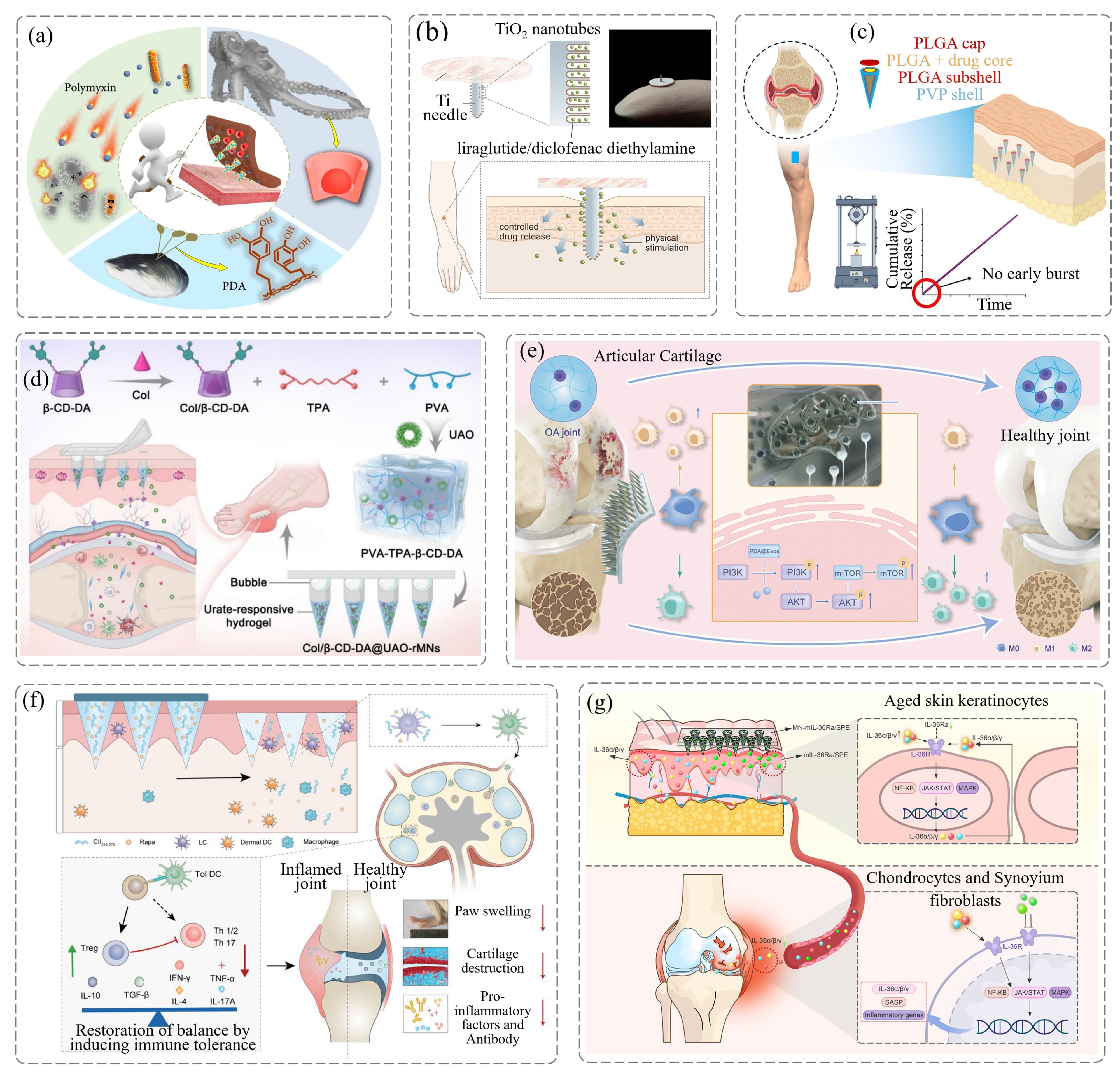
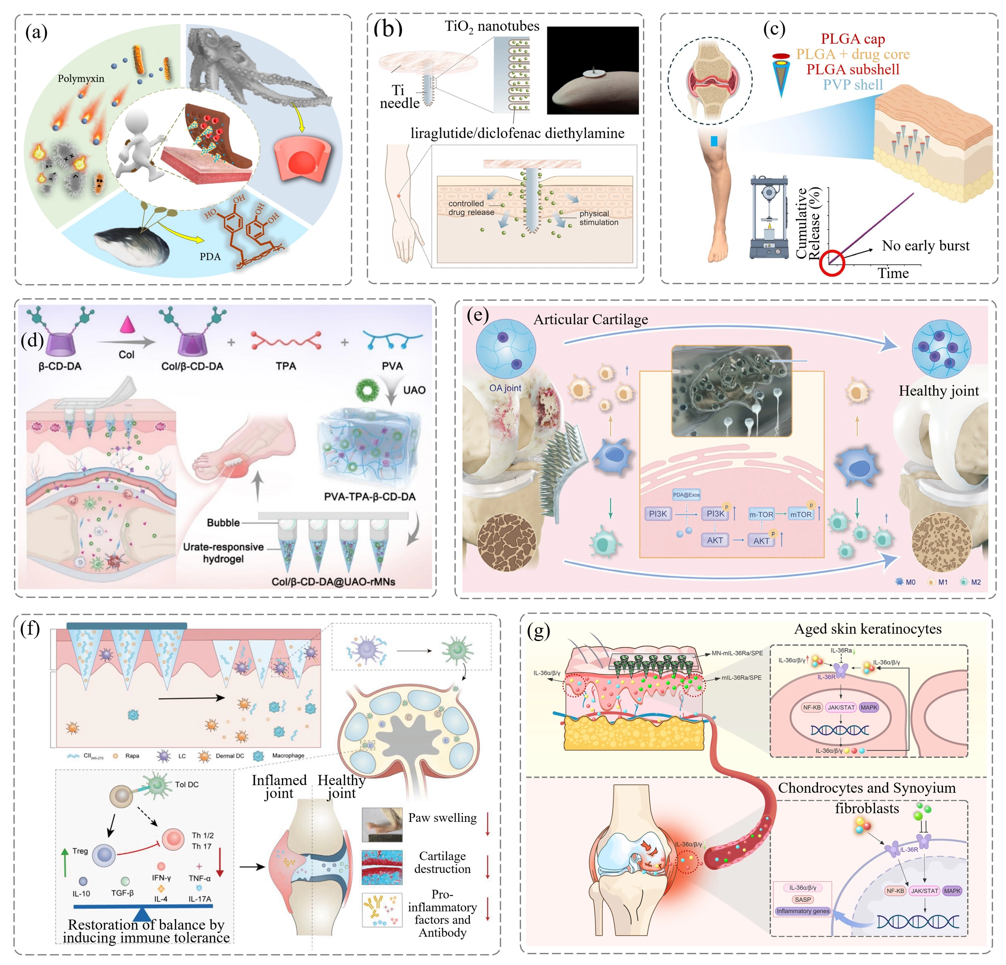
Figure 2. Structural design and therapeutic mechanisms of biochemical factor-controlled release MNs in joint interventions. (a) Bioinspired adhesive MNs employed under joint deformation for cartilage repair in OA. Reproduced from reference[24]. CC BY 4.0; (b) Thumbtack-like MNs integrated with physical needling and sustained drug release for the attenuation of cartilage erosion. Reproduced from reference[25]. CC BY 4.0; (c) Triple-layered core-shell MNs developed with suppressed drug burst release for prolonged analgesia and cartilage protection in OA. Reproduced from reference[26]. CC BY 4.0; (d) Uric acid-responsive MNs engineered for closed-loop, on-demand drug release to suppress acute gout attacks. Reproduced from reference[27]. CC BY 4.0; (e) Exosome-loaded composite MNs applied for inflammatory microenvironment remodeling to promote cartilage and subchondral bone regeneration. Reproduced from reference[29]. CC BY 4.0; (f) Immunomodulatory MNs utilized for the restoration of systemic immune tolerance to eliminate joint swelling in RA. Reproduced with permission from reference[31]. Copyright 2024 Wiley-VCH; (g) MNs targeted to the “skin-joint” axis for monoclonal antibody delivery to block the inflammatory cascade and reverse OA degeneration. Reproduced from reference[32]. CC BY 4.0. MNs: microneedles; OA: osteoarthritis; PLGA: poly(lactic-co-glycolic) acid; TiO2: titanium dioxide; β-CD-DA: β-cyclodextrin dialdehyde; PVA: poly(vinyl alcohol); TPA: terephthalaldehyde; PVP: poly(vinylpyrrolidone); PI3K: phosphoinositide-3 kinase; Akt: protein kinase-B; mTOR: mammalian target of rapamycin; PDA@Exo: polydopamine-exosome.
Leveraging the optimized mechanisms of mechanical anchoring and responsive controlled-release, MNs have emerged as a highly versatile platform for the delivery of biologics (including exosomes, genes, and antigens) to orchestrate the remodeling of synovial, cartilage, and systemic immune networks. For synergistic biochemical intervention in OA, Xiao et al. utilized natural sea cucumber-derived polysaccharide as the MN matrix to co-deliver 3-acetylaconitine liposomes[28]. This approach effectively downregulated pro-inflammatory cytokines, promoted collagen synthesis within cartilage, and elevated mechanical pain thresholds in animal models. To further reverse cartilage degeneration, Li et al. developed an MN-mediated combination therapy encapsulating polydopamine-mesenchymal stem cell exosomes polydopamine-exosome (PDA@Exo)[29]. As illustrated in Figure 2e, the polydopamine component within this system scavenged excess IA ROS, while the delivered exosomes activated the intracellular phosphoinositide-3 kinase-protein kinase-B-mammalian target of rapamycin (PI3K-Akt-mTOR) signaling pathway in chondrocytes. Reductive catechol and imine groups on the PDA nanoparticles efficiently scavenge excessive intracellular ROS to alleviate oxidative stress and restore mitochondrial membrane potential, while the delivered exosomes promote M2 macrophage polarization (upregulating arginase 1 (Arg 1)/cluster of differentiation 206 (CD206) and downregulating inducible nitric oxide synthase (iNOS)). Consequently, this concurrent action not only curtailed apoptotic processes but also instigated the polarization of resident macrophages toward the anti-inflammatory M2 phenotype, thereby achieving synergistic restoration of both cartilage and subchondral bone.
For RA intervention, which is typically accompanied by immune dysregulation, Hua et al. employed dissolving MNs to co-deliver the pro-apoptotic gene p53 upregulated modulator of apoptosis (PUMA) and celastrol[30]. This strategy specifically induced apoptosis in highly invasive fibroblast-like synoviocytes (FLS) while simultaneously blocking the nuclear factor kappa B (NF-κB) inflammatory cascade in macrophages, thereby effectively restoring synovial homeostasis. Additionally, Zhao et al. utilized polymeric MNs for the co-delivery of autoantigens and low-dose rapamycin[31], which enabled the targeted recruitment of epidermal Langerhans cells and induced their phenotypic conversion into tolerogenic dendritic cells. Upon transdermal dissolution of the hyaluronic acid matrix, rapamycin inhibits mTOR in dendritic cells to downregulate maturation markers (CD80/CD86) while upregulating the CCR7 homing receptor. As shown in Figure 2f, following CCR7-mediated homing to draining lymph nodes, these tolerogenic dendritic cells systemically potentiated the differentiation of regulatory T cells (Tregs, elevating interleukin-10 (IL-10)/transforming growth factor beta (TGF-β)) while suppressing Th1/Th17-mediated pro-inflammatory responses (decreasing tumor necrosis factor-alpha (TNF-α), IL-17A, and interferon-gamma (IFN-γ)), thus achieving a translational leap from localized drug administration to the reconstruction of systemic immune tolerance. Aligning with this cross-scale regulatory paradigm, Chen et al. elucidated a “skin-joint” axis whereby aged skin exacerbates OA pathogenesis[32]. In aged skin, reduced IL-36Ra in senescent keratinocytes promotes excessive secretion of IL-36 agonists (IL-36α/β/γ), driving joint NF-κB, mitogen-activated protein kinases (MAPK), and Janus Kinase-Signal transducer and activator of transcription (JAK-STAT) inflammatory cascades. As depicted in Figure 2g, the sustained delivery of recombinant IL-36Ra protein or a specific monoclonal antibody (Spesolimab) into the cutaneous layer using MNs directly blocked the full-scale IL-36R pro-inflammatory signaling cascade, downregulating catabolic enzymes (matrix metalloproteinase-13 (MMP-13), MMP-3, a disintegrin and metalloproteinase with thrombospondin motifs 5 (ADAMTS-5)) and effectively attenuating OA progression. These collective findings demonstrate that biochemical factor-controlled-release MNs possess substantial translational potential for the systemic intervention of complex joint disorders.
2.2 MNs for physical stimuli responsiveness and mechanotransduction
As dynamic microenvironments subjected to multidimensional mechanical stress, joints present formidable physical barriers composed of the dense cartilaginous ECM and hyperplastic synovial linings. Conventional MNs rely predominantly on passive diffusion, which severely constrains deep tissue penetration and cell-specific targeted delivery. Furthermore, in the face of highly dynamic pathological cascades, such as acute gouty flare-ups, OA inflammatory surges, or RA immune storms, passive sustained-release kinetics fail to match the imperative for high-dose interventions during acute phases, often resulting in delayed therapeutic efficacy or off-target toxicity. To overcome these bottlenecks, a new class of physical/mechanical signal-transducing MN platforms has emerged, integrating active physical modalities, including thermal, electromagnetic, acoustic, and mechanical energies, into the device architecture. On the one hand, active physical energy effectively breaches physiological tissue barriers to facilitate deep anti-inflammatory drug permeation and highly specific, on-demand triggered release, thereby precisely aligning with therapeutic windows. On the other hand, the transduced physical signals inherently possess disease-modifying capabilities, directly activating chondrocyte metabolic pathways, improving microcirculation, and modulating immune polarization.
Among the various physical intervention mechanisms, thermodynamic physical fields, specifically photothermal conversion and thermal conduction, have demonstrated significant advantages in reshaping the arthritic inflammatory microenvironment and governing drug release kinetics. For instance, Lu et al. constructed a near-infrared-triggered smart photothermal MN platform that leverages hyperthermia to accelerate matrix phase transitions for the on-demand delivery of loxoprofen and tofacitinib[33]. Concomitantly, the localized mild thermal effect ameliorates RA joint microcirculation and expands physical permeation channels. To overcome the penetration limitations of superficial photothermal therapy, Lin et al. devised dynamic helically embedded heat-transfer MNs by capitalizing on the excellent solid-state thermal conductivity of the MN platform[34]. As shown in Figure 3a, this design directly conducts external thermal energy to deep-seated lesions, physically suppressing mitochondrial apoptosis. Concurrently, the thermal energy serves as a physical switch that triggers the dissociation of embedded hydrogel microspheres, thereby achieving a deep synergy between macroscopic heat conduction and microscopic organelle-targeted regulation.
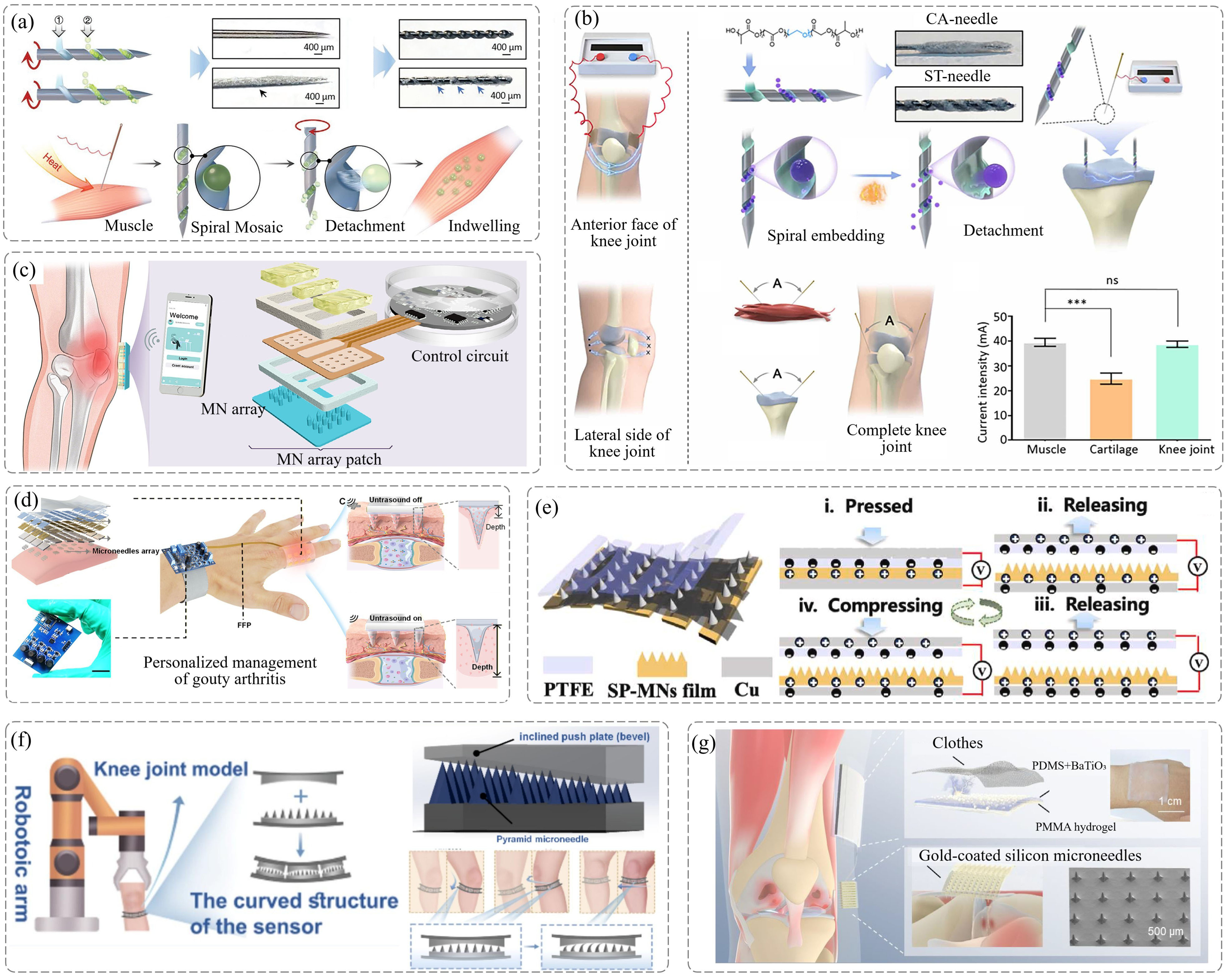
Figure 3. Structural design and therapeutic mechanisms of physical field-responsive and mechanotransductive MNs in multiscale joint interventions. (a) Thermal-conductive MNs targeted to deep tissues for thermal conduction to inhibit mitochondrial apoptosis and repair damaged cartilage. Reproduced with permission from reference[34]. Copyright 2023 Wiley-VCH; (b) Threaded MNs designed for microcurrent constraint via geometric configuration to regulate cellular metabolism and repair degenerated tissues in situ. Reproduced from reference[35]. CC BY 4.0; (c) Wearable electrically controlled MNs driven by iontophoresis for on-demand controlled release of multiple anti-inflammatory drugs in RA. Reproduced with permission from reference[36]. Copyright 2026 Wiley-VCH; (d) Ultrasound-responsive MNs utilized for physical barrier disruption via directional acoustic streaming to achieve rapid detumescence during acute gout phases. Reproduced from reference[37]. CC BY 4.0; (e) Kirigami MNs employed for the conversion of mechanical friction into endogenous electrical stimulation to promote cartilage healing in OA. Reproduced from reference[38]. CC BY 4.0; (f) 3D-printed sensing MNs applied for localized micro-stress amplification to achieve highly sensitive monitoring of joint micro-displacements. Reproduced from reference[39]. CC BY 4.0; (g) Friction-driven MNs utilized for endogenous electrical stimulation generation to synergize with nanozymes for on-demand scavenging of reactive ROS within OA lesions. Reproduced with permission from reference[40]. Copyright 2026 Wiley-VCH. MNs: microneedles; RA: rheumatoid arthritis; ROS: reactive oxygen species; OA: osteoarthritis; PTFE: polytetrafluoroethylene; CA-needle: Chinese acupuncture needle; ST-needle: screw-thread needle; PDMS: polydimethylsiloxane; PMMA: poly(methyl methacrylate).
In the realm of electromagnetic and acoustic actuation, external energy fields have similarly demonstrated robust capabilities in overcoming passive permeation limitations. As shown in Figure 3b, Lin et al. designed physically/chemically dual-constrained screw-threaded MNs based on electromagnetic principles[35]. By leveraging the physical configuration to constrain microcurrent pathways, this system precisely modulated adenosine triphosphate (ATP) and calcium ion fluxes between the mitochondria and endoplasmic reticulum of diseased cells, thereby achieving a combined electro-biochemical intervention on target cells. Extending this intervention to the macroscopic tissue level, Chen et al. developed a wearable electronically controlled MN device integrated with microcircuits[36] (Figure 3c). Leveraging the iontophoresis effect, the system orchestrates the spatiotemporal controlled release of multiple RA therapeutics, remarkably downregulating localized pro-inflammatory cytokine expression. To satisfy the imperative for rapid infiltration during acute gouty flares, Zhang et al. introduced an ultrasound field to construct flexible responsive MNs[37]. Utilizing the tip-directed acoustic streaming effect, this system markedly enhanced deep drug penetration, enabling a closed-loop rapid drug delivery that initiated local anti-inflammatory and anti-edema responses within 10 minutes, as demonstrated in Figure 3d.
In the domain of self-powered mechanical energy conversion and high-sensitivity sensing, directly transducing daily joint kinematics into therapeutic or monitoring signals has carved out a novel pathway for the development of battery-free intelligent devices. As shown in Figure 3e, Li et al. designed a silk fibroin MN-based triboelectric nanogenerator (TENG) leveraging a kirigami metamaterial mechanical design[38]. This platform directly translates macro-scale joint deformations into endogenous electrical stimulation, thereby actively accelerating OA cartilage healing. On the input side of the theranostic closed loop, given that conventional planar sensors fail to capture deep subtle motions, Zhang et al. constructed polyacrylamide hydrogel MN sensors via 3D printing[39]. By utilizing the 3D geometric deformation of the needle body to amplify local stress variations, this system achieves ultra-high sensitivity in detecting subtle knee displacements and prodromal stiffness, thereby providing mechanical support for dynamically adjusting intervention dosages (Figure 3f). To further deepen the cross-disciplinary synergy between mechanomechanics and biochemical intervention, Yue et al. integrated TENG with electroresponsive nanozymes[40]. As depicted in Figure 3g, this strategy harvests mechanical energy from somatic joint movements and converts it into microcurrents that mimic electroacupuncture. Consequently, it exploits an electric field-catalysis dual synergy to scavenge excessive ROS at the OA lesion site on demand, elevating the battery-free, closed-loop paradigm of “exercise-as-therapy” to a new level.
In summary, the emergence of physicomechanical signal-transducing systems signifies that acoustic, optical, electrical, and thermal energy fields no longer serve merely as external triggers for drug release; rather, they have become core therapeutic interventions intrinsically involved in tissue remodeling. Looking toward future clinical translation, the critical challenges lie in further surmounting the integration limits of flexible micropower sources, ensuring the long-term mechanical compatibility of high-hardness MN arrays under repeated cycles of severe joint friction, and achieving deep integration of sensor networks with artificial intelligence algorithms. Addressing these challenges will be pivotal for the development of next-generation closed-loop joint theranostic devices.
3. Macroscale Structural Engineering for Joint Tissue Repair
Beyond microscale biochemical regulation, the reconstruction of macroscopic anatomical morphology and mechanical function is critical for achieving long-term joint repair. The articular cavity is situated in an extreme mechanical environment characterized by high strain, multiaxial cyclic loading, and continuous synovial fluid shearing. Native joint tissues, relying on anisotropic collagen networks and multiphasic gradient interfaces, have established efficient energy dissipation mechanisms. However, conventional homogeneous scaffolds are susceptible to mechanical fatigue, often suffering from stress concentration and structural failure; moreover, lacking spatial physical constraints, these implants are prone to being washed away by synovial fluid. Therefore, the underlying logic of joint repair urgently requires a paradigm shift from “simple biochemical component stacking” toward “cross-scale spatially structured programming”. AM, with its advantages of bottom-up customization of physical boundaries and construction of hierarchical modulus gradients, has emerged as a pivotal tool for physical topological remodeling. Current cutting-edge explorations are primarily centered on two main trajectories: bioinspired architectures and mechanically guided tissue formation, as well as spatially programmed multi-factor regulatory systems.
3.1 Bioinspired architectures and mechanically guided tissue formation
In the tissue engineering of cartilage and fibrocartilaginous tissues such as the meniscus, the primary challenge confronting implants is mechanical decompensation precipitated by severe compression, shear stress, and high-frequency cyclic loading. Evidence shows that simply enhancing the intrinsic properties of homogeneous materials is insufficient to match the complex dynamic mechanical environment. In recent years, leveraging AM to precisely replicate the multilayered biomimetic architectures of native tissues, including load-bearing frameworks, heterogeneous interfaces, and anisotropic fibers, has emerged as a novel paradigm of “structure-directed function”, whereby specific topological configurations are translated into active physical instructions that guide cell proliferation, differentiation, and spatial self-assembly.
To address the bottleneck of conventional hydrogels that are prone to fatigue attenuation due to the lack of rigid support, Li et al. proposed a “hard skeleton-soft matrix” composite strategy[41]. Utilizing fused deposition modeling 3D printing, they constructed a high-modulus polycaprolactone (PCL) scaffold as a load-bearing foundation that closely conforms to the anatomical morphology of native meniscus, while crosslinking magnesium ion-loaded sodium alginate hydrogel within the interstices (Figure 4a). This architecture effectively buffers the shear and compressive stresses of the knee joint while providing a structural sanctuary for the sustained release of magnesium ions. In addition, to address the mechanical mismatch and interfacial delamination commonly occurring across disparate soft-to-hard structural boundaries, Kim et al. developed a wavelength-selective multimaterial digital light processing (DLP) technique utilizing a hybrid epoxy-acrylate resin[42]. By leveraging orthogonal photocuring at 365 nm (cationic/radical dual-curing for rigid domains, E ≈ 1.7 GPa) and 405 nm (radical-only curing for elastic domains, E ≈ 0.6 MPa), this platform realizes monolithic 3D printing of seamless hard-soft composites with an extreme stiffness disparity exceeding 1,000-fold (Figure 4b). At the material mechanics level, this strategy effectively prevents interfacial stress concentration and structural collapse under cyclic loading, enabling programmable compressive damping architectures as well as biomimetic articulating joint constructs featuring rigid bone segments seamlessly interfaced with flexible ligaments. To enhance mechanical tolerance at the microscopic network level, Zhu et al. drew inspiration from the native intervertebral disc to crosslink a peptide-based rigid nanorod (PRN) framework within gelatin methacryloyl (GelMA) hydrogels, as depicted in Figure 4c[43]. This successfully translated nanoscale energy dissipation mechanisms into a remarkable macroscopic compressive strength enhancement of 1,018% along with exceptional fatigue resistance, providing a highly stable physical scaffolding for in situ cartilage regeneration. Furthermore, recapitulating the continuous fiber orientation of native tissues is equally critical for reconstructing load-bearing architectures. To confront the challenge of poor suture tear resistance in meniscus implants, Ma et al. developed a continuous fiber impregnation 3D printing technique[44], directionally integrating high-strength silk fibers within the interior of PCL filaments. This biomimetic architecture not only replicates the anisotropy and suture pull-out strength of the native meniscus, but also serves as a sustained active mechanical stimulation source that activates chondrocyte mechanotransduction pathways, driving long-term ECM deposition and remodeling (Figure 4d).
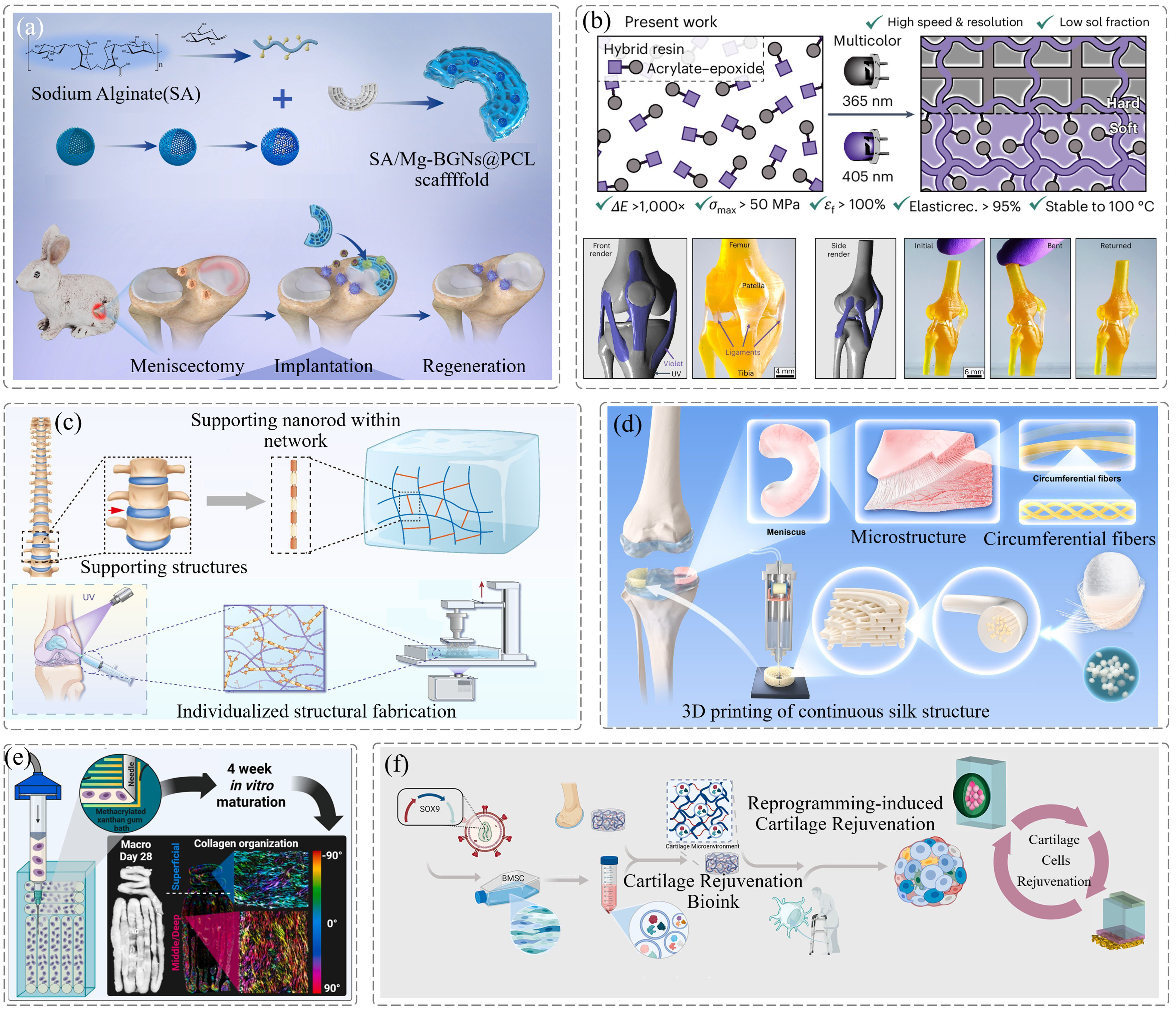
Figure 4. Biomimetic structures and mechanically guided tissue construction. (a) Biomimetic multiphasic PCL-hydrogel composite scaffolds utilized for meniscus reconstruction. Reproduced from reference[41]. CC BY 4.0; (b) Wavelength-selective multi-material 3D printing employed for the reconstruction of heterogeneous joint interfaces. Reproduced with permission from reference[42]. Copyright 2025 Springer Nature; (c) Micro-hydrogels reinforced with peptide nanorods for repair of osteochondral defects. Reproduced from reference[43]. CC BY 4.0; (d) Continuous silk fiber-reinforced scaffolds replicated from the anisotropic characteristics of the meniscus. Reproduced with permission from reference[44]. Copyright 2026 Cell Press; (e) Collagen guided by support baths as physical boundaries for biomimetic arcade-like alignment. Reproduced from reference[46]. CC BY 4.0; (f) BGSC-organoid culture systems driven for the rejuvenation of senescent chondrocytes. Reproduced from reference[47]. CC BY 4.0. PCL: polycaprolactone; BGSC: biomimetic gradient-structured chondrocyte organoid; BMSC: bone marrow-derived mesenchymal stem cell.
As research perspectives evolve from macroscopic mechanical support toward fine physical boundary constraints, 3D printing has demonstrated significant advantages in directly guiding cell fate through the construction of specific spatial topologies. To overcome the challenges of delayed maturation and phenotypic instability in cartilage organoids during in vitro culture, Shen et al. developed a high-precision biomimetic network based on DNA-silk fibroin utilizing DLP bioprinting[45]. This system provides 3D physical confinement and permeable nutrient infiltration pathways for early-stage organoid development, effectively resisting mechanical collapse immediately after implantation while significantly accelerating intercellular 3D communication and the directed secretion of specific matrix components. Furthermore, Karam et al. proposed a novel cell-only bioprinting paradigm based on a suspension support bath, which employs the supporting medium to precisely confine chondroprogenitor cells within predefined macroscopic spatial boundaries (Figure 4e)[46]. By liberating cells from the physical entrapment and spreading constraints imposed by conventional polymeric inks, this strategy relies solely on the mechanical guidance of spatial boundaries to successfully induce an arcade-like collagen fiber self-assembly that closely mimics the superficial zone characteristics of native cartilage, thereby providing crucial architectural support. Additionally, Miao et al. constructed cartilage organoids with macroscopic biomimetic gradients, faithfully recapitulating the hierarchical variations in cell density, matrix composition, and biomechanical modulus characteristic of native cartilage[47]. This structured platform not only closely approximates native tissue in terms of physical load-bearing capability but also modulates the forkhead box O (FOXO) pathway via coupled mechanobiological cues, achieving deep rejuvenation and functional reversal of damaged cartilage, as depicted in Figure 4f.
In summary, the biomimetic architectures fabricated by 3D printing have comprehensively transcended the paradigm of passive physical filling. By precisely recapitulating the mechanical gradients, fiber orientation, and multiscale spatial constraints of native tissues, these topological configurations have been elevated to active physical intervention instructions. Through these instructions, target cells can be precisely guided toward the assembly of fully functional biomechanical networks, with reduced dependence on exogenous biochemical factors. Consequently, these advancements establish a critical scientific pathway for the structural and functional reconstruction of the entire joint load-bearing system. Beyond static structural biomimicry, the integration of 4D printing with stimuli-responsive biomaterials has introduced a dynamic paradigm of shape-morphing scaffolds capable of active spatial reconfiguration and functional adaptation in response to environmental cues, including temperature, magnetic fields, light, and humidity[48,49]. By encoding temporary compact geometries into shape-memory polymer composites (SMPCs), such as magnetic nanoparticle-doped polylactic acid matrices, scaffolds can be delivered through minimally invasive surgical channels and subsequently triggered in situ via remote alternating magnetic fields or physiological thermal shifts to recover their pre-programmed porous architectures, achieving seamless defect-conformal self-fitting and immediate load support[48]. Furthermore, multi-responsive 4D biomass and polymer networks can undergo sequential shape transformation, targeted drug release, and dynamic mechanotransductive signaling, offering an integrated engineering pathway for minimally invasive tissue reconstruction and joint defect repair[49].
3.2 Spatially programmed multi-factor regulatory systems
Despite the stable mechanical support provided by physical biomimetic structures for tissue reconstruction, the pathological progression of degenerative OA or RA is often accompanied by the disruption of multiphasic interfaces and highly heterogeneous inflammatory cascades. Consequently, achieving the heterogeneous reconstruction of multiple distinct components within a single, integrated implant, while simultaneously ensuring the spatiotemporal controlled release of interventional factors, remains a core challenge in joint tissue engineering. The framework of multi-factor spatial programming architectures introduces a promising, state-of-the-art strategy to circumvent this obstacle. By leveraging AM’s superior spatial positioning and heterogeneous customization, this paradigm precisely anchors specific cells, active microspheres, or macromolecular drugs in 3D coordinate networks, thereby constructing regulatory systems characterized by temporal responsiveness and spatial heterogeneity. Another core pathological feature of joint diseases is the cross-interface dissociation of multiphasic tissues and the inflammatory erosion caused by pathological synovial fluid. Within complex lesions involving cartilage-bone or synovium-cartilage interfaces, the repair of a single component often leads to disconnection at the cross-interface junction. As illustrated in Figure 5a, Zhang et al. exploited the localized composition customization capabilities of multi-nozzle 3D bioprinting to synchronously orchestrate articular chondrocyte progenitor cells and bone marrow mesenchymal stem cells into a predefined cellular gradient within a singular physical space[50]. This strategy circumvented the limitations inherent to monophasic repair, effectively driving microscopic crosstalk and seamless structural integration across the cartilage-bone-vascular multiphasic interface at a three-dimensional scale. Furthermore, Zhou et al. utilized 3D printing to precisely deposit a bilayered composite hydrogel, comprising a recellularized type II collagen cartilage base and an overlying layer co-loaded with FLS and multi-phenotype macrophages, thereby constructing a highly biomimetic human synovium-cartilage in vitro model, as depicted in Figure 5b[51]. This system successfully recapitulated the complex spatial pathological evolution of RA, including synovial invasion, macrophage inflammatory polarization, and matrix degradation, providing a high-fidelity 3D testing platform for evaluating the interventional efficacy of joint repair scaffolds.
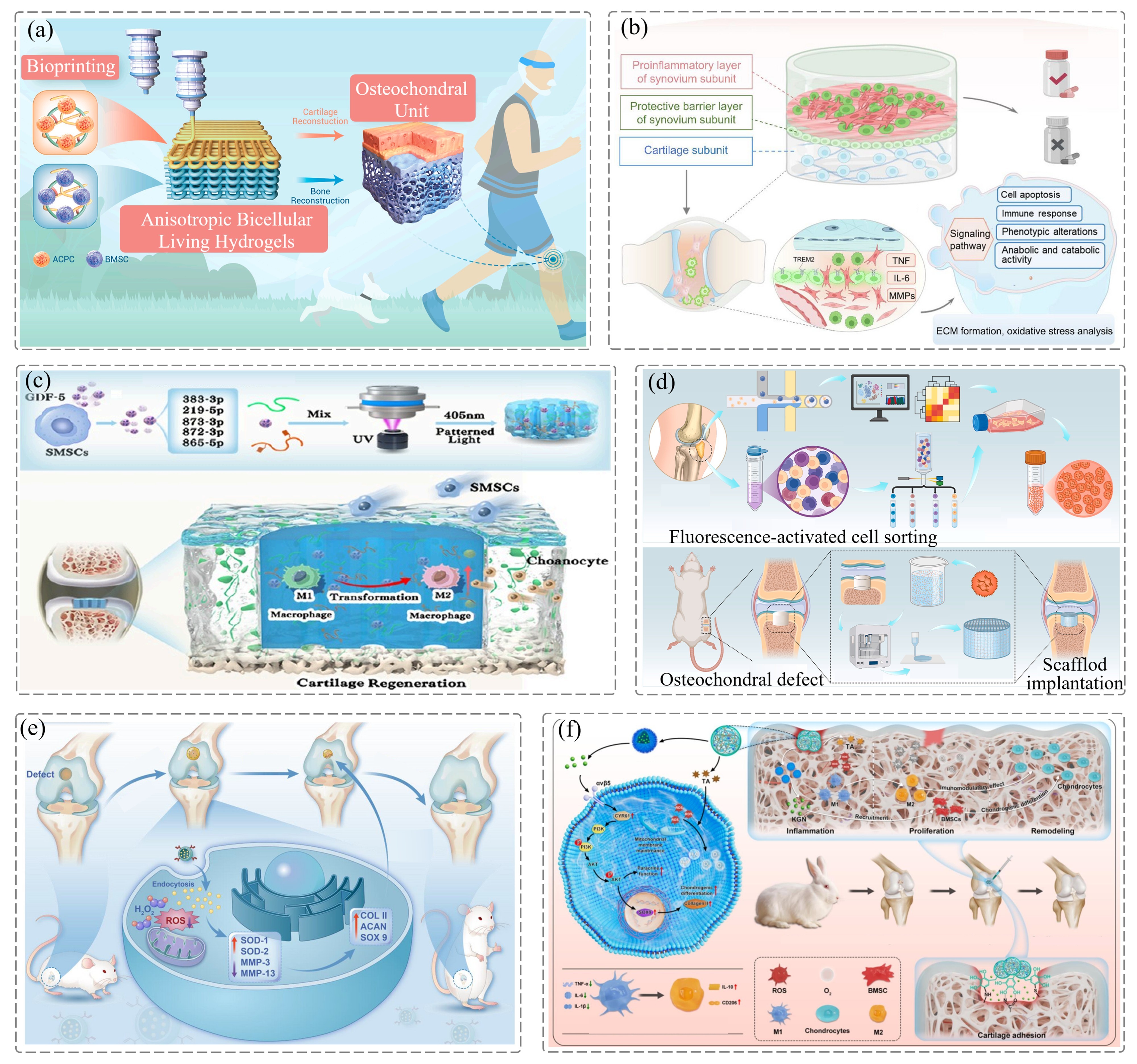
Figure 5. Multi-factor spatially programmed regulation systems. (a) Anisotropic dual-cell-laden active hydrogels applied for in situ reconstruction of the osteochondral biphasic interface. Reproduced from reference[50]. CC BY 4.0; (b) In vitro synovium-cartilage models established for simulating multiphasic pathological interactions in RA. Reproduced from reference[51]. CC BY 4.0; (c) 3D-printed porous physical reservoirs loaded with targeted exosomes from GDF-5-pretreated stem cells. Reproduced with permission from reference[52]. Copyright 2025 American Chemical Society; (d) Schematic illustration presented for the construction of biomimetic 3D-printed scaffolds for osteochondral regeneration. Reproduced from reference[53]. CC BY 4.0; (e) Cell capsule-delivering bioink systems developed for cartilage defect repair. Reproduced with permission from reference[54]. Copyright 2025 Wiley-VCH; (f) 3D-printed integrated bioactive microspheres endowed with multiple pathological stimulus-responsiveness. Reproduced with permission from reference[55]. Copyright 2025 Elsevier. RA: rheumatoid arthritis; GDF-5: growth differentiation factor 5; ROS: reactive oxygen species; MMP-13: matrix metalloproteinase-13; MMP-3: matrix metalloproteinase-3; SOD-1: superoxide dismutase 1; SMSCs: synovium-derived mesenchymal stem cells; SOD-2: superoxide dismutase 2; Sox9: sex determining region Y (SRY)-box 9.
On the other hand, to overcome the bottleneck of rapid synovial washout of free therapeutic agents, multi-factor spatial programming architectures convert 3D-printed scaffolds into “spatial physical reservoirs” that ensure the long-term, steady-state controlled release of biochemical cues. Integrating 3D-printed topological structures with cell-free exosome therapeutics represents a powerful modality to trigger endogenous tissue regeneration. As shown in Figure 5c, Zheng et al. developed a porous composite scaffold using 3D printing and precisely localized growth differentiation factor 5 (GDF-5)-pretreated mesenchymal stem cell exosomes within its framework[52]. By providing a sustained release of miR-383-3p-enriched exosomes, this physical reservoir successfully drives the homing and targeted differentiation of endogenous synovial stem cells into the defect zone. To further implement immunomodulatory control, Lou et al. integrated newly identified human skeletal stem cell exosomes into a 3D-printed scaffold based on single-cell RNA sequencing insights (Figure 5d)[53]. This platform not only matches the damaged anatomy with high-fidelity physical geometry, but also harnesses the sequential release of exosomes to direct the polarization of local macrophages toward a pro-healing M2 phenotype, ultimately achieving deep remodeling of the immune microenvironment alongside mechanical scaffolding.
Concomitantly, the refined orchestration of microvesicles, bioactive microspheres, and macromolecules within 3D-printed spatial coordinate grids confers enhanced structural fidelity and spatiotemporal specificity upon controlled-release platforms. As shown in Figure 5e, Yu et al. engineered a bioink functionalized with antioxidant punicalagin-loaded chondrocyte membrane vesicles, constructing a novel cell-capsule delivery scaffold via high-resolution DLP bioprinting[54]. Within this architecture, the vesicles are stably anchored by the resulting 3D cross-linked polymeric network, establishing a long-acting reactive ROS-scavenging barrier within the highly dynamic IA cavity. To address the long-standing hurdles of polydisperse microsphere dimensions and insufficient IA retention, Zhou et al. implemented DLP bioprinting to develop stimulus-responsive, multifunctional bioactive microspheres (Figure 5f)[55]. Physical programming of their internal microarchitectures successfully enables the pathologically responsive, on-demand controlled release of chondrogenesis-promoting molecules, such as kartogenin. Regarding the precise assembly of multi-component biomaterial inks, Jahani et al. developed 3D-printed alginate-chitosan scaffolds that achieved controlled spatial distribution and sustained delivery of insulin to promote chondrogenic differentiation of stem cells for cartilage regeneration[56]. Concurrently, Su et al. engineered a refined spatial co-assembly of cartilage-protective modified citrus pectin (MCP) macromolecules with GelMA/hyaluronic acid (HAMA) matrices and living cells, where network degradation drives the chronological release of MCP to exert synergistic anti-inflammatory and matrix-degrading inhibitory effects[57].
In summary, whether through the biomimetic structural design that provides mechanotransductive instructions via anisotropic physical architectures, or through the programmatic orchestration systems that construct multi-component spatiotemporal controlled-release networks via digital AM, the breakthroughs achieved by 3D printing in multiscale structural engineering have reshaped the fundamental paradigm of joint tissue repair. The synergistic advantages of these dual pathways, specifically in mechanical functionality adaptation, seamless cross-interfacial integration, and pathological microenvironment remodeling, are steadily propelling joint engineering toward the long-term milestone of structurally and functionally integrated tissue repair.
4. Summary and Perspectives
In summary, the pathology of joint degeneration spans cartilage wear, subchondral bone sclerosis, and synovitis cascades, where long-term therapeutic repair remains bottlenecked by dynamic IA loads and osteochondral interfacial failure. Conventional systemic drug delivery or homogeneous fillers fail to address these multiscale challenges that bridge inflammation regulation and structural reconstruction. To systematically synthesize the emerging platforms discussed herein, Table 1 provides a comprehensive classification framework across several core dimensions: stimulus modality (endogenous pathological triggers and exogenous physical fields), therapeutic scale (microscale bio-interfaces and macroscale structural constructs), drug delivery/action strategy (stimuli-responsive release, active physical permeation, and spatially programmed multicellular bioinks), and primary tissue-regeneration function (immunomodulation, ROS clearance, load bearing, and osteochondral integration). By synergizing micro-scale microenvironmental modulation with macro-scale biomechanical reconstruction, these intelligent platforms establish a versatile foundation for next-generation joint therapeutics. MN systems demonstrate unique advantages in modulating the local joint microenvironment. Specifically, biochemical factor-releasing MNs neutralize IA inflammation, regulate metabolism, and manage pain through the targeted delivery of small molecules and exosomes. Concurrently, physical signal-transducing MNs harness external stimuli, including electrical, thermal, ultrasonic, and triboelectric fields, or biomimetic mechanical sensing to achieve active physical modulation and real-time monitoring. These synergistic approaches provide a localized, on-demand, and long-term management paradigm for joint disorders. At the macroscopic level, 3D-printed scaffolds are dedicated to the structural replacement and regeneration of joint tissues. Biomimetic, mechanics-guided scaffolds utilize anisotropic topologies and gradient porosities to supply mechanical cues that direct cell orientation and matrix deposition. Furthermore, multi-factor spatially programmed scaffolds, fabricated via multi-channel bioprinting, enable the precise localization and programmed release of cellular and biochemical factors, effectively recapitulating natural tissue heterogeneity and reparative kinetics. Such innovations have shown initial success in the regeneration of the meniscus, osteochondral interfaces, and bone-cartilage units, establishing a robust foundation for future joint therapeutic interventions.
Table 1. Systematic multi-dimensional classification and characteristic comparison of smart therapeutic platforms for joint repair.
| Platform | Category | Therapeutic scale | Stimulus | Delivery/Action strategy | Key bio-function | Refs. |
| MNs | Biochemical-releasing MNs | Micro-scale (Local tissue/cellular level) | Endogenous: ROS, urate spikes, synovial pH, enzymes | Stimuli-responsive degradation, core-shell sustained release, rapid bubble separation | Local ROS clearance, M2 macrophage polarization, synovial homeostasis, systemic immune tolerance induction | [24,26,27,29,31,32] |
| Physical field-responsive/transducing MNs | Micro-scale (Cellular to tissue interface) | Exogenous/Motion: NIR/thermal, ultrasound, electric microcurrents, joint kinematics | Photothermal melting, acoustic streaming, iontophoresis, tribo-/piezoelectric stimulation | Deep tissue permeation, mitochondrial apoptosis suppression, electro-catalytic ROS scavenging, real-time kinematics sensing | [33,34,36-40] | |
| 3D-Printed Scaffolds | Bioinspired/mechanically guided scaffolds | Macro-scale (Organ/multiphasic tissue level) | Biomechanical: Dynamic compression, cyclic shear, spatial confinement | Topographical contact guidance, continuous fiber reinforcement, gradient modulus matching | Cell alignment, arcade-like collagen remodeling, mechanical load bearing, senescent chondrocyte rejuvenation | [41-44,46,47] |
| Spatially programmed multi-factor systems | Macro-to-micro hierarchy | Biochemical/Pathological: Spatial cues, cell-matrix crosstalk, MMPs, ROS | 3D coordinate-patterned bioinks, sequential exosome release, anchored microspheres | Cartilage-bonevessel integrated regeneration, continuous ROS scavenging barrier, endogenous stem cell homing | [50,52-55,57] |
ROS: reactive oxygen species; MNs: microneedles.
However, clinical translation still faces multifaceted engineering and biological bottlenecks across several core dimensions:
Biocompatibility and long-term degradation: Macro-scaffolds fabricated from photocurable resins require exhaustive toxicological validation to ensure that photoinitiators, unreacted residual monomers, and degradation byproducts do not provoke chronic synovitis or systemic toxicity. Crucially, the degradation kinetics of 3D-printed scaffolds must dynamically synchronize with the pace of neo-tissue deposition to prevent premature structural collapse or foreign-body encapsulation. For transdermal MNs, long-term repeated application requires non-immunogenic, highly biocompatible matrix polymers that avoid localized dermal irritation, erythema, or micro-scarring.
Dynamic mechanical adaptation and fixation: Articulating joints subject therapeutic scaffolds to complex, multi-axial cyclic loading, necessitating strict biomechanical harmony across distinct physical criteria. Hierarchical modulus matching remains essential to eliminate stress concentrations and mitigate stress shielding across the heterogeneous osteochondral interface, an objective increasingly facilitated by multi-parameter and algorithmic optimization to tailor regional stiffness[58]. Concurrently, enduring millions of dynamic weight-bearing cycles annually demands exceptional fatigue resistance and rapid compressive recovery, which are critical to preventing micro-crack propagation and interfacial shear delamination under repetitive joint locomotion[59]. Furthermore, coupling biomimetic porous architectures with smart shape-memory composites enables scaffolds to provide robust load-bearing support while enabling minimally invasive delivery and defect-conformal self-fitting[48]. For wearable MN arrays, optimizing stress redistribution alongside flexible backing compliance is equally vital to prevent needle fracture and substrate delamination during active joint flexion.
Scalable manufacturing and sterilization protocols: Translating complex 3D-printed scaffolds demands cleanroom-compatible, high-throughput AM systems with validated batch-to-batch reproducibility. Standard terminal sterilization methods (such as autoclaving, ethylene oxide, or gamma irradiation) must be carefully calibrated to avoid compromising the structural architecture of polymer scaffolds or denaturing temperature-sensitive biologics encapsulated within MNs.
Surgical feasibility and implantation procedures: While MNs offer non-invasive patient self-administration, macro-scaffolds must be compatible with minimally invasive arthroscopic delivery or press-fit surgical fixation rather than invasive open arthrotomy. In this regard, 4D-printed stimuli-responsive scaffolds and shape-memory composites provide an attractive route, allowing implants to enter joint cavities in contracted temporary states and self-expand upon thermal or magnetic stimulation to achieve seamless defect filling. Although emerging in situ photopolymerization allows defect-conformal filling, translational hurdles such as light penetration depth, wet tissue adhesion, and the cytotoxicity of in vivo photocuring must be resolved.
Preclinical modeling and human clinical conditions: Preclinical evaluations predominantly rely on small rodent or rabbit models, whose thin stratum corneum, quadrupedal joint mechanics, and thin articular cartilage layers differ drastically from the thick human cartilage and high-load bipedal biomechanics. Establishing standardized evaluation criteria in large animal models (such as sheep or pigs) under authentic physiological weight-bearing conditions is imperative before clinical implementation.
To systematically benchmark these translational disparities and functional attributes, Table 2 provides a comprehensive cross-scale comparison between wearable MN interfaces and 3D-printed structural scaffolds across primary anatomical sites, manufacturing and sterilization protocols, in vivo degradation kinetics, and preclinical evaluation models. Although these two platforms have largely evolved along parallel engineering tracks due to their distinct physical interfaces, they exhibit profound complementarity across the joint disease continuum. Pathologically, joint degeneration is propelled by a self-perpetuating vicious loop: micro-scale biochemical catabolism (synovial inflammation, reactive oxygen species accumulation, and chondrocyte pyroptosis) progressively compromises extracellular matrix integrity, while macro-scale mechanical destabilization and structural defects chronically provoke inflammatory flare-ups. In a prospective clinical continuum, non-invasive wearable MN patches and implantable 3D-printed scaffolds can address distinct stages of this degenerative cascade. Wearable MNs offer an optimal modality for early-stage intervention or pre-operative microenvironment conditioning, delivering targeted therapeutics transdermally to suppress inflammatory cascades and halt chondrocyte loss without invasive joint cavity entry. When irreversible focal tissue loss occurs, 3D-printed biomimetic scaffolds provide the essential macroscale biomechanical support and spatial contact cues for osteochondral or meniscal reconstruction. Following surgical implantation, wearable MN platforms can be reapplied post-operatively for on-demand transdermal delivery of anabolic molecules or anti-fibrotic agents, preventing inflammatory graft resorption and sustaining long-term tissue maturation without repeated joint punctures.
Table 2. Cross-scale characteristic comparison and translational evaluation of wearable microneedle interfaces and 3D-printed structural scaffolds in joint therapy.
| Dimension | Micro-scale interfaces (Wearable MNs) | Macro-scale architectures (3D-printed scaffolds) |
| Primary anatomical site | Cutaneous/Peri-articular (Stratum corneum/dermis) | Intra-articular/Defect area (Cartilage, subchondral bone, meniscus) |
| Delivery/ Implantation procedure | Minimally invasive transdermal self-application; painless patch adhesion without joint cavity entry | Surgical implantation (arthroscopic press-fit, open arthrotomy, or in situ photopolymerization) |
| Mechanical attribute/Durability | Flexible skin conformability; shear-stress dissipation under repetitive joint flexion | Compressive/shear modulus matching; high fatigue resilience; tensile hoop-stress distribution |
| Biocompatibility/Degradation | Non-irritating, low-cytotoxicity backing/matrix; rapid dissolution or non-toxic dermal clearance | Non-cytotoxic resin/crosslinker; synchronized scaffold degradation with de novo tissue ingrowth |
| Sterilization/Scalability | Low-temperature sterilization (such as ethylene oxide or gamma irradiation) avoiding protein/drug denaturation; scalable stereolithography or micromolding | Cleanroom/GMP-compliant high-resolution bioprinting; aseptic manufacturing of cell-laden bioinks |
| Preclinical evaluation/Translation gap | Tested in rodent/rabbit skin (thinner stratum corneum and distinct skin elasticity compared to humans) | Evaluated in small quadrupeds lacking human-like cartilage thickness and upright joint loads |
| Clinical intervention stage | Early disease intervention, flare-up control, or post-op maintenance | Mid-to-late stage structural replacement and focal defect reconstruction |
Looking forward, the field is poised to transition toward higher-order autonomy, intelligence, and self-powered operations. By coupling MN-based sensing with stimulus-responsive scaffold materials and incorporating machine learning, autonomous closed-loop platforms could conceivably be established, enabling adaptive tracking of joint pathological cues (such as localized inflammatory hyperthermia or pH fluctuations) and on-demand intervention. Furthermore, exploring triboelectric or piezoelectric nanogenerators to harvest biomechanical energy from joint kinematics offers promising avenues to potentially drive responsive drug release and electrically assist chondrogenesis, thereby mitigating reliance on bulky external batteries. Beyond mechanical harvesting, flexible thermoelectric devices (TEDs) present an attractive multi-functional paradigm for joint theranostics. Utilizing the Seebeck effect, flexible TEDs hold the potential to harness the continuous temperature gradient between peri-articular skin and the ambient environment for battery-free physiological tracking, while possibly providing biomimetic microcurrents to support cellular repair[60]. Concurrently, utilizing the Peltier effect offers an intriguing modality for localized cooling, which may help mitigate synovial inflammatory hyperemia, downregulate catabolic enzymes, and alleviate joint swelling and pain[61]. Nevertheless, maintaining long-term stability against dynamic bending fatigue and moisture-induced corrosion remains a critical design constraint for practical wearable implementation[62,63]. Integrating these self-powered sensing, electrical modulation, and physical cooling concepts offers valuable insights for designing next-generation joint therapies. Driven by continuous breakthroughs in responsive materials, bioelectronics, and AM, this integrated strategy is anticipated to help overcome current therapeutic bottlenecks, ultimately holding great promise to guide joint engineering beyond mere structural replacement toward fully functional remodeling and long-term tissue homeostasis.
Acknowledgements
The authors declare that Gemini 3.7 was used solely for language polishing during the manuscript preparation process. All research content, including study design, data analysis, interpretations, figures, and tables, is original and was not generated using AI tools.
Authors contribution
Yang C: Writing-original draft, writing-review & editing.
Sun X, Li J, Chen W, Deng J, Xing J, Zhao Z: Writing-review & editing.
Hou Y, Yan X: Conceptualization, supervision.
Conflicts of interest
The authors declare no conflicts of interest.
Ethical approval
Not applicable.
Consent to participate
Not applicable.
Consent for publication
Not applicable.
Availability of data and materials
Not applicable
Funding
This work was supported by the Hubei Provincial Natural Science Foundation of China (Grant No. 2026AFC0156) and the State Key Laboratory of Advanced Technology for Materials Synthesis and Processing, Wuhan University of Technology (Grant No. 2026-KF-19).
Copyright
© The Author(s) 2026.
References
-
17. Li C, Zhang W, Nie Y, Jiang D, Jia J, Zhang W, et al. Integrated and bifunctional bilayer 3D printing scaffold for osteochondral defect repair. Adv Funct Mater. 2023;33(20):2214158.[DOI]
-
19. Lin Y, Chen Y, Deng R, Qin H, Li N, Qin Y, et al. Delivery of neutrophil membrane encapsulated non-steroidal anti-inflammatory drugs by degradable biopolymer microneedle patch for rheumatoid arthritis therapy. Nano Today. 2023;49:101791.[DOI]
-
23. Xu Y, Dai H, Ye Q, Zhang W, Wang N, Xiong L, et al. Nanocellulose-based bio-inks for 3D bioprinting: Advances and prospects in bone and cartilage tissue engineering. Soft Sci. 2026;6(1):7.[DOI]
-
25. Cai R, Zhao S, Lu Z, You J, Wang H, Xie A, et al. Engineered thumbtack needle for augmented acupuncture therapy. Cell Biomater. 2025;1(1):100002.[DOI]
-
34. Lin F, Zhuang Y, Xiang L, Ye T, Wang Z, Wu L, et al. Localization of lesion cells and targeted mitochondria via embedded hydrogel microsphere using heat transfer microneedles. Adv Funct Mater. 2023;33(18):2212730.[DOI]
-
37. Zhang S, Zhou Z, Chen L, Zeng YJ, Xu T. Wearable ultrasound microneedle patch for on-demand and sustained management of gouty arthritis. npj Flex Electron. 2026;10(1):53.[DOI]
-
39. Zhang S, Jia W, Xu T, Guo A, Liu Y, Liu G, et al. High-sensitivity wearable sensor based on micro-needle structure design assisted by 3D printing. Chem Eng J. 2025;520:165966.[DOI]
-
40. Jiang Z, Yue Z, Yao S, Chen W, Tang C, Hu Q, et al. Noninvasive, electro-boosted catalytic anti-inflammation system for on-demand osteoarthritis treatment. Adv Funct Mater. 2026;36(37):e31883.[DOI]
-
41. Li H, Yang Y, Gao T, Li R, Wang C, Wang X, et al. 3D-printed PCL scaffolds combined with injectable sodium alginate/magnesium-doped mesoporous bioactive glass nanosphere hydrogel for meniscus regeneration: in vitro, in vivo, and multiomics-based therapeutic analyses. Bioact Mater. 2025;48:313-335.
-
44. Ma K, Lin W, Liu Z, Chen L, Jie Y, Zheng R, et al. 3D-printed continuous-silk-reinforced scaffolds with biomimetic mechanics for meniscus repair. Matter. 2026;9(3):102630.[DOI]
-
48. Zhao W, Huang Z, Liu L, Wang W, Leng J, Liu Y. Porous bone tissue scaffold concept based on shape memory PLA/Fe3O4. Compos Sci Technol. 2021;203:108563.[DOI]
-
49. Yang Z, Li J, Guo Y, Wang Y, Zhao W, Zhao W, et al. Biomass materials and their application in 4D printing. Int J Extreme Manuf. 2025;7(5):052003.[DOI]
-
54. Yu M, Zhou Z, Zhu W, Chen X, Zhu S, Sun J, et al. Cellular capsule delivery bioink with punicalagin-loaded chondrocyte membrane vesicles for tissue engineering therapy. Adv Funct Mater. 2025;35(43):2504180.[DOI]
-
55. Zhou F, Zhang W, Yang Y, Liu S, Long J, Zhang S, et al. 3D printing programmed diagnostic and therapeutic bioactive “all-in-one” microspheres with multistimuli responsiveness for cartilage regeneration. Chem Eng J. 2025;515:163898.[DOI]
-
56. Jahani A, Nourbakhsh MS, Moradi A, Mohammadi M, Tayebi L. Incorporating insulin into alginate-chitosan 3D-printed scaffolds: A comprehensive study on structure, mechanics, and biocompatibility for cartilage tissue engineering. Carbohydr Polym Technol Appl. 2025;9:100696.[DOI]
-
59. Bai T, Qin X, Wu Z, Su J, Li L, Yao H, et al. 3D-printed resilient biomass-based covered stents for tracheal implants. Adv Compos Hybrid Mater. 2026;9(3):236.[DOI]
-
60. Wu J, Dong Y, Li B, Zhang L, Bao W, Zhang C, et al. Multifunctional flexible thermoelectric fabric for smart wearables. Carbon Energy. 2026.[DOI]
-
62. Wu X, Mori T, Liu W. Thermoelectric stability as a design constraint for energy conversion. Joule. 2026;10(4):102421.[DOI]
-
63. Wu X, Wang L, Wu G, Li A, Mori T. Moisture-stable Mg3(Bi, Sb)2 thermoelectrics enabled by anodic protection. Natl Sci Rev. 2026;13(10):nwag232.[DOI]
Copyright
© The Author(s) 2026. This is an Open Access article licensed under a Creative Commons Attribution 4.0 International License (https://creativecommons.org/licenses/by/4.0/), which permits unrestricted use, sharing, adaptation, distribution and reproduction in any medium or format, for any purpose, even commercially, as long as you give appropriate credit to the original author(s) and the source, provide a link to the Creative Commons license, and indicate if changes were made.
Publisher’s Note
Science Exploration remains a neutral stance on jurisdictional claims in published
maps
and institutional affiliations. The views expressed in this article are solely those
of
the author(s) and do not reflect the opinions of the Editors or the publisher.
Share And Cite


Science Exploration Style
Yang C, Hou Y, Sun X, Li J, Chen W, Deng J, et al. Smart device-mediated joint therapy: From biochemical microenvironment modulation to macro-structural regeneration. Smart Mater Devices. 2026;2:202647. https://doi.org/10.70401/smd.2026.0045
Tips
Copy completed.
Submit a Manuscript
Author Instructions
Cite this Article
Article Metrics
0
View
0
Download
Cited
Article Updates

- Abstract
- Keywords
- 1. Introduction
- 2. Micro-Scale Smart Biointerfaces for Local Joint Modulation
- 3. Macroscale Structural Engineering for Joint Tissue Repair
- 4. Summary and Perspectives
- Acknowledgements
- Authors contribution
- Conflicts of interest
- Ethical approval
- Consent to participate
- Consent for publication
- Availability of data and materials
- Funding
- References
- Copyright
 Get citation
Science Exploration Style

Yang C, Hou Y, Sun X, Li J, Chen W, Deng J, et al. Smart device-mediated joint therapy: From biochemical microenvironment modulation to macro-structural regeneration. Smart Mater Devices. 2026;2:202647. https://doi.org/10.70401/smd.2026.0045
copy
Share Link
copy