TCPP-based metal-organic frameworks for cancer theranostics: Structural design, functional mechanisms, and biomedical applications

Huan Zhang
1

,
Pengyu Li
1
,
Yujie Zhang
1,*
,
Xiaohui Xu
1,2,*
,
Li Qiu
1
*Correspondence to:
Yujie Zhang, Department of Medical Ultrasound, West China Hospital, Sichuan University, Chengdu 610041, Sichuan, China.
E-mail: zhangyujie1997@wchscu.edu.cn
Xiaohui Xu, Department of Medical Ultrasound, West China Hospital, Sichuan University, Chengdu 610041, Sichuan, China. E-mail: xiaohuixu@scu.edu.cn
Xiaohui Xu, Department of Medical Ultrasound, West China Hospital, Sichuan University, Chengdu 610041, Sichuan, China. E-mail: xiaohuixu@scu.edu.cn
Smart Mater Devices. 2027;3:202639. 10.70401/smd.2026.0052
Received: July 13, 2026Accepted: September 30, 2026Published: October 08, 2026
This article belongs to the Special lssue Intelligent Soft and Organic Matter: Fundamentals, Electrochemistry, and Sensing
Abstract
Metal-organic frameworks (MOFs) have emerged as a versatile class of porous nanomaterials for cancer therapy owing to their programmable compositions, ordered architectures, and multifunctional properties. Among them, meso-tetra(4-carboxyphenyl)porphyrin (TCPP)-based MOFs have attracted particular attention because the porphyrinic ligand serves not only as a structural building block but also as an intrinsic photosensitizer and sonosensitizer, while framework metal nodes provide catalytic, imaging, and radiosensitizing functions. The synergistic integration of these components enables TCPP-based MOFs to function as multifunctional theranostic platforms rather than conventional drug carriers. In this Perspective, we organize recent advances around a structure–active center–mechanism–therapeutic outcome framework for interpreting TCPP-based MOFs in cancer theranostics. We first discuss the coordination chemistry of TCPP, representative framework topologies, and the distinct roles of metalloporphyrin centers and framework metal nodes in regulating reactive oxygen species generation, catalytic activity, ferroptosis, and multimodal imaging. We then use this framework to examine their applications in photodynamic, chemodynamic, sonodynamic, radiotherapy/radiosensitization, and radiodynamic strategies, as well as drug-delivery-enabled combination therapy and image-guided theranostics. Finally, we highlight current challenges associated with biosafety, degradation, scalable synthesis, and biological barriers, and outline future opportunities for developing structurally programmable, stimulus-responsive, and clinically translatable TCPP-based MOFs for precision oncology.
Keywords
TCPP, porphyrinic metal-organic frameworks, photodynamic therapy, chemodynamic therapy, sonodynamic therapy, ferroptosis, theranostics
1. Introduction
Despite remarkable advances in oncology, achieving durable tumor eradication while minimizing systemic toxicity remains a central challenge in cancer therapy. Although surgery, radiotherapy, chemotherapy, targeted therapy, and immunotherapy have substantially improved clinical outcomes, their therapeutic efficacy is frequently compromised by tumor heterogeneity, hypoxic and immunosuppressive tumor microenvironments, adaptive antioxidant defenses, and the emergence of drug-resistant cell populations[1]. These intrinsic barriers often limit the effectiveness of single-modality treatments and underscore the growing demand for localized, stimuli-responsive, and multimodal therapeutic strategies capable of precisely modulating the complex tumor microenvironment. In this regard, porphyrinic metal-organic framework (MOF) nanoplatforms have demonstrated the capacity to enhance tumor-selective accumulation, improve therapeutic precision, and integrate multiple therapeutic and diagnostic functions within a single nanosystem[2,3].
Among the diverse nanomaterials developed for cancer therapy, MOFs have attracted exceptional attention owing to their highly ordered porous architectures, ultrahigh surface areas, tunable pore environments, modular compositions, and coordination-bond-mediated biodegradability[4-6]. These unique structural characteristics enable efficient drug encapsulation, controlled cargo release, and flexible integration of multiple functional components. More importantly, MOFs are increasingly recognized as therapeutically active materials rather than merely passive delivery vehicles. Through rational selection of metal nodes and organic linkers, MOFs can intrinsically participate in photodynamic, sonodynamic, radiodynamic, catalytic, chemodynamic, and imaging processes, thereby serving as multifunctional platforms that synergistically combine diagnosis and therapy.
MOF-based cancer detection in these systems spans fluorescence, magnetic resonance imaging (MRI), computed tomography (CT), photoacoustic imaging (PAI), and radionuclide imaging, whose signals arise respectively from emissive/activatable fluorophores, paramagnetic centers, high-atomic-number element (high-Z) X-ray attenuation, optical-to-acoustic conversion, and radionuclide decay[7-11]. These modalities provide complementary rather than uniformly superior information: fluorescence is highly sensitive but depth-limited, MRI and CT provide deeper anatomical information with distinct contrast and sensitivity, PAI couples optical contrast to greater tissue depth, and nuclear imaging is highly sensitive and quantitative but requires a radionuclide and offers lower spatial resolution. Multimodal integration is therefore most useful when complementary signals address a defined limitation, rather than simply because more modalities are combined.
Among these systems, meso-tetra(4-carboxyphenyl)porphyrin (TCPP)-based MOFs have emerged as one of the most versatile and extensively investigated platforms for cancer theranostics[2,3,6]. As a tetratopic porphyrinic ligand, TCPP not only serves as an excellent structural building block for constructing highly ordered frameworks but also functions as an intrinsically active sensitizer capable of generating reactive oxygen species (ROS) upon light or ultrasound activation[2,12-15]. Ionizing-radiation responsiveness is instead introduced primarily through high-atomic-number framework nodes, as shown for Hf- and Lu-containing systems[3,8,16]. Furthermore, its four peripheral carboxyl groups readily coordinate with diverse metal ions and metal-oxo clusters, including Zr and Hf[3,17-19], Fe[20-22], Cu[7], and Mn, Zn, Al, and In[12-15], allowing the construction of a broad family of nanostructures with precisely tunable compositions, morphologies, and physicochemical properties. The synergistic interplay between porphyrinic ligands and framework metal centers further supports ROS generation, glutathione depletion, Fenton/Fenton-like catalysis, and ferroptosis-associated oxidative regulation[20-24]. High-Z or imaging-active components can separately add radiosensitization and multimodal molecular imaging functions[8,16,25], making TCPP-based MOFs highly attractive candidates for precision cancer therapy.
In this Perspective, we advance a structure–active center–mechanism–therapeutic outcome framework for analyzing the structural design principles, functional mechanisms, and recent advances of TCPP-based MOFs for antitumor applications. We first define the structural characteristics of TCPP, its metallation chemistry, and the representative framework topologies constructed with different metal nodes. We then discuss how the coordinated interactions between porphyrinic ligands and framework metal centers govern catalytic activity, ROS generation, redox regulation, ferroptosis induction, and imaging performance. Subsequently, recent progress in TCPP-based MOFs for photodynamic therapy, chemodynamic therapy and ferroptosis, sonodynamic therapy, radiation-related therapy, drug-delivery-enabled combination therapy, and integrated theranostics is critically compared within this framework. Finally, we highlight the current challenges, emerging opportunities, and future perspectives for translating TCPP-based MOFs from laboratory research toward clinically relevant precision oncology.
2. Structural Basis and Functional Origins of TCPP-MOFs
2.1 Structural characteristics of TCPP-based MOFs
TCPP is a tetratopic porphyrinic ligand that integrates excellent photophysical properties with versatile coordination chemistry, making it one of the most widely employed organic linkers for constructing functional MOFs. Structurally, TCPP consists of a rigid porphyrin macrocycle decorated with four meso-positioned 4-carboxyphenyl groups. The extended π-conjugated porphyrin framework affords strong absorption in the visible region and supports photosensitization in porphyrinic MOFs[2,6,17,18]. Consequently, photoexcited TCPP can sensitize molecular oxygen to generate singlet oxygen (1O2) via energy transfer[2]. These intrinsic photodynamic properties establish TCPP as both a structural building block and a therapeutic functional unit.
Equally important are the four peripheral carboxyl groups, which serve as multidentate coordination sites for metal ions or metal-oxo clusters, enabling the rational construction of highly ordered coordination frameworks. Depending on whether the porphyrin core is metal-free or metallated, TCPP can be categorized into free-base H2TCPP and metal-substituted M-TCPP derivatives, such as Fe-, Mn-, Cu-, and Zn-TCPP[6,8,23] (Figure 1a). While H2TCPP primarily provides photosensitizing capability and framework connectivity, metallation can introduce additional electronic, catalytic, and magnetic functionalities by tailoring the electronic configuration of the porphyrin center. Because the inserted ion perturbs porphyrin electronic levels and spin-orbit coupling, changes in intersystem crossing, excited-state behavior, ROS yield, and redox activity are metal-dependent rather than a universal consequence of metallation[6,8,23]. As a result, M-TCPP derivatives can endow MOFs with metal-dependent catalytic activity, regulated charge-transfer behavior, MRI capability, and other metal-dependent physicochemical properties, substantially broadening their biomedical applications.
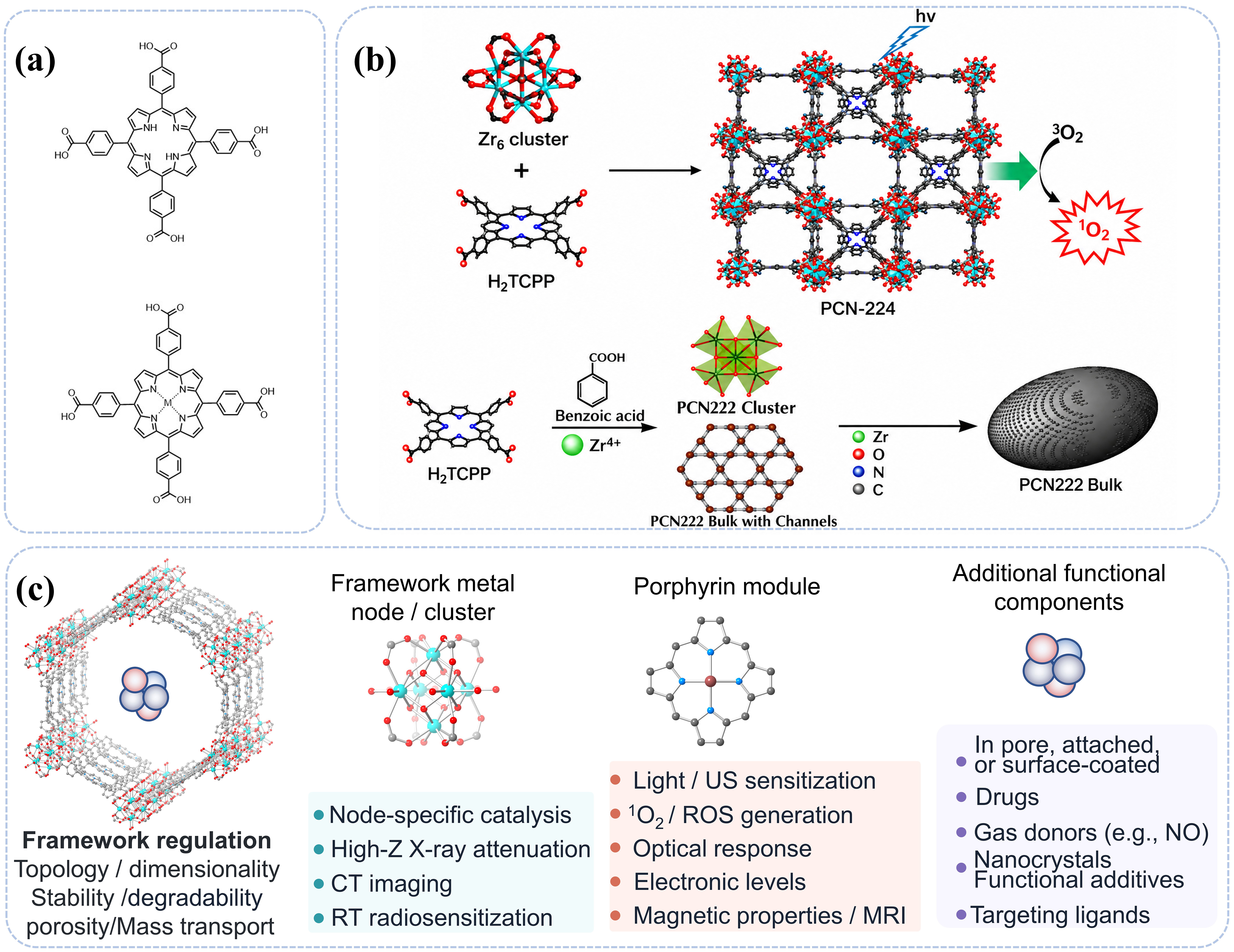
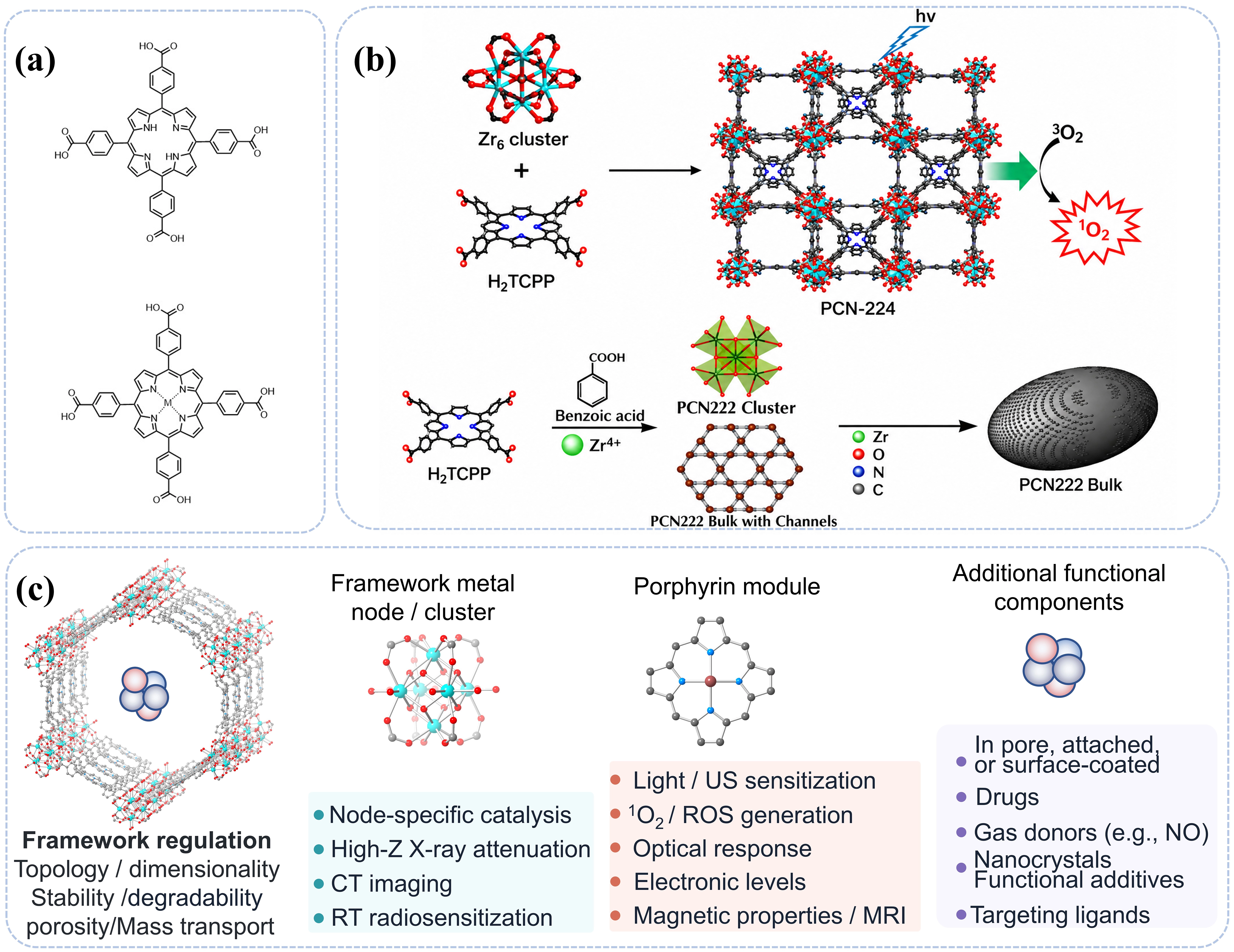
Figure 1. Structural basis and representative architectures of TCPP-based MOFs. (a) Chemical structures of H2TCPP and metallated M-TCPP ligands. Original schematic created by the authors for this Perspective; (b, upper) Schematic construction of PCN-224 from Zr6 clusters and H2TCPP ligands for light-triggered singlet-oxygen generation. Reproduced with permission from reference[2]. Copyright © 2016 American Chemical Society; (b, lower) Schematic representation of the mesoporous PCN-222 nanooval architecture. Reproduced with permission from reference[19]. Copyright © 2020 American Chemical Society; (c) Component-resolved schematic illustrating the distinct functional contributions of the framework metal node/cluster, metallated porphyrin M-N4 center, and additional incorporated components in TCPP-based MOFs. The porphyrin M-N4 center primarily regulates sensitization, charge transfer, redox/catalytic activity, and magnetic properties; the framework metal node/cluster contributes to structural stability, catalytic reactivity, degradability, imaging, and radiosensitization; and additional components provide complementary functions such as drug delivery, gas release, photothermal conversion, targeting, or other therapeutic/imaging capabilities. TCPP: meso-tetra(4-carboxyphenyl)porphyrin; MOFs: metal-organic frameworks; CT: computed tomography; RT: radiotherapy; ROS: reactive oxygen species; MRI: magnetic resonance imaging; NO: nitric oxide; high-Z: high-atomic-number element; US: ultrasound; 1O2: singlet oxygen.
The coordination chemistry between TCPP and different metal nodes further dictates the structural topology and functional characteristics of the resulting MOFs. High-valence metal ions such as Zr4+ and Hf4+ readily assemble into hexanuclear Zr6/Hf6 oxo-cluster secondary building units, which are subsequently interconnected by TCPP linkers to generate robust three-dimensional frameworks, including PCN-224, PCN-222, PCN-223, and Hf-TCPP nanoscale metal-organic framework (NMOF)[2,3,17-19] (Figure 1b). These crystalline architectures spatially isolate individual porphyrin units and support structural stability and porosity[17,18]. In PCN-224, this ordered confinement maintains accessible photoactive porphyrin units and supports 1O2-mediated photosensitization[2]. In contrast, coordination with metal ions such as Cu2+, Al3+, Zn2+/Al3+, or In3+ often affords two-dimensional nanosheets or layered coordination networks. Their reduced thickness and high surface-to-volume ratio facilitate the exposure of catalytically active porphyrin sites, accelerate mass transport, and shorten charge-transfer pathways, thereby enhancing catalytic and photodynamic performance[7,13-15]. These comparisons suggest that topology and dimensionality can influence porphyrin spacing, exposed surface area, and mass-/charge-transfer path length; however, particle size and defect density are not reported on a common quantitative basis across studies, limiting direct structure-ROS rankings[7,13-15].
Beyond structural regulation, the incorporation of redox-active metal centers, particularly Fe and Mn, further expands the functional repertoire of TCPP-based MOFs. These metal ions can be integrated into degradable porous frameworks, nanoneedles, or bimetallic coordination networks that respond dynamically to the tumor microenvironment. Leveraging the variable valence states of Fe and Mn, these materials catalyze Fenton or Fenton-like reactions, consume intracellular glutathione (GSH), amplify oxidative stress, and promote ferroptosis or ferroptosis-associated oxidative damage according to the level of mechanistic evidence[20-23]. Collectively, the modular combination of porphyrinic ligands and diverse metal nodes provides a highly versatile platform in which framework topology, electronic structure, catalytic activity, and therapeutic function can be simultaneously engineered through rational coordination chemistry.
2.2 Active centers of TCPP-based MOFs
The therapeutic functions of TCPP-based MOFs originate from two complementary classes of active centers: metallated porphyrin cores and framework metal nodes. The M-N4 coordination environment within the porphyrin macrocycle regulates intersystem crossing, charge transfer, enzyme-mimetic catalysis[26], and magnetic properties, whereas framework metal nodes determine structural stability, catalytic reactivity, degradability, and stimulus-responsive release. Importantly, these two active centers rarely function independently. Instead, the antitumor performance of TCPP-based MOFs arises from the synergistic interplay among porphyrin-centered photosensitization, metal-node-mediated catalysis, and framework-assisted transport within an integrated coordination architecture. An additional loaded or conjugated component, such as a drug, gas donor, nanocrystal, MXene, or targeting ligand, should be assigned as a separate functional layer rather than attributed to either intrinsic active center[11,15,27,28] (Figure 1c).
Among various metallated porphyrins, Fe-TCPP is one of the most extensively investigated because of its well-established redox chemistry. In the mildly acidic and H2O2-rich tumor microenvironment, Fe3+/Fe2+ redox cycling drives Fenton or Fenton-like reactions to continuously generate highly reactive hydroxyl radicals (·OH). Meanwhile, GSH-mediated reduction of Fe3+ consumes intracellular glutathione, weakens antioxidant defenses, and further amplifies oxidative stress, ultimately promoting lipid peroxidation and ferroptosis-associated oxidative damage. A definitive ferroptosis assignment requires inhibitor-rescue and related pathway evidence. The unique advantage of Fe-TCPP therefore lies in the sustained Fe3+/Fe2+ cycling enabled within tumor cells. Pan et al. developed a cell membrane-coated Fe-TCPP nanoscale MOF that combined GSH depletion, Fenton catalysis, TCPP-mediated photodynamic therapy (PDT), and tirapazamine (TPZ) chemotherapy in a treatment sequence described by the authors as involving ferroptosis, achieving enhanced therapeutic efficacy against triple-negative breast cancer[20]. Likewise, polyethylene glycol (PEG)-modified Fe-MOFs reported by Xie et al. exploited GSH-triggered Fe3+ reduction to amplify Fenton reactions and PDT under MRI/fluorescence imaging guidance[21]. Beyond redox regulation, morphology engineering can further improve catalytic performance. Fe(II)-TCPP nanoneedles increase the exposure of Fe(II) active sites and simultaneously promote ·OH and 1O2 generation, highlighting the synergistic effects of iron-valence regulation and structural optimization on catalytic antitumor activity[22].
Cu-TCPP systems can also drive GSH depletion and ROS-mediated oxidative damage, although their catalytic pathways can differ from Fe-based Fenton chemistry. Wang et al. demonstrated that Cu-TCPP nanosheets generated 1O2 under hypoxic conditions through TCPP peroxidation followed by the Russell mechanism while concurrently consuming intracellular GSH[24]. In addition to catalytic activity, Cu-TCPP exhibits measurable photothermal properties. Li et al. prepared ultrathin Cu-TCPP nanosheets (~5.1 nm) with a photothermal conversion efficiency of approximately 36.8%, enabling synergistic PDT/photothermal therapy (PTT). Moreover, the paramagnetism of Cu(II), together with the near-infrared photothermal effect, enabled MRI- and thermal imaging-guided treatment[7]. These studies illustrate that Cu-TCPP integrates catalytic therapy, photothermal conversion, and imaging across related Cu-TCPP platforms.
Mn-TCPP and Mn-containing TCPP-MOFs are particularly attractive for theranostic applications because Mn simultaneously provides catalytic and imaging functionalities. Bao et al. constructed Hf/Mn-TCPP MOF nanoparticles in which Mn(III)-porphyrin centers shortened the T1 relaxation time of water protons for MRI, while Hf nodes enhanced X-ray attenuation, improving CT imaging and radiotherapeutic energy deposition. Combined with photoacoustic imaging and photothermal conversion, this platform enabled trimodal MRI/CT/PAI-guided PTT/radiotherapy (RT)[8]. Beyond imaging, Mn-porphyrinic MOFs exhibit catalase-like activity that decomposes H2O2 into O2, alleviating tumor hypoxia while reducing GSH/glutathione peroxidase 4 (GPX4) levels. This dual regulation enhances sonodynamic therapy (SDT) and promotes CD8+ T-cell infiltration together with dendritic-cell maturation, demonstrating the multifunctional role of Mn active centers in both tumor catalysis and immune modulation[23]. Xu et al. further reported that the ferroptosis assignment was supported by inhibitor-rescue experiments using the ferroptosis inhibitors UAMC-3203 and ferrostatin-1 together with GPX4/lipid-peroxidation measurements, providing stronger mechanistic evidence than ROS changes alone.
In addition, high-atomic-number metal nodes such as Hf and Lu can enhance X-ray absorption, thereby supporting CT imaging and RT radiosensitization; RDT should be assigned only when radiation-triggered sensitizer chemistry is demonstrated[3,8,16]. Bi-TCPP has instead been investigated as a light-responsive photoimmunotherapy platform[25]. A representative example is the Hf-TCPP NMOF developed by Liu et al., in which Hf nodes increased local radiation energy deposition, whereas TCPP ligands generated 1O2 under light irradiation, resulting in combined RT/PDT rather than X-ray-activated TCPP[3]. In contrast, two-dimensional Al-, Zn-, and In-TCPP frameworks primarily optimize sensitization by exposing more porphyrin active sites and shortening mass- and energy-transfer pathways. ZnAl-TCPP nanosheets generated nearly threefold more ROS than free TCPP under combined light and ultrasound irradiation, demonstrating the structural advantages of heterometallic nanosheets for sono-photodynamic therapy (SPDT)[14]. Similarly, In-TCPP@Zn-NO employed ultrathin In-TCPP nanosheets as a sonosensitizing scaffold to simultaneously enhance ROS generation and nitric oxide (NO) release under ultrasound stimulation, achieving synergistic SDT and gas therapy[15]. Collectively, these studies demonstrate that rational engineering of metallated porphyrin centers and framework metal nodes enables precise regulation of catalytic activity, imaging capability, radiosensitization, and therapeutic modality, providing the mechanistic foundation for the multifunctionality of TCPP-based MOFs.
3. TCPP-MOF Mediated Antitumor Strategies
3.1 Photodynamic therapy
PDT is a minimally invasive therapeutic modality that relies on the generation of ROS by photosensitizers upon irradiation with light at an appropriate wavelength, thereby inducing cytotoxic damage in target tissues. Its spatial selectivity and suitability for repeated local activation have motivated extensive investigation in cancer therapy. As a classical porphyrin-based photosensitizer, TCPP exhibits strong visible-light absorption and efficient singlet oxygen generation. However, free porphyrin molecules often suffer from aggregation-induced quenching and limited tumor-site retention. The TCPP-MOF architecture not only mitigates these limitations but also improves the accessibility and stability of photoactive sites. Consequently, TCPP-MOFs have emerged as an important research focus in tumor PDT, as exemplified by Zr-, Hf-, and Cu-containing systems[2,3,7] and targeted or drug-enabled variants[29-33]. A two-dimensional Yb-TCPP MOF (~200 nm) was also reported to generate 1O2 under 655 nm irradiation and show in vitro PDT activity[34].
The superior photodynamic performance of TCPP-MOFs stems primarily from framework-confined photosensitization and rational structural engineering. Immobilization of TCPP within crystalline frameworks suppresses self-quenching while preserving its intrinsic photophysical properties, whereas the ordered porous structure enhances oxygen accessibility and ROS diffusion. Park et al. developed a Zr-TCPP/PCN-224 system in which TCPP ligands were coordinated with Zr6 oxo-clusters, enabling the periodic distribution of porphyrinic photosensitizing units throughout the framework. This structural arrangement preserved the photosensitizing activity of TCPP while mitigating aggregation-induced quenching. Upon light irradiation, the material continuously generated 1O2, thereby inducing tumor cell death. In addition, folate-targeting moieties were introduced through post-synthetic modification of the Zr6 nodes, which enhanced cellular uptake. These findings demonstrate that TCPP-MOFs can improve photodynamic efficacy through framework confinement and surface-targeted design[2].
Beyond enhancing PDT alone, the modularity of TCPP-MOFs enables seamless integration with complementary therapeutic modalities. Cu-TCPP nanosheets provide a representative example of an integrated PDT/PTT platform. Compared with Zr-cluster-based 3D frameworks, Cu-TCPP nanosheets further extend PDT by introducing photothermal conversion through Cu-associated near-infrared absorption. In this system, TCPP mediates 1O2 generation upon 660 nm light irradiation, whereas Cu-related near-infrared absorption induces a photothermal effect under 808 nm irradiation. The synergistic combination of ROS-mediated photodynamic damage and photothermal ablation markedly enhances tumor cell killing[7].
The versatility of TCPP-MOFs further extends to combination therapy with ionizing radiation. Liu et al. developed an Hf-TCPP NMOF in which TCPP serves as an intrinsic photosensitizer and Hf4+ functions as a high-atomic-number radiosensitizer. Upon visible-light irradiation, TCPP efficiently generates 1O2, whereas Hf nodes enhance local X-ray energy deposition under radiotherapy and thereby amplify secondary radiation damage. This cooperative interaction significantly improves tumor radiosensitivity and illustrates how rational integration of photosensitizing ligands with functional metal nodes enables synergistic PDT/RT within a single nanoplatform[3]. Here, TCPP-derived 1O2 is light-triggered, whereas the X-ray enhancement derives from Hf-mediated energy deposition; this system is therefore best classified as combined PDT plus radiosensitized RT rather than RDT[3].
3.2 Chemodynamic therapy
3.2.1 Fenton/Fenton-like ·OH generation
Chemodynamic therapy (CDT) exploits mildly acidic pH, elevated H2O2 levels, and abundant reducing agents in the tumor microenvironment to trigger in situ Fenton or Fenton-like reactions[20-22,35]. These reactions generate highly reactive ROS, particularly ·OH, thereby inducing oxidative damage in tumor cells[36]. Compared with PDT, which requires external light irradiation, CDT is primarily driven by intrinsic biochemical features of the tumor microenvironment and therefore holds particular promise for the treatment of deep-seated tumors. In TCPP-MOFs, redox-active metal sites, such as Fe and Cu, can participate in redox cycling, catalyze ROS generation, consume GSH, and impair cellular antioxidant defenses. These properties provide a critical materials basis for the development of TCPP-MOF-based CDT nanoplatforms[20-22,35].
Fenton-like reactions typically require the coordinated presence of transition-metal catalytic sites, H2O2, and a weakly acidic microenvironment. Low-valence metal ions, such as Fe2+ and Cu+, catalyze the conversion of H2O2 into ·OH, whereas their high-valence counterparts, such as Fe3+ and Cu2+, can be reduced by GSH and other endogenous reducing species, thereby sustaining metal redox cycling. Accordingly, TCPP-MOFs that incorporate Fe- or Cu-based active centers, expose accessible catalytic sites within the tumor microenvironment, and deplete GSH are particularly well suited for CDT applications[21,35].
Xie et al. reported PEG-functionalized Fe-MOFs, in which PEGylation improved the colloidal stability of the material. After tumor accumulation, the Fe sites underwent valence-state transitions in the presence of GSH, thereby activating Fenton-like reactions. Concurrently, TCPP-mediated PDT further increased intracellular ROS levels, ultimately enabling imaging-guided therapy with ferroptosis enhancement[21]. Cao et al. developed a CuTCPP-MOF that combined Cu-mediated Fenton-like reactions with TCPP-based photosensitization for synergistic PDT/CDT of prostate cancer. This study further demonstrated that Cu-TCPP MOFs can intensify oxidative damage in tumor cells through the synergy between metal-catalyzed ROS generation and porphyrin-mediated photosensitization[35].
3.2.2 Russell-mechanism 1O2 generation
Conventional Fenton-like reactions are highly dependent on H2O2 availability, acidic conditions, and metal valence-state cycling. Thus, ROS generation may be constrained within the complex tumor microenvironment. The Russell mechanism offers a complementary pathway, whereby peroxyl radicals undergo bimolecular recombination to produce 1O2. This process provides an additional light-independent source of 1O2 and may partially bypass limitations associated with hypoxia and restricted light penetration. This pathway is particularly relevant to Cu-TCPP systems[24].
Wang et al. designed Cu-TCPP nanosheets that utilized tumor-associated acidic H2O2 to induce TCPP peroxidation. In the presence of Cu catalytic sites, peroxyl radicals were subsequently generated and underwent Russell-type recombination to produce 1O2. Concurrently, Cu2+ depleted GSH, thereby impairing the ROS-scavenging capacity of tumor cells[24]. This pathway should be distinguished from classical Cu+/H2O2 Fenton-like CDT: Wang et al. reported that light-independent 1O2 generation arose from ligand peroxidation and Russell recombination rather than from a conventional Cu+/H2O2 Fenton cycle[24].
3.3 Sonodynamic therapy
Owing to the limited penetration depth of visible light in biological tissues, SDT has emerged as a promising strategy for extending TCPP-based MOFs to deep-seated tumors. SDT employs ultrasound to activate sonosensitizers and generate ROS. In these systems, porphyrin units can serve as sonodynamically active components[12-15,23]. Possible activation routes include acoustic cavitation/sonoluminescence and ultrasound-assisted charge separation[37], but these physical routes are not universally established; where a study reports only ultrasound-dependent ROS and therapeutic effects, the route should be treated as proposed rather than demonstrated. Additional TCPP-based SDT designs have also been reported[38-40]. PEGylated Al-TCPP nanosheets provide a representative example of ultrasound-dependent ROS generation (Figure 2a)[13].
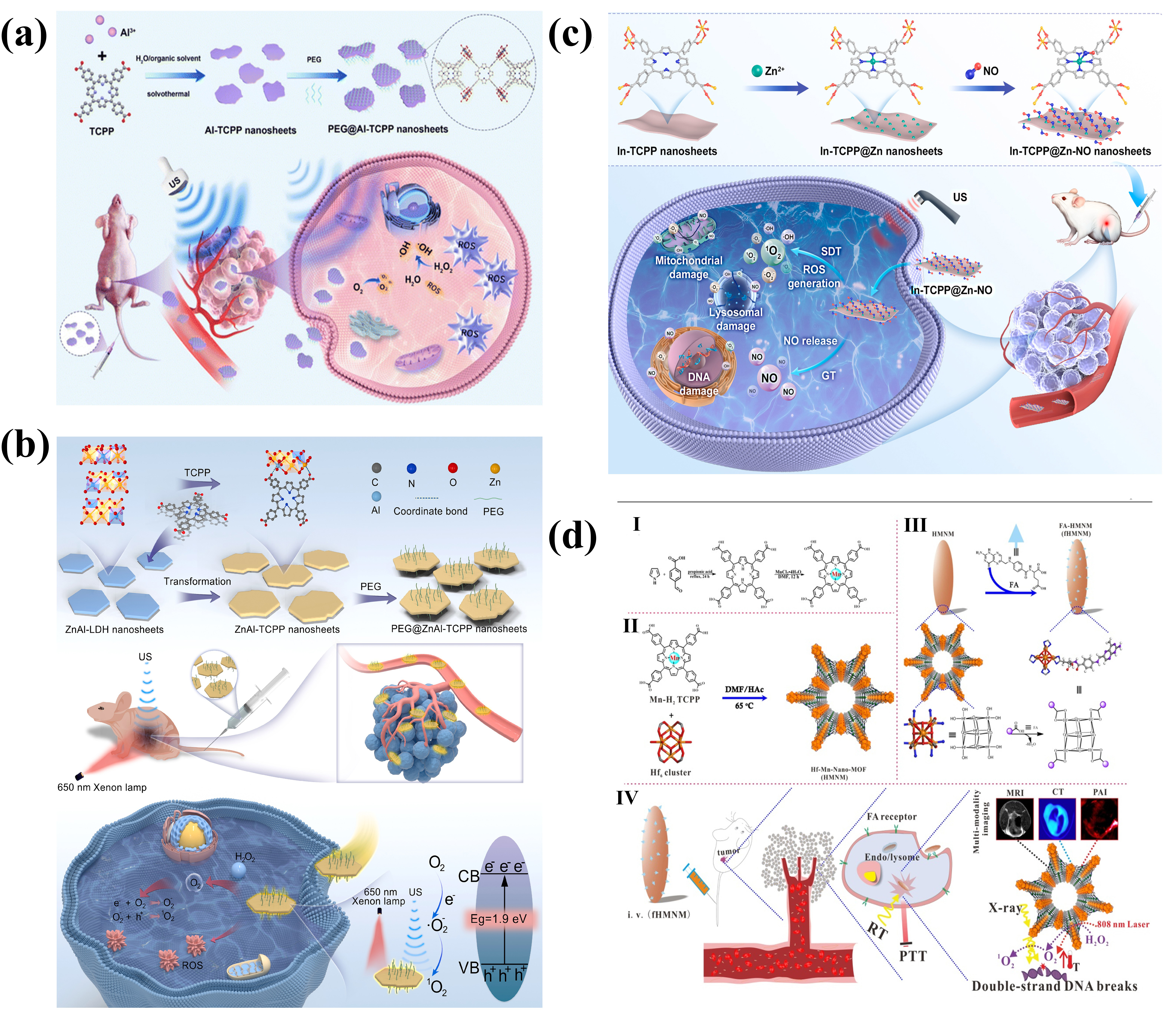
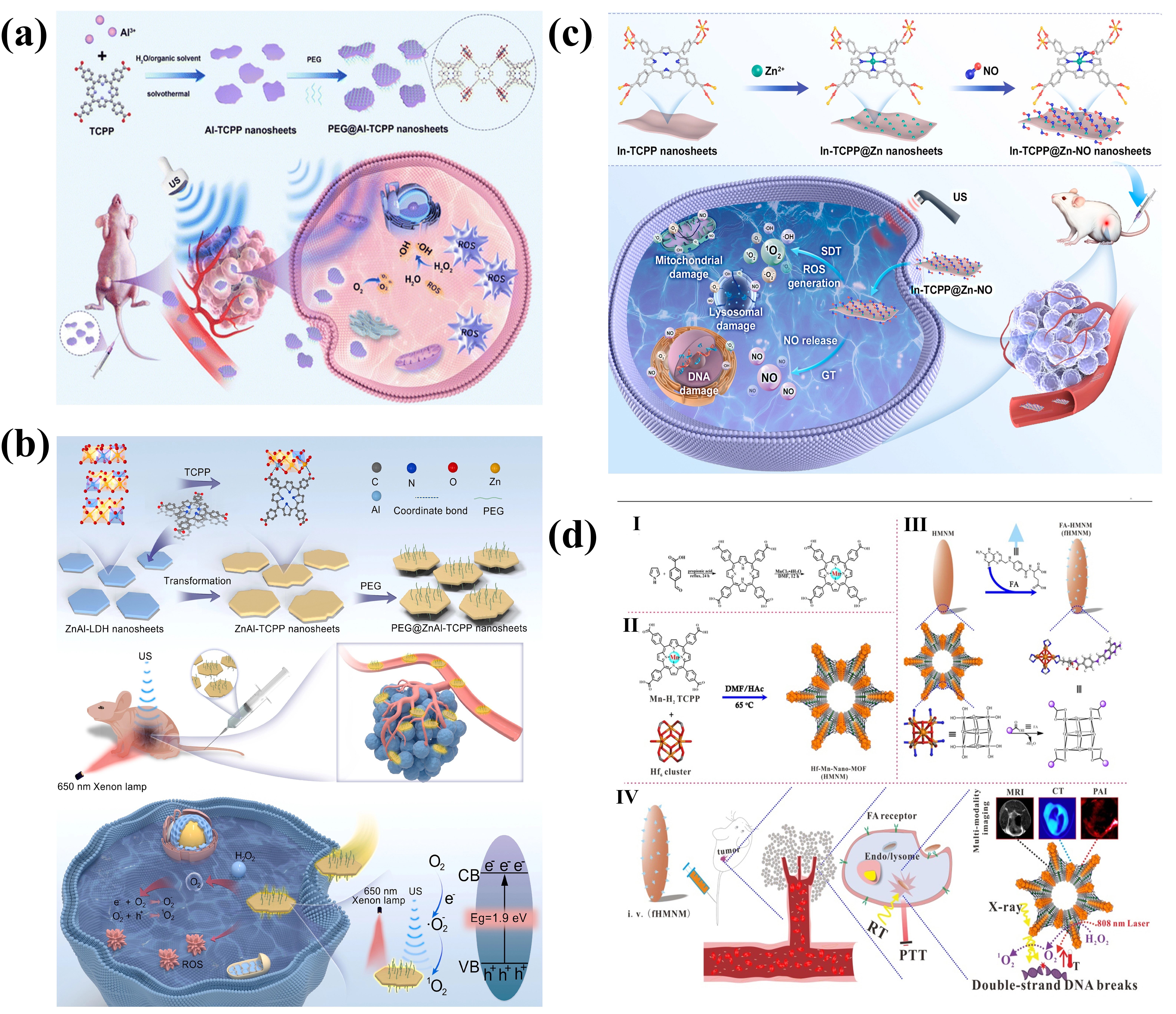
Figure 2. TCPP-based MOF nanoplatforms for sonodynamic, sono-photodynamic, gas/sonodynamic, and radiation-related cancer therapy. (a) Schematic preparation of PEGylated Al-TCPP MOF nanosheets and their ultrasound-triggered sonodynamic antitumor mechanism involving ROS generation and tumor cell damage. Reproduced with permission from reference[13]. Copyright © 2023 Royal Society of Chemistry; (b) Transformation of ZnAl-LDH nanosheets into PEGylated ZnAl-TCPP MOF nanosheets for combined light- and ultrasound-activated sono-photodynamic therapy. Reproduced with permission from reference[14]. Copyright © 2025 Elsevier; (c) Construction of nitric oxide-functionalized In-TCPP@Zn-NO nanosheets for ultrasound-induced synergistic gas/sonodynamic therapy through ROS generation, NO release, mitochondrial damage, lysosomal damage, and DNA damage. Reproduced with permission from reference[15]. Copyright © 2026 Elsevier Ltd; (d) Schematic synthesis and therapeutic mechanism of Hf/Mn-TCPP MOF nanoparticles for MRI/CT/PAI-guided photothermal/radiotherapy synergistic cancer treatment. Reproduced from reference[8]. CC BY-NC 3.0. CT: computed tomography; MRI: magnetic resonance imaging; MOF: metal-organic framework; NO: nitric oxide; PAI: photoacoustic imaging; PTT: photothermal therapy; ROS: reactive oxygen species; RT: radiotherapy; TCPP: meso-tetra(4-carboxyphenyl)porphyrin; SDT: sonodynamic therapy; PEG: polyethylene glycol; US: ultrasound; 1O2: singlet oxygen; ·OH: hydroxyl radicals; DMF: N,N-dimethylformamide; LDH: layered double hydroxide; FA: folic acid.
A major strategy for enhancing SDT is to couple sonodynamic sensitization with catalytic regulation of the tumor microenvironment. Xu et al. constructed a Mn-porphyrin MOF using Mn-TCPP as the sonosensitizing ligand and Zr as the coordination node. Upon ultrasound irradiation, Mn-TCPP produced ROS, thereby inducing oxidative damage in tumor cells. Meanwhile, the Mn sites catalyzed the decomposition of endogenous H2O2 into O2, mitigating the inhibitory effect of tumor hypoxia on SDT. Moreover, the material promoted lipid peroxidation and ferroptosis by downregulating GSH/GPX4 levels. These results suggest that Mn-TCPP MOFs can exert synergistic antitumor effects through oxygen-supplemented SDT and ferroptosis-mediated amplification of oxidative damage[23]. Xu et al. additionally reported inhibitor-rescue evidence using the ferroptosis inhibitors UAMC-3203 and ferrostatin-1 together with GPX4/lipid-peroxidation measurements[23]; however, the physical ultrasound activation route was not directly assigned.
Structural engineering of TCPP-MOFs provides another strategy for improving sonodynamic performance. Zhou et al. prepared two-dimensional Al-TCPP nanosheets in which TCPP ligands were immobilized within an ultrathin coordination framework. The authors associated the two-dimensional architecture with greater exposure of porphyrinic active sites and shorter charge- and energy-transfer pathways. Experimentally, both 1O2 and ·OH were generated under ultrasound irradiation. Following PEGylation, the nanosheets exhibited improved biocompatibility and in vivo stability, leading to effective sonodynamic tumor inhibition in both cellular and animal models[13]. Similarly, Hu et al. transformed ZnAl layered double hydroxides into heterometallic ZnAl-TCPP nanosheets, which generated approximately threefold higher ROS than free TCPP under combined light and ultrasound irradiation. The resulting synergistic PDT/SDT induced severe mitochondrial and lysosomal damage, supporting the benefit of dimensional and compositional engineering under the reported conditions (Figure 2b)[14]. Zhou et al. further incorporated Zn-NO donors into two-dimensional In-TCPP nanosheets, enabling simultaneous ultrasound-triggered ROS generation and NO release. This integrated design combined SDT with gas therapy, providing an additional strategy for amplifying oxidative stress through multimodal activation (Figure 2c)[15].
3.4 RT, radiosensitization, and radiodynamic therapy (RDT)
Radiation-related TCPP-MOF strategies can be distinguished by how the material participates in the X-ray response. In RT/radiosensitization, high-Z components mainly enhance local radiation energy deposition, whereas RDT additionally requires ionizing-radiation-triggered sensitizer chemistry that generates ROS.
In RT-focused applications, TCPP-MOFs commonly exploit high-atomic-number metal nodes for enhanced X-ray absorption and radiosensitization. High-Z energy deposition alone should not be labeled RDT. Bao et al. reported Hf/Mn-TCPP MOF nanoparticles that combined the CT and radiosensitizing functions of Hf with the MRI and photothermal functions of Mn-TCPP, enabling MRI/CT/PAI-guided PTT/RT rather than RDT (Figure 2d)[8]. In a distinct PCN-224@IrNCs/D-Arg platform, Ir nanocrystals and D-arginine were integrated with the porphyrinic framework to combine PDT, nitric-oxide-related therapy, and radiosensitization[27]. These studies illustrate how porphyrinic sensitizers can be combined with high-atomic-number or catalytically active components for multimodal treatment of deep-seated tumors. As noted above, the Hf-TCPP NMOF similarly combines light-triggered TCPP PDT with Hf-mediated RT enhancement rather than X-ray-activated TCPP[3].
LuMix provides a clearer example of RDT. Under X-ray irradiation, Lu absorbs X-rays to generate ·OH and transfers energy to TCPP to produce 1O2, while the TCPP(Fe) ligand catalyzes H2O2 degradation to generate O2 and ·OH, enabling CDT. Thus, LuMix integrates RT, RDT, and CDT within a single nMOF[16].
3.5 Drug delivery and multimodal combination therapy
Owing to their tunable pore structures, functionalizable nanosheet surfaces, and degradable coordination bonds, TCPP-MOFs serve as versatile platforms for the loading of chemotherapeutic agents and bioactive small molecules[4,5,29,30,41]. Degradable or stimulus-responsive variants further extend this loading strategy to additional combination platforms[9,42-45]. These features enable their application in diverse synergistic therapeutic strategies[4,5]. For instance, two-dimensional Zn-TCPP@PEG nanosheets can be loaded with doxorubicin (DOX) and labeled with 99mTc for in vivo monitoring of biodistribution. This design enables both biodistribution monitoring and therapeutic amplification, as the 1O2 generated by TCPP upon light irradiation enhances the therapeutic efficacy of DOX, facilitating radionuclide imaging-guided chemo-photodynamic therapy[9]. Similarly, in DOX@Cu-TCPP systems, DOX is incorporated into Cu-TCPP nanosheets through π-π interactions, and its release is accelerated under mildly acidic conditions. The ROS generated by TCPP synergize with DOX-mediated DNA damage to induce tumor cell death, highlighting the potential of TCPP-MOFs as drug delivery platforms for combined chemotherapy and PDT[5].
However, the therapeutic efficacy of conventional tumor-killing strategies is frequently limited by two major barriers: drug resistance and the hypoxic microenvironment characteristic of solid tumors. To overcome drug resistance, the Fe-TCPP/cisplatin platform exploits Fe sites to induce Fenton reactions, deplete intracellular GSH, and promote lipid peroxidation, while cisplatin mediates DNA crosslinking. The combination of these mechanisms enhances the susceptibility of drug-resistant tumor cells to therapeutic intervention[28]. To overcome hypoxia-associated barriers, oxygen-generating systems such as PCN-224-CaO2-HA can release O2 through the reaction of CaO2 with water or weakly acidic media, thereby alleviating hypoxia-induced limitations in PDT efficacy[32].
Beyond direct tumor cell killing, the integration of TCPP-MOFs with immunotherapeutic agents can facilitate the transformation of highly immunosuppressive “cold” tumors into “hot” tumors characterized by enhanced immune cell infiltration and activation. In the TPP@PTX-CuTCPP system, CuTCPP depletes intracellular GSH, thereby improving the efficacy of paclitaxel against multidrug-resistant tumor cells[10]. Concurrent PD-L1 blockade further enhances antitumor immune responses. Related original studies have combined TCPP/PCN-224 platforms with drug delivery or PDT-induced immunogenic cell death to reinforce antitumor immunity[46-49].
4. Imaging Applications and Theranostic Integration
Molecular imaging plays a central role in precision oncology by enabling tumor visualization, treatment planning, real-time monitoring of nanomaterial biodistribution, and evaluation of therapeutic responses. Owing to their modular composition, TCPP-based MOFs readily integrate diagnostic and therapeutic functions within a single platform. Their imaging capabilities originate from distinct structural components: intrinsic porphyrin fluorescence, paramagnetic metalloporphyrin centers, high-atomic-number framework nodes, and radionuclide-labeling sites. This structural modularity provides a robust foundation for the development of multifunctional theranostic nanoplatforms[4,6,8,9]. Representative MRI/fluorescence and PAI examples are shown in Figure 3a,b,c, respectively. Quantitative metrics are reported below where the cited study provides comparable values; otherwise Table 1 records NR rather than substituting qualitative labels.
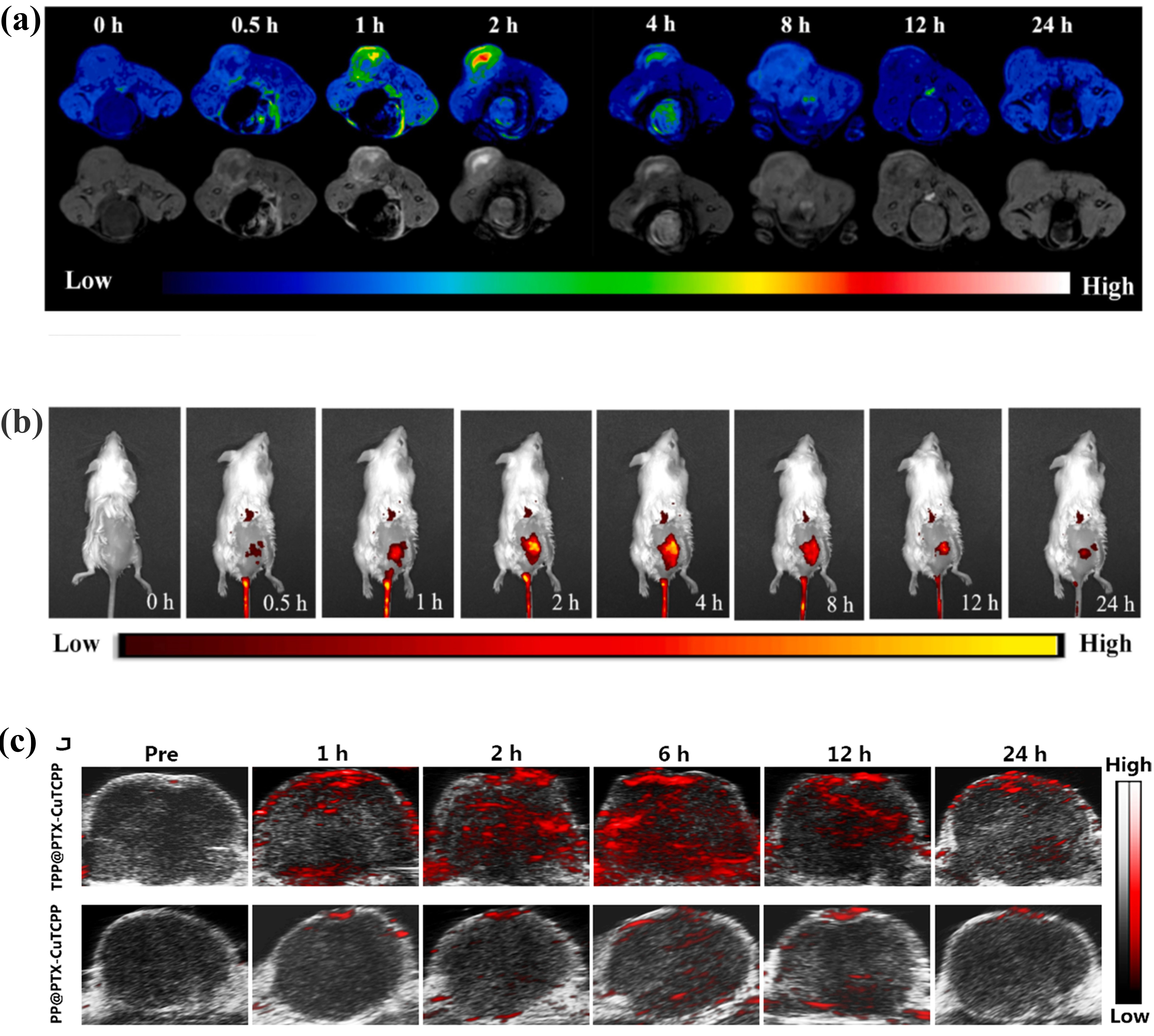
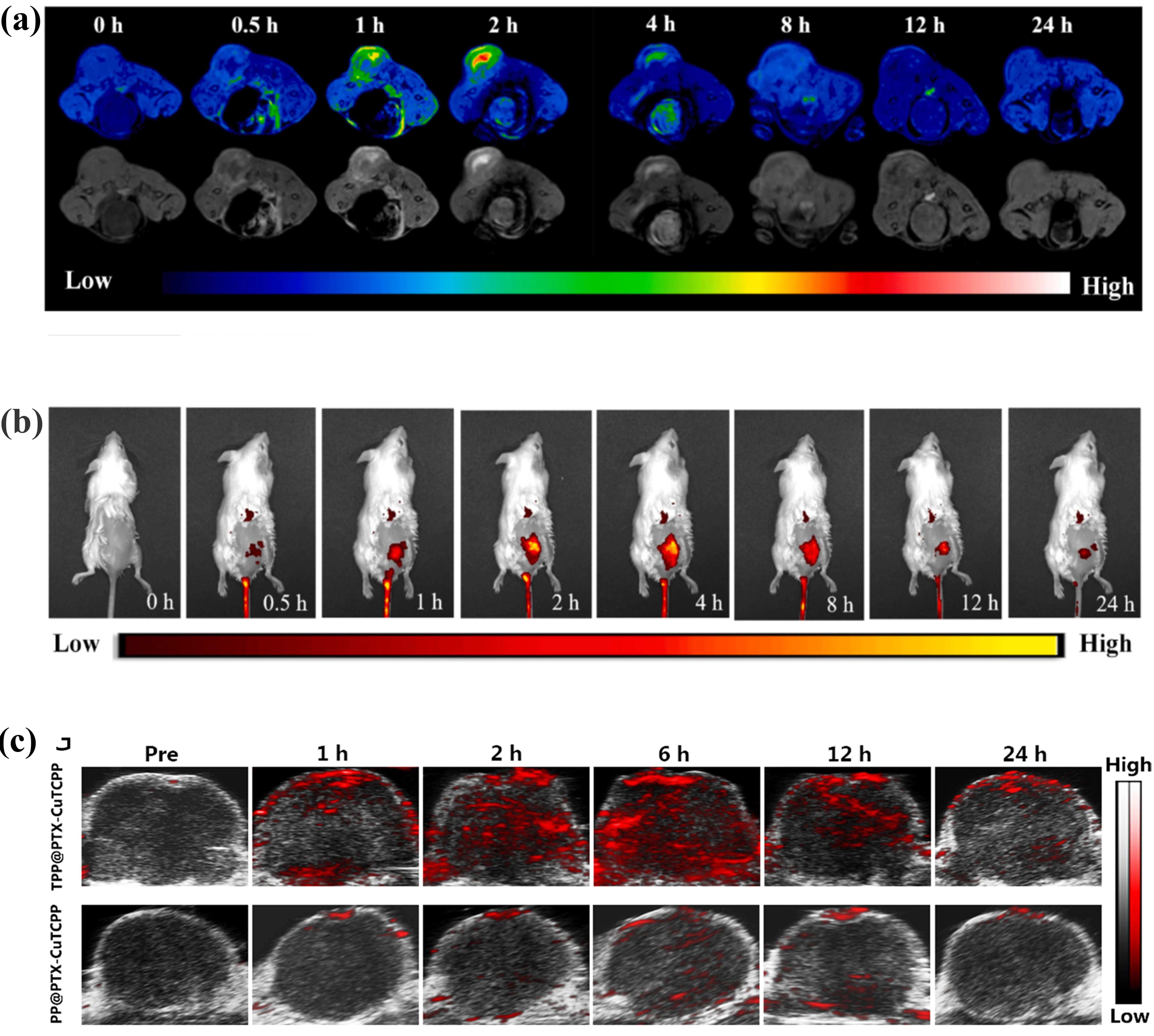
Figure 3. Imaging-guided biodistribution and tumor accumulation of TCPP-based MOF theranostic nanoplatforms. (a) T1-weighted MR imaging of tumors at different time points after intravenous injection of PEG-Fe-MOFs. Reproduced with permission from reference[21]. Copyright © 2025 Elsevier; (b) Fluorescence imaging of 4T1 tumor-bearing nude mice at different time points after intravenous injection of PEG-Fe-MOFs, showing tumor accumulation and imaging-guided photodynamic-enhanced ferroptosis therapy. Reproduced with permission from reference[21]. Copyright © 2025 Elsevier; (c) In vivo PAI images of tumors in MCF-7/Taxol tumor-bearing mice at various time intervals after intravenous injection of TPP@PTX-CuTCPP and PP@PTX-CuTCPP, demonstrating targeted tumor accumulation and providing guidance for therapeutic-window selection. Reproduced from reference[10]. CC BY 4.0. MR: magnetic resonance; MOF: metal-organic framework; PAI: photoacoustic imaging; PEG: polyethylene glycol; PP: PEI-PLGA; PTX: paclitaxel; TCPP: meso-tetra(4-carboxyphenyl)porphyrin; TPP: tLyP-1-PEI-PLGA.
Table 1. Comparative summary of representative TCPP-MOF theranostic systems.
| System/composition | Porphyrin type/center metal | Framework node | Topology/dimensionality | Morphology/size | Therapeutic mechanism | External stimulus | ROS/active species | Imaging modality | Tumor model/major outcome | Irradiation conditions | Biodistribution/clearance/toxicity | Therapeutic outcome/evidence | Key limitation/translation evidence |
| PCN-224[2] | H2TCPP/metal-free core | Zr6 | 3D PCN-224 | Size-controlled NPs; exact size NR here | Framework-confined PDT | Visible light | 1O2 | Fluorescence/PDT guidance | Tumor model NR here; targeted PDT | Light conditions NR here | Long-term biodistribution/clearance NR | Framework confinement improves PDT accessibility | Limited PK and long-term safety evidence |
| Hf-TCPP NMOF[3] | H2TCPP/metal-free core | Hf | 3D nanoscale MOF | NR | Light-triggered PDT + high-Z RT radiosensitization | 661-nm light + X-ray | Light: 1O2; RT: high-Z secondary radiation chemistry | NR | 4T1; combined PDT + RT enhanced tumor control | PDT applied 8 h after RT; light power/X-ray dose NR here | Most Hf reported cleared by ~7 d; longer-term products NR | Combined PDT/RT efficacy | Preclinical mouse evidence; long-term degradation products need study |
| Al-TCPP-PEG[13] | H2TCPP/metal-free core | Al | 2D nanosheet | 160-200 nm lateral; 18.4–26.5 nm thick | SDT | Ultrasound | 1O2 + ·OH | NR | Tumor model NR here; in vivo SDT inhibition | US conditions NR here | Long-term clearance/toxicity NR | In vivo SDT inhibition | Physical US activation route not directly resolved; PK NR |
| ZnAl-TCPP[14] | TCPP; center metal NR | Zn/Al | 2D heterometallic nanosheet | NR | Sono-photodynamic therapy | Light + ultrasound | ROS; ~3× vs. free TCPP under combined stimulation | NR | Tumor model NR here; SPDT | Light/US conditions study-specific; NR here | NR | Enhanced SPDT | Cross-study ROS comparison limited by stimulation conditions |
| In-TCPP@Zn-NO[15] | Zn coordinated at porphyrin center | In | 2D nanosheet | NR | SDT + NO gas therapy | Ultrasound | ROS + NO release | NR | Tumor model NR here; SDT/gas therapy | US conditions NR here | Longitudinal speciation/clearance NR | SDT + NO therapy | Complex metal/NO speciation and PK remain gaps |
| Cu-TCPP nanosheet[7] | TCPP; porphyrin-center metal NR | Cu | 2D nanosheet | 5.1 ± 0.3 nm thick; DLS ~330 nm | PDT + PTT | 660 nm + 808 nm light | 1O2 + heat | MRI + thermal imaging | Saos-2; synergistic PDT/PTT | 808 nm, 1 W cm-2, 10 min; 660 nm, 10 mW cm−2, 30 min | Stable in PBS/RPMI ~1 week with minimal Cu2+ release; 1-month H&E after IV 25 mg kg-1 showed no obvious injury | Synergistic PDT/PTT | Intratumoral therapy and 1-month histology do not establish long-term PK/safety |
| Mn-TCPP/Zr MOF[23] | Mn(III)-TCPP | Zr | Nanoscale porous MOF | DLS ~70 nm | SDT + O2 regulation + ferroptosis/immune modulation | Ultrasound | ROS; O2 generation from H2O2 | NR | H22 and 4T1; SDT/ferroptosis with immune activation | 1 MHz US, 0.9 W cm-2, 30% duty, 10 min | Long-term PK/clearance NR | Ferroptosis component supported by UAMC-3203/Fer-1 rescue + GPX4/lipid-peroxidation evidence | Strong mechanistic ferroptosis evidence; long-term translation data NR |
| PEG-Fe-MOF[21] | TCPP; porphyrin-center metal NR | Fe | Porous nanoscale MOF | 110 ± 22 × 69 ± 13 nm | Fenton-like CDT + PDT; ferroptosis reported | TME + light | ·OH + PDT ROS | T1 MRI + fluorescence | Tumor model NR here; imaging-guided ferroptosis-enhanced therapy | Irradiation NR here | TME-triggered degradation/release of TCPP reported; long-term clearance NR | MRI/fluorescence-guided oxidative therapy | Ferroptosis reported; comparable relaxivity and long-term PK NR |
| Cu-TCPP Russell system[24] | TCPP; porphyrin-center metal NR | Cu | Ultrathin nanosheet | NR | Russell-mechanism 1O2 + GSH depletion; not classical Cu+/H2O2 CDT | Acidic H2O2; light-independent | 1O2 via Russell recombination | NR | Tumor model NR here; oxidative tumor therapy | No light required for Russell pathway | Long-term PK/toxicity NR | Light-independent 1O2 + GSH depletion | Do not infer classical Fenton CDT or ferroptosis without direct evidence |
| Hf/Mn-TCPP MOF nanoparticles[8] | Mn(III)-TCPP | Hf | Nanoscale MOF | NR | PTT + RT radiosensitization; multimodal imaging | 808 nm + X-ray | Secondary ROS/radicals from Hf-enhanced X-ray energy deposition; photothermal heating | MRI/CT/PAI | S180 subcutaneous; PTT/RT | 24 h post-dose: 808 nm, 1 W cm-2, 10 min and/or 4 Gy X-ray; repeated 2 d later | No obvious major-organ damage during reported study; long-term clearance NR | MRI/CT/PAI-guided PTT/RT | Subcutaneous model; short-term safety ≠ long-term toxicity |
| LuMix[16] | TCPP + TCPP(Fe)/Fe center | Lu | Mixed-ligand NMOF | NR | RT + RDT + CDT | Low-dose X-ray | ·OH + radiation-triggered 1O2 | NR | MC38/HN6 reported; RT/RDT/CDT | Low-dose X-ray; exact conditions NR here | Long-term PK/metal release NR | Mechanistically assigned RT/RDT/CDT | Recent preclinical multicomponent platform; manufacturing/PK gaps |
| Zn2+/TCPP-DOX-99mTc[9] | TCPP; center metal NR | Zn | 2D NMOF | NR | Chemo-PDT + radionuclide tracking | Light + radionuclide tracking | 1O2 | SPECT | Tumor model NR here; imaging-guided chemo-PDT | Light conditions NR here | Whole-body tracking demonstrated; long-term clearance/toxicity NR | SPECT-guided chemo-PDT | Quantitative %ID/g and long-term safety NR here |
| Cu-TCPP(Zn)/Ti3C2Tx[11] | Zn-TCPP center | Cu + Ti3C2Tx component | 2D composite | NR | GSH-activated fluorescence + PDT/CDT | GSH + therapeutic irradiation | ROS | Turn-on fluorescence | OSCC; fluorescence-guided combination therapy | NR | Long-term PK/clearance NR | Fluorescence-guided PDT/CDT | Complex multicomponent platform; quantitative fluorescence and long-term translation data NR |
NR, not reported in the cited study or not sufficiently specified in the cited source for reliable extraction in this comparison; no value was inferred. Numerical values should not be directly ranked across studies because material dose, administration route, tumor model, scanner/assay, and irradiation protocols differ. Ferroptosis is treated as mechanistically established only when inhibitor-rescue and related lipid-peroxidation/GPX4 evidence are available; otherwise wording is qualified. CDT: chemodynamic therapy; DLS: dynamic light scattering; GSH: glutathione; GPX4: glutathione peroxidase 4; H&E: hematoxylin and eosin; IV: intravenous; MRI: magnetic resonance imaging; NMOF: nanoscale metal-organic framework; NO: nitric oxide; OSCC: oral squamous cell carcinoma; PAI: photoacoustic imaging; PDT: photodynamic therapy; PK: pharmacokinetics; PTT: photothermal therapy; RDT: radiodynamic therapy; ROS: reactive oxygen species; RT: radiotherapy; SDT: sonodynamic therapy; SPECT: single-photon emission computed tomography; SPDT: sono-photodynamic therapy; TME: tumor microenvironment; US: ultrasound; 1O2: singlet oxygen; ·OH: hydroxyl radicals.
For fluorescence imaging, TCPP exhibits intrinsic fluorescence. However, its emission is frequently quenched within MOF architectures through interactions with metal nodes or other incorporated components. In Cu-TCPP-based systems, the GSH-rich tumor microenvironment can disrupt metal-ligand coordination or trigger reduction reactions, thereby recovering the intrinsic fluorescence of TCPP and enabling GSH-responsive turn-on fluorescence imaging. For instance, Cu-TCPP(Zn)/Ti3C2Tx exploits GSH-activated fluorescence for imaging-guided therapy of oral squamous cell carcinoma (OSCC)[11]. A directly comparable fluorescence quantum yield or turn-on fold is not stated in the present manuscript for this system; Table 1 therefore records the quantitative fluorescence metric as NR[11].
For PAI, the incorporation of metal ions, such as Cu, Hf, and Mn, can improve the photoacoustic properties of TCPP-based MOF systems. Owing to its near-infrared absorption, CuTCPP can function as a PAI contrast agent. Jiang et al. developed TPP@PTX-CuTCPP and observed a linear increase in the PA signal with increasing TCPP concentration. Following intravenous administration, the tumor-associated PA signal reached its maximum at approximately 6 h, allowing visualization of drug accumulation and monitoring of therapeutic responses in multidrug-resistant tumors[10]. Similarly, Hf/Mn-TCPP MOF nanoparticles are suitable for PAI because of their broad absorption across the 600-800 nm wavelength range. In vivo, the PA signal reached its maximum approximately 24 h after injection, thereby facilitating the selection of an appropriate treatment window for combined PTT/RT[8].
MRI commonly relies on paramagnetic metal ions, such as Fe and Mn, which alter the longitudinal (T1) and/or transverse (T2) relaxation times of water protons, thereby enhancing T1- or T2-weighted contrast. PEG-Fe-MOF enables tumor microenvironment-responsive MRI and fluorescence imaging while simultaneously promoting ferroptosis[21]. In the Hf/Mn-TCPP MOF nanoparticles, Mn(III)-porphyrin moieties confer strong T1-weighted MR contrast, facilitating multimodal imaging and therapeutic monitoring using MRI, CT, and PAI[8]. For comparison, Cu-TCPP nanosheets reported r1 = 0.52 mM-1 s-1 at 0.5 T[7], whereas the Hf/Mn-TCPP MOF nanoparticles showed a 3-5-fold greater T1 relaxation rate than the clinical MR comparator used in that study[8]; differing field strengths and protocols preclude direct ranking.
CT imaging and radiation-based therapeutic applications primarily exploit the strong X-ray attenuation of high-atomic-number metals such as Hf and Lu. In addition to enhancing energy deposition during RT or RDT where mechanistically appropriate, these metals can substantially improve CT contrast. The Hf/Mn-TCPP MOF nanoparticles showed approximately 1.7-fold greater X-ray attenuation than the clinical CT comparator[8], while Lu-based nMOFs have also been incorporated into radiation-responsive theranostic platforms[16]. Because concentration ranges and scanner settings differ across reports, CT attenuation values should not be directly ranked without matched conditions.
The porphyrin frameworks and metal coordination sites of two-dimensional TCPP-MOFs can also be utilized for radionuclide labeling, enabling noninvasive tracking of their in vivo biodistribution. Compared with optical imaging, radionuclide imaging is particularly useful for whole-body tracking of material distribution. For example, two-dimensional Zn2+/TCPP NMOFs can be radiolabeled with 99mTc. Subsequent single-photon emission computed tomography (SPECT) imaging confirmed preferential tumor accumulation following intravenous administration, while the same platform also enabled combined chemotherapy and PDT[9]. A directly comparable %ID/g value is not stated in the present manuscript for this platform and is therefore recorded as NR in Table 1 rather than inferred[9].
5. Translational Considerations
5.1 Physiological stability and speciation
Physiological stability should be tested across neutral pH/phosphate-buffered saline (PBS) or phosphate, serum proteins, and tumor-relevant GSH/H2O2 because coordination exchange can alter framework integrity, metal/ligand release, oxidation state, and active-species speciation. Existing data are system-specific: Cu-TCPP nanosheets remained dispersed in PBS/RPMI-1640 for approximately one week with minimal Cu2+ release[7], PEG-Fe-MOFs were reported to degrade in the tumor microenvironment and release TCPP[21], and Hf-TCPP NMOFs showed relatively rapid in vivo clearance, with most Hf eliminated within approximately 7 days[3]. Comparable longitudinal release/speciation datasets are not reported for most platforms and should therefore be treated as a knowledge gap. Future studies should combine inductively coupled plasma mass spectrometry (ICP-MS) or release assays with X-ray diffraction (XRD)/transmission electron microscopy (TEM)/dynamic light scattering (DLS) and spectroscopy/valence-state characterization to distinguish true framework degradation from colloidal or surface changes; these are recommended validation approaches, not results already demonstrated.
5.2 Cross-study comparability and clinical translation
Therapeutic efficacy cannot be ranked independently of material dose, administration route, irradiation wavelength/power/time (or ultrasound/X-ray dose), and tumor model. Subcutaneous xenografts are useful for screening but do not reproduce all delivery barriers of orthotopic or metastatic disease; accordingly, tumor regression under a high local dose should not be interpreted as direct evidence of clinical translatability. Likewise, short-term body-weight curves and endpoint histology are not substitutes for pharmacokinetics, long-term organ biodistribution, degradation products, metal/ligand release, clearance, immunotoxicity, or off-target toxicity. Manufacturing assessment should include reproducibility of particle size and crystallinity, scalable synthesis, batch comparability, release specifications, and standardized quality control[50-52]. Table 1 therefore separates therapeutic outcome from translation evidence and records NR when a requested parameter was not reported.
6. Conclusion
TCPP-based MOFs have emerged as a versatile class of multifunctional nanoplatforms by integrating the photosensitizing and sonosensitizing properties of porphyrins, the catalytic activity of metal centers, and the structural advantages of highly programmable framework architectures. Their therapeutic efficacy is governed by ligand-centered sensitization and framework effects[2,7,24], redox/catalytic regulation including ferroptosis-associated mechanisms[20-23], and radiation/imaging functions contributed by high-Z or paramagnetic components[3,9,16]. Viewed through the structure-active center-mechanism-therapeutic outcome framework, these examples show that a useful design claim should identify both the responsible component and the evidence linking it to the observed therapeutic outcome. Moreover, the periodic confinement of porphyrin units within crystalline frameworks suppresses aggregation-induced quenching while providing well-defined catalytic sites and transport pathways, enabling programmable antitumor therapy.
Nevertheless, TCPP-MOFs still face several translational challenges, including incompletely characterized in vivo degradation, the long-term safety of released metal species, and limited batch-to-batch reproducibility. Renal-clearable porphyrinic MOF nanodots indicate that reducing particle size can improve elimination, although broader long-term biodistribution and degradation studies remain necessary[53]. An interlaboratory study and a recent Perspective have highlighted that nominally similar Zr-porphyrin syntheses may yield materially different products and that phase-pure synthesis requires careful control[50,51], whereas Zr(IV) alkoxide precursors can enable rapid, high-yield, and continuous-flow preparation of porphyrinic nanocrystals[52]. Therapeutic performance also remains constrained by limited light penetration and tumor hypoxia; ultrasound-based systems address deep-tissue activation[12-15], while additional sonodynamic designs have been reported[38-40]. X-ray-responsive systems should be distinguished as radiosensitization or RDT according to mechanism[3,16,27]. Integration with MRI, CT, PAI, fluorescence, or radionuclide imaging may further support biodistribution monitoring, treatment-window selection, and response assessment[8-11,21].
For clinical translation, three evidence domains deserve priority: pharmacokinetics; long-term biodistribution, degradation products, metal/ligand release, clearance pathways, and off-target toxicity; and reproducible particle size/crystallinity together with scalable manufacturing and standardized quality control. Most current evidence remains preclinical, so these requirements should be treated as gaps to be tested rather than inferred from short-term tumor-control studies.
Overall, future research on TCPP-MOFs should focus on developing theranostic platforms with precisely defined structures, well-characterized mechanisms of action, and rigorously validated safety profiles. Continued advances in materials engineering and tumor microenvironment modulation are expected to further expand the potential of TCPP-MOFs for precise and effective antitumor therapy. Future studies should explicitly test whether improved preclinical efficacy is accompanied by reproducible pharmacology, defined biodegradation, and manufacturable quality attributes rather than inferring translation from tumor-control efficacy alone.
Acknowledgements
The authors declare the usage of ChatGPT 5.6 (OpenAI) for language polishing only. All literature searches, reference assessments, scholarly judgments, figures, and conclusions were independently performed, reviewed, and approved by the authors. The authors reviewed, revised, and approved the final manuscript and take full responsibility for its content.
Authors contributions
Zhang H, Li P: Investigation, data curation, writing-original draft.
Zhang Y: Conceptualization, supervision, writing-review & editing.
Xu X, Qiu L: Supervision, writing-review & editing, funding acquisition.
All authors read and approved the final manuscript.
Conflicts of interest
The authors declare no conflicts of interest.
Ethical approval
Not applicable.
Consent to participate
Not applicable.
Consent for publication
Not applicable.
Availability of data and materials
Not applicable.
Funding
This study was supported by the National Natural Science Foundation of China (Grant Nos. 82302224 and W2511096).
Copyright
© The Author(s) 2026.
References
-
7. Li B, Wang X, Chen L, Zhou Y, Dang W, Chang J, et al. Ultrathin Cu-TCPP MOF nanosheets: A new theragnostic nanoplatform with magnetic resonance/near-infrared thermal imaging for synergistic phototherapy of cancers. Theranostics. 2018;8(15):4086-4096.[DOI]
-
8. Bao J, Zu X, Wang X, Li J, Fan D, Shi Y, et al. Multifunctional Hf/Mn-TCPP metal-organic framework nanoparticles for triple-modality imaging-guided PTT/RT synergistic cancer therapy. Int J Nanomed. 2020;15:7687-7702.[DOI]
-
9. Zhu W, Yang Y, Jin Q, Chao Y, Tian L, Liu J, et al. Two-dimensional metal-organic-framework as a unique theranostic nano-platform for nuclear imaging and chemo-photodynamic cancer therapy. Nano Res. 2019;12(6):1307-1312.[DOI]
-
12. Liu S, Meng Q, Liu Z, Wang J, Li J, Ma X, et al. Engineered metal–organic framework with stereotactic anchoring and spatial separation of porphyrins for amplified ultrasound-mediated pyroptosis and cancer immunotherapy. Angew Chem Int Ed. 2025;64(10):e202421402.[DOI]
-
13. Zhou Z, Wang T, Hu T, Cheng C, Yu S, Li H, et al. Facile synthesis of 2D Al-TCPP MOF nanosheets for efficient sonodynamic cancer therapy. Mater Chem Front. 2023;7(8):1684-1693.[DOI]
-
14. Hu T, Wang T, Yang Y, Yu C, Yu J, Liang R, et al. 2D MOFs nanosheets derived from 2D LDHs as a multifunctional sensitizer for sono-photodynamic therapy-mediated tumor treatment. Chem Eng J. 2025;505:159771.[DOI]
-
15. Zhou Y, Pan X, Hu T, Tang W, Xie Z, Li M, et al. Nitric oxide-functionalized 2D metal-organic framework nanosheets for ultrasound-induced synergistic gas/sonodynamic cancer therapy. Biomaterials. 2026;328:123855.[DOI]
-
16. Zhai S, You Z, Li J, Zheng B, Zhao X, Jiang W, et al. Hybrid-ligand metal–organic frameworks enabling radio–radiodynamic–chemodynamic therapy primes checkpoint blockade immunotherapy in hypoxic tumors. ACS Nano. 2025;19(33):30100-30114.[DOI]
-
17. Feng D, Gu ZY, Li JR, Jiang HL, Wei Z, Zhou HC. Zirconium-metalloporphyrin PCN-222: Mesoporous metal–organic frameworks with ultrahigh stability as biomimetic catalysts. Angew Chem Int Ed. 2012;51(41):10307-10310.[DOI]
-
20. Pan WL, Tan Y, Meng W, Huang NH, Zhao YB, Yu ZQ, et al. Microenvironment-driven sequential ferroptosis, photodynamic therapy, and chemotherapy for targeted breast cancer therapy by a cancer-cell-membrane-coated nanoscale metal-organic framework. Biomaterials. 2022;283:121449.[DOI]
-
21. Xie M, Jiang C, Zhang C, Wu Y, Zhang X, Yao R, et al. Tumor microenvironment triggered iron-based metal organic frameworks for magnetic resonance imaging and photodynamic-enhanced ferroptosis therapy. J Colloid Interface Sci. 2025;685:382-395.[DOI]
-
22. Wang C, Sharma KS, Goo YT, Grigoriev V, Raitmayr C, Souza APM, et al. Structurally engineered ferrous metal–organic framework as a chemodynamic therapy nanoagent for concurrent hydroxyl radical and singlet oxygen generation. Adv Funct Mater. 2026;36(35):e29194.[DOI]
-
25. Lu J, Wang Q, Du J, Wu S, Miao Q, Li Y, et al. Bismuth-based porphyrin metal organic framework specific nano-inducer for tumor photoimmunotherapy. Chem Eng J. 2025;503:158546.[DOI]
-
26. Ren J, Aini G, Lei X, Yang H, Guo J, Zhou H, et al. Nanocarbon supporting porous porphyrin-Ru-functionalized vascular grafts for antioxidative stress, anti-inflammation, and prorepair of blood vessel injury. Adv Fiber Mater. 2025;7(6):1909-1928.[DOI]
-
27. Zou YM, Li RT, Yu L, Huang T, Peng J, Meng W, et al. Reprogramming of the tumor microenvironment using a PCN-224@IrNCs/D-Arg nanoplatform for the synergistic PDT, NO, and radiosensitization therapy of breast cancer and improving anti-tumor immunity. Nanoscale. 2023;15(25):10715-10729.
-
28. Li W, Meng X, Xie F, Jia W, Yao J, Wang P. Macrophage-membrane-camouflaged Fe-TCPP MOF nanoplatform for biomimetic cisplatin delivery and ferroptosis-enhanced therapy in triple-negative breast cancer. Cancer Nanotechnol. 2025;17(1):3.[DOI]
-
33. Yuan H, Chen K, Geng J, Wu Z, Wang C, Shi P. Metal-organic framework PCN-224 combined cobalt oxide nanoparticles for hypoxia relief and synergistic photodynamic/chemodynamic therapy. Chem Eur J. 2024;30(36):e202400319.[DOI]
-
37. He Z, Du J, Miao Y, Li Y. Recent developments of inorganic nanosensitizers for sonodynamic therapy. Adv Healthc Mater. 2023;12(22):2300234.[DOI]
-
41. Kong J, Cai M, Zhu R, Zhang Y, Du Y, Jing X, et al. CPP10-targeted photoactivatable MOF nanosystem for combined photodynamic Therapy-Chemotherapy of cancer. J Sci Adv Mater Devices. 2024;9(3):100761.[DOI]
-
42. Sun Y, Du X, Liang J, Wang D, Zheng J, Bao Z, et al. A multifunctional metal-organic framework nanosystem disrupts redox homeostasis for synergistic therapy. J Colloid Interface Sci. 2023;645:607-617.[DOI]
-
47. Liang X, Mu M, Chen B, Fan R, Chen H, Zou B, et al. Metal-organic framework-based photodynamic combined immunotherapy against the distant development of triple-negative breast cancer. Biomater Res. 2023;27(1):120.[DOI]
-
50. Boström HLB, Emmerling S, Heck F, Koschnick C, Jones AJ, Cliffe MJ, et al. How reproducible is the synthesis of Zr–porphyrin metal–organic frameworks? An interlaboratory study. Adv Mater. 2024;36(15):2304832.[DOI]
-
51. Hemmer K, Boström HLB, Krause S, Lotsch BV, Fischer RA. Strategies to achieve reproducible synthesis of phase-pure Zr-porphyrin metal-organic frameworks. Commun Mater. 2024;5:264.[DOI]
-
52. Ceballos M, Zampini G, Semyonov O, Funes-Hernando S, Vila-Fungueiriño JM, Martínez-Giménez S, et al. Ultrafast synthesis of zirconium-porphyrin framework nanocrystals from alkoxide precursors. Cell Rep Phys Sci. 2024;5(12):102318.[DOI]
Copyright
© The Author(s) 2026. This is an Open Access article licensed under a Creative Commons Attribution 4.0 International License (https://creativecommons.org/licenses/by/4.0/), which permits unrestricted use, sharing, adaptation, distribution and reproduction in any medium or format, for any purpose, even commercially, as long as you give appropriate credit to the original author(s) and the source, provide a link to the Creative Commons license, and indicate if changes were made.
Publisher’s Note
Science Exploration remains a neutral stance on jurisdictional claims in published
maps
and institutional affiliations. The views expressed in this article are solely those
of
the author(s) and do not reflect the opinions of the Editors or the publisher.
Share And Cite


Science Exploration Style
Zhang H, Li P, Zhang Y, Xu X, Qiu L. TCPP-based metal-organic frameworks for cancer theranostics: Structural design, functional mechanisms, and biomedical applications. Smart Mater Devices. 2027;3:202639. https://doi.org/10.70401/smd.2026.0052
Tips
Copy completed.
Submit a Manuscript
Author Instructions
Cite this Article
Article Metrics
0
View
0
Download
Cited
Article Updates

- Abstract
- Keywords
- 1. Introduction
- 2. Structural Basis and Functional Origins of TCPP-MOFs
- 3. TCPP-MOF Mediated Antitumor Strategies
- 4. Imaging Applications and Theranostic Integration
- 5. Translational Considerations
- 6. Conclusion
- Acknowledgements
- Authors contributions
- Conflicts of interest
- Ethical approval
- Consent to participate
- Consent for publication
- Availability of data and materials
- Funding
- References
- Copyright
 Get citation
Science Exploration Style

Zhang H, Li P, Zhang Y, Xu X, Qiu L. TCPP-based metal-organic frameworks for cancer theranostics: Structural design, functional mechanisms, and biomedical applications. Smart Mater Devices. 2027;3:202639. https://doi.org/10.70401/smd.2026.0052
copy
Share Link
copy