Advances and challenges in biosensors for point-of-care detection of Alanine/Aspartate Aminotransferase: A review

Jie Cheng
1

,
Yuanyuan Zou
1
,
Jinhong Guo
1,2,*
*Correspondence to:
Jinhong Guo, School of Automation and Intelligent Sensing, Shanghai Jiao Tong University, Shanghai 200240, China; Research Center of Shaoxing Keqiao Laboratory Medicine, Chongqing Medical University, Shaoxing 312030, Zhejiang, China.
E-mail: guojinhong@sjtu.edu.cn
Smart Mater Devices. 2027;3:202643. 10.70401/smd.2026.0058
Received: July 23, 2026Accepted: October 19, 2026Published: November 02, 2026
This article belongs to the Special lssue Micro-Nano Probes and Biosensors for Advanced Diagnostics and Therapeutics
Abstract
Alanine aminotransferase (ALT) and aspartate aminotransferase (AST) are routinely measured serum biochemical markers used primarily for the assessment of hepatocellular injury and the screening and monitoring of liver diseases. Although ultraviolet-visible spectrophotometry and automatic biochemical analyzers in traditional central laboratories possess high accuracy, they are limited by equipment cost, sample pretreatment and detection duration, and cannot meet the demands of primary healthcare, resource-limited regions, and point-of-care testing (POCT). In recent years, diverse electrochemical, optical, and microfluidic biosensing strategies have been developed for point-of-care aminotransferase detection; this review summarizes these representative advances. It emphatically discusses electrochemical and optical signal transduction mechanisms, deeply analyzes recognition strategies such as oxidase/dehydrogenase cascade reactions, enzyme-free electrode monitoring, and direct capture by high-affinity aptamers, and explores the integrated application of nanomaterials in signal amplification, while stressing that activity-based (U/L) and affinity-based outputs are not interchangeable. Glutamate oxidase and pyruvate oxidase are the most frequently adopted key enzymes in aminotransferase activity detection, and electrochemical biosensors remain the most extensively investigated technology in this field. Distinct from biosensors for metabolites such as glucose and uric acid, aminotransferase biosensors generally require substrate incubation and strict temperature control, which elevates the complexity of miniaturization and point-of-care device development. Paper-based colorimetric sensors provide useful reagent-loading capacity and reaction space, but their POCT readiness still depends on standardized whole-blood handling, reaction control, and clinical validation. This review provides systematic guidance and design strategies for developing next-generation low-cost, portable, and clinically practical aminotransferase POCT diagnostic devices.
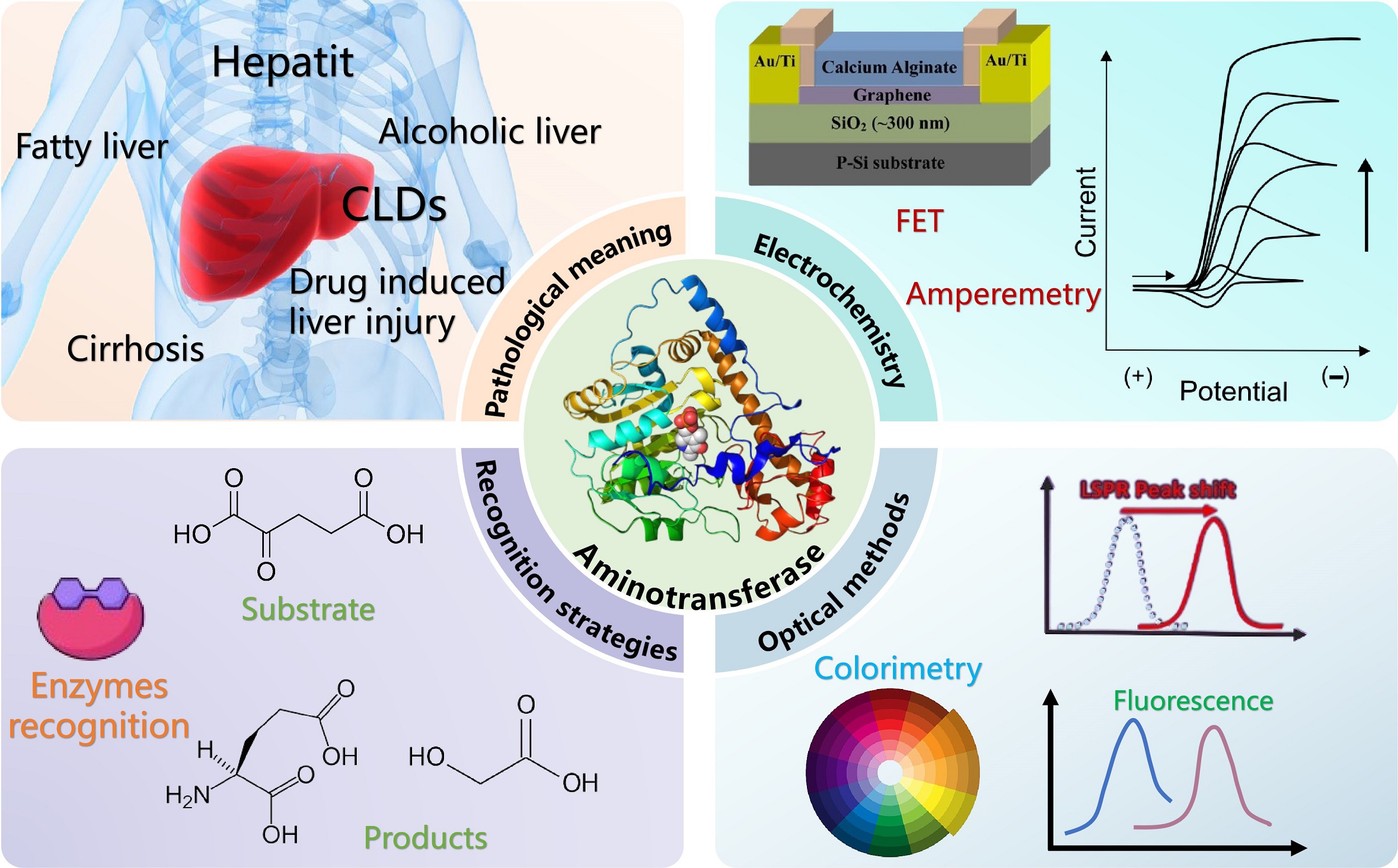
Graphical Abstract
Keywords
Aminotransferase, biosensor, enzyme electrode, point-of-care testing, healthcare
1. Introduction
Aminotransferases are a family of pyridoxal-5'-phosphate-dependent enzymes that catalyze reversible transamination reactions between amino acids and α-keto acids, and play vital roles in amino acid metabolism, energy homeostasis, and nitrogen balance regulation[1]. Among them, alanine aminotransferase (ALT, also known as glutamic-pyruvic transaminase (GPT)) and aspartate aminotransferase (AST, also known as the glutamic-oxaloacetic transaminase (GOT)) are the two most crucial aminotransferases in the human body, serving as core biomarkers for clinical assessment of hepatic health status[2,3]. Although both enzymes are widely distributed in multiple tissues, they differ in tissue specificity and intracellular localization: ALT is predominantly concentrated in the cytoplasm of hepatocytes, whereas a considerable proportion of AST resides in mitochondria in addition to its cytoplasmic distribution[4,5]. Accordingly, alterations in their serum activities can sensitively reflect the nature and severity of tissue injury[6,7]. Clinically, ALT and AST are not only fundamental biochemical markers of hepatocellular injury but also among the most routinely detected indicators for liver disease screening, condition monitoring, and therapeutic efficacy evaluation. Furthermore, the AST/ALT ratio (De Ritis ratio) assists in the differential diagnosis of various liver injury patterns[8,9]. Therefore, simultaneous and accurate detection of these two enzymes is indispensable for the diagnosis, severity grading, and therapeutic monitoring of liver diseases.
Globally, the disease burden of chronic liver diseases, drug-induced liver injury, metabolic-associated fatty liver disease, alcoholic liver disease, and other hepatic disorders is continuously rising[10], which further highlights the significance of aminotransferase detection. Statistics show that approximately 844 million people worldwide suffer from chronic liver disease (CLD), with an annual mortality of 2 million, rendering CLD one of the leading causes of global mortality and morbidity[11-13]. Meanwhile, metabolic disorders including obesity, diabetes mellitus, and hyperlipidemia are frequently accompanied by abnormal aminotransferase levels[14-16], which further expands the population requiring routine monitoring. For these patients, ALT and AST detection is not a one-off laboratory parameter but a dynamic monitoring indicator throughout the entire process of screening, diagnosis, therapeutic follow-up, and long-term management[12]. Accordingly, there is an especially urgent demand for point-of-care testing (POCT) in ALT/AST detection. On one hand, early identification of acute liver injury, critical illness-related hepatic dysfunction, and drug-induced liver injury (DILI) is highly time-sensitive; delayed detection results may directly affect decisions on drug withdrawal, risk stratification, and the timing of clinical intervention[17]. On the other hand, patients with chronic liver diseases, individuals undergoing treatment with potentially hepatotoxic drugs, as well as populations in primary healthcare settings and resource-limited regions, generally require high-frequency monitoring yet cannot sustainably rely on central laboratories for repeated assays[18,19]. Limitations of conventional laboratory paradigms in sample transportation, equipment dependence, operational complexity, and result turnaround time[20] have remarkably impaired the practical application value of aminotransferases as dynamic biomarkers. Therefore, for indicators such as ALT and AST that demand rapid detection, frequent re-testing, and widespread accessibility, the shift of detection scenarios from central laboratories to patient-side testing has emerged as a practical and urgent clinical requirement.
Currently, ALT and AST quantification is still predominantly based on colorimetry, kinetic assays, and automated biochemical analytical platforms. Although these methods are accurate and reliable, they generally require expensive instruments, professional operators, and relatively complicated procedures, which make it difficult to simultaneously fulfill the requirements of rapidity, portability, low cost, and user-friendliness for POCT[21,22]. In recent years, with the rapid advancement of micro-electromechanical systems (MEMS), microfluidics, nanomaterials, electrode modification, and portable readout devices, a variety of biosensors for aminotransferase detection have been successively developed, involving electrochemical, fluorescent, colorimetric, electrochemiluminescent, and localized surface plasmon resonance (LSPR) strategies[23-27]. These approaches exhibit prominent advantages in response speed, sample consumption, system miniaturization, and on-site detection potential, laying an important technical foundation for POCT of ALT and AST.
Previous reviews have provided important overviews of aminotransferase detection from different perspectives. Huang et al. summarized the principles of conventional and biosensor-based methods, Yadav et al. focused primarily on electrochemical biosensors, and Mruga et al. recently presented a comprehensive classification of aminotransferase biosensors according to their recognition elements, immobilization strategies, transduction mechanisms, and analytical performance[28-30]. Building on these studies, the present review examines aminotransferase biosensors specifically from a POCT perspective. Electrochemical, optical, paper-based, and microfluidic platforms are discussed not only in terms of sensing mechanisms and analytical performance, but also with regard to their potential for integration into rapid, portable, and user-friendly testing systems. Particular attention is given to clinically relevant measurands, sample types and volumes, whole-blood compatibility, sample pretreatment, assay time, operating conditions, device integration, clinical validation, and comparison with reference methods. Because many reported aminotransferase biosensors remain laboratory-stage analytical prototypes rather than complete POCT devices, their current limitations and the evidence required for practical translation are critically examined. Through this POCT-oriented framework, this review aims to clarify the progress, remaining barriers, and future development priorities for clinically deployable ALT and AST testing.
2. Recognition and Detection Pathways for Aminotransferase Biosensors
Clinically, ALT and AST are conventionally reported as catalytic activity concentrations rather than protein concentrations. The IFCC primary reference procedures define their activities at 37 °C under specified substrate, cofactor, pH, temperature, and measurement conditions[31-33]. One enzyme unit (U) represents the catalytic amount converting 1 μmol of substrate per minute under the specified assay conditions; thus, U/L is a procedure-dependent activity concentration. A universal conversion from U/L to molar concentration is therefore not scientifically valid unless the molar catalytic activity, isoenzyme composition, fraction of catalytically active protein, and assay conditions are known. This distinction has experimental support: ALT isoforms and splice variants differ in catalytic competence, including a splice variant of ALT2 (ALT2_2) whose recombinant protein does not possess detectable enzymatic activity[34], while AST mass and catalytic activity have been measured as distinct quantities[35]. Accordingly, this review retains U/L for activity-based assays and discusses direct affinity recognition separately; results from the two routes should not be treated as interchangeable.
The detection of aminotransferase catalytic activity is generally based on the formation or consumption of reaction products. ALT catalyzes the transfer of an amino group from L-alanine to α-ketoglutarate, producing pyruvate and L-glutamate, whereas AST catalyzes the corresponding reaction between L-aspartate and α-ketoglutarate, producing oxaloacetate and L-glutamate[32,33]. Thus, glutamate is a common product of both reactions, while pyruvate is produced directly only by ALT. In AST assays, pyruvate can be monitored only after an additional conversion of oxaloacetate, for example, through oxaloacetate decarboxylase-mediated decarboxylation[36]. These distinct product pathways provide the biochemical basis for downstream GluOx-, PyOx-, LDH-, and MDH-coupled signal-transduction strategies (Figure 1).
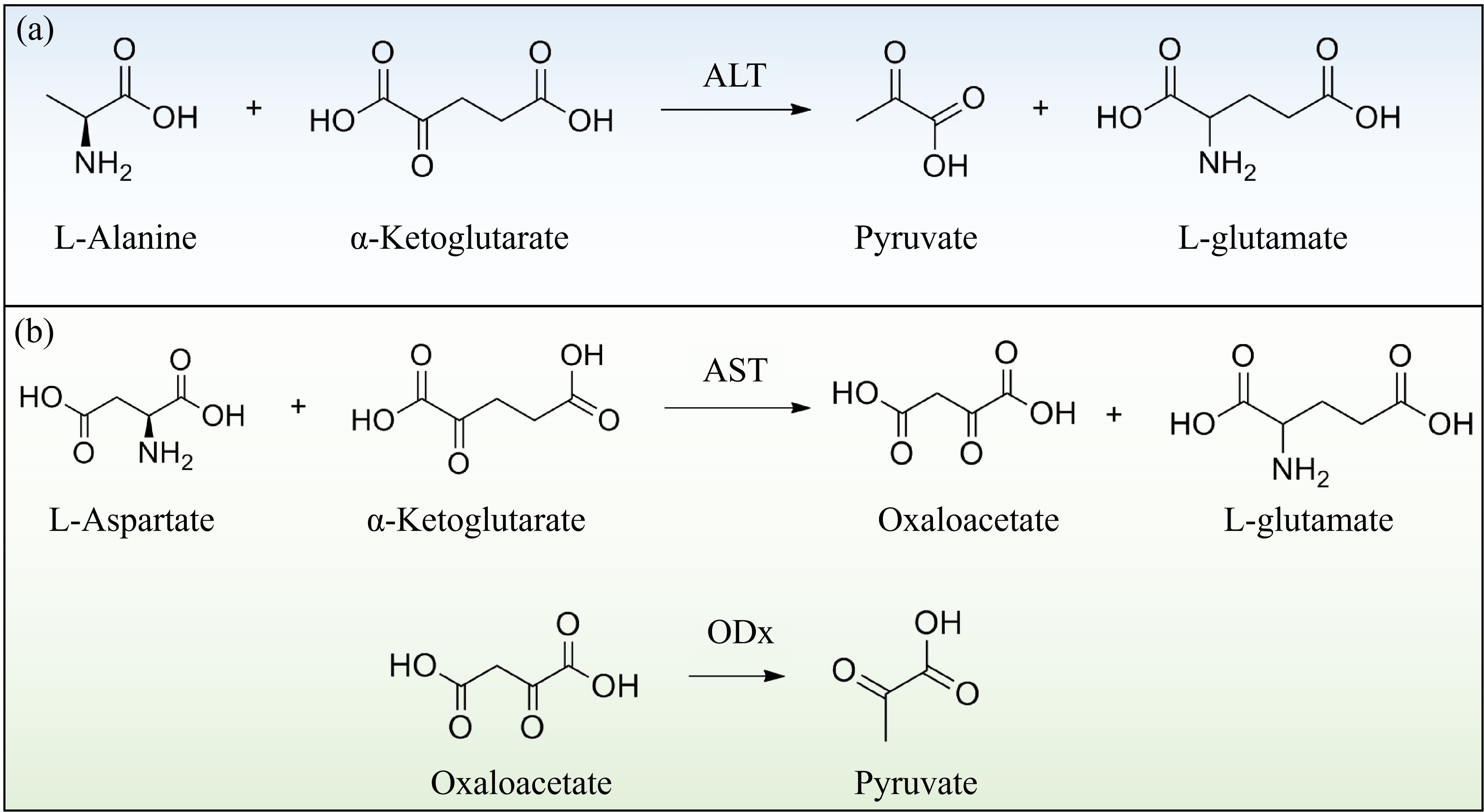
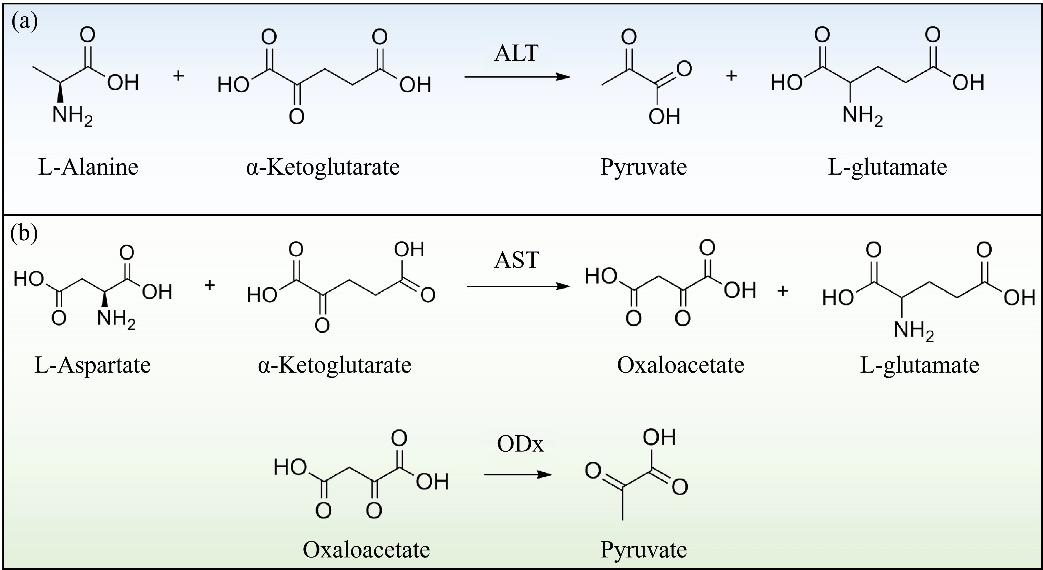
Figure 1. (a) Catalytic reaction equation of ALT; (b) Catalytic reaction equation of AST. ALT: alanine aminotransferase; AST: aspartate aminotransferase.
2.1 Enzyme cascade reaction strategy
The catalytic reactions of ALT and AST themselves cannot directly generate readily detectable signals such as color, current, or fluorescence. Accordingly, their detection generally relies on a series of elaborately designed enzyme-linked cascade reactions.
The most prevalent approaches use glutamate oxidase (GluOx) to oxidize glutamate generated by either ALT or AST, or use pyruvate oxidase (PyOx) to oxidize ALT-derived pyruvate. For AST detection through the PyOx pathway, oxaloacetate must first be converted into pyruvate, typically using oxaloacetate decarboxylase. As shown in Figure 2a, these enzymes catalyze the oxidation of the glutamate or pyruvate generated by the aminotransferases to produce hydrogen peroxide (H2O2), which is subsequently detected via colorimetric, fluorescent, or electrochemical signals. Compared with GluOx-mediated reactions, PyOx-catalyzed reactions require more rigorous conditions involving cofactors including Mg2+ and thiamine pyrophosphate (TPP). Therefore, the GluOx pathway possesses superior storage stability relative to the PyOx pathway[37,38]. Nevertheless, since oxidase-catalyzed H2O2 generation consumes dissolved oxygen as a co-substrate, insufficient ambient oxygen supply can limit H2O2 production and thereby induce signal fluctuations and detection errors[39,40].
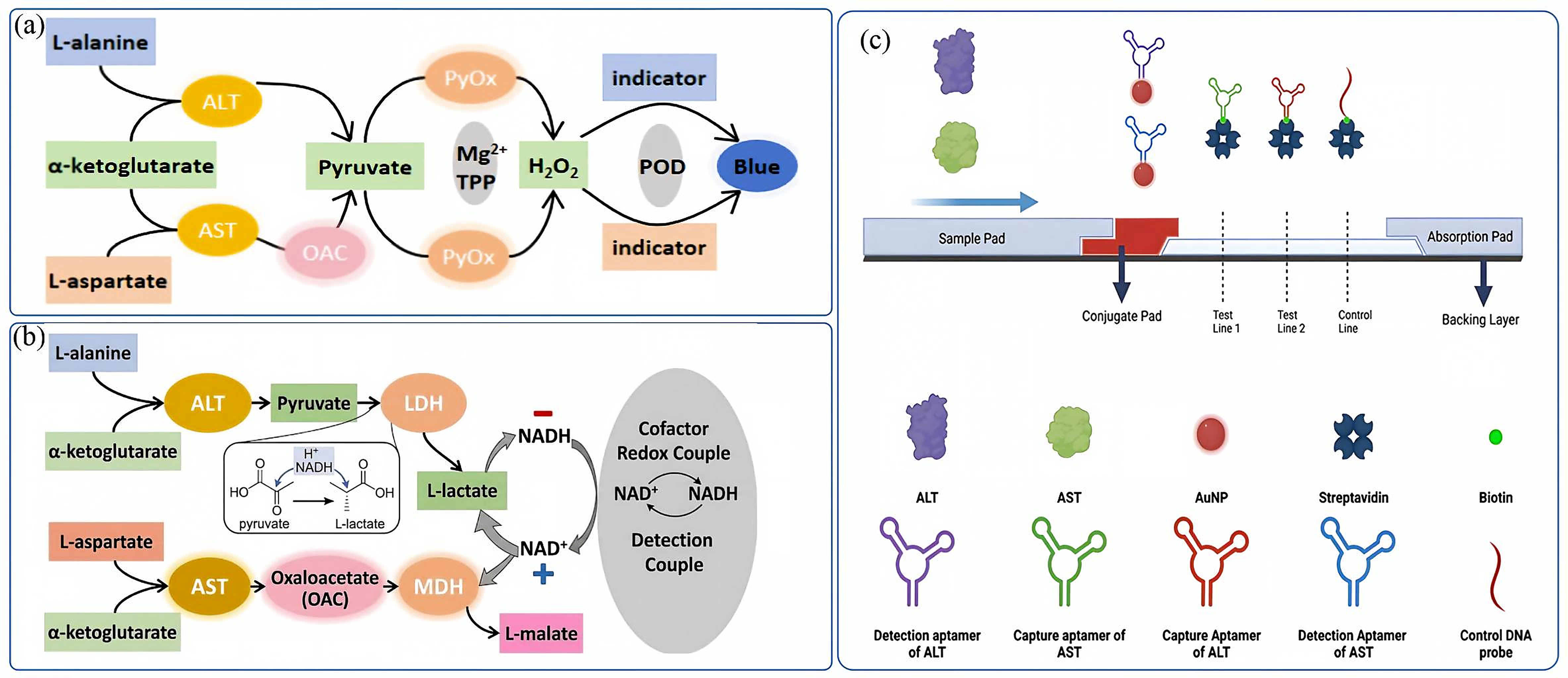
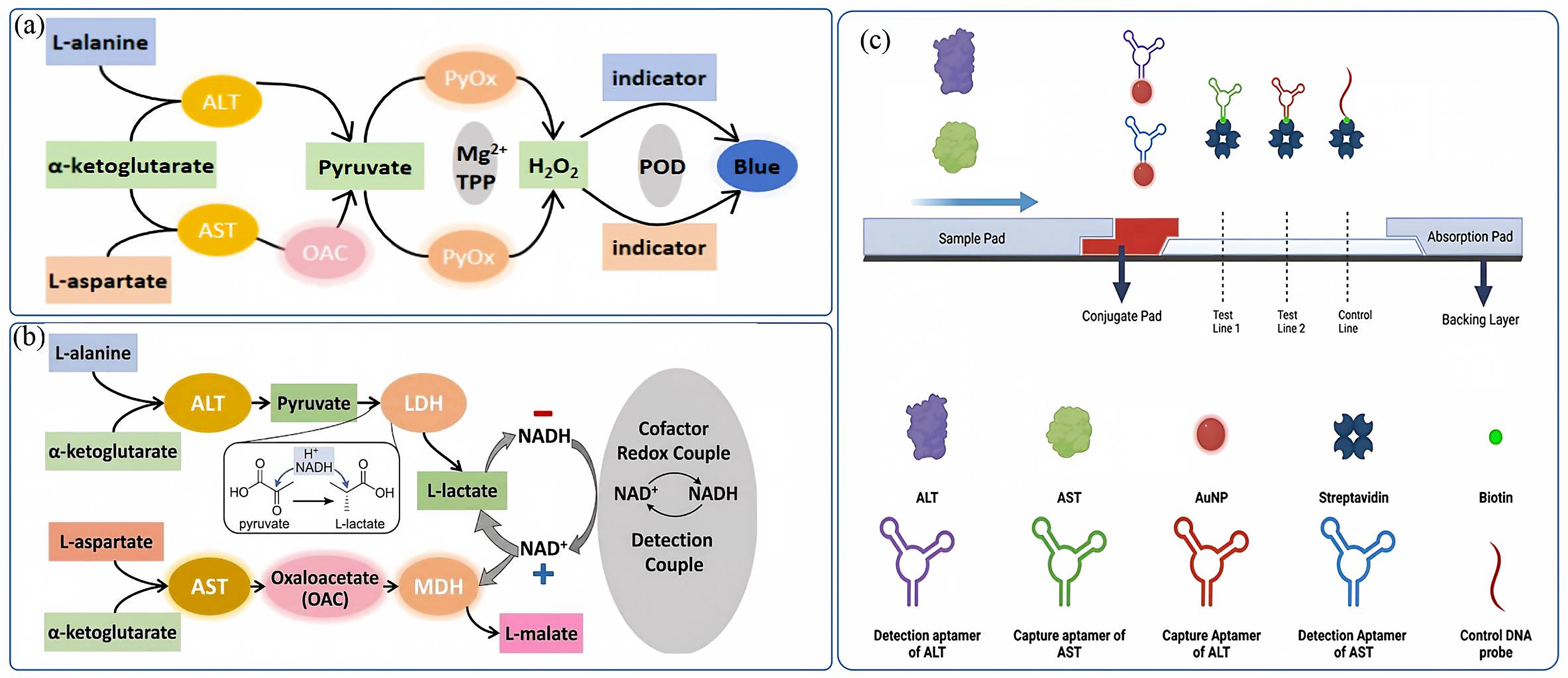
Figure 2. Design of recognition and detection pathways for aminotransferase biosensors. (a) Colorimetric detection principle based on glutamate oxidase and pyruvate oxidase for recognizing aminotransferase-catalyzed products; (b) Colorimetric detection principle using LDH and MDH to identify aminotransferase-catalyzed products; (c) Conceptual schematic of a lateral flow assay employing high-affinity aptamers for direct capture of ALT and AST. Reproduced from reference[21]. CC BY 4.0. LDH: lactate dehydrogenase; MDH: malate dehydrogenase; ALT: alanine aminotransferase; AST: aspartate aminotransferase; OAC: oxaloacetate decarboxylase; PyOx: pyruvate oxidase; TPP: thiamine pyrophosphate; POD: peroxidase; NADH: nicotinamide adenine dinucleotide reduced form.
Another classical strategy employs dehydrogenases such as lactate dehydrogenase (LDH) and malate dehydrogenase (MDH) to reduce pyruvate or oxaloacetate substrates produced by aminotransferases into lactate or malate. This process consumes the nicotinamide adenine dinucleotide reduced form (NADH) coenzyme[41,42], and the activities of ALT and AST can be indirectly quantified by monitoring concentration variations of NADH, as shown in Figure 2b[41,43]. This strategy exhibits favorable selectivity in complex serum matrices and reduces dependence on H2O2. However, this method imposes stringent requirements on substrate supply and reaction condition stability. Strict control over the ratio of enzymes to coenzymes is essential to guarantee the reliability of detection results.
Such cascade reaction designs serve as the cornerstone of enzyme activity detection for POCT. The selection of distinct downstream pathways directly determines the type of biosensors. For instance, PyOx- and GluOx-based pathways are highly suitable for developing electrochemical and colorimetric sensors, since H2O2 is electroactive and capable of participating in chromogenic reactions. In contrast, NADH consumption-based pathways are primarily applicable to spectrophotometric assays[28]. A comparative study on two H2O2-dependent amperometric ALT biosensors revealed that PyOx-based sensors exhibited higher sensitivity and broader linear ranges[38]. Although GluOx-based sensors possess relatively lower sensitivity, they demonstrate superior stability in complex matrices with lower fabrication costs. This finding highlights that trade-offs among analytical performance, stability, and cost are indispensable in POCT device design.
2.2 Enzyme-free electrode strategy
The enzyme-free electrode strategy achieves detection by directly monitoring concentration changes of aminotransferase substrates or their chemical reaction products. For instance, Moed and Zaman developed a Prussian blue-modified enzyme-free electrode in 2019, which indirectly quantified ALT levels by detecting the consumption of α-ketoglutarate[44]. In their design, α-ketoglutarate acts as an antioxidant and directly reacts with H2O2 in solution to produce succinate, water, and carbon dioxide[45], thereby reducing the H2O2 content in the system. Subsequently, H2O2 is coupled with Prussian blue (PB) as an electron transfer mediator and detected via amperometry. This approach eliminates the requirement for enzymes, simplifying electrode fabrication and operational procedures. Nevertheless, enzyme-free methods are generally sensitive to experimental conditions, demanding precise control of initial reagents and reaction duration. Meanwhile, their feasibility for POCT in whole blood or complex biological matrices remains limited.
2.3 Direct protein recognition using high-affinity aptamers
Aptamers recognize molecular epitopes on ALT or AST and would therefore report target binding or protein abundance rather than catalytic turnover. Chinnappan et al.[21,22] proposed screening high-affinity nucleic-acid aptamers by SELEX and incorporating them into lateral flow assay (LFA) devices for point-of-care applications, as illustrated in Figure 2c. However, both cited articles present a hypothesis or perspective rather than an experimentally established ALT/AST aptasensor: no selected ALT/AST aptamer sequence, binding affinity, quantitative limit of detection, analytical range, clinical sample validation, or comparison with a standardized activity method was reported. Consequently, this route should be described as a conceptual proposal rather than as a platform at the laboratory validation stage. If realized, it could provide rapid enzyme-independent recognition, but its output would correspond to protein abundance rather than catalytic activity; accordingly, such results could not be interpreted using existing U/L reference intervals, and clinical interpretation would require new reference intervals established against standardized ALT/AST activity assays in matched clinical samples.
2.4 Comparison of different recognition strategies
Enzyme-cascade, enzyme-free, and affinity-based strategies exhibit different characteristics in aminotransferase detection. At present, enzyme-cascade assays remain the most widely investigated approach. Their major advantage is that they retain catalytic activity as the detection target, allowing ALT and AST results to be expressed in U/L and compared with conventional clinical assays. Several integrated systems have further achieved whole-blood analysis, short detection times, and comparison with clinical biochemical analyzers[26,46,47]. However, these methods generally require multiple enzymes, substrates, and cofactors. Their analytical performance is also influenced by reaction temperature, incubation time, oxygen concentration, and reagent stability.
Enzyme-free strategies attempt to simplify the reaction system by directly monitoring the consumption of substrates or the formation of chemical products. For instance, the Prussian blue-modified electrode developed by Moed and Zaman[44] provided a detection range of 40-1,990 U/L and a limit of detection (LOD) of 20.6 U/L using 50 μL buffer-based ALT aliquots intended to simulate a finger-prick sample volume; actual whole-blood analysis was not experimentally demonstrated. Nevertheless, the assay required approximately 100 min and involved several manual reagent-addition steps. In addition, no independent clinical comparison was reported. Therefore, further simplification of the assay procedure is still required before this strategy can be applied to rapid POCT. It should also be noted that the elimination of biological enzymes does not necessarily indicate a lower total assay cost, because electrode fabrication, chemical reagents, instrumentation, and user operation must also be considered.
Aptamer-based detection is still at an earlier stage of development. The reported studies[21,22] proposed the screening of ALT- and AST-binding aptamers by SELEX and their integration into lateral-flow devices. However, experimentally validated aptamer sequences, binding affinities, analytical ranges, limits of detection, selectivity, and clinical performance have not yet been reported. Thus, the advantages of aptamer recognition in rapidity, stability, and cost remain to be experimentally demonstrated. Overall, enzyme-cascade strategies currently possess a higher level of analytical and clinical validation, whereas enzyme-free and aptamer-based methods require further investigation of assay workflow, matrix compatibility, and clinical performance. This comparison also involves different measurands: enzyme-cascade assays report catalytic activity in U/L and can be interpreted against clinical reference intervals, whereas aptamer-based assays would report ALT/AST protein abundance, for which no validated reference intervals currently exist.
3. Electrochemical Biosensors for Aminotransferase Detection
Benefiting from high sensitivity, rapid response, facile miniaturization, and low cost[48], electrochemical biosensors hold promising application prospects in aminotransferase detection[49]. Compared with conventional spectrophotometric methods, electrochemical approaches can directly convert variations including H2O2 generation, NADH/NAD+ transformation, local pH fluctuations, and interfacial charge changes induced by aminotransferase reactions into current, potential, or conductance signals, rendering them more suitable for portable and point-of-care testing. In recent years, electrochemical platforms for aminotransferase detection have expanded from traditional enzyme electrodes to various configurations including thin-film electrodes, microelectrode arrays, field-effect transistors, and intelligent terminal-coupled chips[36,42,50,51]. Nevertheless, aminotransferase detection is markedly more complicated than the analysis of small molecules such as glucose and lactic acid. The core reason is that aminotransferase quantification is essentially an enzymatic activity assay, which generally relies on continuous coupling of substrate conversion, coupled enzymatic reactions, and terminal electrical signal transduction. Accordingly, biosensors are required to achieve not only high sensitivity, but also favorable enzyme/substrate loading capacity, electron transfer efficiency, matrix interference resistance, and interfacial stability. Therefore, effective integration of multi-step enzymatic reactions is the crucial bottleneck for translating electrochemical aminotransferase detection into practical POCT devices.
In terms of detection principles, existing electrochemical aminotransferase biosensors can be roughly classified into three categories. First, amperometric and voltammetric sensors based on multi-enzyme cascade reactions, which achieve quantification by monitoring variations in H2O2 or NADH. Second, field-effect transistor sensors based on local pH shifts or interfacial charge alterations. Third, enzyme-free or quasi-enzyme-free strategies relying on indirect changes of substrates or byproducts. These three technical routes correspond to distinct interfacial designs and device integration strategies.
Table 1 summarizes the configurations and analytical performances of diverse electrochemical aminotransferase biosensors. Overall, performance improvement is not driven by a single factor, but originates from the synergistic optimization of immobilization strategies, nanomaterials, and signal transduction modes.
Table 1. Summary of electrochemical biosensors for ALT or AST activity analysis.
| Analytical method | Electrode/sensing interface | Recognition enzyme | Detection range (U/L) | LOD(U/L) | Sample/preparation | Assay time/conditions | Calibration | Clinical comparison | Storage stability | User steps/device integration | POCT readiness | Ref. |
| SWV | PB/Mo2C/SPE | GluOx | 5~200 | 2.76 | Rabbit serum, 1:9 in 150 μL assay | NR | Buffer standards; log fit (R2 = 0.9954) | 3 rabbits vs ELISA; no significant difference (95% CI) | 4 °C; duration NR | Semiautomated sensor array + external electrochemical reader | + | [52] |
| Amperometry | P-meta-PD/Pt disk | GluOx | 10~500 | 2.5 | Buffer/simulated serum ions (2 mL) | ~15 min (~60 s response) | ALT standards in buffer | NR | 3-day operational test | Modified Pt disk + benchtop potentiostat | + | [53] |
| CV | PyOx MIP/Pt | PyOx | 25~700 | 2.97 | Human plasma/serum, 100 μL (+ 50 μL ALT standard) | 10 min, 25 °C | Standards; matrix testing | n = 12, vs commercial ALT kit | 1 month at 4 °C (~90% response) | MIP microelectrode + external potentiostat | ++ | [54] |
| SWV | PyOx/Graphene@MXene/GCE | PyOx | 0.5~400 | 0.16 | Human serum, 1:10 dilution (5 mL) | 40 min, 37 °C | ALT standards; serum recovery | NR | Composite: approximately 92% at 2 months, 4 °C | Modified GCE + external potentiostat | + | [55] |
| FET | APTES/SiNW-FET | PyOx | 0~450 | 4.1 | ALT-spiked/ depleted human serum (volume NR) | 20 min, 37 °C | ALT-spiked serum series (50-450 U/L) | NR | NR | 7-SiNW-FET array + multiplexed electronics and flow chamber; complete POCT workflow not validated | ++ | [18] |
| DPV | Pt | GluOx | 5~120 | NR | Buffer (100 μL reaction) | 15 min, ~25 °C | H2O2/ALT standards in buffer | NR | NR | Thin-film Pt electrodes + external potentiostat | + | [56] |
| Amperometry | PB/SPE | N/A | 40~1,990 | 20.6 | Buffer ALT standards (50 μL) | ~100 min (multi-step incubations), 60 °C | Buffer ALT standards | NR | NR | PB/SPE + external potentiostat; not self-contained | + | [44] |
| CV | Nafion/ER-GO/SPE | GLDH | 5~300 | 2.2 | Spiked human serum (~20 μL) | 120 min, 25 °C | ALT-spiked serum | 3 spiked sera vs ALT ELISA kit | NR | Modified SPE + external potentiostat | + | [43] |
| CV | SPE | GLDH | 10~250 | 9.25 | Buffer (~20 μL/assay) | 30 min (dual 10 μL/min streams) | ALT standards in buffer | NR | NR | Microfluidic chip + workstation | + | [57] |
| DPV | [TBA]2[Ni(mnt)2]/thiol-functionalized graphene/Au | GluOx | 5~140 | NR | Buffer (volume NR) | NR | ALT standards in buffer | NR | Glutamate response: 81.4% at 3 weeks, 4 °C | Nanofilm/Au electrode + external potentiostat | + | [58] |
| Amperometry | Poly(methylene blue)-CNT/SPCE | LDH | 10~640 | 10 | Human serum, 1:1 dilution; 50 μL readout | 20 min reaction; ~60 s readou | Standards; serum sex-classification model | n = 39, sex classification; no clinical ALT comparison | NR | Electrochemical chip interfaced to smartphone | ++ | [42] |
| Amperometry | GluOx/overoxidized PPy-Nafion/micro-Pt | GluOx | 10~900 | 8.48 | ALT-spiked PBS (volume NR) | 10 min pre-reaction; ~5 s response | ALT standards | NR | 8 weeks at -20 °C | Micro-Pt wire electrode + external potentiostat | + | [59] |
| CV | CNT/SPCE | GLDH | 60~300 | NR | Buffer (~6 mL) | 2.5 h, 25 °C | ALT standards in buffer | NR | NR | SPCE + external potentiostat | + | [60] |
| CV | Graphite/Poly(4-aminophenol)/PyOx | PyOx | 3 × 10⁻⁵~3 | 2.68 × 10⁻⁵ | Buffer (volume NR) | 25 min at 37 °C; ~200 s scan | ALT standards in buffer | NR | 30 days, 8 ± 1 °C, dark (~97%) | Modified graphite electrode + external potentiostat | + | [61] |
| FET | GFET | N/A | 10~100 | NR | Buffer standards (volume NR) | NR | ALT standards in buffer | NR | NR | Graphene FET + semiconductor parameter analyzer | + | [50] |
| Amperometry | Ir/C SPE | PyOx | ~0-80 (reported: 0-544 ng/mL) | 2.18 | Human serum, 200 μL; 1:2 dilution (10-fold for high ALT) | 20 s at 21-23 °C | PBS/calf-serum ALT standards | Human serum vs Vista 1500 | NR | Single-use Ir/C SPE + benchtop workstation | ++ | [62] |
| CV | PyOx and OAC/Au electrode | PyOx | 43~480 (ALT) 30~240 (AST) | 25 (ALT) 20 (AST) | Spiked plasma (1 mL; 20-fold dilution) | 25 min at 37 °C | Spiked-plasma calibration | Spiked-plasma R2: ALT 0.92, AST 0.94 | NR | Dual-enzyme Au electrode + external reader | ++ | [36] |
| CV/Amperometry | Pt/Au bimetallic hierarchical structure array | GluOx | 20~140 (ALT) 20~180 (AST) | 20 | Buffer (volume NR) | Real-time, steady state within seconds | ALT/AST standards in buffer | NR | Stable; duration NR | Pt/Au array + external potentiostat | + | [63] |
| Amperometry | GluOx/Pt | GluOx | 10~1,000 | 3.29 | Buffer (volume NR) | 180 s | ALT standards in buffer | NR | 6 months, 4 °C (~79%) | GluOx/Pt electrode + external reader | + | [51] |
| Amperometry | GluOx/APTES/Pt | GluOx | 1.3~250 | 1.3 | Buffer standards (~40 μL) | ~20 s response | ALT/AST standards in buffer | NR | No loss at 120 days, -4 °C; < 75% at 130 days | Electrochemical array + external reader | + | [64] |
POCT-readiness score: +++ = minimum POCT-readiness threshold met, i.e., (i) native biological specimen tested (whole blood, serum, or plasma), (ii) total assay time ≤ 30 min, (iii) portable or integrated readout (handheld/portable analyzer, smartphone, or equipment-free visual reading; an external benchtop potentiostat does not qualify), and (iv) comparison with a reference or automated clinical method; ++ = native specimen and ≤ 30 min assay, but lacking portable integration and/or clinical comparison; + = remaining prototypes (buffer-only validation, assay time > 30 min or not reported, and/or no native-specimen testing). A “+++” rating denotes the minimum evidence level defined in this review, not regulatory or full clinical readiness. ALT: alanine aminotransferase; AST: aspartate aminotransferase; SWV: square-wave voltammetry; CV: cyclic voltammetry; DPV: differential pulse voltammetry; FET: field-effect transistor; GFET: graphene field-effect transistor; SPE/SPCE: screen-printed (carbon) electrode; GCE: glassy-carbon electrode; PB: Prussian blue; MIP: molecularly imprinted polymer; SAM/LbL: self-assembled monolayer/layer-by-layer assembly; APTES: (3-aminopropyl)triethoxysilane; CNT: carbon nanantube; PPy: polypyrrole; ER-GO: electrochemically reduced graphene oxide; GluOx: glutamate oxidase; PyOx: pyruvate oxidase; GLDH: glutamate dehydrogenase; LDH: lactate dehydrogenase; OAC: oxaloacetate decarboxylase; TBA: tetrabutylammonium; mnt: maleonitriledithiolate; NR: not reported; POCT: point-of-care testing; Pt: platinum.
3.1 Enzyme immobilization and interfacial engineering
The primary challenge for electrochemical aminotransferase biosensors is to construct a stable and efficient microenvironment for interfacial reactions. Early studies mostly adopted membrane entrapment or physical adsorption to immobilize coupled enzymes including pyruvate oxidase and glutamate oxidase. Although such methods are straightforward, they commonly suffer from enzyme leaching, excessively thick membrane layers, and restricted mass transfer, resulting in unsatisfactory sensitivity and reproducibility[28,65-68]. To address the instability of physically adsorbed membranes, research has gradually shifted toward the strategy of a polymer matrix combined with cross-linking immobilization[37,69-71]. A representative example was reported by Jamal et al.[51], who immobilized GluOx on platinum (Pt) electrodes via glutaraldehyde/BSA cross-linking, and introduced Nafion to enhance membrane stability and selectivity. This study demonstrated an evident trade-off among enzyme loading capacity, cross-linking degree, and protective membrane thickness: excessive enzyme loading causes interfacial blockage, while overly thick Nafion layers increase diffusion resistance. Building on such cross-linking strategies, Mruga et al.[38] recently developed two amperometric ALT biosensors based on PyOx and GluOx cascades, respectively, and systematically optimized their electrode-modification schemes (Figure 3a). Their results quantitatively revealed how immobilization conditions govern the sensitivity, response speed, and immobilization reproducibility of cascade-based ALT sensing. Accordingly, the core objective of immobilization optimization is not merely to increase enzyme quantity, but to improve the overall interfacial reaction efficiency.
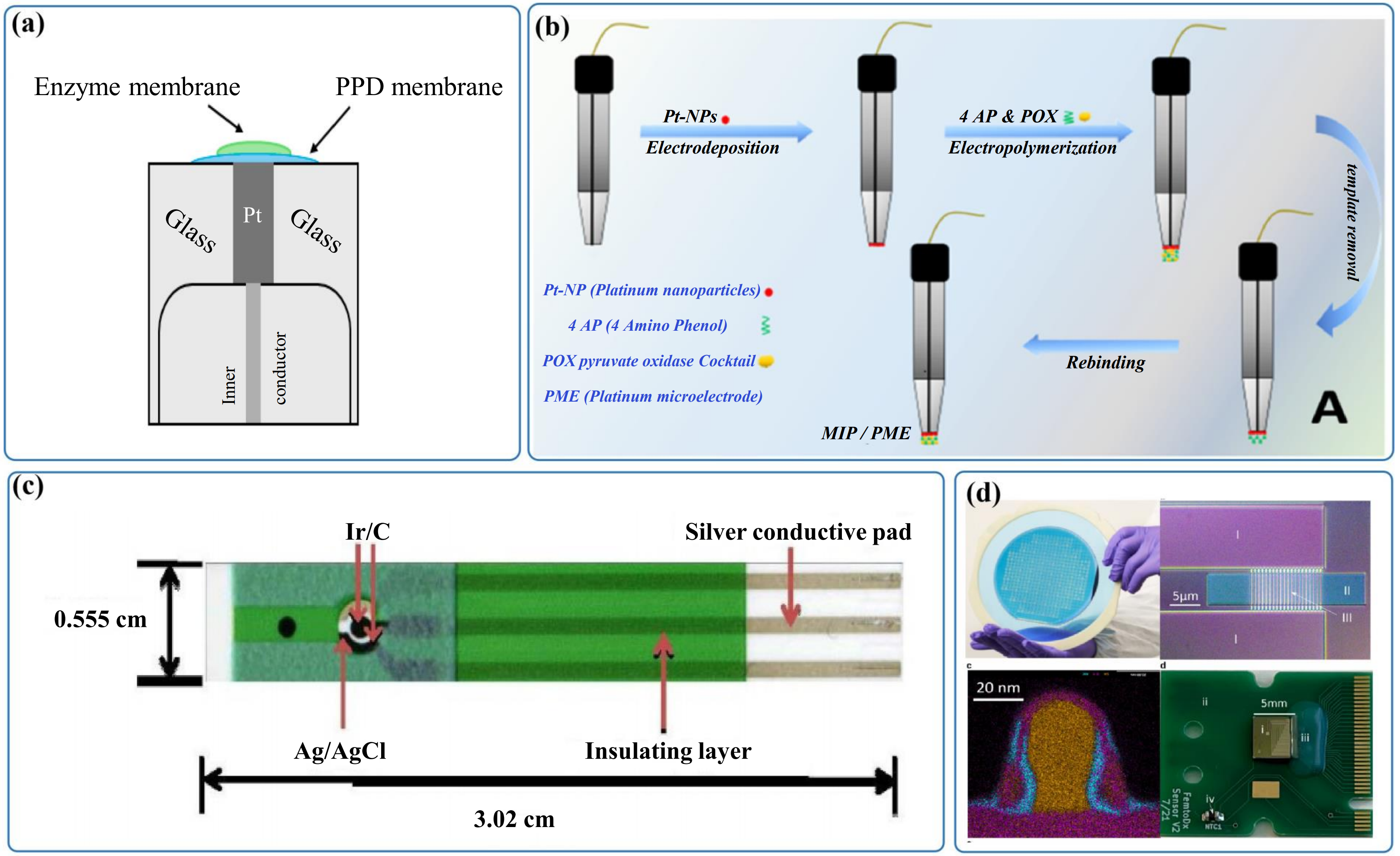
Figure 3. (a) Electrode-modification schemes of POx- and GluOx-based amperometric ALT biosensors. Reproduced from reference[38]. CC BY 4.0; (b) Molecularly imprinted microelectrode for point-of-care ALT sensing. Reproduced from reference[54]. CC BY 4.0; (c) Screen-printed electrochemical Ir-C-ALT sensor. Reproduced from reference[62]. CC BY 4.0; (d) Alanine aminotransferase biosensor based on a silicon nanowire field-effect transistor. Reproduced from reference[18]. CC BY 4.0. GluOx: glutamate oxidase; ALT: alanine aminotransferase; Pt-NPs: platinum nanoparticles; 4 AP: 4 amino phenol; POX: pyruvate oxidase cocktail; PME: platinum microelectrode; MIP: molecularly imprinted polymer.
Another important route involves oriented immobilization strategies centered on self-assembled monolayers (SAMs) and layer-by-layer (LbL) assembly. Han et al.[36] developed SAM/LbL multi-layer enzyme electrodes based on PyOx. They sequentially immobilized PyOx via the BS3 cross-linker and further co-immobilized oxaloacetate decarboxylase for AST detection, thereby constructing a surface architecture suitable for multi-enzyme cascade reactions. This system realized aminotransferase detection within the range of 7.5-720 U/L, which fully covers the concentration range required for human plasma sample analysis. More importantly, this work revealed that multi-layer immobilization does not necessarily yield improved limits of detection. Although increased enzyme loading accompanies the growth of immobilized layers, the diffusion pathways of products toward electrodes and electron transfer distances are simultaneously prolonged, which may counteract or even diminish the analytical benefits. Accordingly, high-performance interfacial engineering should not one-sidedly pursue high enzyme loading, but prioritize molecular accessibility and hierarchical coupling efficiency. For cascade-reaction-dependent systems such as aminotransferase detection, the spatial configuration of immobilized layers is generally more crucial than the absolute quantity of immobilized enzymes.
Recent studies have further expanded the immobilization targets from merely detection enzymes to the integrated design of substrates, cofactors, and reaction microenvironments. For instance, Her et al.[50] did not immobilize detection enzymes on graphene field-effect transistors (GFETs). Instead, they encapsulated L-alanine and α-ketoglutarate within an alginate hydrogel layer to construct a one-step ALT detection interface coupled with GFETs. Essentially, this design transfers substrate reactions from bulk solution to a semi-solid interfacial system, which simplifies experimental procedures and facilitates device integration. This indicates that POCT-oriented interfacial engineering has evolved from simple enzyme immobilization toward the construction of self-contained microsystems that can initiate reactions directly. In addition, Samy et al.[54] fabricated molecularly imprinted microelectrodes (Figure 3b), achieving a linear range of 25-700 U/L and a limit of detection of 2.97 U/L. Their work also demonstrates that templated interfacial design contributes to improved selectivity and portability for ALT detection.
Overall, the development of aminotransferase sensing interfaces has evolved from simple physical adsorption to cross-linking immobilization, oriented assembly, and collaborative preloading of substrates and enzymes. For such systems relying on multi-step cascade reactions, the core of interfacial engineering lies in balancing enzyme activity preservation, rapid mass transfer, and subsequent system integration.
3.2 Nanomaterial modification and signal amplification
Following effective enzyme immobilization, improving signal readout efficiency constitutes another critical issue. Since aminotransferase detection generally involves multi-step enzymatic reactions accompanied by inevitable signal attenuation, nanomaterials with high activity and electrical conductivity have been widely adopted to enhance electron transfer and electrocatalytic performance.
Noble metal nanostructures represent the most commonly adopted enhancement strategy[72-74]. Ren et al.[63] fabricated hierarchical flower-like Pt/Au bimetallic micro/nanoarrays, which remarkably enlarged the specific surface area of electrodes and enzyme immobilization sites, thereby improving the detection response toward AST/ALT. Hsueh et al.[62] constructed disposable ALT biosensors (Figure 3c) using iridium nanocarbon composites, achieving a limit of detection as low as 2.18 U/L in diluted serum. This demonstrates that the combination of noble metal nanoparticles with mass-producible printed electrodes is a practical route balancing analytical performance and fabrication cost[69-71,74].
Beyond noble metals, carbon materials provide more cost-effective alternatives. Xu et al.[60] reported that screen-printed carbon electrodes (SPCEs) modified with carbon nanotubes (CNTs) exhibited a superior NADH response compared with unmodified electrodes. On this basis, Deng et al.[42] adopted CNT/polyMB-modified SPCEs to realize low-potential electrocatalytic signal readout integrated with smartphone chips. Graphene and its derivatives possess two-dimensional highly conductive frameworks, surface modifiability, and excellent biocompatibility, leading to their rapidly expanding applications in aminotransferase detection in recent years. Her et al.[50] achieved label-free ALT detection by exploiting the high sensitivity of graphene toward variations in local ionic environments. Yao et al.[43] fabricated graphene-Nafion-modified screen-printed electrodes, yielding a linear detection range of 5-300 U/L with a limit of detection of 2.2 U/L. In addition, Jiang et al.[58] coupled the metal dithiolene complex [TBA]2[Ni(mnt)2] with thiol-functionalized graphene to construct uniformly dispersed composite membranes with ordered nanocubic structures. After modification on Au electrodes, the resulting sensor delivered an ALT sensitivity of 0.636 nA⋅U/L, which outperformed numerous previously reported GluOx-based electrodes. These results verify that composite catalytic membranes can significantly amplify signals via synergistic effects among heterogeneous materials.
Notably, signal amplification is not merely confined to increasing current intensity but also involves suppressing background signals. Jamal et al.[51] integrated the low-potential mediator ferrocene carboxylic acid with the GluOx/Pt system to realize ALT detection at 0.4 V. Combined with a background subtraction strategy, this design alleviated interference from ascorbic acid and uric acid, achieving a linear range of 10-1,000 U/L, an LOD of 3.29 U/L, and favorable long-term stability. In terms of anti-interference performance, Nafion remains one of the most widely used selective membrane materials[75-77]. It can repel anionic interfering species and is commonly coated outside the enzyme layer or blended with the enzyme layer to form composite membranes. Similarly, size-exclusion membranes such as poly(m-phenylenediamine)[53,78] are adopted to prevent macromolecular and charged interferents from approaching the working electrode. Accordingly, the core of high-performance aminotransferase sensing interfaces lies not in the single optimal material but in the synergistic matching among catalytic layers, immobilization layers, and size-screening layers.
3.3 Electrochemical detection methods
At present, electrochemical aminotransferase biosensors have developed multiple technical routes, including amperometric/voltammetric detection, field-effect transistors, and portable integrated platforms. Among these, amperometric and voltammetric assays based on multi-enzyme cascade reactions remain the most mature. Such methods generally take H2O2, glutamate, or NADH as terminal signal molecules and achieve quantification via constant-potential current responses or pulsed voltammetry. For instance, Saygili et al.[56] combined platinum thin-film electrodes with differential pulse voltammetry (DPV) to realize ALT detection in the range of 5-120 U/L, demonstrating the potential of thin-film electrodes for miniature circuit integration.
Distinct from conventional Faradaic current readout, FET sensors accomplish detection via interfacial charge variations or local pH shifts. Her et al.[50] developed a GFET that realized label-free detection by monitoring local pH and surface charge changes induced by ALT reactions. The device exhibited a pH sensitivity of 23.12 mV/pH at low operating voltages, with an ALT linear range of 10-100 U/L. This strategy avoids partial interference arising from direct high-potential oxidation of H2O2 and facilitates chip integration and low-power operation. Nevertheless, local pH dependence also renders such sensors more susceptible to buffer systems, ionic strength, and surface drift. Silicon nanowire field-effect transistor (SiNW-FET) platforms (Figure 3d) provide another route for kinetic activity detection, but the available evidence does not justify ranking them as more clinically practical than GFETs or other electrochemical formats. Muratore et al.[18] coupled ALT-generated pyruvate to pyruvate oxidase-mediated ferricyanide reduction and monitored the resulting local charge changes using a SiNW-FET array. The assay covered 50-450 U/L in ALT-spiked, ALT-depleted human serum. Electrical signals were recorded for 20 min, although the proposed maximum-slope calculation used data acquired within 300-600 s. The experiments required off-chip reagent mixing, syringe delivery through a flow chamber, and temperature control at 37 °C. No fingerstick or whole-blood operation, independent clinical cohort, or comparison with a standardized reference analyzer was reported. Therefore, the study demonstrates progress in serum-matrix compatibility and manufacturable sensor-array design, but its complete POCT readiness remains to be established.
In terms of portable device development, Deng et al.[42] integrated ALT detection into a smartphone-interfaced chip, achieving a standalone ALT detection range of 10-640 U/L with a detection time of approximately 20 minutes. Although this work was applied in forensic scenarios, it technically verified the practical feasibility of the POCT route consisting of enzymatic cascade reactions, printed electrodes, and smartphone terminals. By comparison, Moed and Zaman[44] proposed a quasi-enzyme-free strategy that indirectly quantifies ALT levels by monitoring variations in key components in the system, delivering a broad linear range of 40-1,990 U/L. This approach simplifies enzyme immobilization on electrode surfaces yet requires rigorous reaction control. Accordingly, it is more suitable for conceptual verification rather than direct translation into household POCT products.
Overall, existing electrochemical aminotransferase biosensors have achieved remarkable progress in the limit of detection, linear range, and device miniaturization, yet most designs remain at the laboratory prototype stage. Their primary bottlenecks do not lie in individual electrical performance, but in systematic challenges including preloading of substrates, stabilization of cofactors, incubation control, temperature adaptation, and complex blood sample pretreatment. Future development should shift from merely pursuing ultra-low limits of detection toward dry pre-storage of substrates, microfluidic/paper-based integration, low-potential anti-interference designs, and validation using genuine whole-blood samples. Only when these issues are comprehensively addressed can electrochemical aminotransferase biosensors be practically translated into point-of-care clinical and household testing applications.
Analytical sensitivity should not be equated with clinical or POCT readiness. Limits of detection far below clinically relevant ALT/AST activity ranges provide limited additional value when serum separation, extensive dilution, prolonged incubation, tightly controlled temperature, or multiple off-chip operations are still required. Evidence obtained in buffer or spiked serum/plasma primarily demonstrates analytical feasibility; stronger translational evidence requires undiluted whole-blood or fingerstick operation, coverage of clinically relevant ranges, evaluation of interference and hematocrit effects, reagent and calibration stability, and agreement with standardized reference methods in adequately sized clinical cohorts.
4. Optical Biosensors for Aminotransferase Detection
Whereas electrochemical platforms encode aminotransferase-coupled reactions as changes in current, potential, or conductance, optical platforms convert related biochemical pathways into absorbance, reflectance, fluorescence, or luminescence signals. Thus, the two platform classes differ primarily in signal transduction and device integration rather than in their fundamental recognition chemistry. The following section shifts from electrode-interface engineering to optical reaction and readout architectures, with particular attention to paper-based reagent storage, standardized imaging, and portable instrumentation.
Optical biosensors quantify ALT and AST activities by recording changes in absorbance, reflected light intensity, fluorescence, or luminescence signals induced by aminotransferase reactions. These strategies offer intuitive readout, multi-channel integration, and compatibility with paper-based devices and smartphone imaging, but their POCT suitability depends on the complete assay workflow and validation level rather than signal modality alone. A recent review[30] has indicated that optical aminotransferase sensing mainly consists of colorimetry, fluorescence spectroscopy, electrochemiluminescence, reflectometry, and a small number of photoelectric conversion methods. Paper-based colorimetric assays and portable optical readers have progressed toward integrated POCT formats, although whole-blood compatibility, workflow automation, and clinical validation vary substantially among studies. Analogous to electrochemical biosensors, aminotransferases themselves are generally not directly optically active analytes. Thus, most optical detections rely on cascade reactions: ALT or AST first catalyzes the generation of intermediates such as pyruvate and glutamate, and then enzyme activity variations are converted into differences in color, fluorescence, or luminescence via pyruvate oxidase, glutamate oxidase, lactate dehydrogenase, nanozymes, or chromogenic/luminescent probes. For this reason, the performance of optical biosensors depends not only on the signal readout module but more importantly on reaction system design, substrate preloading, and sample adaptability. For ALT and AST, their clinical reference ranges are approximately 5-35/36 U/L and 5-40/59 U/L, respectively[79,80]. Therefore, biosensors are required to cover the ranges of normal clinical levels, mild-to-moderate elevation, and obvious pathological abnormality.
4.1 Colorimetric and reflectance sensors
Colorimetry is among the most extensively developed techniques for optical aminotransferase detection. Its core advantage lies in direct visual interpretation via color changes, making it particularly suitable for resource-limited regions and rapid point-of-care screening[81,82]. Most early and current paper-based platforms follow the principle of chromogenic reactions of aminotransferase products. By spatially separating reaction zones, ALT and AST assays are confined to distinct detection areas to achieve dual-index differentiation[46,82]. For instance, Pollock et al.[82] fabricated two patterned regions on paper via wax printing and screen printing. Combined with stacked plasma separation membranes and polyester films, they constructed a three-dimensional (3D) paper analytical device containing separate ALT/AST test zones and three control zones (Figure 4a). Nevertheless, the accuracy of semi-quantitative visual grading was only 84%[83]. Recent reviews also confirm that optical multi-zone colorimetric platforms dominate among prototype systems capable of simultaneous ALT and AST detection[30]. Resmi et al.[84] reported that pyruvate generated by ALT reacted with 2,4-dinitrophenylhydrazine to form pyruvate hydrazone, leading to progressive deepening of the pale yellow color in the detection zone. The sensor achieved a linear range of 20-140 U/L with a limit of detection of 4.12 U/L in serum. By integrating plasma separation membranes into test strips, the system realized whole-blood detection, and quantitative analysis was realized via ImageJ combined with machine learning. Jain et al.[85] further evaluated paper-based ALT devices using clinical specimens. The device enabled rapid semi-quantitative analysis of fingertip blood and serum, showing favorable consistency with the Abaxis platform for serum samples, and demonstrated the viability of remote quantitative analysis based on smartphone imaging. In a subsequent perspective, Chinnappan et al.[22] proposed a double-layer microfluidic paper device concept that would integrate whole-blood plasma separation with multi-zone colorimetric readout for semi-quantitative ALT/AST analysis directly from venipuncture blood (Figure 4b). Similarly, Wang et al.[41] deposited reagents via inkjet printing to realize low-cost, mass-producible fabrication of ALT/AST paper-based devices. The detection ranges were 5.38-86.1 U/L for ALT and 5.4-91.2 U/L for AST, with results in good agreement with clinical spectrophotometric assays.
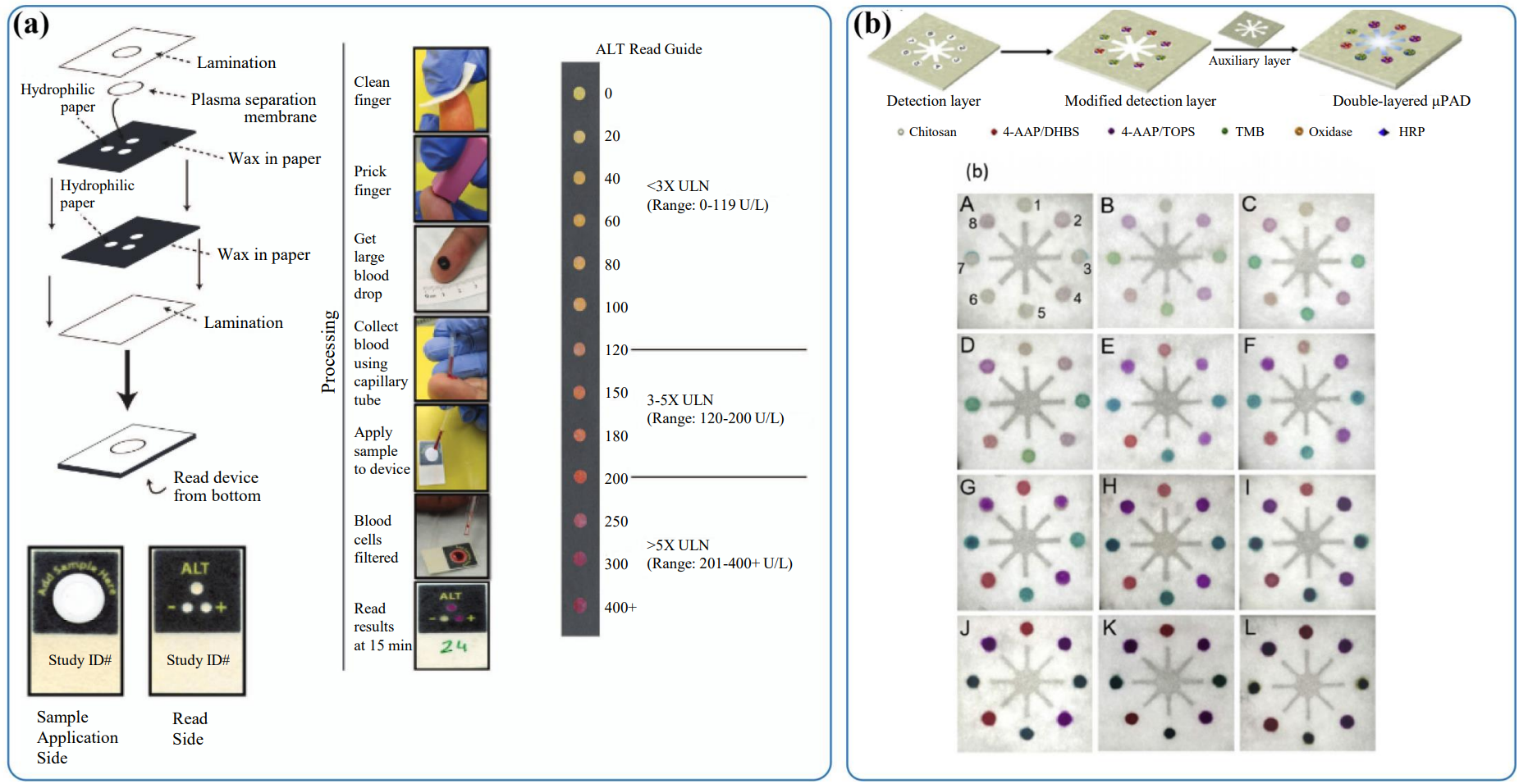
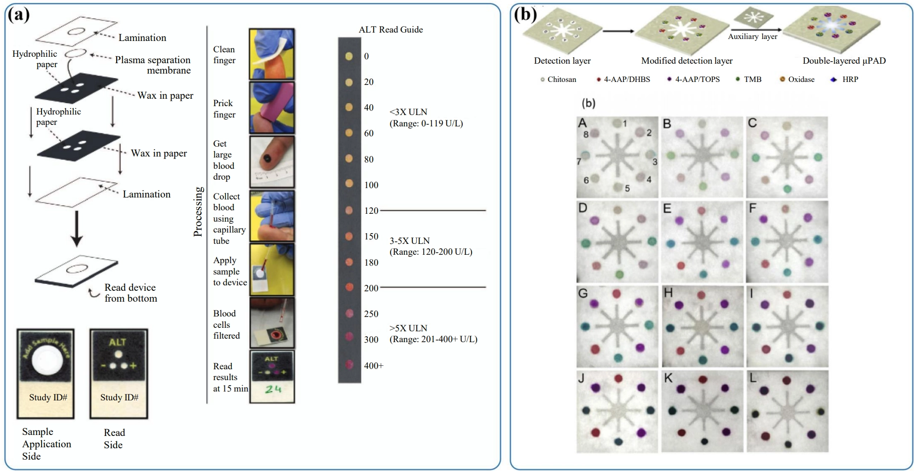
Figure 4. (a) Schematic of the three-zone paper-based transaminase test and its field evaluation. Reproduced from reference[83]. CC BY 4.0; (b) Double-layer microfluidic paper device for semi-quantitative ALT/AST analysis from whole blood. Reproduced from reference[22]. CC BY 4.0. ALT: alanine aminotransferase; AST: aspartate aminotransferase; ULN: upper limit of normal; 4-AAP: 4-aminoantipyrine; DHBS: 3,5-dichloro-2-hydroxy-benzenesulfonic acid; TMB: 3,3’,5,5’-tetramethylbenzidine.
In recent years, colorimetric platforms have been further integrated with nanozyme and hydrogel systems to enhance signal intensity and multi-index compatibility. Liu et al.[27] embedded Au-loaded layered double oxide nanozymes into agarose hydrogels to construct a smartphone-assisted colorimetric platform for ALT, AST, and ALP detection. In preliminary absorbance-based assays, the ALT and AST calibration ranges were 7-200 and 10-150 U/L, with calculated LODs of 2.56 and 7.5 U/L, respectively. After integration into the hydrogel-microdevice platform, smartphone grayscale analysis provided working ranges of 10-180 U/L for ALT and 15-150 U/L for AST; platform-specific LODs were not separately reported. The device was evaluated using diluted human plasma through standard addition (n = 3), but no independent clinical cohort or correlation with a standardized reference analyzer was reported. The reported 12 h result represents short-term signal stability rather than storage shelf life.
Besides paper-based chromogenic assays, reflectometry also constitutes an important branch of optical sensing[26]. Sang et al.[46] developed a dual-channel portable blood enzyme analyzer that quantifies ALT and AST according to reflected light intensity, with precise temperature control maintained at 37 °C ± 0.1 °C. For ALT and AST, the system achieved linear detection ranges of 4-350 U/L and 4-250 U/L, with limits of detection of 3.5 U/L and 4 U/L, respectively. The detection results exhibited favorable correlation with those obtained from the Roche Cobas C702 clinical analyzer. This work demonstrates that optical strategies can be fabricated not only as disposable test strips but also into miniaturized portable analytical devices integrated with temperature control modules.
Overall, colorimetric and reflectance methods possess advantages including simple operation, convenient multi-index integration, and smartphone readout for aminotransferase detection. Nevertheless, their quantitative performance is susceptible to illumination conditions, sample turbidity, background color, and reaction duration, thereby necessitating standardized image acquisition and algorithmic correction. The smartphone microfluidic deep learning platform reported by Ingole et al.[86] further demonstrates that image standardization and artificial intelligence analysis have become crucial approaches for improving the accuracy of optical point-of-care testing.
4.2 Fluorescence, electrochemiluminescence and other high-sensitivity optical strategies
Fluorescence and electrochemiluminescence (ECL) methods can provide high analytical sensitivity and low background under controlled assay conditions, but these benefits depend on probe design, excitation or electrochemical driving, and detector configuration. They are compatible with quantum dots, nanozymes, and paper-based luminescent electrodes, while typically requiring more specialized instrumentation than visual colorimetry. ECL platforms are included in the optical section because their quantitative output is based on photon emission, although signal generation requires electrochemical excitation.
In terms of fluorescence sensing, Liu et al.[87] designed nitrogen-doped graphene quantum dots with high quantum yield and multicolor tunability for fluorescent detection of AST. The system displayed favorable linearity within the range of 2.0-60.0 U/L, with a limit of detection as low as 0.065 U/L, and was applicable for in vivo imaging of zebrafish. Although this work focused on AST detection, it demonstrates that optical aminotransferase sensing has expanded from in vitro colorimetric assays toward integrated in vitro and in vivo fluorescence imaging.
ECL represents another high-sensitivity strategy. For instance, Zhi et al.[88] demonstrated the feasibility of ECL detection for ALT. The system adopted AgNPs/N,S-doped graphene quantum dot (N,S-GQDs) modified paper-based electrodes to generate stable luminescent responses under dual-potential scanning. In the supplementary materials, the concentrations of persulfate, the pH value, and the scanning rate were systematically optimized. Meanwhile, it was verified that various reactants exerted limited interference on ECL signals, revealing that the platform possesses both high sensitivity and favorable system stability. In addition, Lai et al.[23] recently developed a dry-chemistry-based electrochemiluminescence chip by integrating three-electrode ECL, dry chemistry, and POCT technologies. Combined with a self-developed automatic ECL analyzer, the chip was applied for ECL quantification of ALT. Under optimized conditions, the dry chemistry-ECL (DC-ECL) device was successfully utilized for ALT detection in serum, delivering a broad linear range of 5-1,000 U/L and a limit of detection of 1.702 U/L. The study demonstrates integration of dry reagents with automated ECL readout, but it does not establish commercial POCT readiness. Validation was limited to serum, with no reported whole-blood operation or sufficiently characterized independent clinical comparison. Integrating upstream blood-cell separation, as demonstrated in other paper-based dry-chemistry and optode systems, could enable whole-blood operation, but such integration was not reported for this device[89-91].
In addition, a small number of photoelectric conversion methods are also noteworthy. Kozinetz et al.[92] developed photoelectric sensors based on buried silicon barrier structures and realized real-time detection of ALT/AST activities by monitoring photocurrent variations induced by interfacial adsorption. Distinct from conventional colorimetric or fluorescence assays, this strategy converts interfacial charge changes triggered by aminotransferase reactions into photoelectric signals, indicating that optical aminotransferase sensing can be further extended toward semiconductor photoelectric interfaces.
Overall, fluorescence, ECL, and photoelectric methods can achieve sensitive, low background readout, but their practical performance depends on material design, excitation and detection hardware, calibration, and specimen compatibility. Compared with visual paper-based colorimetry, they often require more specialized instrumentation; conversely, they may provide valuable analytical sensitivity when the intended use justifies the added complexity. Their POCT suitability should therefore be evaluated case by case rather than ranked solely by signal modality.
4.3 Optical microfluidic chips
Optical aminotransferase biosensors are summarized in Table 2. Integration with paper-based microfluidics, portable imaging, and smartphone analysis represents one practical development route for optical aminotransferase biosensors. Smartphone cameras, LED sources, and image-processing algorithms can facilitate portable signal acquisition, whereas electrochemical platforms offer different advantages in compact electronic readout and low-power integration. Neither route is inherently more suitable for telemedicine or resource-limited settings without standardized sample handling, robust calibration, and validation in the intended population.
Table 2. Summary of the optical aminotransferase biosensors reported in the literature.
| Analytical method | Description | Enzyme | Detection range (U/L) | LOD (U/L) | Sample/preparation | Assay time/conditions | Calibration method | Clinical comparison | Storage stability | User steps/device integration | POCT readiness | Ref |
| LSPR | TIT-assisted four-tapered-fiber probe | GluOx | 0-1,000 | 4.84 | Buffer standards (volume NR) | 2 min mix + 20 min measurement, ~24 °C | Buffer standards | NR | 15 reuses; shelf life NR | Fiber probe + optical spectrum analyzer | + | [93] |
| ECL | disposable dry chemistry-based ECL chip | PyOx | 5-1,000 | 1.702 | Human serum; 10-fold dilution (volume NR) | Dry reagents; 10 min; automated ECL | Standards; serum spike recovery 94.2-104.9% | n = 9 vs AU5800; R2 = 0.9936 | 37 °C/36 h: 64.37%; estimated 4 °C: ~2.5 months | Disposable chip + automatic portable analyzer | +++ | [23] |
| Colorimetry | B-doped core-shell Fe@BC nanozyme | PyOx, Fe@BC nanozyme | 10-1,000 | 4 | Human serum/ALT standards (50 µL) | RT; two 10 min steps; acid stop | 450 nm; triplicate, RSD < 5% | n = 8 vs hospital; r = 0.991 | NR | Two-step assay + microplate reader | ++ | [94] |
| Colorimetry | smartphone-assisted hydrogel platform | PyOx, GluOx, Au/LDO | Abs.: ALT 7-200; AST 10-150 Phone: ALT 10-180; AST 15-150 | Abs.: ALT 2.56; AST 7.5 Phone: NR | Diluted plasma; 1 µL/min × 50 s | 30 min, 30 °C | Absorbance; standard addition | Kit-based recovery only (n = 3): ALT 98.0-104.5%, AST 105.5-109.0% | 12 h signal stability; shelf life NR | Hydrogel device + smartphone; automated reagent addition | ++ | [27] |
| Colorimetry | Multi-layer paper-based test strip/LED reflectophotometry | PyOx, POD | 4-480 | 3.62 | Whole blood, 30 µL | 2 min, 37 °C | POC-strip calibration | n = 50 vs analyzer; r = 0.9749 | NR | Strip + portable reader + smartphone | +++ | [26] |
| LSPR | TIT fiber structure-based probe | GluOx | 10-1,000 | 10.61 | PBS standards (volume NR) | 3 min (Methods), 20 min (selectivity tests) | Peak shift; mean of 3 probes; R2 = 0.9599 (figure; text: 95.62%) | NR | 10 repeats, SD = 0.0145; shelf life NR | Fiber probe + spectrometer; wash/dry | + | [24] |
| Fluorescence | Visual fluorescence sensor based on dye-functionalized MOF | N/A | 1-1,500 | 0.5 | Goat serum, 0.5 mL, 10-fold dilution | 3 min (after pretreatment) | Standards; goat-serum recovery | Spike recovery 96.7-105.3% | Material: 48 h in water; assay shelf life NR | Solution assay + fluorescence reader | ++ | [25] |
| Colorimetry | PAD with Inkjet Printer-Sprayed Reagents | LDH, MDH | ALT 5.38-86.1 AST 5.4-91.2 | NR | Human serum, 2 µL | ALT 7 min; AST 6 min; 37 °C | Serum calibration; R2: ALT 0.9944, AST 0.9932 | n = 20 vs hospital; clinical, R2: ALT 0.9588, AST 0.9319 | NR | PAD + manual pipetting/image analysis | +++ | [41] |
| Colorimetry | Disposable semi-quantitative multiplex paper sensor | PyOx, HRP | 40-200 | 53 (ALT)84 (AST) | Whole blood/serum, 30 µL | 15 min, RT; visual bins | Clinical-bin calibration | 223 venous + 10 fingerstick samples vs analyzer; > 90% accuracy | 11 weeks, 25 °C; color change ≤ 8% | Single addition; equipment-free visual readout | +++ | [82] |
| Fluorescence | MB-Based Bioassay | LDH | 0-3.13 | 0.25 | Rat serum, 1 µL (~1,000-fold dilution) | Real-time response; total time NR | ECL calibration; r = 0.993 | One rat-serum recovery test: 89.2-91.8% | 8 days, 4 °C (91.6%) | 3-electrode ECL + PMT/computer | + | [95] |
| ECL | a CNTs pre-functionalized Pt electrode | PyOx | 0.00475-350 | NR | Buffer standards (volume NR) | 2 min mix + 20 min measurement, ~24 °C | Buffer standards | NR | 15 reuses; shelf life NR | Fiber probe + optical spectrum analyzer | + | [96] |
POCT-readiness score: +++ = minimum POCT-readiness threshold met, i.e., (i) native biological specimen tested (whole blood, serum, or plasma), (ii) total assay time ≤ 30 min, (iii) portable or integrated readout (handheld/portable analyzer, smartphone, or equipment-free visual reading; a benchtop spectrometer/microplate reader does not qualify), and (iv) comparison with a reference or automated clinical method; ++ = native specimen and ≤ 30 min assay, but lacking portable integration and/or clinical comparison; + = remaining prototypes (buffer-only validation, assay time > 30 min or not reported, and/or no native-specimen testing). A “+++” rating denotes the minimum evidence level defined in this review, not regulatory or full clinical readiness. LOD: limit of detection; POCT: point-of-care testing; ALT: alanine aminotransferase; AST: aspartate aminotransferase; CNTs: carbon nanotubes; ECL: electrochemiluminescence; Pt: platinum; LSPR: localized surface plasmon resonance; PAD: paper-based analytical device; MOF: metal-organic framework; TIT: as defined in the original report; GluOx: glutamate oxidase; PyOx: pyruvate oxidase; LDH: lactate dehydrogenase; MDH: malate dehydrogenase; POD/HRP: peroxidase; PMT: photomultiplier tube; RT: room temperature; NR: not reported; Au/LDO: Au-decorated CoAl-layered double oxide.
Ingole et al.[86] developed a smartphone-integrated microfluidic colorimetric platform that combines microfluidic flow cells, controlled illumination, and convolutional neural network analysis to realize quantitative detection of ALT and AST. The platform achieved linear ranges of 10-300 U/L for both enzymes, with limits of detection of 2.97 U/L for ALT and 2.5 U/L for AST. In addition, robust cross-smartphone transfer was enabled via a two-point calibration strategy. This study illustrates the value of combining chromogenic reactions with a closed-loop workflow encompassing sample handling, standardized imaging, algorithmic quantification, and result output.
Li and Cheng[47] further demonstrated the potential of optical multiplex detection for liver-function assessment. Their microfluidic paper-based device enables simultaneous quantitative measurement of ALT, AST, and ALB in fingertip blood and whole-blood samples. The results correlated well with those obtained using a Mindray BS-350s analyzer, and the device performance was further evaluated under challenging environmental conditions, including 35 °C and 90% relative humidity. More recently, Liang et al.[97] reported an equipment-free platform based on parallel liquid manipulation for quantitative multiplexed liver-function monitoring, further demonstrating the potential of integrated microfluidic architectures for point-of-care applications.
Overall, optical biosensors can support intuitive signal readout, multi-channel parallel detection, and integration with paper substrates and mobile terminals. Fluorescence and ECL may provide enhanced analytical sensitivity but require controlled excitation, detection hardware, and more complex reagents. Colorimetric and reflectance formats may simplify visual or camera-based readout but are more susceptible to illumination, reaction time, temperature, sample color, and image-acquisition variability. Suitability for primary-care or household testing therefore depends on the integrated workflow and validation evidence rather than on modality-wide assumptions.
The next-generation deployable systems are unlikely to merely pursue ultra-low limits of detection. Instead, they will integrate paper-based/microfluidic reactors, blood sample pretreatment, standardized imaging, and artificial intelligence algorithms into stable, low-cost, mass-producible diagnostic devices. Optical strategies provide useful routes for AST/ALT differentiation, multi-index detection, and mobile readout, but further progress is required in long-term stability, standardized manufacturing, and large-scale clinical validation. Accordingly, optical platforms should be judged by the same evidence hierarchy as electrochemical systems: buffer and spiked-matrix studies establish analytical feasibility, whereas POCT claims require integrated sample-to-result operation and independent clinical comparison in the intended specimen matrix.
4.4 Comparison of electrochemical and optical biosensors for POCT
Electrochemical and optical biosensors mainly differ in their signal-transduction and readout methods. Electrochemical platforms convert the changes in H2O2, NADH/NAD+, local pH, or interfacial charge generated by aminotransferase-related reactions into electrical signals. These methods generally exhibit rapid response, convenient miniaturization, and compatibility with compact and low-power electronic readers. Screen-printed electrodes, microelectrode arrays, and field-effect transistors also provide useful routes for portable device integration. However, their performance may be affected by electroactive interferents, electrode fouling, reference-electrode instability, and variations in the electrode interface. In addition, many reported systems still require serum samples, dilution, or off-chip reagent mixing.
Optical platforms quantify aminotransferase activity through changes in absorbance, reflectance, fluorescence, or luminescence. Colorimetric and reflectance methods are readily integrated with paper-based devices, LED/photodiode readers, and smartphone imaging, and spatially separated reaction zones facilitate multiplex detection. Fluorescence and ECL methods can provide high sensitivity and low background, but usually require excitation sources, optical filters, or specialized detectors. Optical signals may also be influenced by hemoglobin, sample turbidity, ambient light, and image-acquisition conditions. Although several paper-based and reflectance systems have achieved whole-blood detection and comparison with clinical analyzers, the validation level differs considerably among individual studies.
From the perspective of POCT, neither electrochemical nor optical detection possesses an absolute advantage. Most systems rely on similar upstream enzyme-cascade reactions and therefore share common requirements for substrate storage, reaction time, temperature control, calibration, and reagent stability. Their practical performance depends not only on the signal-transduction method, but also on sample pretreatment, device integration, operational complexity, and clinical validation. Therefore, the selection of an electrochemical or optical platform should be based on the intended application, sample type, required sensitivity, instrument configuration, and testing environment.
5. Conclusion and Outlook
This review evaluates electrochemical and optical aminotransferase biosensors from a POCT-oriented perspective. Existing studies demonstrate diverse mechanisms and increasingly portable formats, but most remain analytical prototypes rather than clinically deployable systems. Analytical sensitivity alone is insufficient to establish POCT readiness: for ALT and AST, the decisive evidence includes clinically relevant range coverage, compatibility with fingerstick or undiluted whole blood, minimal pretreatment and off-chip handling, controlled assay time and temperature, stable preloaded reagents and calibration, and agreement with standardized reference methods in adequately sized clinical cohorts. It should be emphasized that activity-based assays and affinity- or mass-based assays are not interchangeable: the former quantify catalytic function and are expressed in U/L, whereas the latter quantify protein binding or abundance. Affinity-based outputs therefore cannot be interpreted using existing U/L reference intervals, and their clinical adoption would require new reference intervals together with agreement studies against standardized activity assays. Electrochemical platforms offer direct electrical readout and straightforward miniaturization, whereas optical, paper-based, and microfluidic platforms provide useful routes for reagent storage, visual or smartphone readout, and multiplex integration; neither modality is inherently more clinically translatable without a complete sample-to-result workflow and robust validation. Future development should therefore prioritize integrated blood processing, dry-reagent stabilization, automated reaction control, hematocrit and interference correction, traceable calibration, and prospective clinical evaluation rather than further pursuit of ultra-low detection limits alone. These advances will be essential for converting current ALT/AST biosensor concepts into reliable tools for decentralized liver-function screening and longitudinal monitoring.
An emerging route beyond conventional enzyme-cascade and aptamer-based formats is modular protein engineering. Cui et al. used mRNA display and ribosome display to generate large libraries of monobody and designed ankyrin repeat protein binders against cytosolic AST, mitochondrial AST, and ALT1, and subsequently incorporated selected binders into a two-component allosteric biosensor architecture coupled to a β-lactamase reporter[98]. This work demonstrates that recognition of aminotransferase proteins can be directly converted into an enzymatic output through modular binder selection and biosensor assembly. Importantly, however, such systems measure the presence or abundance of ALT/AST proteins rather than their catalytic activities. Their results therefore cannot be directly expressed as or converted into clinically standardized U/L values without experimentally establishing the relationships among protein concentration, catalytically active fraction, isoform composition, and reference-method activity measurements, and their clinical interpretation would require new reference intervals specific to the measured protein species. Nevertheless, isoform-selective engineered binders may provide information complementary to conventional activity assays and could simplify reagent integration in future POCT devices.
Generative artificial intelligence and structure-guided protein design may further expand this strategy by enabling the de novo design of binding pockets and ligand-responsive protein switches. Recent work has demonstrated deep-learning-assisted design of proteins capable of recognizing structurally diverse small molecules and converting ligand binding into nanopore or protein-assembly outputs[99]. Although these approaches have not yet been established for aminotransferase POCT, analogous designs targeting ALT/AST proteins or their reaction products, such as glutamate, pyruvate, and oxaloacetate, could potentially convert molecular recognition directly into optical or electrical signals, thereby reducing reliance on multienzyme coupling cascades. Here again, the measurand must be defined explicitly: designs that bind ALT/AST proteins would report protein abundance, whereas those targeting reaction products would retain an activity-based readout. Translation of these concepts will require rigorous evaluation of selectivity among related metabolites, endogenous background interference, response kinetics, reversibility, matrix compatibility, reagent stability, and agreement with standardized activity assays. Thus, AI-based design should be regarded as an accelerator of candidate generation rather than a substitute for experimental screening and clinical validation.
Acknowledgements
The authors declare that ChatGPT-5 was used solely for language polishing during the manuscript preparation process. All research content, including study design, data analysis, interpretations, figures, and tables, is original and was not generated using AI tools. The authors reviewed, revised, and approved the final manuscript and take full responsibility for its content.
Authors contribution
Cheng J: Conceptualization, methodology, investigation, data curation, formal analysis, visualization, writing-original draft.
Zou Y: Investigation, data curation, validation.
Guo J: Conceptualization, supervision, project administration, funding acquisition, writing-review & editing.
Conflicts of interest
Author Jinhong Guo is a member of the Editorial Board of Smart Materials and Devices and a Guest Editor for this Special Issue. The other authors declare no conflicts of interest.
Ethical approval
Not applicable.
Consent to participate
Not applicable.
Consent for publication
Not applicable.
Availability of data and materials
Not applicable.
Funding
The authors acknowledge financial support from Natural Science Foundation of Chongqing, China (Grant No. CSTB2024NSCQ-JQX0012), and National Natural Science Foundation of China (Grant No. 62122017).
Copyright
© The Author(s) 2026.
References
-
4. Sherman KE. Alanine aminotransferase in clinical practice. A review. Arch Intern Med. 1991;151(2):260-265.[PubMed]
-
5. Aulbach AD, Amuzie CJ. Biomarkers in nonclinical drug development. In: Faqi AS, editor. A comprehensive guide to toxicology in nonclinical drug development. Amsterdam: Elsevier; 2017. p. 447-471.[DOI]
-
7. Moriles KE, Zubair M, Azer SA. Alanine amino transferase (ALT) test. In: StatPearls [Internet]. Treasure Island (FL): StatPearls Publishing. 2024.[PubMed]
-
18. Muratore KA, Zhou D, Du JJ, Chlystek JS, Motesadi K, Larsen EK, et al. Alanine aminotransferase assay biosensor platform using silicon nanowire field effect transistors. Commun Eng. 2023;2:8.[DOI]
-
24. Wang Z, Singh R, Marques C, Jha R, Zhang B, Kumar S. Taper-in-taper fiber structure-based LSPR sensor for alanine aminotransferase detection. Opt Express. 2021;29(26):43793-43810.[DOI]
-
28. Huang XJ, Choi YK, Im HS, Yarimaga O, Yoon E, Kim HS. Aspartate aminotransferase (AST/GOT) and alanine aminotransferase (ALT/GPT) detection techniques. Sensors. 2006;6(7):756-782.[DOI]
-
29. Yadav S, Jangra R, Sharma BR, Sharma M. Current advancement in biosensing techniques for determination of alanine aminotransferase and aspartate aminotransferase-a mini review. Process Biochem. 2022;114:71-76.[DOI]
-
30. Mruga D, Berketa K, Dzyadevych S, Soldatkin O. Biosensors for transaminase detection: Their diversity and trends - a comprehensive review. TrAC Trends Anal Chem. 2025;193:118460.[DOI]
-
32. Schumann G, Bonora R, Ceriotti F, Férard G, Ferrero CA, Franck PFH, et al. IFCC primary reference procedures for the measurement of catalytic activity concentrations of enzymes at 37 degrees C. International Federation of Clinical Chemistry and Laboratory Medicine. Clin Chem Lab Med. 2002;40(7):718-724.
-
33. Schumann G, Bonora R, Ceriotti F, Férard G, Ferrero CA, Franck PFH, et al. IFCC primary reference procedures for the measurement of catalytic activity concentrations of enzymes at 37 degrees C. International Federation of Clinical Chemistry and Laboratory Medicine. Clin Chem Lab Med. 2002;40(7):725-733.
-
37. Mizutani F, Sato Y, Sawaguchi T, Yabuki S, Iijima S. Rapid measurement of transaminase activities using an amperometric l-glutamate-sensing electrode based on a glutamate oxidase–polyion complex-bilayer membrane. Sens Actuators B Chem. 1998;52(1-2):23-29.[DOI]
-
42. Deng W, Dou Y, Song P, Xu H, Aldalbahi A, Chen N, et al. Lab on smartphone with interfaced electrochemical chips for on-site gender verification. J Electroanal Chem. 2016;777:117-122.[DOI]
-
43. Yao F, Zheng P, Huang H, Yu Y, Mei L, Jia R. Electrochemical sensing platform for glutamic pyruvic transaminase determination. Int J Electrochem Sci. 2018;13(3):2263-2271.[DOI]
-
50. Her JL, Pan TM, Lin WY, Wang KS, Li LJ. Label-free detection of alanine aminotransferase using a graphene field-effect biosensor. Sens Actuators B Chem. 2013;182:396-400.[DOI]
-
53. Mruga D, Dzyadevych S, Soldatkin O. High-selective alanine transaminase-sensitive biosensor based on nanosize semipermeable poly-meta-phenylenediamine membrane. Appl Nanosci. 2023;13(10):6939-6949.[DOI]
-
54. Samy MA, Abdel-Tawab MAH, Abdel-Ghani NT, El Nashar RM. Application of molecularly imprinted microelectrode as a promising point-of-care biosensor for alanine aminotransferase enzyme. Chemosensors. 2023;11(5):262.[DOI]
-
57. Wang J, Xu Z, Zou HQ, Hu SL, Wang DZ, Liu JS, et al. Electrochemical determination of glutamic pyruvic transaminase using a microfluidic chip. Microfluid Nanofluid. 2017;21(2):27.[DOI]
-
58. Jiang D, Li C, Liu T, Li L, Chu Z, Jin W, et al. A regular nanostructured dithiolene metal complex film for ultrasensitive biosensing of liver enzyme. Sens Actuators B Chem. 2017;241:860-867.[DOI]
-
60. Xu Z, Hu SL, Tian WF, Wang DZ, Liu JS, Liu C, et al. Cyclic voltammetric determination of glutamic-pyruvic transaminase activity based on transdeamination. Anal Methods. 2015;7(22):9421-9425.[DOI]
-
61. Paraíso LF, de Paula LF, Franco DL, Madurro JM, Brito-Madurro AG. Bioelectrochemical detection of alanine aminotransferase for molecular diagnostic of the liver disease. Int J Electrochem Sci. 2014;9(3):1286-1297.[DOI]
-
65. Wollenberger U, Scheller FW, Böhmer A, Passarge M, Müller HG. A specific enzyme electrode for l-glutamate-development and application. Biosensors. 1989;4(6):381-391.[DOI]
-
66. Mizutani F, Tsuda K, Karube I, Suzuki S, Matsumoto K. Determination of glutamate pyruvate transaminase and pyruvate with an amperometric pyruvate oxidase sensor. Anal Chim Acta. 1980;118(1):65-71.[DOI]
-
68. Kihara K, Yasukawa E, Hayashi M, Hirose S. Determination of glutamate-pyruvate transaminase activity in blood serum with a pyruvate oxidase/poly(vinyl chloride) membrane sensor. Anal Chim Acta. 1984;159:81-86.[DOI]
-
70. Chang KS, Hsu WL, Chen HY, Chang CK, Chen CY. Determination of glutamate pyruvate transaminase activity in clinical specimens using a biosensor composed of immobilized l-glutamate oxidase in a photo-crosslinkable polymer membrane on a palladium-deposited screen-printed carbon electrode. Anal Chim Acta. 2003;481(2):199-208.[DOI]
-
71. Chang KS, Chen CY. Effect of L-aspartate concentration on the response of the amperometric L‐glutamate sensor for the measurement of L‐glutamate and aspartate aminotransferase activity in serum. Anal Lett. 2007;40(5):933-945.[DOI]
-
74. Guo X, Liang B, Jian J, Zhang Y, Ye X. Glucose biosensor based on a platinum electrode modified with rhodium nanoparticles and with glucose oxidase immobilized on gold nanoparticles. Microchim Acta. 2014;181(5):519-525.[DOI]
-
77. Kakhki RM. Nafion based biosensors: A review of recent advances and applications. Int J Polym Mater Polym Biomater. 2024;73(17):1470-1487.[DOI]
-
78. Pyeshkova VM, Dudchenko OY, Soldatkin OO, Alekseev SA, Seker T, Kurc BA, et al. Development of three-enzyme lactose amperometric biosensor modified by nanosized poly (meta-phenylenediamine) film. Appl Nanosci. 2022;12(4):1267-1274.[DOI]
-
80. Ceriotti F, Henny J, Queraltó J, Ziyu Z, Özarda Y, Chen B, et al. Common reference intervals for aspartate aminotransferase (AST), alanine aminotransferase (ALT) and γ-glutamyl transferase (GGT) in serum: Results from an IFCC multicenter study. Clin Chem Lab Med. 2010;48(11):1593-1601.
-
87. Liu C, Qi L, Zhang S, Wang X, Guo X, Liu H, et al. Color-tunable graphene quantum dots via one-step nitrogen-induced intramolecular charge transfer for in vivo/vitro aminotransferase fluorescence sensing. Sens Actuators B Chem. 2024;418:136245.[DOI]
-
88. Zhi S, Zou H, Bao S, Liu J, Yang Z, Shi J, et al. A dual-potential electrochemiluminescence sensor for glutamate pyruvate transaminase detection based on AgNPs/N, S-GQDs modified paper-based electrode. Microchem J. 2024;206:111442.[DOI]
-
89. Cheng J, Fu Y, Guo J, Guo J. A low-cost paper-based blood urea nitrogen optical biosensor for renal surveillance in fingertip blood. Sens Actuators B Chem. 2023;387:133795.[DOI]
-
96. Chu H, Wu M, Cai X, Tu Y. A nano-functionalized real-time electrochemiluminescent biosensor for alanine transaminase assay. Sci China Chem. 2011;54(5):816-821.[DOI]
Copyright
© The Author(s) 2026. This is an Open Access article licensed under a Creative Commons Attribution 4.0 International License (https://creativecommons.org/licenses/by/4.0/), which permits unrestricted use, sharing, adaptation, distribution and reproduction in any medium or format, for any purpose, even commercially, as long as you give appropriate credit to the original author(s) and the source, provide a link to the Creative Commons license, and indicate if changes were made.
Publisher’s Note
Science Exploration remains a neutral stance on jurisdictional claims in published
maps
and institutional affiliations. The views expressed in this article are solely those
of
the author(s) and do not reflect the opinions of the Editors or the publisher.
Share And Cite


Science Exploration Style
Cheng J, Zou Y, Guo J. Advances and challenges in biosensors for point-of-care detection of Alanine/Aspartate Aminotransferase: A review. Smart Mater Devices. 2027;3:202643. https://doi.org/10.70401/smd.2026.0058
Tips
Copy completed.
Submit a Manuscript
Author Instructions
Cite this Article
Article Metrics
0
View
0
Download
Cited
Article Updates

- Abstract
- Keywords
- 1. Introduction
- 2. Recognition and Detection Pathways for Aminotransferase Biosensors
- 3. Electrochemical Biosensors for Aminotransferase Detection
- 4. Optical Biosensors for Aminotransferase Detection
- 5. Conclusion and Outlook
- Acknowledgements
- Authors contribution
- Conflicts of interest
- Ethical approval
- Consent to participate
- Consent for publication
- Availability of data and materials
- Funding
- References
- Copyright
 Get citation
Science Exploration Style

Cheng J, Zou Y, Guo J. Advances and challenges in biosensors for point-of-care detection of Alanine/Aspartate Aminotransferase: A review. Smart Mater Devices. 2027;3:202643. https://doi.org/10.70401/smd.2026.0058
copy
Share Link
copy