Lanthanide-doped nanoparticles: An emerging platform for theranostic applications in neurodegenerative diseases
Jialin Liu
Lihua Li
*

*Correspondence to:
Lihua Li, MOE Key Laboratory of Laser Life Science & Institute of Laser Life Science, Future Institute of Technology, South China Normal University, Guangzhou 510631, Guangdong, China.
E-mail: lilihua@scnu.edu.cn
BME Horiz. 2026;4:202613. 10.70401/bmeh.2026.0030
Received: February 13, 2026Accepted: June 11, 2026Published: June 11, 2026
Abstract
Lanthanide-doped nanoparticles (LNPs) offer an emerging non-invasive theranostic platform for monitoring and treating neurodegenerative diseases (NDs) own to their high optical stability, deep tissue penetration, and excellent biocompatibility. Their key advantage lies in the ability to produce upconversion or downshifting luminescence under near-infrared excitation, enabling high-resolution deep-tissue imaging and in situ monitoring, particularly suitable for tracking pathological progression and studying molecular interactions in disorders such as Alzheimer’s disease and Parkinson’s disease. By integrating functional components such as organic dyes, noble metal nanoparticles, or therapeutic agents, LNPs can be engineered into multifunctional theranostic nanoplatforms capable of simultaneous diagnosis and targeted therapy. Moreover, their precisely tunable emission properties open new avenues for deep-brain imaging and optical modulation. This review systematically summarizes the luminescence mechanisms of LNPs and recent advances in their applications for biosensing and diagnosis in NDs. It covers the detection of key biomarkers, including metal ions, nucleic acids, proteins, and reactive oxygen species. The discussion further extends to the therapeutic strategies targeting intracellular and microenvironmental factors, as well as synergistic approaches, with a particular emphasis on the role of LNPs in targeted drug delivery and combined theranostics. Finally, the review discusses future prospects for leveraging this platform to improve clinical outcomes in NDs.
Keywords
LNPs, neurodegenerative diseases, theranostics, bioimaging, targeted drug delivery
1. Introduction
Neurodegenerative diseases (NDs) are characterized by the progressive loss of neurons[1], primarily affecting middle-aged and elderly populations[2]. Common ND include Alzheimer’s disease (AD)[3], Parkinson’s disease (PD)[4,5], and Huntington’s disease, with symptoms ranging widely across cognitive, motor, and behavioral impairments, hallmark features of these disorders[1]. The pathological changes in NDs are typically progressive[6]. Despite significant research advances, the underlying mechanisms, early diagnosis, and effective therapeutic strategies remain substantial challenges. For instance, PD often presents insidious symptoms that become evident only after substantial disease progression, complicating early detection[7-9]. Furthermore, current treatments may alleviate symptoms but do not halt disease progression or reverse neuronal loss, underscoring the need for preventive strategies and targeted therapies[7]. Enhancing understanding of ND mechanisms and improving diagnostic tools are critical for advancing both prevention and treatment.
To address these challenges, luminescent materials have been widely employed in studying ND mechanisms and regulation[10]. Traditional organic fluorescent dyes such as fluorescein, rhodamine, and indocyanine green are valued for their excellent biocompatibility and bright fluorescence[11]. However, they suffer from broad emission spectra, which limit biomarker specificity, and are prone to photobleaching and photodegradation, reducing their practical utility. In contrast, lanthanide-doped nanoparticles (LNPs) offer a non-invasive approach for sensing and monitoring disease progression. These nanoparticles can absorb low-energy photons and convert them into high-energy photons, emitting visible or near-infrared (NIR) light[12-15]. A unique feature of LNPs is their ability to perform upconversion, transforming NIR (e.g., 808 nm and 980 nm) into visible light (400-700 nm) through radiative transitions[16-18]. This technology offers two major advantages: first, the tunable full-spectrum emission of upconversion nanoparticles (UCNPs) allows easy integration with various dyes, molecules, and functional groups, meeting the stringent requirements for biological stimulus-response in photomediated biosensing, imaging, and therapeutics[13]. Second, remote photoactivation enables non-invasive, low-phototoxicity imaging and sensing[19]. These properties overcome the limitations of traditional luminescent materials, enhance diagnostic sensitivity, and hold significant promise for advancing non-invasive imaging in ND research and management. To provide a clearer quantitative benchmark for researchers, Table 1 compares LNPs with quantum dots (QDs), traditional organic small-molecule dyes, and other nanocarriers in terms of sensitivity, penetration depth, toxicity, and other key parameters.
Table 1. Quantitative comparison of LNPs with other imaging and therapeutic agents.
| Property | LNPs | QDs | Organic Dyes | AuNPs |
| Excitation wavelength | NIR (808/980 nm)[20] | UV-Vis-NIR (size-dependent)[21,22] | UV-Vis-NIR (structure- and solvent-dependent)[23] | Vis (localized surface plasmon resonance, ~520 nm)[24,25] |
| Emission window | UV-Vis-NIR-II (highly tunable via ion doping)[26] | UV-Vis-NIR-III (size-tuned emission)[27] | Vis-NIR-II (organic fluorophores can reach ~1,700 nm)[28] | Weak native fluorescence (mainly as scattering/enhancement substrate)[25] |
| Quantum yield | 0.1-10%[26,29] | > 80%[30] | 97% (rhodamine)[31-33] | - |
| Fluorescence lifetime | 100 μs-10 ms[29,34] | 20-100 ns[27] | 1-10 ns[28,32] | - |
| Sensitivity (Detection Limit) | pM to fM range, background-free[26] | pM range, but high background[27,30] | nM to μM range[31] | Low (depends on cargo)[35] |
| Penetration Depth | Deep (NIR-I ~2 mm; NIR-II photoacoustic imaging)theoretically up to ~5 cm)[26] | Shallow (UV-Vis or single-photon NIR excitation: < 1 mm)[30] | Shallow (mm scale, photobleaching)[28] | Depends on excitation wavelength (behaves like small-molecule/scattering probes)[36] |
| Photostability | Excellent (no photobleaching)[37] | Moderate (blinking, photobleaching)[27] | Poor (rapid photobleaching)[31,32] | Not applicable[35] |
| Toxicity | Low (biocompatible, but long-term unknown)[26] | Moderate to High (heavy metal release, e.g., Cd, Pb)[30] | Low to Moderate (some are toxic)[38] | Low (biocompatible)[36] |
| Emission Tunability | High (size, doping, core-shell)[29,37] | High (size-dependent)[27] | Moderate (chemical modification)[28] | Not applicable[35] |
| Multiplexing Capacity | High (multiple narrow emission bands)[26,34] | High (multiple colors)[27] | Low (broad spectra)[31,38] | Not applicable[39] |
LNPs: lanthanide-doped nanoparticles; QDs: quantum dots; AuNPs: gold nanoparticles; NIR: near-infrared; UV-Vis: ultraviolet-visible.
As a platform for ND theranostics, LNPs offer significant advantages in penetration depth, background suppression, photostability, time-resolved capability, and multimodal functionalisation. Despite uncertainties related to low quantum yield and long-term bioretention, LNPs are currently the only nanoplatform with the potential for deep-brain theranostics, including deep-brain imaging, neuroimmune modulation, and NIR-responsive synergistic therapy[40]. It is therefore recommended to prioritise the synthesis of low-toxicity, high-brightness LNPs combined with time-gated imaging systems to further improve the in vivo signal-to-noise ratio[41,42]. QDs are recommended for multiplex in vitro diagnostics; organic dyes are suitable for rapid, low-cost preliminary screening; and AuNPs are suitable for non-quantitative colorimetric detection[24]. Future designs need to strike a balance among NIR-II penetration, renal/hepatobiliary clearance, and targeting specificity.
Beyond their superior optical properties, LNPs can also serve as drug carriers, delivering therapeutic agents directly to lesion sites. This capability improves treatment efficacy and minimises off-target effects, which are key requirements for precise intervention in ND[16,43-45]. Moreover, LNPs possess a high atomic number and strong X-ray absorption, making them suitable for various biological imaging applications[46]. In addition, techniques such as luminescence resonance energy transfer (LRET) and the internal filter effect (IFE) have opened new avenues for detecting molecular interactions, locating specific genes or proteins, and tracking in vivo drug distribution and metabolism[47,48]. These advances provide deeper insights into the complex mechanisms underlying ND. For clinical treatment, LNPs can be functionalized with specific antibodies or ligands to enable targeted drug delivery and NIR-activated release[18]. This allows for precise labeling and visualization of cells, tissues, or biological samples, facilitating microscopic observation of morphological, distributional, and dynamic changes, which are key to understanding neurodegeneration.
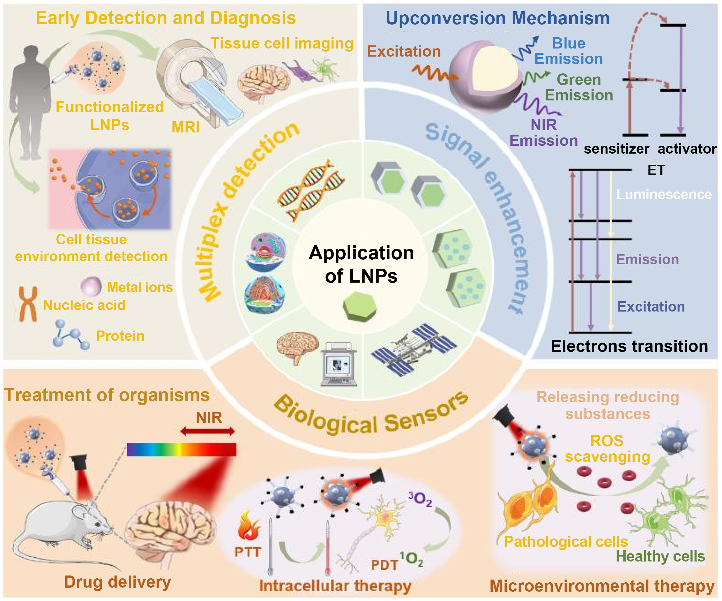
This review comprehensively examines recent advances in the luminescence mechanisms of LNPs and their applications in the detection and treatment of NDs. The structure of the review is outlined in Figure 1: Chapter 1 introduces fundamental concepts of LNPs luminescence and common luminescence phenomena; Chapter 2 discusses recent progress in using LNPs based luminescence to detect neurodegenerative changes, aiming to improve the detection and diagnosis of specific cells and tissues; Chapter 3 explores the development of functionalized nanomedicines utilizing LNPs, with an emphasis on their ability to effectively target particular cells and tissues; Chapter 4 analyzes the current bottlenecks in the application of LNPs for the diagnosis and treatment of ND, and proposes multi-dimensional integrated strategies to facilitate their clinical translation; Chapter 5 systematically summarizes the upconversion and downconversion luminescence (DCL) mechanisms of LNPs as well as associated energy transfer processes. It provides a comprehensive review of their research progress in the detection (including high-sensitivity and high-specificity imaging of metal ions, nucleic acids, protein aggregates, reactive oxygen species, and other biomarkers) and treatment (including intracellular drug delivery, photodynamic therapy (PDT), microenvironment modulation, and synergistic therapy) of NDs, aiming to offer both theoretical foundations and practical references for the clinical translation of precision theranostic technologies.
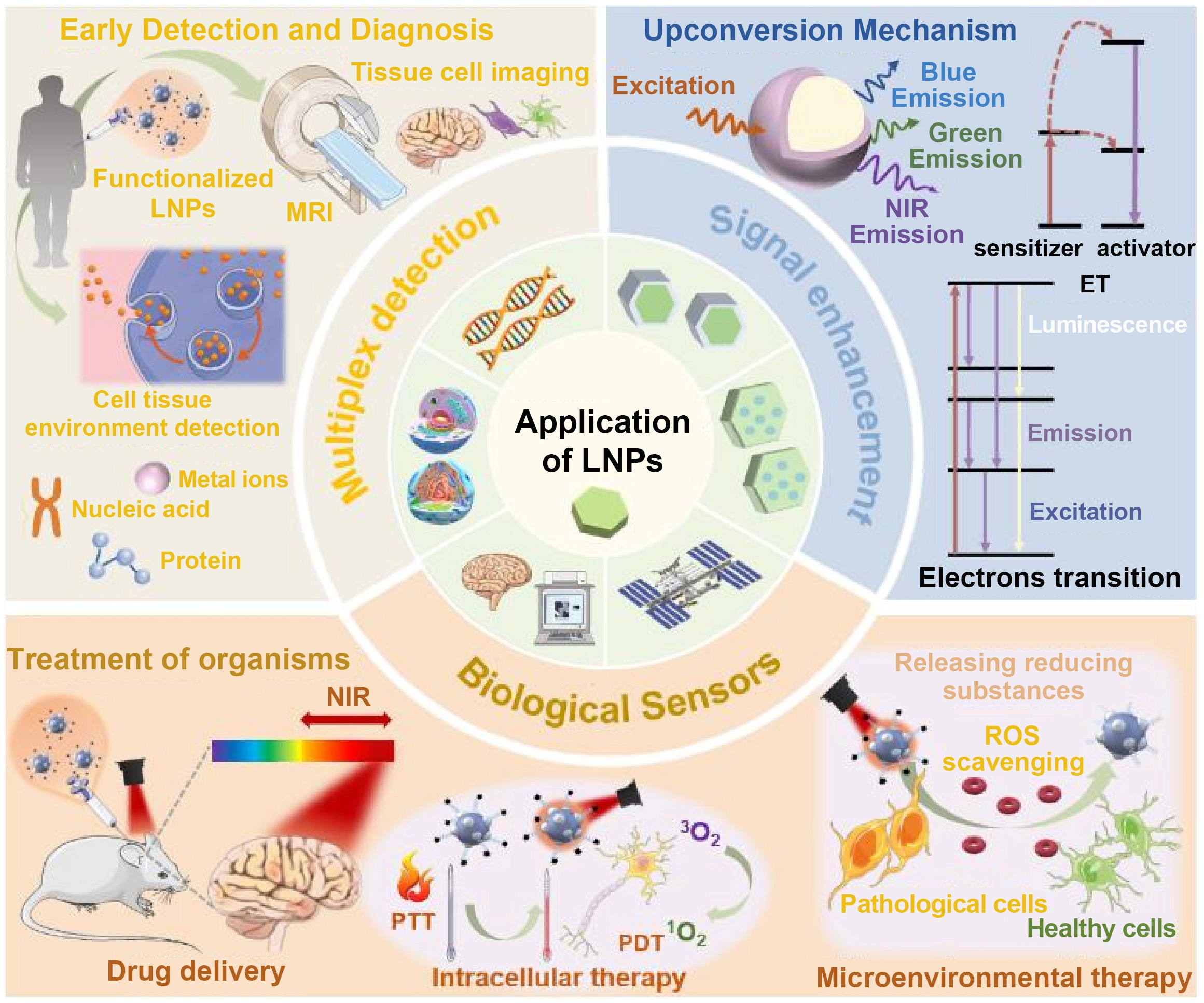
Figure 1. Schematic illustrating the application of LNPs based luminescence mechanisms in the diagnosis, detection, and treatment of neurodegenerative diseases. LNPs: lanthanide-doped nanoparticles; MRI: magnetic resonance imaging; NIR: near-infrared; PTT: photothermal therapy; PDT: photodynamic therapy; ET: energy transfer upconversion; ROS: reactive oxygen species.
2. Luminescence Mechanism
2.1 Luminescence mechanism of LNPs
Rare earth elements comprise 17 elements, including 15 lanthanides along with scandium and yttrium (Y), located in Group III B of the periodic table[49,50]. These elements, often referred to as “RE”, share similar chemical properties due to their electronic configurations[51-53]. When lanthanides lose electrons to form +3 valence ions, they exhibit specific electronic configurations denoted as RE3+: [Xe] 4fn 5s2 5p6. LNPs consist of three main components: the matrix, sensitizer, and activator[53]. The matrix is optically inert, lacking 4f electrons, and serves to provide a suitable crystal field for rare earth ions. Ions such as Y3+ and Gd3+ can modify the local environment around activators, enhancing their luminescent properties. Sensitizers, such as Yb3+, exhibit high absorption capabilities and can efficiently transfer the absorbed energy to activators[54]. The activator acts as the luminescence center in LNPs, where unpaired electrons in the f-orbital enable the absorption and emission of energy as fluorescence. Common activators include Er3+ (green emission), Ho3+ (red emission), and Tm3+ (blue emission). Co-doped systems like Nd3+/Yb3+ are also utilized in LNPs stimulated at 808 nm[55]. Rare earth ions interact with radiation across various wavelengths, yielding diverse fluorescence characteristics[56,57]. Based on emission mode, rare earth luminescent materials can be classified into downconversion and upconversion types. The following sections introduce these luminescence mechanisms according to the type of rare earth material.
2.1.1 Upconversion luminescence (UCL)
Luminescence in UCNPs arises from energy transitions among their constituent ions. As shown in Figure 2, this process involves three stages: (1) the matrix lattice absorbs excitation energy; (2) this energy is transferred to activator ions, exciting them; and (3) excited rare earth ions emit fluorescence as they return to their ground state within the matrix[58,59]. Upconversion can be explained through four primary mechanisms: excited-state absorption (ESA), energy transfer upconversion (ET), two-photon absorption (TPA), and photon avalanche (PA), each with distinct efficiency characteristics[60-62].
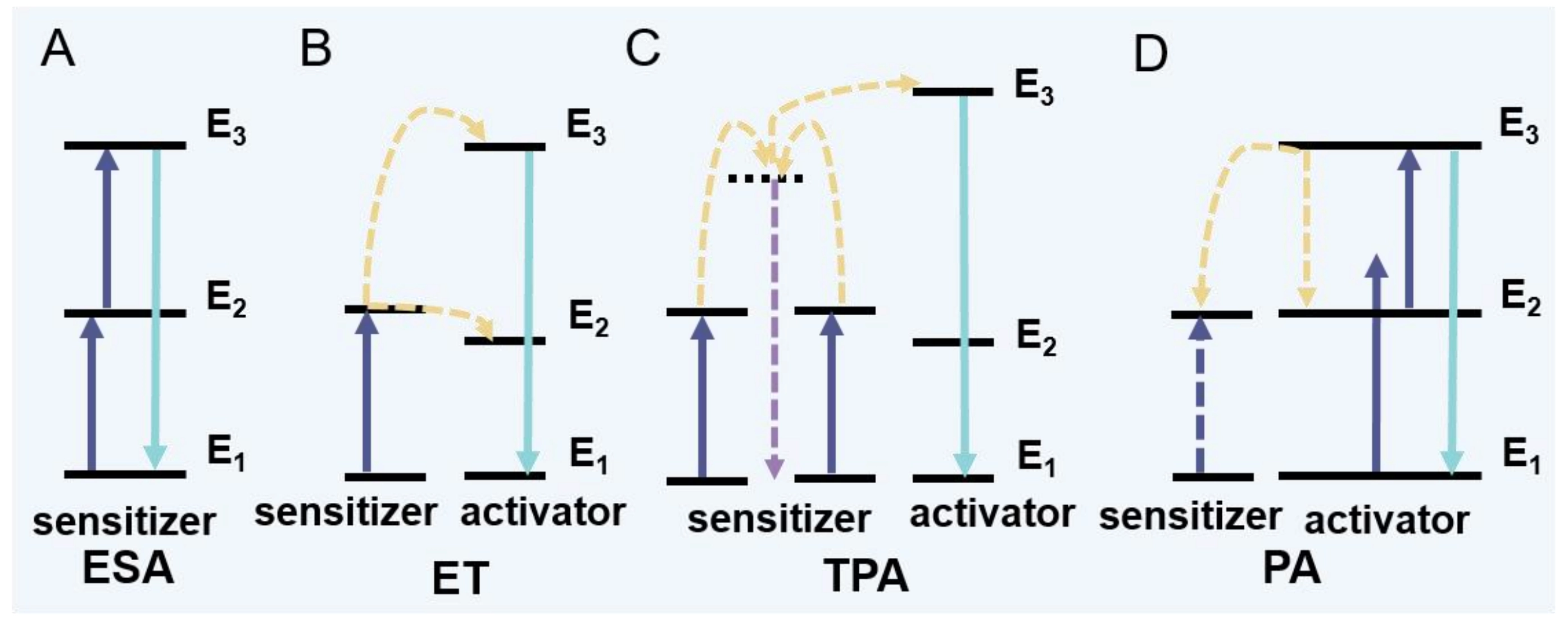
Figure 2. (A) ESA; (B) ET, (C) TPA; (D) PA. ESA: excited state absorption; ET: energy transfer upconversion; TPA: two-photon absorption; PA: photon avalanche.
ESA: Initially, ions in the ground state (E1) absorb a photon and transition to an intermediate metastable energy level (E2) (Figure 2A)[63]. If the vibrational energy matches the difference between E2 and a higher level (E3), ions can absorb additional energy at E2 and transition to E3, enabling TPA[64]. This process may also lead to transitions to even higher energy states, resulting in UCL. ESA operates independently of rare earth ion concentration and typically requires dual-wavelength pumping in rare earth-doped crystals, one wavelength to excite ions from the ground state to a metastable state, and another to promote them to a higher energy level[63,65,66].
ET: In ET, energy transfers from a donor ion to an acceptor ion (Figure 2B). This transfer reduces the number of excited-state electrons in the donor, potentially leading to luminescence reduction or quenching. When a donor electron relaxes from an excited state to a lower-energy ground state, it transfers energy to an acceptor ion[67,68].
TPA: Under high excitation power, an ion can simultaneously absorb two photons (Figure 2C). The ion transitions from the ground state to a final state via a virtual intermediate state before emitting UCL. The energy of the emitted photon equals the sum of the energies of the two absorbed photons. TPA is less common in conventional upconversion systems due to its complexity[69,70].
PA: PA combines ESA and ETU processes, with the pump wavelength matching an excited-state energy rather than the ground state (Figure 2D). UCL via PA is highly dependent on pump power: below a certain threshold, emission is minimal; above it, intensity increases dramatically[71,72].
Energy transfer systems using NIR excitation and visible emission enable biomass sensing through monitoring energy transfer signals. Numerous studies have focused on enhancing UCL to improve signal contrast in UCNP-based analytical probes. Most luminescence detection systems employing UCNPs utilize LRET and IFE in combination with dyes or other nanomaterials such as QDs and AuNPs[73,74].
2.1.2 DCL
DCL, which typically demonstrates higher energy conversion efficiency than upconversion, is more broadly applied in practice. Following Stokes’ law, this process involves the absorption of high-energy photons and their conversion into emitted photons of lower energy. Downconversion nanoparticles (DCNPs) can be classified into two primary types: downshifting, where a single high-energy photon is converted into one lower-energy photon, and quantum cutting, in which one high-energy photon is transformed into multiple lower-energy photons[75-77].
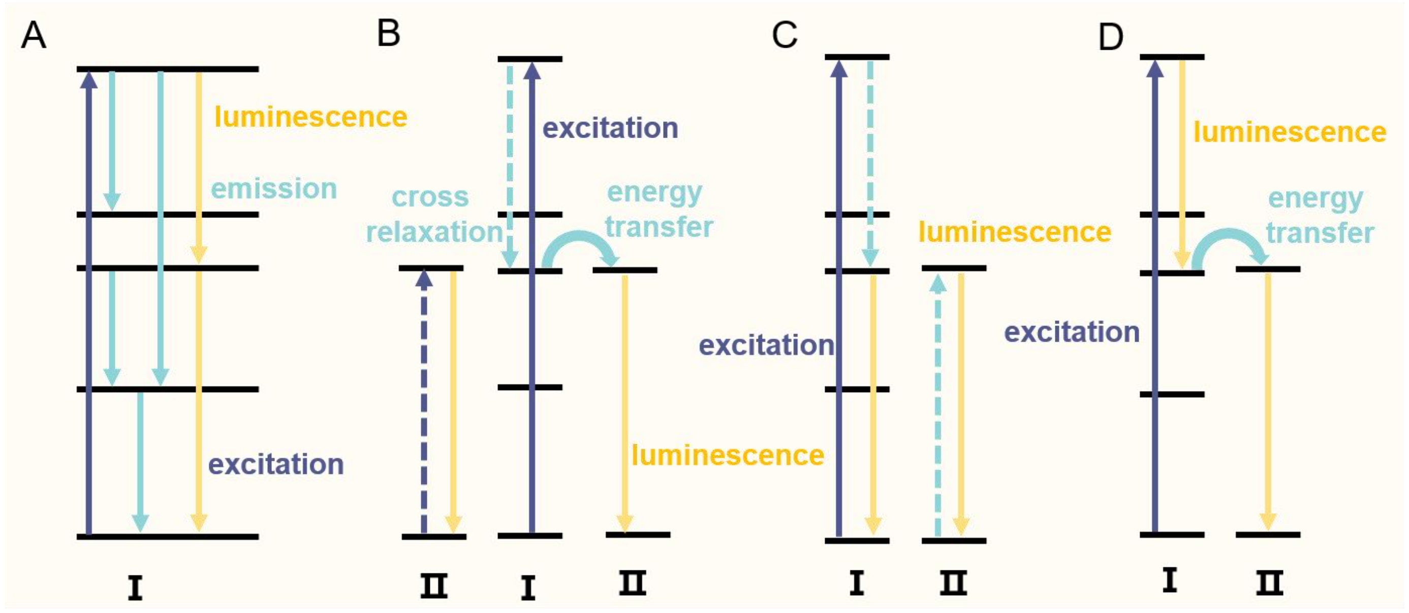
As illustrated in Figure 3, the underlying mechanism encompasses several pathways: (A) electrons, upon photon absorption, transition from the ground state to an excited state and subsequently undergo further transitions, emitting light at varying wavelengths from different energy levels; (B) cross-relaxation between two ions occurs alongside energy transfer, where after electrons return to the ground state, fluorescence is emitted while residual energy is transferred to neighboring ions; (C) a process similar to (B) based on cross-relaxation results in the simultaneous emission of multiple photons and luminescence; and (D) electrons absorb energy to transition from the ground state to an excited state, relax to a lower excited state with the emission of low-energy photons, and, upon returning to the ground state, transfer energy to other ions, yielding visible light emission[75-77].
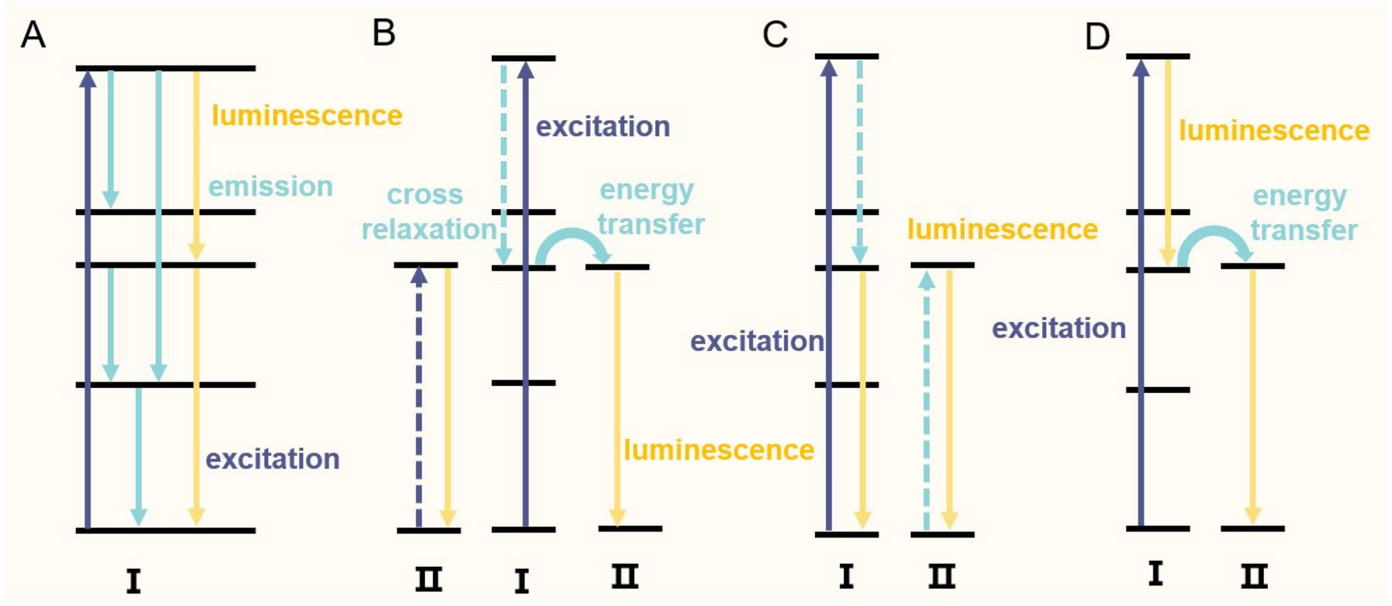
Figure 3. Downconversion luminescence mechanisms. (A) Electron transitions; (B) Cross-relaxation between two ions; (C) Multi-photon emission via cross-relaxation; (D) Electron transition and energy transfer.
2.2 LRET effect
LRET is a photophysical process that facilitates energy transfer between a donor and an acceptor, requiring an excitation light source to induce luminescence. In LRET systems, LNPs typically serve as energy donors. Their surfaces are modified with recognition units such as organic dyes, noble metal nanoparticles, graphene oxide, or QDs, enabling sensitive detection of biological substances through the generation or quenching of resonance energy. For LRET to occur, two key conditions must be satisfied: (1) the acceptor’s absorption spectrum must overlap with the emission spectrum of the LNPs, and (2) the distance between the acceptor and the LNPs must be sufficiently short. LRET efficiency can be modulated by adjusting spectral overlap and donor-acceptor distance[78-82].
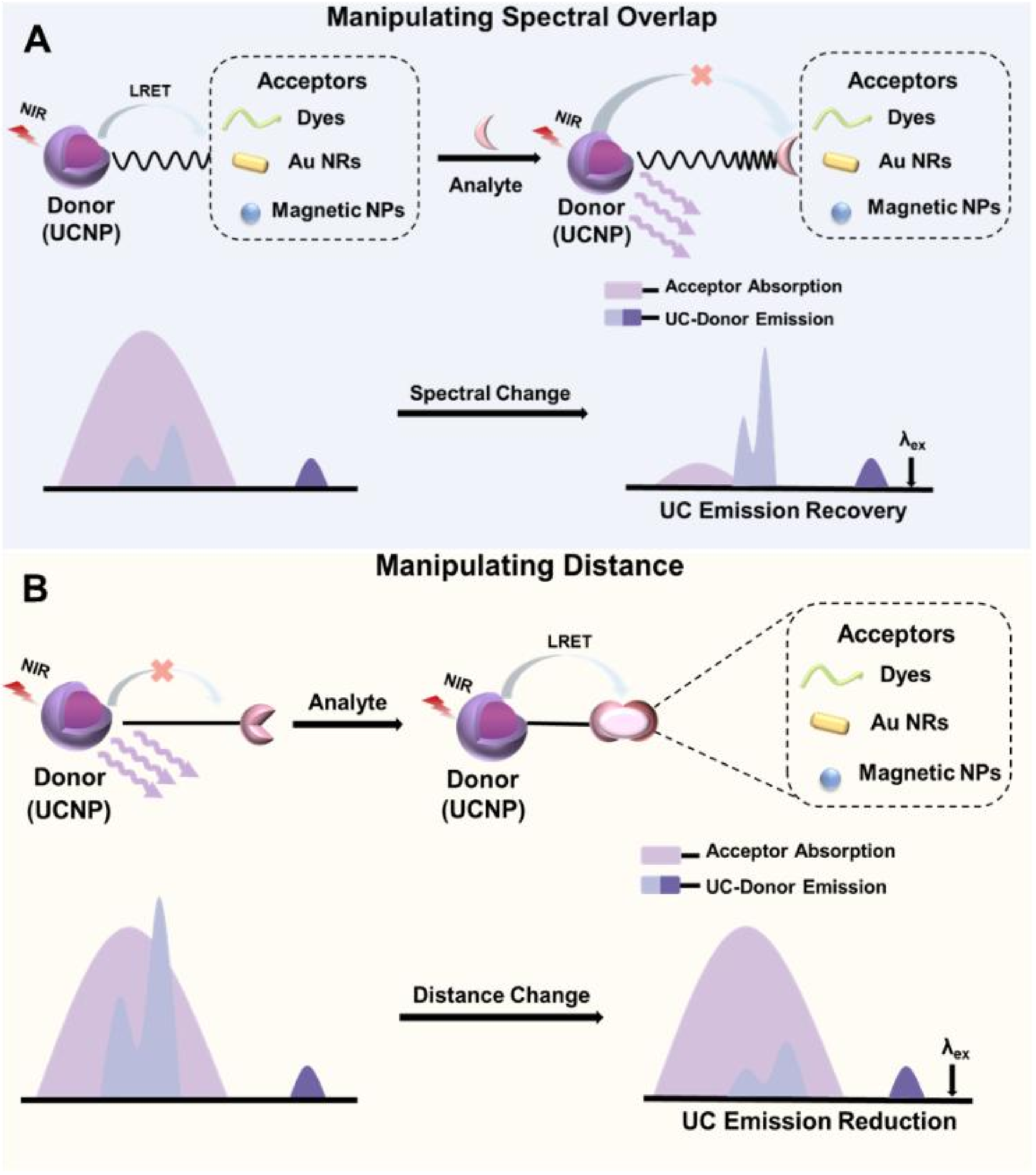
(1) Tuning Spectral Overlap: LRET efficiency can be optimized by altering the acceptor’s absorption intensity or wavelength upon interaction with an analyte (Figure 4A). When the analyte binds to a recognition element, it disrupts the LRET process, leading to enhanced LNPs emission[82]. This allows precise control over the LRET mechanism. Considerable research has focused on functionalizing LNP surfaces with suitable sensing elements to develop LRET-based probes. For example, Zhao et al. utilized LRET by exploiting spectral overlap between the green luminescence of LNPs at 540 nm and the absorption peaks (544 nm and 572 nm) of an organic dye upon reaction with cysteine (Cys) and homocysteine (Hcy)[83]. In the presence of Cys and Hcy, the absorption peak of reacted acceptors overlaps with the LNPs emission, quenching the green luminescence. This mechanism enables selective detection of Cys and Hcy, enhancing sensitivity and selectivity while allowing cellular imaging of target molecules.
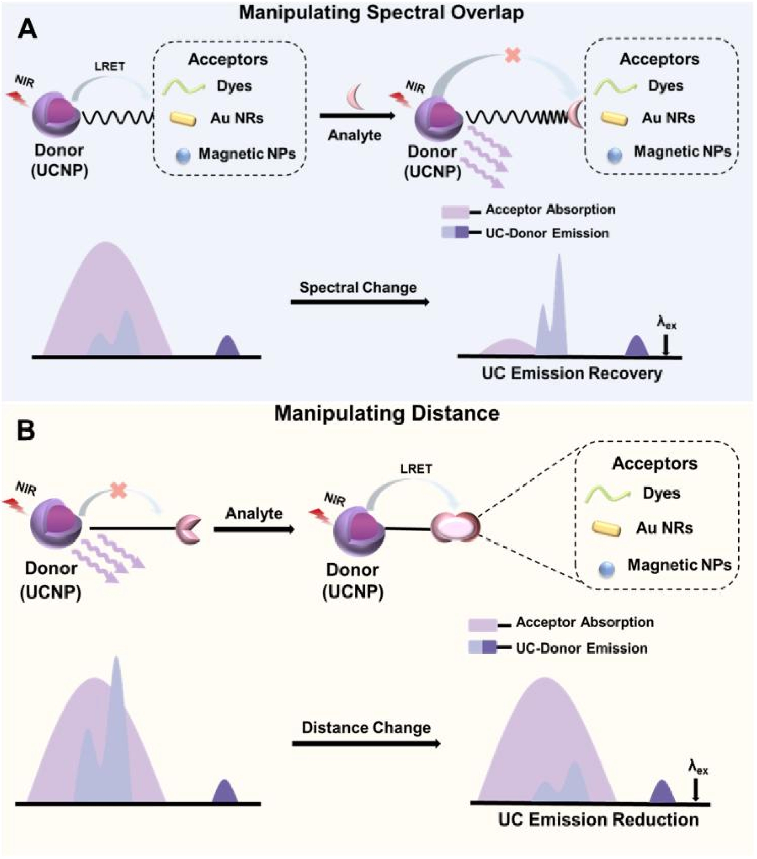
Figure 4. Schematic diagram of the main strategies for the fabrication of LRET-based LNPs. (A) Manipulating the spectral overlap: upon addition of the analyte, LNPs emission is recovered caused by the suppressed LRET process; (B) Manipulating the distance between energy donor and acceptor: upon addition of the analyte, LNPs luminescence is decreased since the distance between the donor and acceptor is close enough for an effective energy transfer. LRET: luminescence resonance energy transfer; LNPs: lanthanide-doped nanoparticles; UCNP: upconversion nanoparticle; Au NRs: gold nanorods; NIR: near-infrared.
(2) Adjusting Distance: Another strategy to enhance LRET efficiency involves modulating the distance between LNPs and the acceptor (Figure 4B). Analyte binding can disrupt the linkage between LNPs and the acceptor, restoring upconversion fluorescence. Wu et al. demonstrated that LRET efficiency could be improved by controlling the distance between LNPs and DNA-modified silver nanoparticles (DNA-AgNPs)[84]. By removing oleic acid ligands from the LNP surface and directly conjugating DNA-AgNPs, they achieved up to 93% quenching of the upconversion signal. This LRET-based approach represents a promising strategy for biosensing applications.
2.3 IFE effect
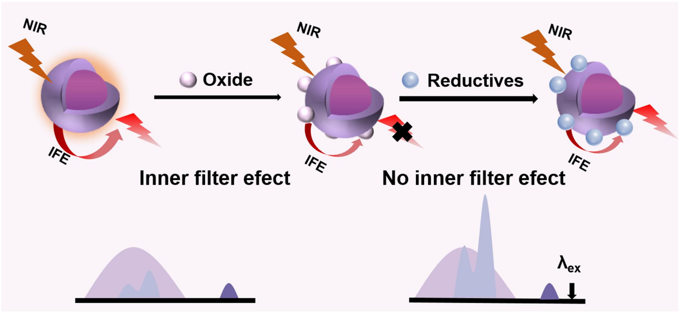
IFE is a photophysical phenomenon that modulates luminescence by the competitive absorption of excitation and/or emission light by absorbers present in the measurement system[85]. In the context of LNPs, IFE can attenuate both the incident excitation light and the emitted UCL, leading to a reduction in the detected fluorescence intensity. This occurs when absorbers in the surrounding medium or within the LNPs matrix itself (Figure 5) spectrally overlap with the excitation or emission bands of the LNPs. Unlike resonance energy transfer processes, IFE is a non-interactive, inner-filter phenomenon; it reduces the effective photon flux reaching or emanating from the luminophore without altering its intrinsic photophysical properties, such as emission lifetime. This characteristic allows IFE to be exploited in the design of radiometric or intensity-based sensors, where changes in the absorber concentration directly correlate with measurable fluorescence modulation.
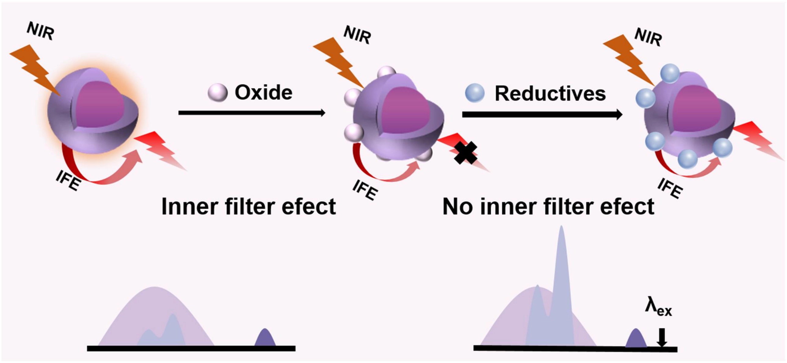
Figure 5. Schematic diagram of the main strategies for the fabrication of IFE-based LNPs. IFE: internal filter effect; LNPs: lanthanide-doped nanoparticles; NIR: near-infrared.
A representative application is the endogenous hydrogen sulfide (H2S)-activated NIR-II probe DCNPs@Prussian blue (PB) reported by Wang et al., which integrates IFE for specific tumor imaging[86]. In this system, the dense coating of PB on the surface of LNPs results in strong spectral overlap between PB’s broad absorption and the excitation/emission profiles of DCNPs. This leads to significant luminescence quenching via both IFE and LRET. In the tumor microenvironment, however, elevated H2S reduces Fe(III) in PB to Fe(II), triggering the decomposition of the PB shell. The consequent decrease in absorber concentration diminishes the IFE and LRET quenching, thereby restoring the NIR-II fluorescence for precise image-guided surgery.
Similarly, Guo et al. developed a dual-channel sensor based on the IFE quenching mechanism for the detection of xanthine (XA) in biological samples. In this design, red-emitting LNPs serve as the fluorescent reporter, while the oxidation product of 3,3′,5,5′-tetramethylbenzidine (TMB) generated via XA oxidase-catalyzed reaction, acts as a strong absorber[87]. The increasing absorbance of oxidized TMB during the reaction progressively quenches the LNPs emission through IFE. This approach achieved detection limits of 0.58 μM (fluorescence channel) and 1.19 μM (colorimetric channel), with excellent recovery rates (96.3-104.3%) in serum samples, demonstrating high practicality for rapid biosensing.
A critical aspect of IFE-based probe design is the mandatory spectral overlap between the UCL emission band and the absorption profile of the target-responsive absorber. Since IFE is a distance-independent, “through-space” effect, it does not require nanoscale proximity or specific binding between the absorber and the LNPs. Consequently, IFE-based probes often employ a “mix-and-detect” format without the need for covalent conjugation, simplifying preparation and preserving the colloidal stability of the nanoparticles. However, this same distance independence also introduces susceptibility to interference from background absorbers in complex matrices, which can limit selectivity in cellular or in vivo applications.
In contrast, LRET-based probes function through dipole–dipole coupling, which requires close proximity (typically 1-10 nm) between the donor and acceptor[47,88]. The larger donor–acceptor separation in typical LRET configurations naturally minimizes IFE interference. Furthermore, LRET systems often involve purification steps (e.g., centrifugation) to remove unbound acceptors, leading to a well-defined and lower surface density of acceptors compared to the homogeneous absorber distribution common in IFE systems. This fundamental difference in operating principle and construction makes LRET generally more suitable for intracellular and in vivo imaging where precise spatial resolution and minimized background are crucial, whereas IFE-based strategies excel in homogeneous assay formats for rapid, sensitive detection in transparent media.
3. Biosensing and Diagnosis
Functionalization of LNPs with specific targeting ligands enables their selective binding to disease-associated cells or tissues, enhancing diagnostic precision through site-specific accumulation[89-91]. This targeted delivery not only increases imaging contrast but also provides more accurate insights into pathological processes, ultimately supporting the development of personalized treatment strategies[91,92]. This section reviews recent advances in LNP-based detection and diagnosis, focusing on four key analyte categories: metal ions, nucleic acids, proteins, and reactive oxygen species (ROS)[92].
3.1 Optical detection of metal ions based on LNPs
LNPs exhibit unique advantages for the in situ tracing of metal ions in biological systems, owing to their tunable luminescence properties, ion recognition capabilities, and suitability for long-term live cell imaging[19,54,92]. Upon entry into a biological environment, ion-recognition groups immobilized on the surface of LNPs can selectively bind to target metal ions. This binding modulates the luminescence signal by altering mechanisms such as LRET or the IFE, thereby enabling sensitive detection of a variety of metal ions.
For example, Liu et al. constructed an efficient upconversion nanoprobe for the detection and imaging of Ca2+ based on a sandwich structure (Figure 6A)[93]. The probe utilized LNPs as the luminescent core, polyacrylic acid as the intermediate layer, and the Ca2+-sensitive dye Fluo-4 as the recognition receptor. By removing the surface oleic acid ligands, the donor–acceptor distance in the LRET system was significantly reduced, greatly enhancing the detection sensitivity for Ca2+. Building on this strategy, Li et al. successfully designed and synthesized a novel class of core–shell–shell lanthanide-doped upconversion nanoparticles (cssUCNPs)[94]. By integrating the Fluo4 calcium indicator with the cssUCNPs, they constructed a dual-modal nanoprobe (cssUCNPs@Fluo4) based on the LRET effect. This nanoprobe enabled highly sensitive, real-time, and long-term (> 24 h) simultaneous monitoring of Ca2+ (detection limit: 1 μM) and temperature (sensitivity: 0.5 K-1). In MPP+-induced cellular and zebrafish models of PD, the probe revealed, for the first time, three distinct phases of Ca2+ and temperature dynamics during PD pathogenesis: an early phase (0-3 h) characterized by a rapid surge in both parameters (Δ[Ca2+] ≈ 112 nM, ΔT ≈ 6.8 K); a middle phase (3-18 h) marked by oscillatory fluctuations; and a late phase (18-24 h) during which both parameters stabilize at elevated levels. These findings directly link mitochondrial dysfunction with Ca2+ dysregulation and propose Ca2+-sensitive and thermosensitive therapeutic targets for PD intervention, thereby providing a powerful nanoscale sensing platform for real-time dynamic monitoring and mechanistic dissection of NDs.
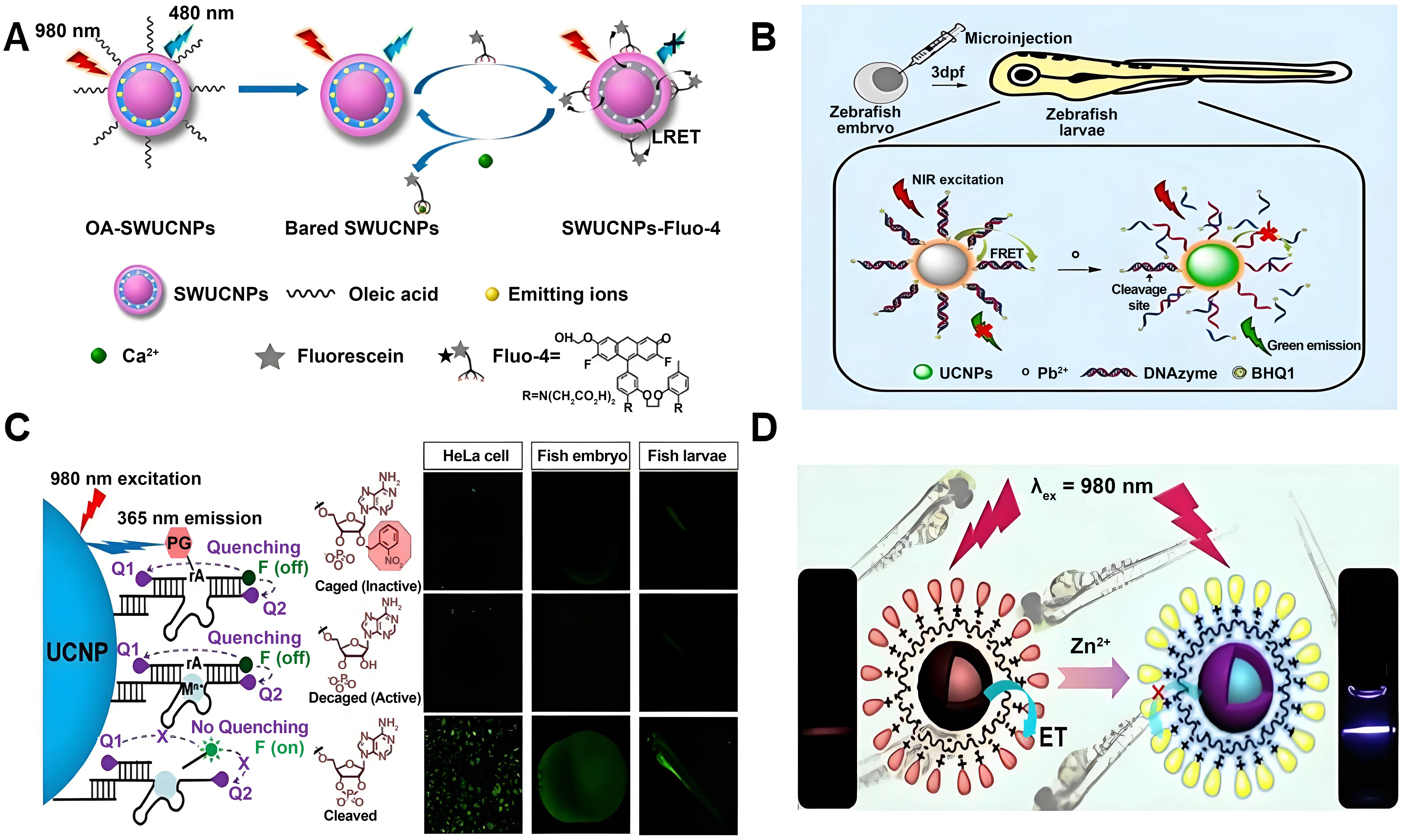
Figure 6. (A) Schematic illustration of the NIR-980 nm nanoprobe for calcium based on LRET from UCNP with bared surface to Fluo-4 in vitro system. Adapted with permission from reference[93]. Copyright © 2015 American Chemical Society; (B) Schematic diagram of detection of Pb2+ using DNAzyme-modified UCNPs and its biological application in early-stage zebrafish based on LRET. Adapted with permission from reference[95]. Copyright © 2021 Elsevier; (C) Schematic illustration showing the synthesis of a photo-controllable UCNP and DNAzyme-based nanosensor and its response to Zn2+. Adapted with permission from reference[96]. Copyright © 2018 American Chemical Society; (D) Schematic depicting the synthesis of chromophore-assembled UCNPs and their response to Zn2+. Adapted with permission from reference[97]. Copyright © 2015 American Chemical Society. NIR: near-infrared; LRET: luminescence resonance energy transfer; UCNP: upconversion nanoparticle.
Beyond calcium detection, LNPs have also been widely applied to the sensing of other metal ions. For instance, Huang et al. developed a Pb2+-responsive LNPs-DNAzyme nanosensor based on the LRET effect. This sensor employed a BHQ1 quencher, LNPs, and DNAzyme to form a donor-acceptor bilayer, achieving highly specific detection and imaging of Pb2+ in live cells and early-stage zebrafish (Figure 6B)[95]. Yang et al. utilized LNPs to convert NIR light into ultraviolet light, which photolytically removed photocaging groups on a substrate chain and restored its enzymatic cleavage activity in the presence of specific metal ions such as Zn2+ (Figure 6C)[96]. By introducing the LNPs-DNAzyme probe into live cells and zebrafish embryos, this method enabled high-resolution detection of both endogenous and exogenous metal ions while exhibiting low phototoxicity and good biocompatibility. Similarly, based on the LRET strategy, Chang et al. constructed a nanoprobe for rapid Zn2+ detection by loading Zn2+-responsive chromophores onto the surface of NaYF4:Yb/Tm@NaYF4 nanoparticles (Figure 6D)[97]. The UCL of the probe could be quenched via a LRET process and restored upon Zn2+ binding, allowing for quantitative analysis of Zn2+ levels relevant to the pathogenesis of AD. This strategy cleverly integrates the NIR luminescence properties of LNPs with in vivo microdialysis sampling technology, enabling dynamic and quantitative monitoring of Zn2+, an ion closely associated with the pathogenesis of AD, thereby providing a paradigm for the practical application of LNPs in real-time analyte detection within the brain.
3.2 Nucleic acid detection based on LNPs
Messenger RNAs (mRNAs) play a central role in protein synthesis and serve as critical biomarkers for ND, where their expression levels can reflect disease progression[98,99]. Recent advances in sensing technologies have improved the sensitivity and specificity of mRNA detection in complex biological environments.
Vilela et al. reported a sensing platform that integrates graphene oxide with LNPs for the specific detection of mRNA-related oligonucleotides (Figure 7A)[100]. This system exhibited high sensitivity and successfully identified AD associated mRNAs in diverse biological fluids. The fluorescence-based format enables real-time monitoring of mRNA levels, highlighting its potential as a diagnostic tool for neurodegenerative disorders. Similarly, Kuang et al. constructed DNA-directed satellite-type assemblies composed of gold-nanorod-coated platinum-LNPs for the analysis of intracellular thymidine kinase 1 mRNA (Figure 7B)[101]. The probe operates via a light-restoration mechanism upon target binding, allowing both imaging and quantitative detection with a remarkably low limit of detection (0.67 fmol per 10 µg RNA), which is particularly valuable for early-stage clinical diagnostics. Furthermore, Tan et al. explored DNA-UCNPs composites for integrated diagnostic and therapeutic applications (Figure 7C)[102]. By employing the LRET effect, they developed a multicolor LNPs-MXene receptor system utilizing LNPs@AuNP conjugates for RNA detection. This design demonstrated enhanced sensitivity and specificity compared with conventional monochromatic LNPs-MXene configurations. The same group also combined solid-phase and liquid-phase detection strategies, enabling both qualitative and quantitative analysis of targets via portable devices. This versatile approach supports the detection of diverse analytes, including RNA, DNA, antibodies, and antigens, thereby broadening its utility across multiple biomedical fields. These advances underscore the growing potential of LNPs based platforms in nucleic-acid detection and diagnostics, especially for applications demanding high sensitivity and specificity in complex biological environments.
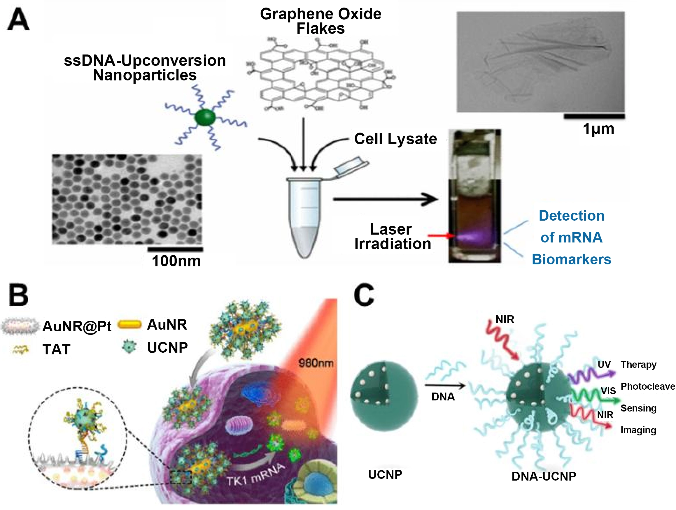
Figure 7. (A) Schematic illustration of UCNP-based platforms for the targeted detection of AD-associated mRNA biomarkers. Adapted with permission from reference[100]. Copyright © 2016 American Chemical Society; (B) Schematic illustration of UCNPs satellite assemblies ultrasensitive detection of mRNA in living cells. Adapted with permission from reference[101]. Copyright © 2018 American Chemical Society; (C) Schematic illustration of DNA-functionalized UCNP composites developed for diagnostic and therapeutic applications. Adapted with permission from reference[102]. Copyright © 2021 American Chemical Society. UCNP: upconversion nanoparticle; AD: Alzheimer’s disease; mRNA: messenger RNA; NIR: near-infrared; UV: ultraviolet.
3.3 Protein detection based on LNPs
LNPs have become important biomedical research tools in the field of protein detection due to their efficient coupling with antibodies, peptides, or other targeting molecules[103]. This targeting capability enables precise localization within specific cells or tissues, thereby facilitating high spatial-resolution visualization of protein distribution, dynamics, and interactions in living systems[104,105].
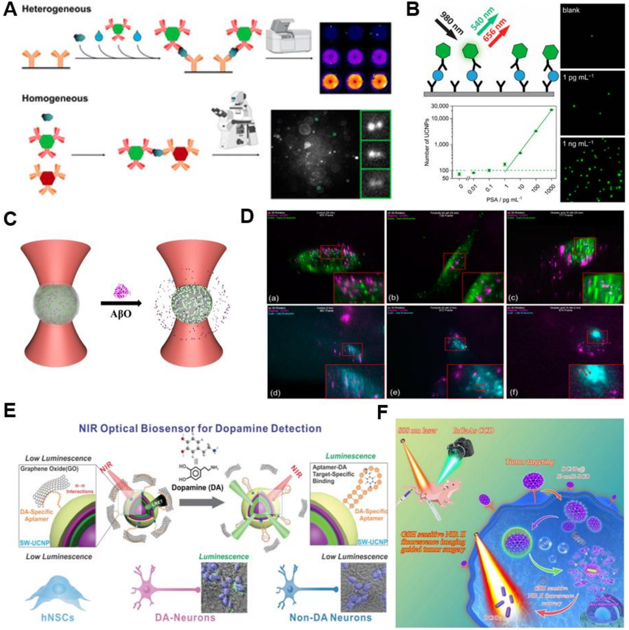
For example, Hlaváček et al. achieved the detection of blood-based biomarkers by modifying the surface of UCNPs with silica and conjugating them with streptavidin or antibodies (Figure 8A)[106]. Furthermore, the combination of therapeutic agents with UCNPs can enhance the specificity of drug delivery to diseased cells or tissues, thereby reducing off-target effects and improving therapeutic efficacy. Such targeted probes also enable real-time imaging of tumor progression, providing crucial insights into drug resistance mechanisms and tumor recurrence. In a representative application, Zdeněk Farka et al. utilized UCNPs as optical markers to achieve the detection and quantification of prostate-specific antigen under wide-field microscopy with 980 nm laser excitation (Figure 8B)[107]. Fang et al. developed metal-organic framework/black hole quencher (H-USM/BHQ1) microspheres for optical tweezer-based microscopic imaging (Figure 8C)[108]. In this system, the fluorescence ratio of highly doped UCNP-SiO2 was positively correlated with the concentration of amyloid-β oligomers (AβOs) associated with AD. In the presence of varying concentrations of AβOs, the decomposition of the ZIF-8 framework triggered the release of BHQ1, leading to a significant enhancement of fluorescence at 540 nm. Moreover, Lee et al. constructed aptamer-functionalized, high-brightness core-shell-shell structured UCNPs, which enabled dynamic tracing of neurotransmitters released from dopamine-derived neural stem cells at the single-cell level via the LRET mechanism (Figure 8D)[109]. To this end, Song et al. employed UCNPs with diameters of approximately 100 nm for bioimaging, enabling the visualization of tau protein aggregation in neurons through NIR irradiation (Figure 8E)[110]. Tracking neurotransmitter release during neural differentiation and modulation is essential for understanding disease pathways and developing targeted therapies.
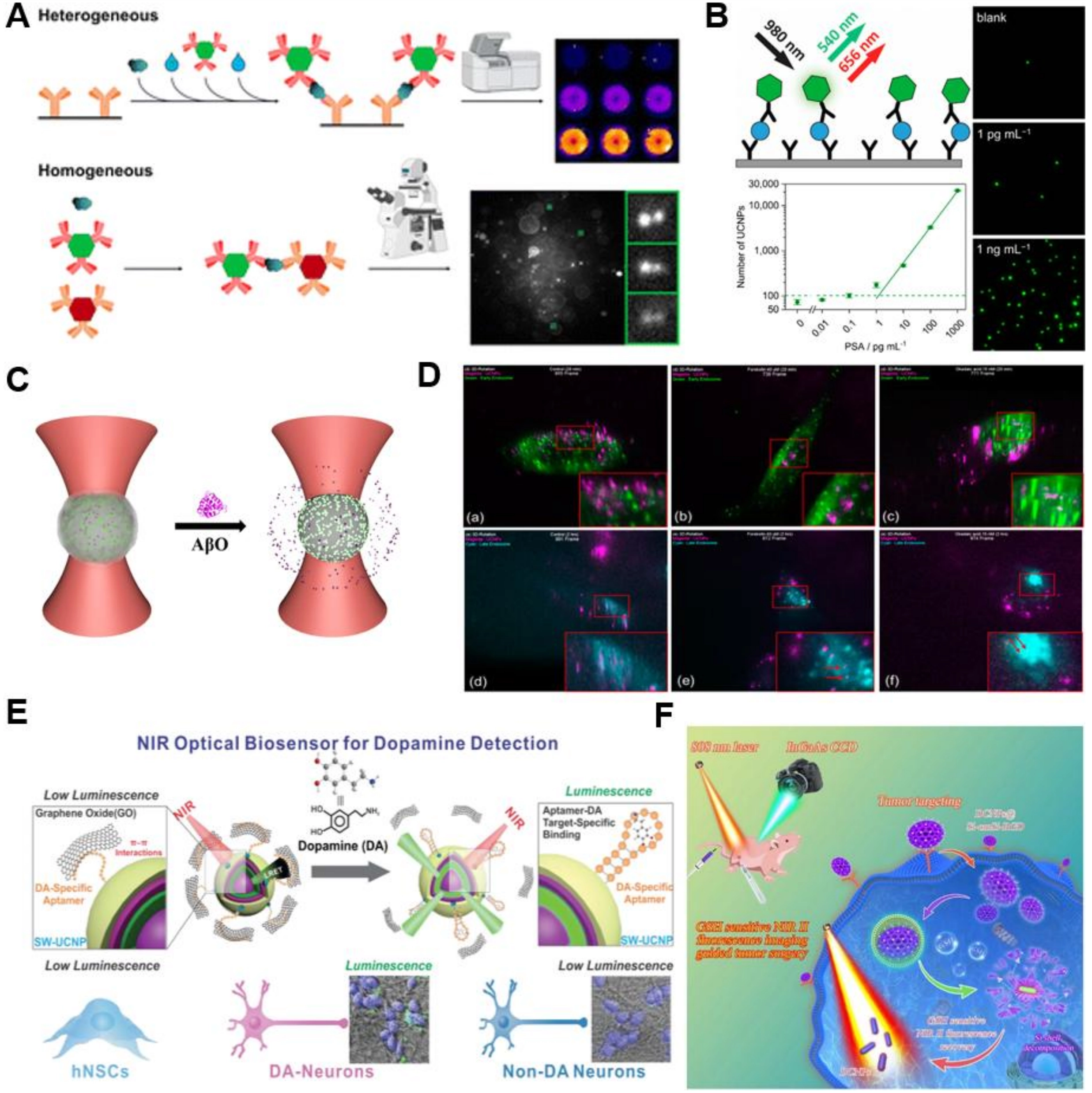
Figure 8. (A) Schematic illustration of purification of protein-conjugated UCNPs and analysis of aggregation. Adapted with permission from reference[106]. Copyright © 2022 Springer Nature; (B) Schematic illustration of a microtiter plate immunoassay for single-molecule counting of PSA using UCNPs as background-free fluorescent labels. Adapted with permission from reference[107]. Copyright © 2017 American Chemical Society; (C) Schematic illustration of amyloid-β oligomer detection via a fluorescence ratio strategy based on optically trapped, highly UCNPs microspheres. Adapted with permission from reference[108]. Copyright © 2021 American Chemical Society; (D) Schematic illustration of early state colocalizations of UCNPs with early endosomes or late endosomes in tau aggregated cell. Adapted from reference[109]. CC BY 4.0; (E) Schematic illustration of a NIR-based dopamine sensor constructed using UCNPs and its application for real-time dopamine sensing in living neurons. Adapted with permission from reference[110]. Copyright © 2019 John Wiley & Sons; (F) Schematic illustration of DCNP fabrication with enhanced signal-to-background ratio for precise liver tumor resection guided by NIR-II fluorescence imaging. Adapted with permission from reference[111]. Copyright © 2022 Elsevier. UCNPs: upconversion nanoparticles; NIR: near-infrared; DCNP: downconversion nanoparticle; PSA: prostate-specific antigen.
In summary, these advances demonstrate the expanding role of UCNPs platforms in protein detection and biomarker analysis, highlighting their potential for real-time, high-resolution imaging in complex biological environments. Beyond the visible to NIR-I range, Wu et al. fabricated biocompatible nanoprobes based on rare-earth DCNPs using layer-by-layer assembly for fluorescence imaging in the NIR-II (Figure 8F)[111]. The long-wavelength emission characteristics of DCNPs provide deep tissue penetration and minimize autofluorescence interference, resulting in superior imaging contrast. The layer-by-layer coating further endows the nanoparticles with tumor-targeting capability and prolonged circulation time, enabling non-invasive detection of orthotopic ovarian tumors. This strategy offers a new approach for receptor-targeted imaging, contributing to early cancer diagnosis and precision therapy.
3.4 Detection of ROS based on LNPs
LNPs have emerged as powerful tools for imaging ROS production in neuronal cells and monitoring oxidative stress in the brain, particularly in the context of NDs[112-114]. ROS play key roles in various physiological and pathological processes, such as cellular signal transduction, inflammation, and oxidative stress responses[114]. Elevated levels of ROS, including hydrogen peroxide (H2O2), superoxide anion (O2-), and hypochlorous acid (HClO), are closely associated with numerous biological processes and the progression of neurodegenerative disorders[115-117]. Therefore, tracking and visualizing abnormal ROS levels are crucial for early diagnosis and gaining deeper insights into the pathogenesis of these diseases[118-120].
High concentrations of HClO in biological systems can trigger various inflammatory diseases, driving significant interest in optical methods for ClO- detection. For example, cyanine-sensitized UCNPs systems, constructed by coupling cyanine dyes with UCNPs, enable highly selective detection of ClO- via the LRET effect. Li’s group successfully imaged ClO- in living mice using a UCNPs-hCy3 composite probe combined with LRET and a filtering mechanism. When ClO- interacts with UCNPs-hCy3, oxidation of the dye alters its absorption spectrum, thereby reducing the LRET effect. This method effectively detected ClO- in both living cells and an arthritis mouse model, achieving a detection limit of 27 ppb (Figure 9A)[121].

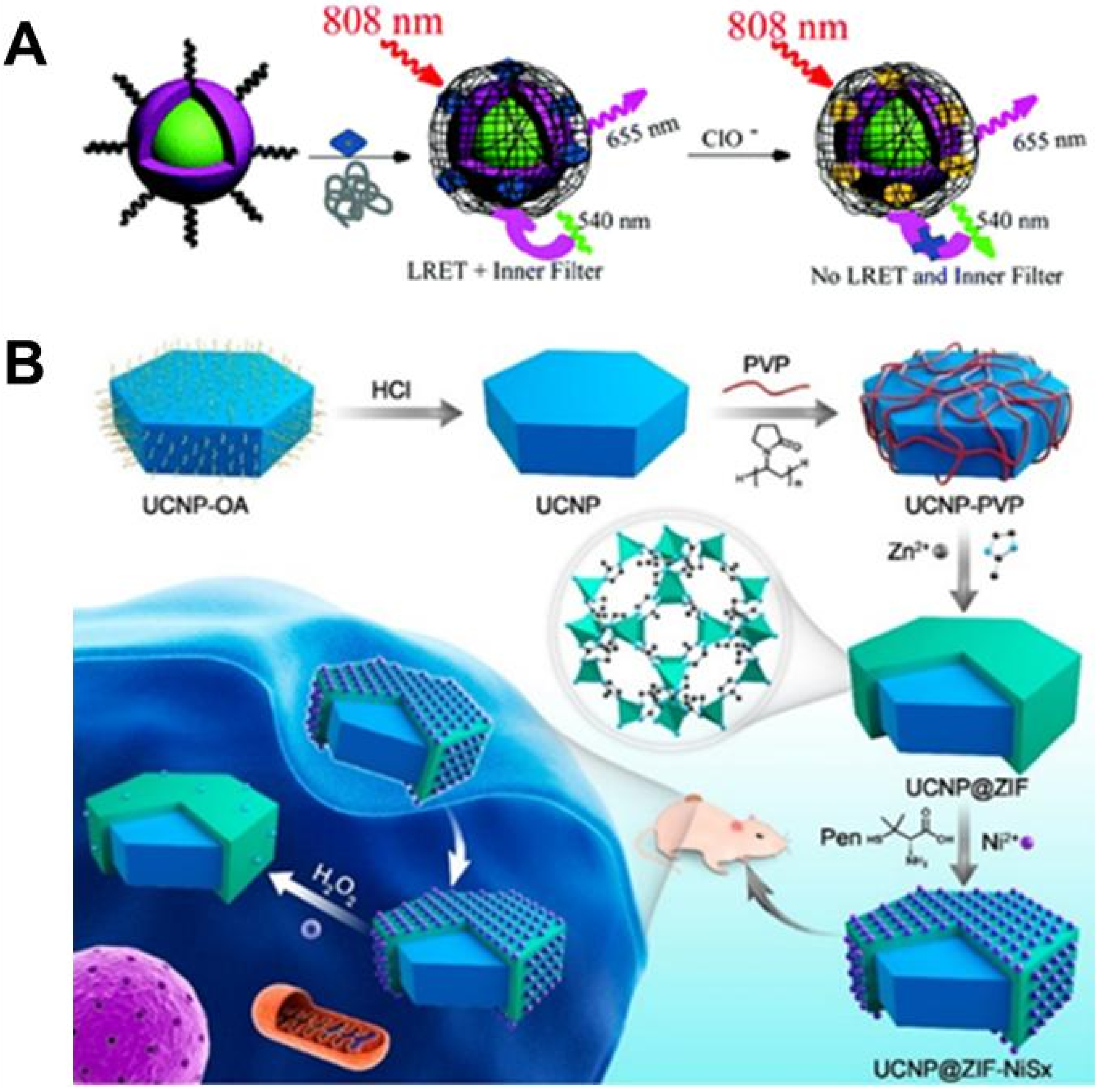
Figure 9. (A) Schematic illustration of An Nd-sensitized upconversion nanophosphor modified with a cyanine dye for the ratiometric upconversion luminescence bioimaging of hypochlorite in living cells (RAW 264.7 macrophages) and a mouse model of arthritis under 808 nm NIR excitation via LRET (detection limit: 27 ppb in aqueous solution). Adapted with permission from reference[121]. Copyright © 2015 Royal Society of Chemistry; (B) ROS detection using UCNP@MOF-NiSx nano assemblies in PCS-460-010 cells (in vitro) and in vivo applications, where H2O2 is used as a proof-of-concept ROS target and a chiral-optical/fluorescent dual-mode readout enables selective and quantitative monitoring based on NiSx degradation during the detection process. Adapted with permission from reference[122]. Copyright © 2019 American Chemical Society. NIR: near-infrared; LRET: luminescence resonance energy transfer; ROS: reactive oxygen species; UCNP: upconversion nanoparticle; MOF: metal-organic framework; PVP: poly(vinylpyrrolidone); OA: oleic acid; ZIF: zeolitic imidazolate framework-8.
Recent studies have further developed various novel UCNPs-based sensors capable of selectively detecting and quantifying ROS in cells and tissues. By functionalizing UCNPs with ROS-responsive probes, researchers can monitor ROS production in real time within specific cells or tissues, enhancing understanding of oxidative stress dynamics. A representative example is the UCNPs@ZIF-NiSx nanoassembly reported by Kuang et al., which exhibits strong circular dichroism (CD) signals at 440 and 530 nm, indicating effective chiral properties (Figure 9B)[122]. Studies revealed that NiSx nanoparticles significantly quench the UCL of UCNPs at 540 nm, while emission at 660 nm remains largely unchanged. This interaction between UCNPs and NiSx nanoparticles can be leveraged to monitor ROS levels. Based on this dual-mode response of chiral optical signals and fluorescence signals, the nanocomponent enables quantitative tracking of ROS dynamics, particularly H2O2, in living cells. The findings confirm that UCNP-based sensors can elucidate the dynamic behavior and mechanistic roles of ROS in cellular processes. For instance, elevated H2O2 levels correlate with the activation of cellular stress pathways and may serve as early indicators of cellular damage, underscoring the value of UCNPs sensors in studying NDs.
These advances highlight the promising prospects of UCNPs technology in the field of ROS detection, with broad potential applications in biomedical research, particularly in elucidating the mechanisms of diseases associated with oxidative stress.
4. LNDs in ND Therapeutics
LNPs have significantly advanced imaging technologies by enhancing the sensitivity and specificity of fluorescence imaging, magnetic resonance imaging, and computed tomography. Recent research developments indicate that LNPs can be functionally adapted for multimodal imaging applications, including UCL, positron emission tomography, and single photon emission computed tomography[121,122]. These imaging capabilities are critical for real-time observation of biological processes, contributing to improved disease monitoring and therapeutic response assessment[123]. LNPs are also increasingly utilized in PDT and targeted drug delivery systems[117,118]. By conjugating LNPs with specific ligands or therapeutic agents, drugs can be precisely delivered to diseased tissues or cells, thereby reducing off-target effects and enhancing therapeutic efficacy[123-125]. Furthermore, LNPs can generate ROS upon photoexcitation, expanding their applicability in PDT for cancer and other diseases[118,126,127]. The integration of LNPs into therapeutic strategies not only refines existing treatments but also opens new avenues for innovative therapeutic approaches[125].
4.1 Intracellular therapy
Protein overexpression and misfolding are central to the pathogenesis of NDs, such as amyloid-β (Aβ) aggregation in AD and α-synuclein aggregation in PD[128,129]. Understanding protein aggregation mechanisms and developing strategies to prevent or reverse this process are therefore critical in biomedical research[130,131]. Consequently, disaggregation of pathological protein assemblies has become a major therapeutic focus. LNPs serve as versatile tools for studying protein aggregation dynamics and developing novel therapeutic interventions[131-133]. Their non-invasive, remote-controllable nature makes them particularly suitable for neuronal regulation and optogenetics applications. For example, Zhang et al. described a UCNPs-based platform capable of activating optogenetic proteins and selectively ablating deep-tissue neurons to regulate motor behavior (Figure 10A)[128]. Their results demonstrated that neuronal loss significantly compromises reverse locomotion, highlighting the essential contribution of specific neuronal circuits to motor control. Beyond neuromodulation, UCNPs also enable real-time visualization of protein aggregation in both cellular and in vivo settings.
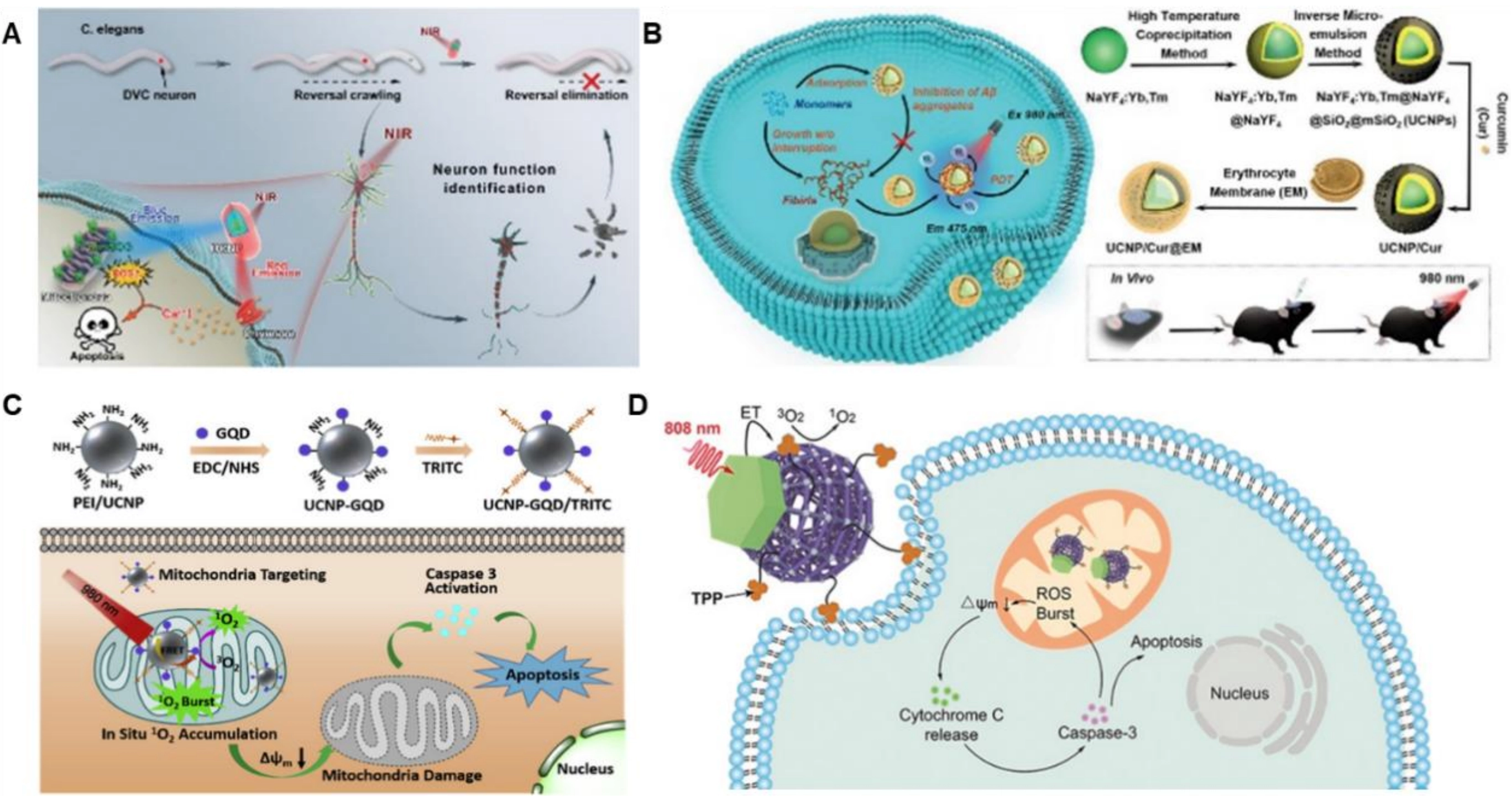
Figure 10. (A) Schematic illustration of NIR-980 nm light-induced neuron function identification strategy based on UCNPs-multiplex optogenetic protein activation system in C. elegans (locomotion regulation). Adapted with permission from reference[128]. Copyright © 2020 John Wiley & Sons; (B) Schematic illustration of biomimetic upconversion nanobait-based PDT for the inhibition of Aβ aggregates, evaluated in an AD cell model (SH-SY5Y cells expressing Aβ) by measuring Aβ aggregation via fluorescence and cell viability via MTT assay. Adapted with permission from reference[134]. Copyright © 2023 John Wiley & Sons; (C) Schematic illustration of the fabrication of mitochondria-targeting UCNP-GQD/TRITC, which upon laser irradiation triggers an in situ1O2 burst in mitochondria to initiate highly efficient apoptosis of tumor cells, as evaluated in cancer cell lines (HeLa, MCF-7) by measuring 1O2 generation via SOSG probe, mitochondrial membrane potential via staining, and apoptosis via Annexin V assay. Adapted with permission from reference[135]. Copyright © 2017 Elsevier Ltd; (D) Schematic of 808 nm NIR light-activated, mitochondria-targeted upconversion MOFs for amplified PDT, evaluated in cancer cell lines (4T1, HeLa) by measuring PDT efficacy via CCK-8 cell viability assay and ROS detection via DCFH-DA. Adapted with permission from reference[136]. Copyright © 2020 John Wiley & Sons. NIR: near-infrared; UCNPs: upconversion nanoparticles; PDT: photodynamic therapy; AD: Alzheimer’s disease; GQD: graphene quantum dot; MOFs: metal-organic frameworks; ROS: reactive oxygen species; ET: energy transfer upconversion; TRITC: tetramethylrhodamine isothiocyanate; CCK-8: cell counting kit-8; MCF-7: michigan cancer foundation-7; SOSG: singlet oxygen sensor green.
PDT has shown considerable promise in targeting protein aggregates. Guan et al. designed UCNPs@SiO2-THS, a nanostructure functionalized with the Aβ-targeting peptide KLVFF, to alleviate Aβ neurotoxicity. Under NIR irradiation, this system suppresses Aβ42 monomer aggregation and disrupts pre-formed Aβ42 fibrils through selective photo-oxidation, exhibiting minimal off-target effects and enhanced specificity. In a separate study, Gao et al. developed a UCNPs/C3N4/CoP photocatalytic system that generates hydrogen (H2) from water under NIR illumination for the treatment of AD[129]. In this configuration, UCNPs act as optical transducers, while CoP serves as a cocatalyst to improve catalytic efficiency and reduce Aβ deposition. Wang et al. engineered a core-shell UCNPs/Cur@erythrocyte membrane (EM) construct in which curcumin is surface-immobilized and encapsulated within a mesoporous silica layer modified with EM (Figure 10B)[134]. Upon NIR exposure, curcumin produces ROS, whereas the EM promotes the recruitment and entrapment of Aβ peptides, thereby inhibiting Aβ monomer elongation and facilitating the disassembly of existing aggregates. Song et al. demonstrated that polyelectrolyte-coated UCNPs are internalized by tau-aggregated neurons with efficiency comparable to that observed in healthy neurons. Zhang et al. constructed a UCNPs-graphene quantum dot (GQD)/TRITC nanocomposite in which NIR laser irradiation triggers efficient energy transfer from UCNPs to GQDs, leading to substantial singlet oxygen (1O2) generation for mitochondria-targeted induction of programmed cell death (Figure 10C)[135]. Similarly, Zhao et al. integrated porphyrin-based metal-organic frameworks (MOFs) with UCNPs to amplify energy transfer under NIR light, thereby activating PDT to induce cell death (Figure 10D)[136].
The unique optical properties of UCNPs further minimize undesirable ROS generation during imaging, making them ideal for long-term mitochondrial investigations. Li et al. created a multifunctional nanoprobe for tracking the translocation of human apurinic/apyrimidinic endonuclease 1 during NIR-triggered, mitochondria-directed PDT[137]. Together, these diverse applications highlight the considerable potential of UCNPs to refine therapeutic approaches and advance the mechanistic understanding of NDs.
4.2 Microenvironmental therapy
Upon NIR excitation, LNPs emit higher-energy photons that can activate therapeutic agents or generate ROS, both of which are central to PDT[117,118,138]. ROS play important roles in cellular signaling and immune responses, but excessive ROS levels, such as those induced by ultraviolet light or thermal stress, can trigger oxidative damage to cells[118]. This oxidative injury is a key contributor to numerous pathologies, including cancer, inflammation, and NDs[120,139]. Consequently, harnessing ROS-mediated cytotoxicity has emerged as a promising strategy in PDT, enabling the selective elimination of pathogenic cells while sparing healthy tissue.
By conjugating LNPs with appropriate photosensitizers, researchers can achieve spatially controlled ROS generation within specific cells or tissues[140,141]. However, careful modulation of ROS levels is essential, as overproduction may compromise cellular integrity and exacerbate disease progression[142]. For instance, Li et al. developed a CeO2-x-LNPs nanozyme system that exhibits potent ROS-scavenging and antioxidant activities, effectively mitigating oxidative stress and protecting neurons in models of PD[143]. In the context of AD, where Aβ aggregation drives oxidative damage, another study designed a chitosan-modified, NIR-responsive upconversion nanoplatform (UCNPs(Tm/Er)@SiO2@GPS@CH) that efficiently loads polyoxometalates via electrostatic interactions. Under NIR irradiation, this platform generates abundant ROS to achieve targeted photo-oxidation of Aβ fibrils. ROS production in the Aβ fibril group was approximately twice that of the control group, markedly enhancing therapeutic specificity.
Beyond direct ROS regulation, LNP-based microenvironmental modulation strategies have recently become more diverse and refined. One notable approach involves molecular hydrogen, which selectively scavenges highly toxic ROS such as hydroxyl radicals (•OH). Zhang et al. developed a biomimetic upconversion nanoreactor that encapsulates ascorbic acid, chlorophyll a, an indocyanine dye, and platinum nanoparticles within cross-linked vesicles, while UCNPs are immobilized on the vesicle surface as light-harvesting antennas. Under NIR irradiation, this nanoreactor catalyzes in situ hydrogen production in the brains of AD mice, effectively clearing local excess ROS and significantly inhibiting tau hyperphosphorylation, thereby providing a proof-of-concept for hydrogen-based AD therapy.
Another emerging frontier in LNP-mediated microenvironmental therapy is the modulation of microglial phenotype polarization. Microglia, the resident immune cells of the central nervous system, play a dual role in ND progression: the M1 phenotype promotes neuroinflammation, whereas the M2 phenotype exerts anti-inflammatory and reparative functions. Studies have shown that UCNPs can exert anti-inflammatory effects by scavenging intracellular ROS and promoting microglial polarization from M1 to M2. Yu et al. designed a ROS-responsive cascade-targeting nanoplatform (KMAI@NPs) that efficiently crosses the blood–brain barrier (BBB) and selectively targets activated microglia. By activating the Nrf2/GPX4 axis, this platform regulates microglial polarization and inhibits ferroptosis, significantly reducing neuroinflammation, iron overload, and amyloid pathology in APP/PS1 transgenic mice. These findings suggest that combining LNP-mediated ROS modulation with microglial immune reprogramming may enable multi-target synergistic intervention in NDs.
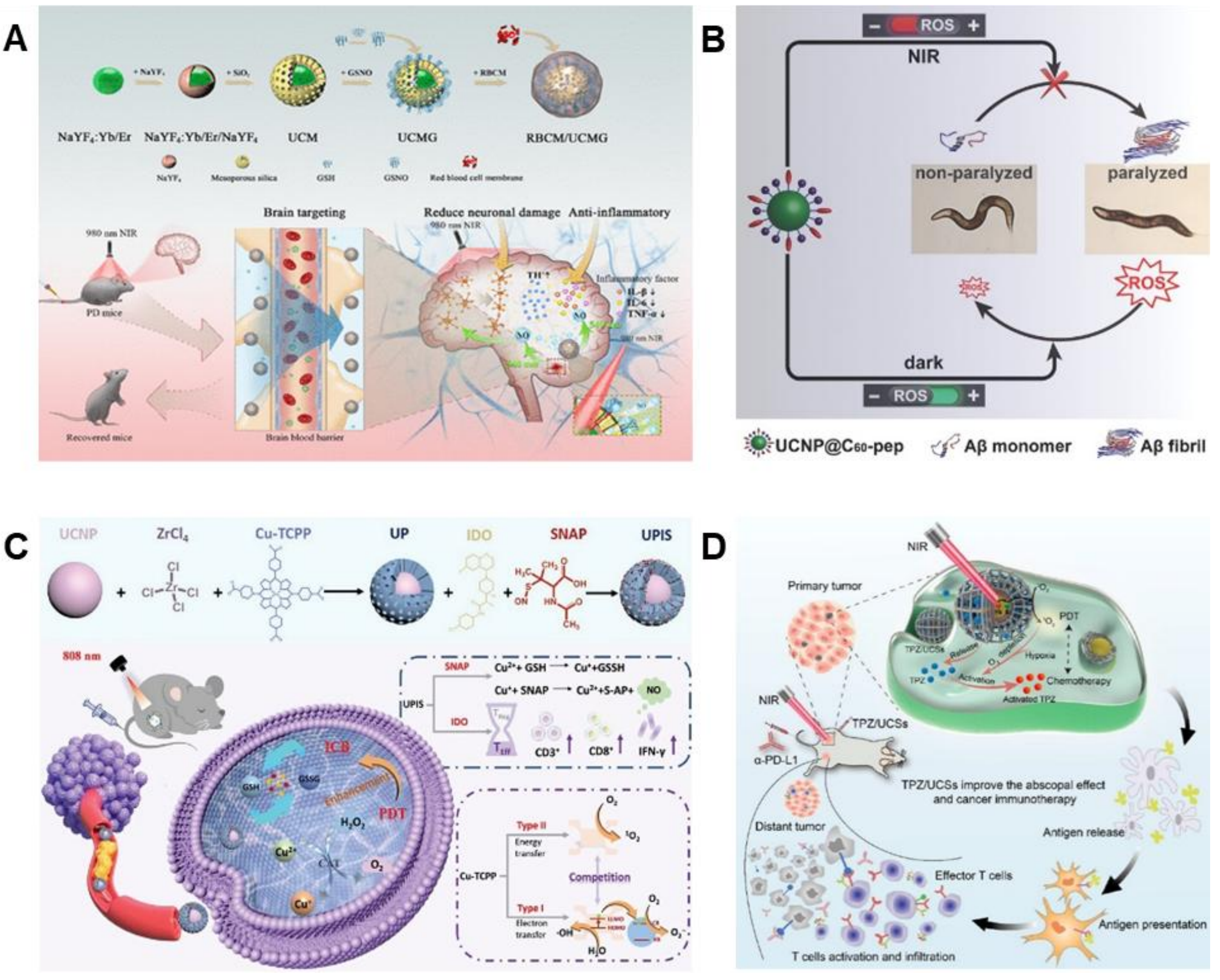
Parallel efforts have focused on BBB-crossing delivery systems for PD therapy. For instance, Wuang et al. designed a UCNP-based delivery system co-loaded with nitric oxide and a therapeutic agent to target the PD microenvironment and cross the BBB (Figure 11A)[144]. This system conferred robust neuroprotection in PD cellular models, primarily through the clearance of excess ROS and suppression of neuroinflammation, leading to improved motor function and preservation of dopaminergic neurons in the substantia nigra and striatum of PD mice. Recent studies further indicate that localized ROS production via UCNPs can enhance the efficacy of anticancer therapies.
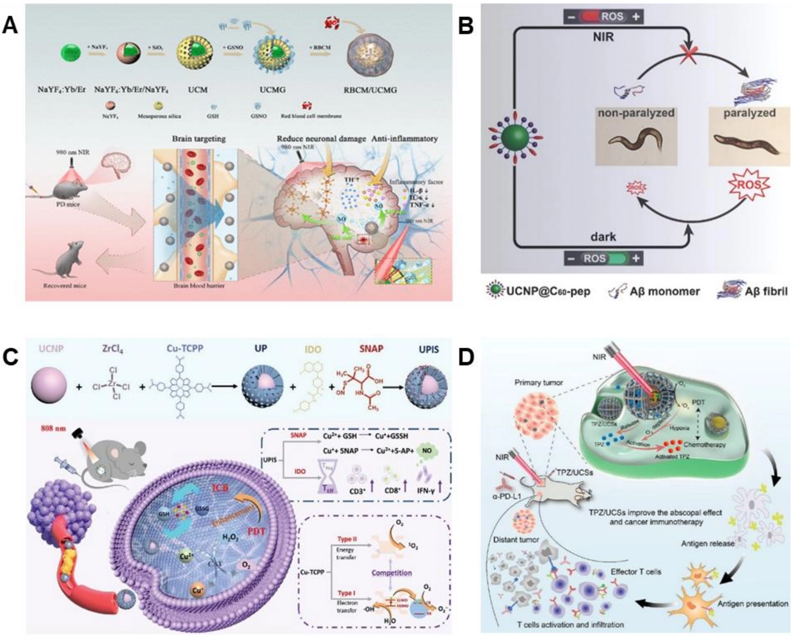
Figure 11. (A) Schematic illustration of the synthesis of RBCM/UCMG. By utilizing the surface CD47 protein that modifies the erythrocyte membrane to exert immune evasion, UCNPs could rapidly pass through the BBB and target brain lesionsa. Adapted with permission from reference[144]. Copyright © 2023 American Chemical Society; (B) Scheme UCNP@C60-pep inhibited Aβ aggregation in vivo and attenuated the oxidative stress to prolong the lifespan of the CL2006 strain. UCNP@C60-pep produced ROS under NIR to disturb Aβ aggregation and scavenged the overproduced ROS in the dark. As a result, UCNP@C60-pep could ameliorate Aβ-triggered paralysis to CL2006 worms. Adapted with permission from reference[145]. Copyright © 2018 John Wiley & Sons; (C) A schematic illustration of the construction of the designated core-shell UCNPs@MOF heterostructure, and the involved mechanisms of synergistic photodynamic and immunotherapy for primary and distant tumors. Adapted with permission from reference[146]. Copyright © 2024 John Wiley & Sons; (D) Schematic illustration of the structure of TPZ/UCSs and their application to tumor treatment through a combination of NIR light-triggered PDT and hypoxia-activated chemotherapy with immunotherapy. Adapted with permission from reference[147]. Copyright © 2020 American Chemical Society. RBCM: red blood cell membrane; UCMG: upconversion nanoparticle-methylene green; UCNPs: upconversion nanoparticles; BBB: blood–brain barrier; ROS: reactive oxygen species; NIR: near-infrared; MOF: metal-organic framework; TPZ: tirapazamine; UCSs: core–shell upconversion nanoparticle@porphyrinic MOFs; PDT: photodynamic therapy.
4.3 Synergistic therapy
In recent years, the integration of LNPs into multimodal treatment regimens has attracted considerable attention in biomedical research[148,149]. LNPs can be functionalized with a variety of therapeutic agents, such as photosensitizers, chemotherapeutic drugs, and immune modulators, thereby augmenting the effectiveness of combined modalities including PDT, photothermal therapy, chemotherapy, and immunotherapy[149-151].
The incorporation of photosensitizers into LNPs systems enables targeted ROS generation under NIR irradiation, offering a powerful approach for eradicating cancer cells or pathogens[119,152-154]. For example, Qu et al. developed UCNP@C60-pep, in which the Aβ-targeting peptide KLVFF is coupled to C60 (Figure 11B)[145]. This NIR-responsive platform selectively oxidizes Aβ aggregates associated with AD. Under NIR illumination, UCNP@C60-pep enhances the hydrophilicity of Aβ, promotes ROS production, reduces Aβ aggregation, and alleviates paralysis in Aβ-overexpressing nematode models.
Further advances were demonstrated by Du et al., who engineered a core–shell UCNPs@MOF nanoagent capable of performing both type I and type II PDT under 808 nm NIR irradiation (Figure 11C)[146]. This system leverages elevated glutathione levels in the tumor microenvironment to reduce Cu2+ ions in the copper-based MOF shell, thereby amplifying ROS generation. The MOFs are further loaded with indoleamine 2,3-dioxygenase inhibitors and S-nitroso-N-acetylpenicillamine to promote tumor-infiltrating lymphocytes and enhance antitumor immunity, illustrating a combined photodynamic-immunotherapeutic strategy.
Additionally, chemo-immunotherapeutic combinations have been explored using UCNPs platforms[154]. Yan et al. reported a UCNP-based delivery system that co-encapsulates the chemotherapeutic agent gemcitabine with an immune checkpoint inhibitor, yielding improved outcomes in hypoxic tumor models (Figure 11D)[147]. This platform not only potentiates the cytotoxic effect of gemcitabine but also activates an immune response against residual tumor cells, resulting in enhanced survival rates.
The versatile application of UCNPs in synergistic therapies underscores their potential as multifunctional platforms capable of augmenting existing treatment paradigms. Continued investigation into UCNP functionalization and combination strategies will likely unveil further innovative approaches for improving therapeutic outcomes across a range of diseases.
5. Research Limitations and Challenges
The application of LNPs based diagnostic and therapeutic technologies for NDs is confronted with multi-dimensional challenges, spanning patient variability, pharmacokinetic barriers, monitoring limitations, and economic constraints[155-157].
First, the significant genetic and neuropathological heterogeneity observed across patient populations necessitates a personalized approach to diagnosis and treatment[157]. The unique genomic and neurobiological characteristics of each individual require tailored therapeutic strategies[158]. Consequently, clinicians must not only identify the specific etiological factors in each case but also design individualized treatment regimens. This complexity impedes the development of standardized, universally applicable therapeutic protocols[120]. Pharmacokinetic limitations further complicate the translation of nanotheranostic approaches[159]. The relatively short systemic circulation time of nanoparticles often leads to insufficient accumulation at target sites[160]. Kim et al. have shown that suboptimal dosing or delivery methods can result in nanoparticle sequestration by non-target tissues, thereby reducing delivery efficiency to pathological regions[161]. In diseases such as AD and PD, where precise targeting of affected brain areas is critical, low accumulation rates significantly constrain therapeutic efficacy. A major barrier is the low delivery efficiency across the BBB. Current evidence indicates that only a minimal fraction of administered nanoparticles reaches the intended lesions, limiting the practical utility of such systems.
To overcome the formidable challenge posed by the BBB, researchers have explored various strategies. Receptor-mediated transcytosis involves modifying LNPs with ligands such as transferrin, lactoferrin, or angiopep-2 that target receptors highly expressed on brain capillary endothelial cells[162,163]. For example, Huang et al. developed a biomimetic erythrocyte membrane-coated UCNP system (RBCM/UCMG) that leverages surface CD47 proteins for immune evasion and enhanced BBB penetration[144]. In a PD mouse model, this system achieved significant accumulation in the substantia nigra and striatum, leading to improved motor function and preservation of dopaminergic neurons. Cell-penetrating peptides (CPPs) offer a non-receptor-mediated route; conjugation with CPPs such as trans-activator of transcription (TAT) (derived from HIV-1) facilitates transcytosis. Zhang et al. used TAT-functionalized LNPs to deliver optogenetic proteins across the BBB in living mice, enabling remote control of deep-tissue neurons[128]. Another promising approach is exosome/cell membrane biomimetic camouflage, where LNPs are coated with membranes derived from erythrocytes or stem cells, significantly improving biocompatibility and BBB crossing. Wang et al. constructed a curcumin-loaded, erythrocyte membrane-coated LNPs (UCNPs/Cur@EM) that not only enhanced BBB penetration but also effectively inhibited Aβ aggregation in an AD mouse model[134]. Beyond these biological strategies, physical methods such as focused ultrasound combined with microbubbles can transiently open the BBB. Although this technique has not yet been widely applied to LNPs, it has been successfully used with other nanoparticles for brain delivery and holds considerable promise for LNP-based theranostics. Finally, intranasal administration completely bypasses the BBB by delivering LNPs directly to the brain via the olfactory or trigeminal nerve pathways. Therefore, LNPs are not only therapeutic tools but also versatile probes for fundamental mechanistic studies.
A related challenge is the absence of reliable real-time monitoring systems, which hinders clinical assessment and adaptation of treatment. Without the ability to dynamically track therapeutic responses and disease progression, clinicians lack the data needed for dose optimization or regimen adjustment, ultimately affecting both the safety and effectiveness of interventions. Moreover, the high costs associated with research, development, and clinical implementation present a major economic barrier. As noted by Diou et al., repeated setbacks in translational research have escalated expenditures and elevated the eventual costs of clinical adoption[161].
To overcome these barriers and facilitate the clinical translation of nanotheranostic platforms, integrated strategies are essential. These include the development of smart targeted delivery systems to enhance lesion-specific accumulation, the implementation of real-time monitoring technologies to visualize treatment dynamics, and the establishment of standardized manufacturing processes to reduce production costs. Such multi-faceted advances continue to garner focused attention within both academic and translational research communities, underscoring a collective effort toward more effective and accessible ND therapies.
6. Summary and Outlook
NDs are characterized by progressive neuronal degeneration and functional deterioration and present a significant and escalating global health challenge[9,164]. Current therapies mainly focus on managing symptoms and providing support for brain function but do not reverse the underlying damage[3,165,166]. This highlights the need for preventive measures and targeted therapies[167]. Over the past decade, LNPs have emerged as a transformative platform at the interface of nanotechnology and biomedicine, offering new pathways to advance the diagnosis and treatment of NDs[3,167-169]. This review examined the fundamental luminescence mechanisms of LNPs, encompassing UCNPs, DCNPs, and energy transfer processes such as LRET and IFE. The unique optical properties of LNPs, including tunable emission, deep-tissue penetration enabled by NIR excitation, and exceptional photostability, allow them to surpass the limitations of conventional organic fluorophores, establishing LNPs as advanced tools for high-sensitivity detection and high-resolution bioimaging.
In the detection and diagnosis of NDs, LNPs have demonstrated broad utility and high performance. They enable sensitive and specific detection of various disease biomarkers, including metal ions, nucleic acids, protein aggregates, and ROS[7,170]. By attaching targeting molecules like antibodies, peptides, or aptamers to LNPs’ surfaces, these nanoparticles can be precisely directed to affected cells and tissues, significantly improving diagnostic accuracy and allowing real-time monitoring of disease development. LNPs are playing an important role in therapeutic development. Their applications range from intracellular drug delivery to modulation of the disease microenvironment and combined treatment strategies. As drug carriers, LNPs can transport therapeutic agents, such as antioxidants, neuroprotective compounds, or gene regulators, directly to compromised neural regions, increasing treatment effectiveness while minimizing side effects. Their application in PDT, where NIR light activates localized ROS production to break down toxic protein aggregates or destroy abnormal cells, shows particular promise. Furthermore, by engineering multifunctional LNPs complexes, combined treatment approaches, such as PDT with immunotherapy or chemotherapy, become feasible, creating new opportunities for comprehensive disease management.
Despite these advances, the clinical translation of LNPs based nanotheranostics still faces several key challenges. Differences in genetic background and disease manifestation among patients make it difficult to develop universal treatment protocols, requiring more personalized approaches. Pharmacokinetic issues, including rapid clearance from circulation and limited accumulation at target sites, reduce treatment bioavailability. The absence of reliable, real-time systems for monitoring treatment responses in living organisms hinders optimal dosing and treatment adjustment. Moreover, the high costs associated with research, development, and large-scale production present significant economic barriers to widespread clinical adoption.
Looking forward, continued progress of LNPs technology in NDs will require coordinated, interdisciplinary collaboration. A particularly exciting direction is the use of LNPs as a novel class of “photoactivatable” chemical tools to directly participate in and elucidate the molecular pathological mechanisms of NDs[171,172]. Phase separation and aberrant aggregation of biomolecular condensates are now recognized as key pathological events in AD, PD, and other NDs. However, characterizing these condensates currently relies on complex proteomic technologies. For instance, Kappel et al. employed a high-throughput in situ imaging sequencing method, to systematically decode the “molecular grammar” by which protein sequences drive nuclear condensate formation, providing a scalable platform for dissecting condensate sequence determinants[173]. Such advances open new dimensions for future LNPs applications: by integrating LNPs with these cutting-edge proteomic strategies, one could exploit the NIR optical window of LNPs for in situ, non-invasive photochemical labeling and precise capture of specific condensate components deep within living cells or brain tissue[174]. Subsequent coupling with proteomic techniques such as mass spectrometry would enable unbiased, high-coverage identification of dynamic condensate components. This cross-modal technological fusion holds promise for overcoming the bottlenecks of conventional chemical biology tools in ND research, namely, poor optical penetration and low precision of chemical intervention, and offers a new perspective for understanding the interaction networks between LNPs and various misfolded proteins or signaling macromolecules.
At present, optimizing the targeted delivery and biocompatibility of LNPs remains a key focus and cornerstone of research. Building on this foundation, important future directions also include: (1) designing next-generation smart LNPs with improved targeting ability, longer circulation times, and controlled drug release properties; (2) developing integrated real-time imaging and sensing platforms to dynamically visualize treatment delivery and biological responses; (3) establishing standardized, scalable manufacturing and quality control processes to ensure consistency, safety, and cost-effectiveness; and (4) creating innovative combination therapies that harness the unique properties of LNPs to address the complex, multifaceted nature of NDs.
In conclusion, LNPs are at the forefront of ongoing advances in nanomedicine. By combining sophisticated optical properties with therapeutic functions, they offer a versatile and effective approach for understanding disease processes, enabling early and accurate diagnosis, and developing targeted treatments. Achieving their full clinical potential will require continued collaboration among researchers in chemistry, materials science, neuroscience, and clinical medicine. With sustained innovation and thorough clinical evaluation, LNPs based technologies hold significant promise for transforming patient care and improving outcomes in the challenging area of NDs.
Acknowledgements
We thank the supporting from the Key Laboratory of Laser Life Science, Ministry of Education and Guangdong Key Laboratory of Laser Life Science.
Authors contribution
Liu J: Investigation, writing-original draft.
Li L: Funding acquisition, project administration, resources, supervision, validation, writing-review & editing.
Conflicts of interest
The authors declare no conflicts of interest.
Ethical approval
Not applicable.
Consent to participate
Not applicable.
Consent for publication
Not applicable.
Availability of data and materials
Not applicable.
Funding
This work was supported by the national natural Science Foundation of China (Grant Nos. 62527816 and 52002133), and the fundings for young scholars at South China Normal University (SCNU) (This program does not have a grant number).
Copyright
© The Author(s) 2026.
References
-
1. Ciurea AV, Mohan AG, Covache-Busuioc RA, Costin HP, Glavan LA, Corlatescu AD, et al. Unraveling molecular and genetic insights into neurodegenerative diseases: Advances in understanding Alzheimer’s, Parkinson’s, and Huntington’s diseases and amyotrophic lateral sclerosis. Int J Mol Sci. 2023;24(13):10809.[DOI]
-
3. Su D, Cui Y, He C, Yin P, Bai R, Zhu J, et al. Projections for prevalence of Parkinson’s disease and its driving factors in 195 countries and territories to 2050: Modelling study of Global Burden of Disease Study 2021. BMJ. 2025;388:e080952.[DOI]
-
4. Foltynie T, Bruno V, Fox S, Kühn AA, Lindop F, Lees AJ. Medical, surgical, and physical treatments for Parkinson’s disease. Lancet. 2024;403(10423):305-324.[DOI]
-
7. Ito H. Symptoms and signs of Parkinson’s disease and other movement disorders. In: Itakura T, editor. Deep brain stimulation for neurological disorders. Cham: Springer; 2015. p. 21-37.[DOI]
-
8. Jankovic J, Lang AE. Diagnosis and assessment of Parkinson disease and other movement disorders. In: Bradley’s neurology in clinical practice. Amsterdam: Elsevier; 2021. p. 310-333. Available from: https://www.vitalsource.com/products/bradley-39-s-neurology-in-clinical-practice-joseph-jankovic-john-c-v9780323642637
-
10. Cao C, Hong C, Li Y, Li G, Jiang G. A long-term and stable surface modification method for lanthanide doped upconversion nanoparticles by oxidized alginate. Zeitschrift Anorg Allge Chemie. 2020;646(19):1607-1610.[DOI]
-
11. Ding Z, He Y, Rao H, Zhang L, Nguyen W, Wang J, et al. Novel fluorescent probe based on rare-earth doped upconversion nanomaterials and its applications in early cancer detection. Nanomaterials. 2022;12(11):1787.[DOI]
-
12. Yu Z, Deng Y, Ye J, van Turnhout L, Liu T, Tew A, et al. Triplets electrically turn on insulating lanthanide-doped nanoparticles. Nature. 2025;647(8090):625-631.[DOI]
-
15. Ling H, Zhang W, Zhang Y, Shen J, Liu Q. Lanthanide-doped upconversion nanoparticles for single-particle imaging. ChemBioChem. 2025;26(11):e202400942.[DOI]
-
17. Xie X, Li Z, Zhang Y, Guo S, Pendharkar AI, Lu M, et al. Emerging ≈800 nm excited lanthanide-doped upconversion nanoparticles. Small. 2017;13(6):1602843.[DOI]
-
18. Ge X, Wei R, Sun L. Lanthanide nanoparticles with efficient near-infrared-II emission for biological applications. J Mater Chem B. 2020;8(45):10257-10270.[DOI]
-
21. Yang D, University Y, Li L, University Y, Zhang X, University Y, et al. Achieving excitation wavelength dependence of cesium cadmium halogen quantum dots with multi-excitonic emission center. J Phys Chem Lett. 2025;16(22):5480-5487.[DOI]
-
22. Xu Q, Gao J, Wang S, Wang Y, Liu D, Wang J. Quantum dots in cell imaging and their safety issues. J Mater Chem B. 2021;9(29):5765-5779.[DOI]
-
24. Chen H, Liu X, Xi X, Chen H, Yan L, Fu Z, et al. Upconversion luminescence of NaYF4: Ln3+ nanoparticles on gold nanorod array with dual-wavelength excitation. Nanomaterials. 2026;16(4):277.[DOI]
-
26. Jeong M, Lee Y, Park J, Jung H, Lee H. Lipid nanoparticles (LNPs) for in vivo RNA delivery and their breakthrough technology for future applications. Adv Drug Deliv Rev. 2023;200:114990.[DOI]
-
27. Bian F, Sun L, Cai L, Wang Y, Zhao Y. Quantum dots from microfluidics for nanomedical application. WIREs Nanomed Nanobiotechnol. 2019;11(5):e1567.[DOI]
-
28. Yang M, Ji C, Yin M. Aggregation-enhanced photothermal therapy of organic dyes. WIREs Nanomed Nanobiotechnol. 2024;16(3):e1960.[DOI]
-
29. Alejo T, Toro-Córdova A, Fernández L, Rivero A, Stoian AM, Pérez L, et al. Comprehensive optimization of a freeze-drying process achieving enhanced long-term stability and in vivo performance of lyophilized mRNA-LNPs. Int J Mol Sci. 2024;25(19):10603.[DOI]
-
30. Le N, Zhang M, Kim K. Quantum dots and their interaction with biological systems. Int J Mol Sci. 2022;23(18):10763.[DOI]
-
32. Alguacil FJ, Alonso M, Robla JI. Removal of hazardous organic dyes from liquid wastes using advanced nanomaterials. Int J Mol Sci. 2024;25(17):9671.[DOI]
-
33. Lan D, Zhu H, Zhang J, Li S, Chen Q, Wang C, et al. Adsorptive removal of organic dyes via porous materials for wastewater treatment in recent decades: A review on species, mechanisms and perspectives. Chemosphere. 2022;293:133464.[DOI]
-
34. Di J, Du Z, Wu K, Jin S, Wang X, Li T, et al. Biodistribution and non-linear gene expression of mRNA LNPs affected by delivery route and particle size. Pharm Res. 2022;39(1):105-114.[DOI]
-
35. Chen Y, Feng X. Gold nanoparticles for skin drug delivery. Int J Pharm. 2022;625:122122.[DOI]
-
36. Wu M, Xiao Y, Wu R, Lei J, Li T, Zheng Y. Aggregable gold nanoparticles for cancer photothermal therapy. J Mater Chem B. 2024;12(33):8048-8061.[DOI]
-
37. Khare P, Edgecomb SX, Hamadani CM, Tanner EE, Manickam DS. Lipid nanoparticle-mediated drug delivery to the brain. Adv Drug Deliv Rev. 2023;197:114861.[DOI]
-
38. Uddin MJ, Ampiaw RE, Lee W. Adsorptive removal of dyes from wastewater using a metal-organic framework: A review. Chemosphere. 2021;284:131314.[DOI]
-
40. Xue Y, Wang C, Li H, Du S, Zhong Y, Zhang Y, et al. Lipid nanoparticles enhance mRNA delivery to the central nervous system upon intrathecal injection. Adv Mater. 2025;37(27):2417097.[DOI]
-
44. Wang M, Hu C, Su Q. Luminescent lifetime regulation of lanthanide-doped nanoparticles for biosensing. Biosensors. 2022;12(2):131.[DOI]
-
45. Jethva P, Momin M, Khan T, Omri A. Lanthanide-doped upconversion luminescent nanoparticles: Evolving role in bioimaging, biosensing, and drug delivery. Materials. 2022;15(7):2374.[DOI]
-
48. Hu G, Gao S, Han X, Yang L. Comparison of immunochromatographic strips using colloidal gold, quantum dots, and upconversion nanoparticles for visual detection of norfloxacin in milk samples. Food Anal Meth. 2020;13(5):1069-1077.[DOI]
-
49. Wysokińska E, Cichos J, Kowalczyk A, Karbowiak M, Strządała L, Bednarkiewicz A, et al. Toxicity mechanism of low doses of NaGdF4: Yb3+, Er3+ upconverting nanoparticles in activated macrophage cell lines. Biomolecules. 2019;9(1):14.[DOI]
-
50. Li Y, Xu J, Hu T, Shi G, Peng Y, Qiao F, et al. Synthesis and upconversion luminescence fine-tuning of Yb3+/Ho3+-doped indium and gallium oxide nanoparticles. Inorg Chem. 2024;63(37):17032-17042.[DOI]
-
52. Li J, Xie Y, Sun R, Zhou J, Sun L. Exploring luminescence quenching on lanthanide-doped nanoparticles through changing the spatial distribution of sensitizer and activator. Nano Res. 2024;17(5):4517-4524.[DOI]
-
53. Ling H, Guan D, Wen R, Hu J, Zhang Y, Zhao F, et al. Effect of surface modification on the luminescence of individual upconversion nanoparticles. Small. 2024;20(26):2309035.[DOI]
-
54. Loo JF, Chien YH, Yin F, Kong SK, Ho HP, Yong KT. Upconversion and downconversion nanoparticles for biophotonics and nanomedicine. Coord Chem Rev. 2019;400:213042.[DOI]
-
55. Liu B, Li C, Yang P, Hou Z, Lin J. 808-nm-light-excited lanthanide-doped nanoparticles: Rational design, luminescence control and theranostic applications. Adv Mater. 2017;29(18):1605434.[DOI]
-
56. Li H, Wang X, Huang D, Chen G. Recent advances of lanthanide-doped upconversion nanoparticles for biological applications. Nanotechnology. 2020;31(7):072001.[DOI]
-
57. Arumugam GM, Karunakaran SK, Galian RE, Pérez-Prieto J. Recent progress in lanthanide-doped inorganic perovskite nanocrystals and nanoheterostructures: A future vision of bioimaging. Nanomaterials. 2022;12(13):2130.[DOI]
-
58. Yao J, Huang C, Liu C, Yang M. Upconversion luminescence nanomaterials: A versatile platform for imaging, sensing, and therapy. Talanta. 2020;208:120157.[DOI]
-
61. Li Z, Liang T, Wang Q, Liu Z. Strategies for constructing upconversion luminescence nanoprobes to improve signal contrast. Small. 2020;16:1905084.[DOI]
-
62. Li Y, Wang X, Yang L, Xie X, Yao S, Min T, et al. Developing FRET-based UCNP@SiO2-AIE/AIE@DSPE-PEG2000 nanomaterials for lysosome-targeted NIR photodynamic cancer therapy. Mikrochim Acta. 2025;192(12):813.[DOI]
-
64. Fischer SA, Cramer CJ, Govind N. Excited state absorption from real-time time-dependent density functional theory. J Chem Theory Comput. 2015;11(9):4294-4303.[DOI]
-
68. Lamon S, Wu Y, Zhang Q, Liu X, Gu M. Nanoscale optical writing through upconversion resonance energy transfer. Sci Adv. 2021;7(9):eabe2209.[DOI]
-
69. Shaw PA, Forsyth E, Haseeb F, Yang S, Bradley M, Klausen M. Two-photon absorption: An open door to the NIR-II biological window? Front Chem. 2022;10:921354.[DOI]
-
71. Madonini F, Villa F. Single photon avalanche diode arrays for time-resolved Raman spectroscopy. Sensors. 2021;21(13):4287.[DOI]
-
73. Ma S, Wang Y, Wang C, Wang L, Miao Q, Liu Y, et al. Accurate photoactivation monitoring via the construction of an intramolecular synergistic counteracting mechanism of FRET and IFE. Chem Commun. 2023;59(37):5599-5602.[DOI]
-
75. Chen Y, Wei J, Zhang J, Qiu H, Zhang Y, Zhang J, et al. Boosting the downconversion luminescence of Tm3+-doped nanoparticles for S-band polymer waveguide amplifier. Nano Lett. 2024;24(4):1399-1405.[DOI]
-
76. Wang Z, Meijerink A. Dye-sensitized downconversion. J Phys Chem Lett. 2018;9(7):1522-1526.[DOI]
-
79. Kang D, Lee S, Shin H, Pyun J, Lee J. An efficient NIR-to-NIR signal-based LRET system for homogeneous competitive immunoassay. Biosens Bioelectron. 2020;150:111921.[DOI]
-
81. Chen Q, Tang K, Luo D, Han L, Yu C, Shen Y, et al. Paper-based LRET sensor for the detection of total heavy rare-earth ions. Front Chem. 2022;10:1028441.[DOI]
-
82. Kang D, Ahn HJ, Lee J, Kim SK, Pyun J, Song CS, et al. An NIR dual-emitting/absorbing inorganic compact pair: A self-calibrating LRET system for homogeneous virus detection. Biosens Bioelectron. 2021;190:113369.[DOI]
-
85. Wu H, Wang Q, Zhu L, Zhang H, Liu L. Regulating the aromatic structure in carbon dots to enhance the internal filter effect for sensitive on-site monitoring of anthrax biomarkers. Talanta. 2025;291:127854.[DOI]
-
87. Guo Y, Zhao T, Guo Q, Ding M, Chen X, Lin J. Highly sensitive detection for xanthine by combining single-band red up-conversion nanoparticles and cycle signal amplification strategy based on internal filtration effect. Spectrochim Acta Part A Mol Biomol Spectrosc. 2024;306:123566.[DOI]
-
91. Grebenik EA, Kostyuk AB, Deyev SM. Upconversion nanoparticles and their hybrid assemblies for biomedical applications. Russ Chem Rev. 2016;85(12):1277-1296.[DOI]
-
92. Lin W, Li J, Feng H, Qi F, Huang L. Recent advances in triplet–triplet annihilation upconversion for bioimaging and biosensing. J Anal Test. 2023;7(4):327-344.[DOI]
-
94. Liu J, Li N, Chen Y, Pu R, Wei W, Lin Y, et al. Real-time biosensing of Parkinson’s disease biomarkers via NIR-activated upconversion nanoparticles. BMEMat. 2026.[DOI]
-
95. Huang L, Chen F, Zong X, Lu Q, Wu C, Ni Z, et al. Near-infrared light excited UCNP-DNAzyme nanosensor for selective detection of Pb2+ and in vivo imaging. Talanta. 2021;227:122156.[DOI]
-
96. Yang Z, Loh KY, Chu YT, Feng R, Satyavolu NSR, Xiong M, et al. Optical control of metal ion probes in cells and zebrafish using highly selective DNAzymes conjugated to upconversion nanoparticles. J Am Chem Soc. 2018;140(50):17656-17665.[DOI]
-
98. Belur NR, Bustos BI, Lubbe SJ, Mazzulli JR. Nuclear aggregates of NONO/SFPQ and A-to-I-edited RNA in Parkinson’s disease and dementia with Lewy bodies. Neuron. 2024;112(15):2558-2580.[DOI]
-
99. Salemi M, Lanza G, Mogavero MP, Cosentino FII, Borgione E, Iorio R, et al. A transcriptome analysis of mRNAs and long non-coding RNAs in patients with Parkinson’s disease. Int J Mol Sci. 2022;23(3):1535.[DOI]
-
102. Zhang D, Peng R, Liu W, Donovan MJ, Wang L, Ismail I, et al. Engineering DNA on the surface of upconversion nanoparticles for bioanalysis and therapeutics. ACS Nano. 2021;15(11):17257-17274.[DOI]
-
103. Zhang J, Zheng Y, Luo Y, Du Y, Zhang X, Fu J. Curcumin inhibits LPS-induced neuroinflammation by promoting microglial M2 polarization via TREM2/TLR4/NF-κB pathways in BV2 cells. Mol Immunol. 2019;116:29-37.[DOI]
-
104. Brandstetter D, Helbig C, Osawa K, Minemura H, Anzai Y, Torisu T, et al. Three-dimensional homodyne light detection (3D-HLD) for high-throughput submicron particle analysis in (highly concentrated) protein biopharmaceuticals, viral vectors, and LNPs. J Pharm Sci. 2024;113(4):891-899.
-
106. Hlaváček A, Farka Z, Mickert MJ, Kostiv U, Brandmeier JC, Horák D, et al. Bioconjugates of photon-upconversion nanoparticles for cancer biomarker detection and imaging. Nat Protoc. 2022;17(4):1028-1072.[DOI]
-
109. Song YH, De R, Lee KT. Uptake of polyelectrolyte functionalized upconversion nanoparticles by tau-aggregated neuron cells. Pharmaceutics. 2021;13(1):102.[DOI]
-
111. Li J, Zhu F, Lou K, Tian H, Luo Q, Dang Y, et al. Tumor microenvironment enhanced NIR II fluorescence imaging for tumor precise surgery navigation via tetrasulfide mesoporous silica-coated Nd-based rare-earth nanocrystals. Mater Today Bio. 2022;16:100397.[DOI]
-
112. Chen S, Zhang C, Jia G, Duan J, Wang S, Zhang J. Size-dependent cytotoxicity of europium doped NaYF4 nanoparticles in endothelial cells. Mater Sci Eng C. 2014;43:330-342.[DOI]
-
113. Yan S, Xing G, Yuan X, Cui E, Ji K, Yang X, et al. Upconversion nanoparticles-CuMnO2 nanoassemblies for NIR-excited imaging of reactive oxygen species in vivo. J Colloid Interface Sci. 2025;677:666-674.[DOI]
-
116. Zhang Y, Zhu X, Zhang J, Wu Y, Liu J, Zhang Y. Synergistic upconversion photodynamic and photothermal therapy under cold near-infrared excitation. J Colloid Interface Sci. 2021;600:513-529.[DOI]
-
117. Ding S, Wu W, Peng T, Pang W, Jiang P, Zhan Q, et al. Near-infrared light excited photodynamic anticancer therapy based on UCNP@AIEgen nanocomposite. Nanoscale Adv. 2021;3(8):2325-2333.[DOI]
-
118. Güleryüz B, Ünal U, Gülsoy M. Near infrared light activated upconversion nanoparticles (UCNP) based photodynamic therapy of prostate cancers: An in vitro study. Photodiagnosis Photodyn Ther. 2021;36:102616.[DOI]
-
119. Liu Z, Li M, Xie Q, Liu Y, Huang J, Zeng Q, et al. Eradicating fungal biofilm-based infections by ultrasound-assisted semiconductor sensitized upconversion photodynamic therapy. Nat Commun. 2025;16:6499.[DOI]
-
121. Zou X, Liu Y, Zhu X, Chen M, Yao L, Feng W, et al. An Nd3+-sensitized upconversion nanophosphor modified with a cyanine dye for the ratiometric upconversion luminescence bioimaging of hypochlorite. Nanoscale. 2015;7(9):4105-4113.[DOI]
-
122. Hao C, Wu X, Sun M, Zhang H, Yuan A, Xu L, et al. Chiral core–shell upconversion Nanoparticle@MOF nanoassemblies for quantification and bioimaging of reactive oxygen species in vivo. J Am Chem Soc. 2019;141(49):19373-19378.[DOI]
-
123. Moise-Crintea A, Constantin AM, Jianu EM, Orlea IM, Manea M, Cojocariu RO, et al. Advancing lung cancer treatment: A comprehensive review of photodynamic therapy and nanoparticle applications. Pharmaceutics. 2025;17(12):1579.[DOI]
-
124. Bae GH, Shin S, Park JD, Koh EY, Ko S, Han J, et al. Photosensitizing lipid nanoparticles for ferroptosis-enhanced photodynamic cancer therapy via GPX4 silencing. Adv Healthc Mater. 2026;15(7):e03748.[DOI]
-
125. Simões MM, Paiva KL, de Souza IF, Mello VC, Martins da Silva IG, Souza PEN, et al. The potential of photodynamic therapy using solid lipid nanoparticles with aluminum phthalocyanine chloride as a nanocarrier for modulating immunogenic cell death in murine melanoma in vitro. Pharmaceutics. 2024;16(7):941.
-
126. Akhtar N, Chen CL, Chattopadhyay S. PDT-active upconversion nanoheaters for targeted imaging guided combinatorial cancer phototherapies with low-power single NIR excitation. Biomater Adv. 2022;141:213117.[DOI]
-
127. Qiu H, Tan M, Ohulchanskyy TY, Lovell JF, Chen G. Recent progress in upconversion photodynamic therapy. Nanomaterials. 2018;8(5):344.[DOI]
-
129. Ge K, Li Z, Wang A, Bai Z, Zhang X, Zheng X, et al. An NIR-driven upconversion/C3N4/CoP photocatalyst for efficient hydrogen production by inhibiting electron–hole pair recombination for Alzheimer’s disease therapy. ACS Nano. 2023;17(3):2222-2234.[DOI]
-
130. Guryev EL, Smyshlyaeva AS, Shilyagina NY, Sokolova EA, Shanwar S, Kostyuk AB, et al. UCNP-based photoluminescent nanomedicines for targeted imaging and theranostics of cancer. Molecules. 2020;25(18):4302.[DOI]
-
131. Qu A, Wu X, Xu L, Liu L, Ma W, Kuang H, et al. SERS- and luminescence-active Au–Au–UCNP trimers for attomolar detection of two cancer biomarkers. Nanoscale. 2017;9(11):3865-3872.[DOI]
-
134. Wang X, Zhang W, Hou L, Geng W, Wang J, Kong Y, et al. A biomimetic upconversion nanobait-based near infrared light guided photodynamic therapy alleviates Alzheimer’s disease by inhibiting β-amyloid aggregation. Adv Healthc Mater. 2024;13(9):2303278.[DOI]
-
135. Zhang D, Wen L, Huang R, Wang H, Hu X, Xing D. Mitochondrial specific photodynamic therapy by rare-earth nanoparticles mediated near-infrared graphene quantum dots. Biomaterials. 2018;153:14-26.[DOI]
-
138. Liu YQ, Qin LY, Li HJ, Wang YX, Zhang R, Shi JM, et al. Application of lanthanide-doped upconversion nanoparticles for cancer treatment: A review. Nanomedicine. 2021;16(24):2207-2242.[DOI]
-
139. Wang K, Zheng J, Li R, Chen T, Ma Y, Wu P, et al. Single-cell multi-omics assessment of spinal cord injury blocking via cerium-doped upconversion antioxidant nanoenzymes. Adv Sci. 2025;12(8):2412526.[DOI]
-
140. Ghosh A, Singh A, Guha S, Saha B, Das G, Sahu SK. A multifunctional NIR responsive upconversion porphyrin nanoprobe for Fenton/FRET induced ROS amplification and targeted drug delivery. J Colloid Interface Sci. 2025;700:138311.[DOI]
-
141. Xu C, Law SK, Leung AWN. Comparison of the differences between two-photon excitation, upconversion, and conventional photodynamic therapy on cancers in in vitro and in vivo studies. Pharmaceuticals. 2024;17(6):663.[DOI]
-
142. Chen Y, Yang Y, Du S, Ren J, Jiang H, Zhang L, et al. Mitochondria-targeting upconversion Nanoparticles@MOF for multiple-enhanced photodynamic therapy in hypoxic tumor. ACS Appl Mater Interfaces. 2023;15(30):35884-35894.[DOI]
-
145. Du Z, Gao N, Wang X, Ren J, Qu X. Near-infrared switchable fullerene-based synergy therapy for Alzheimer’s disease. Small. 2018;14(33):1801852.[DOI]
-
146. Du J, Jia T, Li F, Li Y, Wang Q, He L, et al. MOF-coated upconversion nanoparticle agents enable synergistic photodynamic therapy and immunotherapy. Adv Funct Mater. 2024;34(36):2401272.[DOI]
-
147. Shao Y, Liu B, Di Z, Zhang G, Sun LD, Li L, et al. Engineering of upconverted metal–organic frameworks for near-infrared light-triggered combinational photodynamic/chemo-/immunotherapy against hypoxic tumors. J Am Chem Soc. 2020;142(8):3939-3946.[DOI]
-
149. Sohrabi M, Babaei Z, Haghpanah V, Larijani B, Abbasi A, Mahdavi M. Recent advances in gene therapy-based cancer monotherapy and synergistic bimodal therapy using upconversion nanoparticles: Structural and biological aspects. Biomed Pharmacother. 2022;156:113872.[DOI]
-
150. Li Z, Lu S, Liu W, Dai T, Ke J, Li X, et al. Synergistic lysozyme-photodynamic therapy against resistant bacteria based on an intelligent upconversion nanoplatform. Angew Chem. 2021;133(35):19350-19355.[DOI]
-
151. Zhou J, Ma L, Li Z, Chen B, Wu Y, Meng X. Synthesis of lenvatinib-loaded upconversion@polydopamine nanocomposites for upconversion luminescence imaging-guided chemo-photothermal synergistic therapy of anaplastic thyroid cancer. RSC Adv. 2023;13(38):26925-26932.[DOI]
-
152. Lv Z, Cao Y, Xue D, Zhang H, Zhou S, Yin N, et al. A multiphoton transition activated iron based metal organic framework for synergistic therapy of photodynamic therapy/chemodynamic therapy/chemotherapy for orthotopic gliomas. J Mater Chem B. 2023;11(5):1100-1107.[DOI]
-
153. Wang J, Wang W, Shen Q, Lan L, Guan C, Xu X, et al. Cell-surface photochemistry mediated calcium overload for synergistic tumor therapy. J Nanobiotechnol. 2023;21(1):335.[DOI]
-
155. Mettenbrink EM, Yang W, Wilhelm S. Bioimaging with upconversion nanoparticles. Adv Photonics Res. 2022;3(12):2200098.[DOI]
-
156. Zou L, Xu K, Tian H, Fang Y. Remote neural regulation mediated by nanomaterials. Nanotechnology. 2022;33(27):272002.[DOI]
-
160. Lai WF, Rogach A, Wong WT. Molecular design of upconversion nanoparticles for gene delivery. Chem Sci. 2017;8(11):7339-7358.[DOI]
-
162. Moosavi SG, Rahiman N, Jaafari MR, Arabi L. Lipid nanoparticle (LNP) mediated mRNA delivery in neurodegenerative diseases. J Control Release. 2025;381:113641.[DOI]
-
164. Aarsland D, Kurz MW. The epidemiology of dementia associated with Parkinson’s disease. Brain Pathol. 2010;20(3):633-639.[DOI]
-
165. Li L, Zhou F, Gao Q, Lu Y, Xu X, Hu R, et al. Visualizing dynamic performance of lipid droplets in a Parkinson’s disease model via a smart photostable aggregation-induced emission probe. iScience. 2019;21:261-272.[DOI]
-
166. Raza C, Anjum R, Shakeel NUA. Parkinson’s disease: Mechanisms, translational models and management strategies. Life Sci. 2019;226:77-90.[DOI]
-
168. Yulug B, Velioglu HA, Sayman D, Cankaya S, Hanoglu L. Brain temperature in healthy and diseased conditions: A review on the special implications of MRS for monitoring brain temperature. Biomed Pharmacother. 2023;160:114287.[DOI]
-
169. Monzio Compagnoni G, Di Fonzo A, Corti S, Comi GP, Bresolin N, Masliah E. The role of mitochondria in neurodegenerative diseases: The lesson from Alzheimer’s disease and Parkinson’s disease. Mol Neurobiol. 2020;57(7):2959-2980.[DOI]
-
172. Sun R, Huang Y, Feng H, Zhao N, Wan W, Shen D, et al. 1000 fold ultra-photosensitized fluorescent protein mimics toward photocatalytic proximity labeling and proteomic profiling functions. Adv Sci. 2025;12(15):2413063.[DOI]
-
174. Zhang S, Owyong TC, Sanislav O, Englmaier L, Sui X, Wang G, et al. Global analysis of endogenous protein disorder in cells. Nat Meth. 2025;22(1):124-134.[DOI]
Copyright
© The Author(s) 2026. This is an Open Access article licensed under a Creative Commons Attribution 4.0 International License (https://creativecommons.org/licenses/by/4.0/), which permits unrestricted use, sharing, adaptation, distribution and reproduction in any medium or format, for any purpose, even commercially, as long as you give appropriate credit to the original author(s) and the source, provide a link to the Creative Commons license, and indicate if changes were made.
Publisher’s Note
Science Exploration remains a neutral stance on jurisdictional claims in published
maps
and institutional affiliations. The views expressed in this article are solely those
of
the author(s) and do not reflect the opinions of the Editors or the publisher.
Share And Cite


Science Exploration Style
Liu J, Li L. Lanthanide-doped nanoparticles: An emerging platform for theranostic applications in neurodegenerative diseases. BME Horiz. 2026;4:202613. https://doi.org/10.70401/bmeh.2026.0030
Tips
Copy completed.
Submit a Manuscript
Author Instructions
Cite this Article
Article Metrics
0
View
0
Download
Cited
Article Updates

- Abstract
- Keywords
- 1. Introduction
- 2. Luminescence Mechanism
- 3. Biosensing and Diagnosis
- 4. LNDs in ND Therapeutics
- 5. Research Limitations and Challenges
- 6. Summary and Outlook
- Acknowledgements
- Authors contribution
- Conflicts of interest
- Ethical approval
- Consent to participate
- Consent for publication
- Availability of data and materials
- Funding
- References
- Copyright
Science Exploration Style
Liu J, Li L. Lanthanide-doped nanoparticles: An emerging platform for theranostic applications in neurodegenerative diseases. BME Horiz. 2026;4:202613. https://doi.org/10.70401/bmeh.2026.0030
copy
Share Link
copy