Overcoming three-layered tumor microenvironmental barriers to photodynamic therapy-induced systemic antitumor immunity

Yuqing Pan
Xiangdong Xue
*

*Correspondence to:
Xiangdong Xue, Shanghai Frontiers Science Center of Drug Target Identification and Delivery, National Key Laboratory of Innovative Immunotherapy, School of Pharmaceutical Sciences, Shanghai Jiao Tong University, Shanghai 200240, China.
E-mail: xuexd@sjtu.edu.cn
BME Horiz. 2026;4:202624. 10.70401/bmeh.2026.0032
Received: June 10, 2026Accepted: July 30, 2026Published: July 31, 2026
This article belongs to the Special lssue Nanomaterials for Advanced Molecular Imaging in Oncology
Abstract
Photodynamic therapy (PDT) induces immunogenic cell death (ICD) through reactive oxygen species (ROS) generation, bridging localized tumor ablation with systemic antitumor immunity. However, the tumor microenvironment (TME) erects three barriers that intercept the PDT-immune cascade at distinct levels. The biochemical barrier, defined by chronic hypoxia and elevated antioxidant defenses, restricts ROS accumulation below the ICD threshold. The physical barrier, constructed by a dense extracellular matrix and activated cancer-associated fibroblasts, confines photosensitizers to the tumor periphery and excludes effector immune cells. The immunosuppressive barrier, mediated by tumor-associated macrophages, regulatory T cells, and myeloid-derived suppressor cells, neutralizes immune activation signals even after successful ICD induction. These barriers are deeply coupled: breaching any single layer yields limited benefit when the others remain intact. This review examines how barrier-oriented PDT-immunotherapy strategies, particularly nanoplatform-enabled approaches, can relieve these three barriers and restore the PDT-immune cascade.
Keywords
Photodynamic therapy, tumor microenvironment barriers, immunogenic cell death, immunotherapy, nanoplatform
1. Introduction
Photodynamic therapy (PDT) generates reactive oxygen species (ROS) through the activation of photosensitizers (PSs) under specific wavelengths of light, inducing immunogenic cell death (ICD) that simultaneously ablates tumors locally and activates systemic antitumor immune responses[1,2]. This capacity to convert localized tumor ablation into systemic immune protection has positioned PDT-immunotherapy as a promising strategy for solid tumor treatment[3]. However, the multilayered cooperative defense system established by the tumor microenvironment (TME) severely constrains the clinical realization of these theoretical advantages[4,5].
Three core barriers intercept the PDT-immune cascade at distinct levels. (i) The biochemical barrier: chronic hypoxia and elevated concentrations of reducing agents (e.g., glutathione (GSH)) restrict effective ROS accumulation at its source, preventing oxidative stress from reaching the threshold required to trigger ICD[6]. (ii) The physical barrier: the fibrotic network composed of a dense extracellular matrix (ECM) and activated cancer-associated fibroblasts (CAFs) not only impedes the uniform distribution of PSs into deep tumor regions but also restricts the physical infiltration of effector T cells[7]. (iii) The immunosuppressive barrier: tumor-associated macrophages (TAMs), regulatory T cells (Tregs), and myeloid-derived suppressor cells (MDSCs), among others, continuously secrete immunosuppressive factors that neutralize the immune activation signals generated by PDT[8].
This three-layered barrier framework is complementary to, rather than a replacement for, established paradigms of antitumor immunity. The Cancer-Immunity Cycle defines the sequential immune events required for antigen release, dendritic cell (DC) priming, T-cell trafficking, tumor infiltration, and cancer cell killing; immune-excluded tumor models emphasize the spatial restriction of effector T cells; and cancer ecosystem concepts highlight dynamic interactions among malignant, stromal, vascular, and immune compartments. However, these paradigms do not specifically identify where PDT-induced immune activation is interrupted within the TME. The framework proposed here reorganizes these concepts around PDT-specific failure points: insufficient ROS-driven ICD initiation, impaired spatial colocalization of PSs, light, oxygen, and immune cells, and downstream neutralization of antigen presentation and effector immune responses. It therefore provides a PDT-specific way to identify where local phototoxicity fails to progress into systemic antitumor immunity.
These barriers are not independent. Hypoxia, stromal restriction, and immunosuppressive signaling can reinforce one another as treatment proceeds[9]. Hypoxia not only impairs ROS generation but also promotes immunosuppressive signaling. The physical barrier simultaneously obstructs PS delivery and effector cell infiltration, creating a spatial mismatch between PS distribution, light irradiation, oxygen availability, and immune-cell access. The immunosuppressive network then intercepts immune signals released after PDT-induced tumor damage, thereby preventing local ICD from developing into durable systemic immunity. This multilayered cooperative defense explains why single-target or single-modality interventions frequently fail to sustain the complete PDT-immune cascade.
Addressing this multilayered defense therefore requires strategies that can restore the PDT-immune cascade across multiple levels of the TME. Nanoplatforms provide a practical way to implement such coordinated interventions because they can improve the pharmacokinetic behavior and intratumoral delivery of PSs while allowing additional functions to be incorporated into the same therapeutic system[10,11]. Compared with direct systemic administration of free PSs, nanocarriers can prolong circulation, reduce premature clearance, and increase tumor enrichment through passive accumulation or ligand-assisted targeting. Their stimulus-responsive designs can further restrict PS release or activation to tumor-associated conditions, such as acidity, enzyme overexpression, redox imbalance, or elevated ROS[12]. This tumor-preferential activation is particularly relevant for reducing off-target phototoxicity.
Therefore, this review centers on how the three-layered TME barriers can be overcome to allow PDT-induced local oxidative damage to progress into systemic antitumor immunity. We discuss the biochemical barrier that suppresses ROS accumulation and ICD initiation, the physical barrier that limits PS distribution and immune-cell infiltration, and the immunosuppressive barrier that prevents ICD-derived signals from being amplified into systemic immune responses. Within each barrier, we summarize representative PDT-immunotherapy strategies, with particular attention to nanoplatform-based approaches that improve delivery, activation, and immune modulation (Scheme 1).
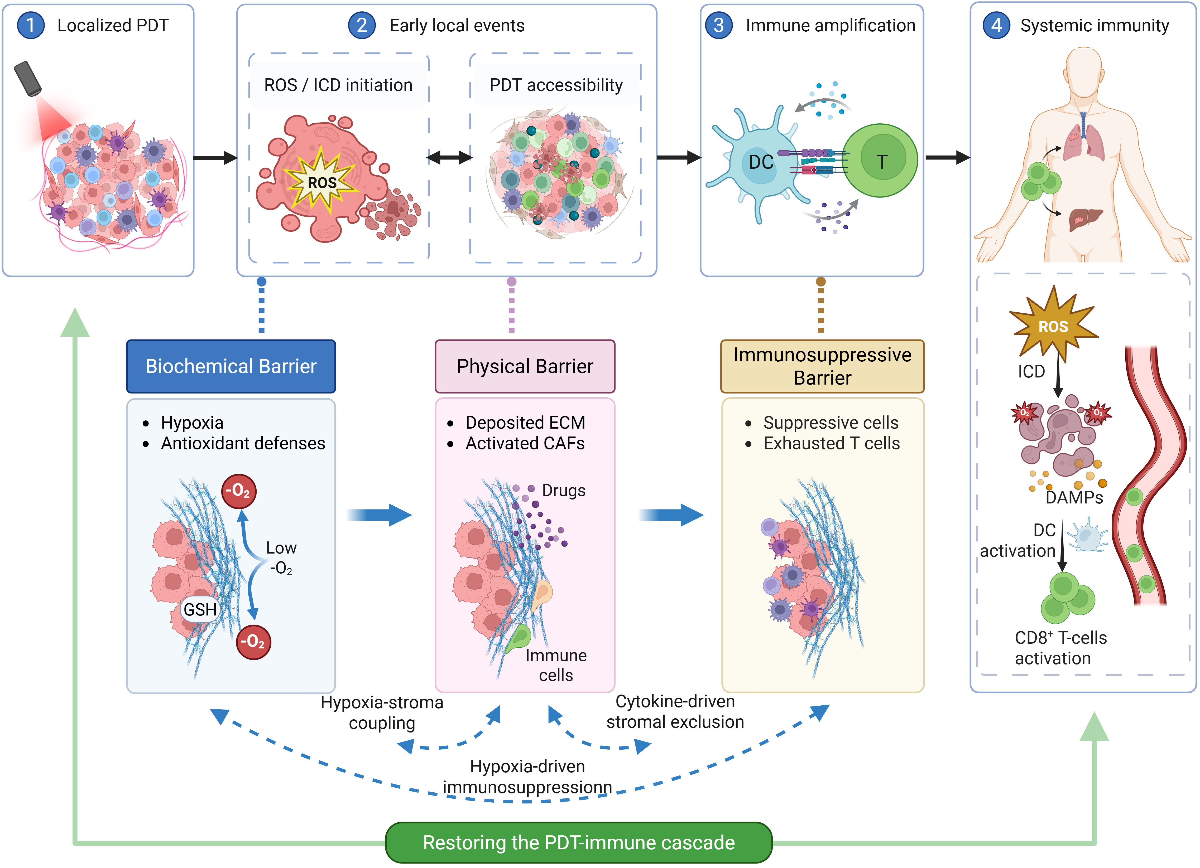
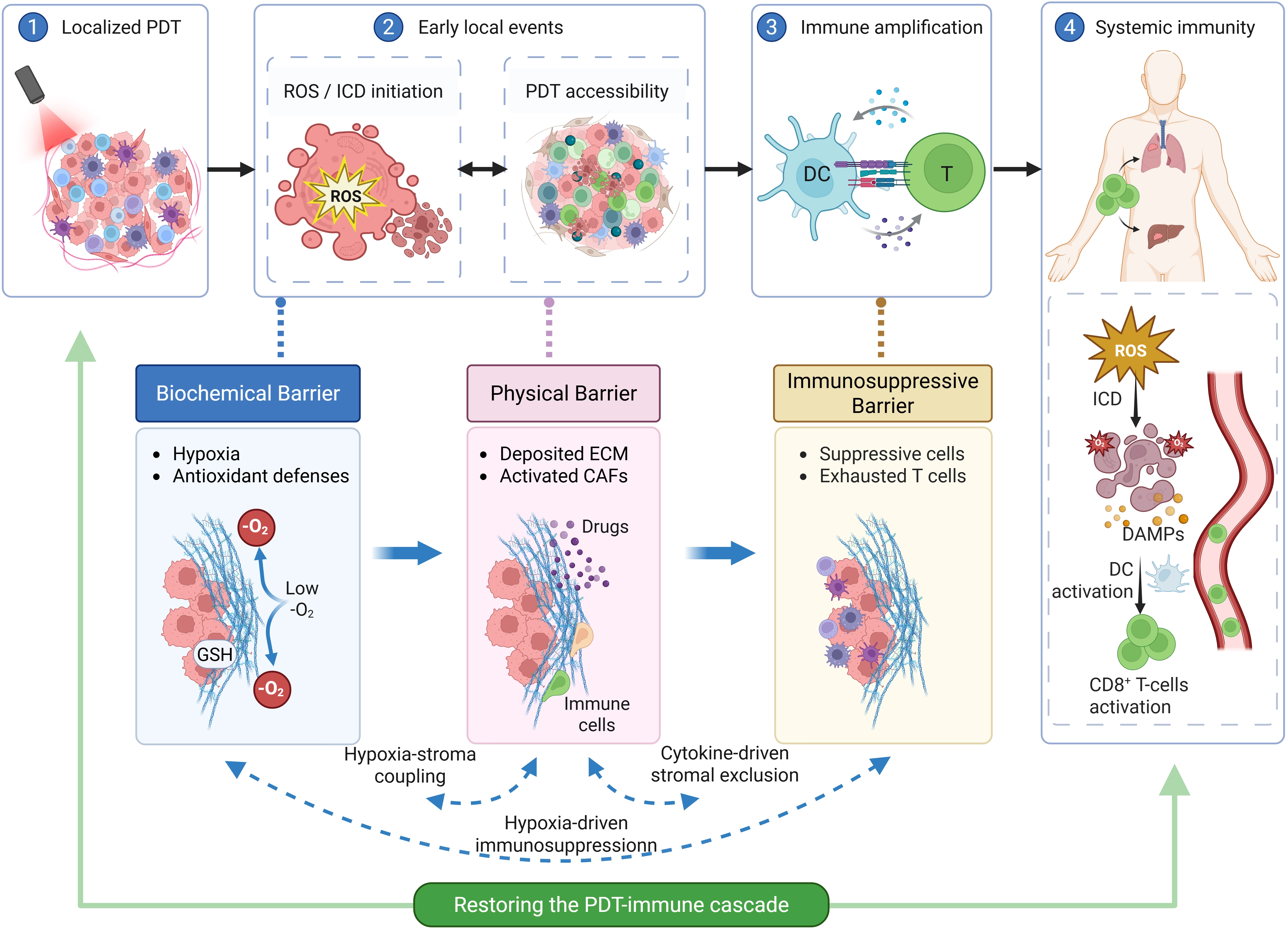
Scheme 1. Three interconnected TME barriers interrupting the PDT-immune cascade. The biochemical barrier limits ROS accumulation, the physical barrier restricts PDT accessibility, and the immunosuppressive barrier weakens antigen presentation and effector-cell activation. These barriers reinforce one another and limit the progression from local PDT to systemic antitumor immunity. Created in BioRender. Xue, L. (2026) https://BioRender.com/8b5h8gc. TME: tumor microenvironment; PDT: photodynamic therapy; ROS: reactive oxygen species; ICD: immunogenic cell death; DC: dendritic cell; GSH: glutathione; ECM: extracellular matrix; CAFs: cancer-associated fibroblasts; DAMPs: damage-associated molecular patterns.
2. Biochemical Barrier: Restoring PDT-ROS Efficacy
Effective PDT depends not only on the photochemical generation of ROS, but also on whether oxidative stress can accumulate long enough and reach a sufficient amplitude to induce ICD. In this sense, the biochemical barrier is not simply a problem of oxygen shortage; it represents a redox-threshold barrier that determines whether PDT-induced oxidative damage can be converted into immune activation. This barrier arises from a fundamental mismatch between the oxygen-dependent nature of type II PDT and the metabolic characteristics of solid tumors. Chronic hypoxia limits the generation efficiency of singlet oxygen (1O2) and promotes tumor adaptation to oxidative stress through hypoxia-inducible factor-related pathways. At the same time, antioxidant networks centered on GSH and heme oxygenase-1 (HO-1) rapidly scavenge the ROS generated during irradiation[13]. By coupling insufficient ROS production with excessive ROS clearance, these processes prevent oxidative stress from crossing the ICD threshold. Therefore, strategies for overcoming the biochemical barrier should not merely increase oxygen supply, but should coordinate oxygen metabolism remodeling with disruption of antioxidant defenses[14].
2.1 Regulating oxygen availability
In situ catalytic oxygen generation. In situ catalytic oxygen production is the most straightforward approach to alleviating the oxygen dependence of PDT. Tang et al. constructed Ir-Mn(II/III) coordination-assembled nanoparticles (NPs) in which high-valence manganese ions, released under lysosomal acidic conditions, exhibited catalase-like activity. These ions catalyzed the decomposition of excess intratumoral H2O2 to generate O2 in situ, effectively increasing the local oxygen concentration within the irradiated region and enhancing the 1O2 yield of type II PDT[15]. However, the efficacy of such catalytic oxygen supply strategies is constrained by the actual intratumoral H2O2 concentration, and the generated O2 may be metabolically consumed before reaching the PS excitation site.
Reducing oxygen consumption. Oxygen supplementation strategies alone may be offset by the high oxygen consumption rate within the TME. Reducing cellular oxygen consumption at the metabolic level is therefore a complementary approach. Qu et al. designed charge-reversible crosslinked NPs that suppressed oxidative phosphorylation through targeted disruption of tumor mitochondria, reducing cellular oxygen consumption and making more molecular oxygen available for 1O2 generation during PDT[16]. This strategy complemented in situ oxygen generation; while the latter increased supply, the former reduced consumption, thereby collectively enhancing the oxygen utilization efficiency of PDT.
2.2 Disrupting antioxidant defenses
Restoring ROS production addresses only one dimension of breaching the biochemical barrier. If the intracellular antioxidant system is not effectively suppressed, ROS generated by PDT will be rapidly quenched before reaching the oxidative damage threshold necessary to trigger ICD. Dismantling the antioxidant defense system of tumor cells is therefore a critical step in ensuring effective transduction of PDT-induced oxidative stress signals.
Direct GSH depletion. Direct depletion of intracellular GSH is a common approach for reducing the ROS-buffering capacity of tumor cells. Li et al. designed an H2O2-triggered self-immolative nanoassembly (Pyz/TPPS) that released quinone methide within tumor cells, irreversibly depleting GSH through nucleophilic addition reactions and weakening the intracellular buffering capacity against PDT-generated 1O2 and hydroxyl radicals[17]. Similarly, Huang et al. developed FeBMnDC NPs containing MnO2, ferritin, and the PS Ce6. MnO2 degradation and ferric ion reduction consumed intracellular GSH, thereby weakening antioxidant buffering during PDT[18].
GSH synthesis blockade. The rapid regeneration of intracellular GSH often limits the durability of direct depletion strategies. Tang et al. developed a prodrug nanoassembly (NOSP) with dual GSH and cysteine depletion capability. By consuming GSH synthesis precursors, NOSP blocked the GSH regeneration pathway at its source, achieving sustained collapse of antioxidant defenses. This prolonged the accumulation of PDT-generated ROS over an extended temporal window, enhancing the conversion efficiency from oxidative damage to ICD signaling[19].
Antioxidant enzyme downregulation. Besides GSH, tumor cells rely on antioxidant enzyme systems such as HO-1 to maintain redox homeostasis, forming a multi-layered buffer against PDT-induced stress. Zou et al. utilized ZnPP to specifically downregulate intratumoral HO-1 expression, disrupting the enzymatic tier of antioxidant defense. Simultaneously, quercetin was employed to remodel CAFs and alleviate tumor hypoxia. Together, these interventions achieved comprehensive amplification of PDT oxidative stress through combined suppression of antioxidant defense, enhanced 1O2 generation, and hypoxia mitigation[20].
2.3 Reducing oxygen dependence
The strategies discussed above optimize oxygen utilization efficiency within the framework of conventional type II PDT. A more radical solution is to shift toward the type I PDT mechanism, which exhibits substantially lower oxygen dependence. Type I PDT generates superoxide anion radicals (O2-) and hydroxyl radicals (•OH) through direct electron transfer between the PS and substrate, significantly reducing dependence on O2 concentration. This mechanism maintains effective ROS output even in severely hypoxic regions deep within solid tumors. Xu et al. developed NO-producing multifunctional NPs that integrated type I photodynamic processes with NO gas therapy, maintaining substantial ROS generation and ICD induction capacity under hypoxic conditions[21]. This approach reduces the oxygen dependence of PDT and may preserve ROS generation under hypoxic conditions.
2.4 Current limitations and mechanistic considerations
Although many approaches have improved PDT-associated ROS production in experimental models, the biochemical barrier remains difficult to control in a predictable way. One reason is that tumor redox status is highly heterogeneous. Hypoxia severity, GSH abundance, and antioxidant enzyme activity vary among tumor types, among patients, and even among different regions of the same tumor. These parameters may also change during treatment. As a result, increasing oxygen supply or consuming GSH at one time point does not necessarily maintain oxidative stress at a level sufficient for ICD throughout the tumor.
Another issue is how redox remodeling is evaluated. Many studies use increased oxygen content, reduced GSH levels, or enhanced ROS fluorescence as evidence of improved PDT efficacy. These readouts are useful, but they do not always show whether ROS is generated in the right region, persists for a sufficient duration, and reaches the threshold required to induce ICD. Excessive ROS amplification may also damage adjacent normal tissues, especially when the response is not tightly confined to the tumor. Thus, the key challenge is not simply to produce more ROS, but to reshape the tumor redox state in a controlled manner so that PDT-induced oxidative stress can be converted into ICD.
Overall, breaching the biochemical barrier requires a shift from transient ROS enhancement to sustained redox remodeling. Oxygen supplementation, reduced oxygen consumption, antioxidant depletion, inhibition of GSH regeneration, and type I PDT each address different parts of this problem (Scheme 2). Together, these strategies increase the likelihood that PDT-induced oxidative stress will cross the ICD threshold. However, even when ROS generation is restored, the therapeutic effect may remain restricted if PSs are mainly retained at the tumor periphery. The next barrier is therefore spatial: PSs, light, oxygen, and immune cells must be brought into the same tumor regions for PDT-immunotherapy to proceed effectively.
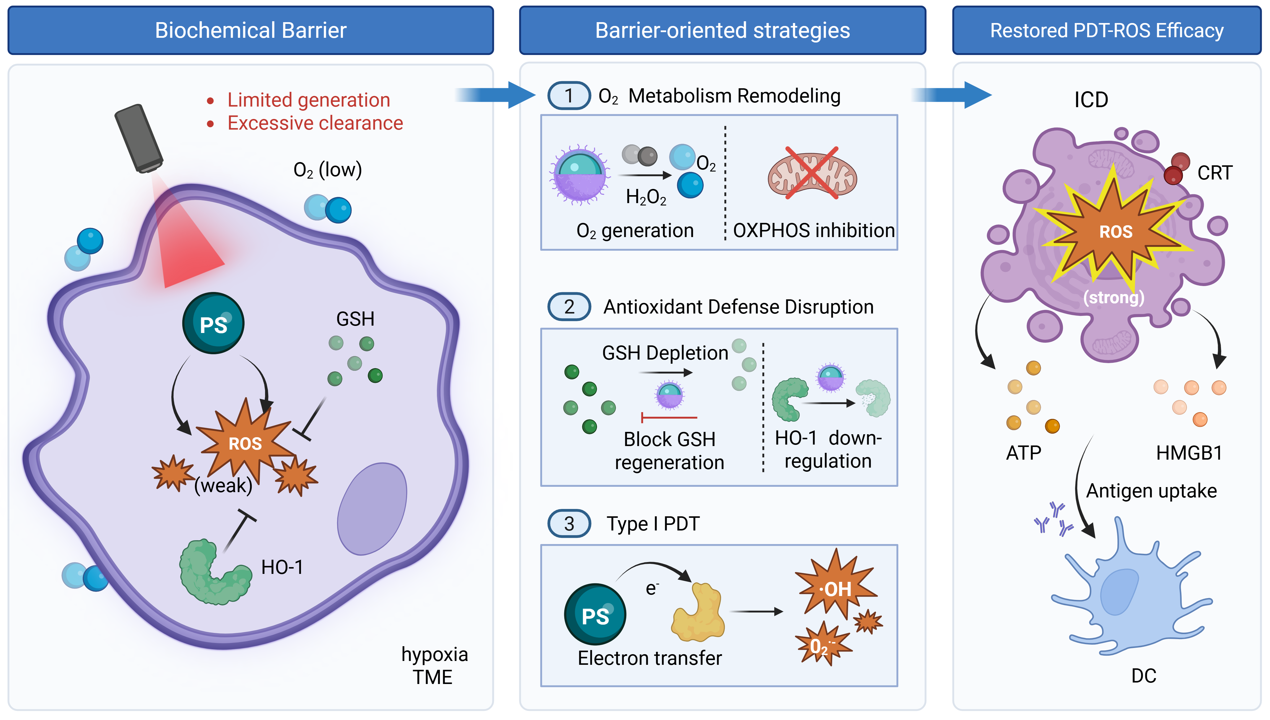
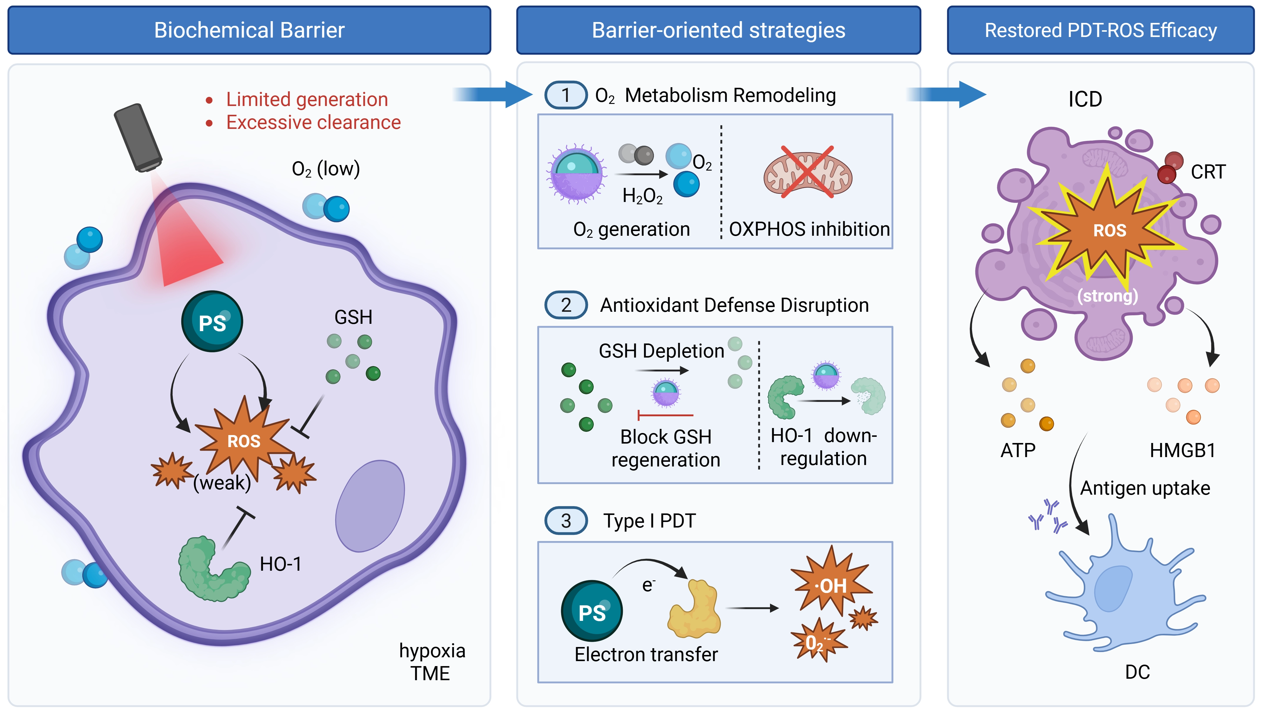
Scheme 2. Barrier-oriented strategies for restoring PDT-ROS efficacy. Hypoxia and antioxidant defenses limit ROS accumulation below the ICD threshold. Strategies that improve oxygen availability, disrupt GSH/HO-1-mediated antioxidant buffering, or shift PDT toward type I ROS generation can strengthen PDT-induced oxidative stress and support ICD initiation. Created in BioRender. Xue, L. (2026) https://BioRender.com/w5dys84. PDT: photodynamic therapy; ROS: reactive oxygen species; ICD: immunogenic cell death; PS: photosensitizer; GSH: glutathione; HO-1: heme oxygenase-1; CRT: calreticulin; ATP: adenosine triphosphate; HMGB1: high mobility group box 1; DC: dendritic cell; OXPHOS: oxidative phosphorylation.
3. Physical Barrier: Enhancing PDT Accessibility
The physical barrier limits where PDT can act within the tumor. For PDT, impaired delivery is not merely a question of how many PS molecules enter the tumor, but whether PSs, light, oxygen, ROS action zones, and immune cells can be spatially coordinated within the same therapeutic region. This barrier is primarily constructed by pathologically deposited ECM and activated CAFs. Activated CAFs continuously reinforce the fibrotic meshwork through excessive secretion and crosslinking of collagen, hyaluronic acid, and other stromal components[22,23]. As a result, the dense ECM impedes the uniform distribution of PSs into deep tumor regions, causing them to accumulate predominantly at the tumor periphery and in perivascular areas. Elevated interstitial fluid pressure further limits the permeation and retention of therapeutic agents within the tumor parenchyma[24]. Even when ROS is generated, its short lifetime and limited diffusion range, with singlet oxygen acting only within approximately 20-200 nm, restrict oxidative damage to regions close to the activated PS[25]. Meanwhile, immune-excluding stroma prevents effector cells from entering the tumor interior after PDT, interrupting the transmission of ICD-derived immune signals[26]. Therefore, breaching the physical barrier should be evaluated not only by improved PS accumulation, but also by enhanced PDT accessibility, expanded effective PDT coverage, better colocalization between PSs and irradiation, and deeper immune-cell infiltration.
3.1 Remodeling the tumor stroma
ECM degradation. To address the spatial constraints that the dense ECM imposes on PS distribution, the most direct strategy is to create matrix channels through enzymatic degradation or signal modulation, establishing physical conditions for deep PS penetration. Sun et al. developed a smart nanosystem that encapsulated hyaluronidase (HAase) within metal-polyphenol networks. This nanosystem hydrolyzed excessively deposited hyaluronic acid in situ, promoted uniform distribution of PSs into deep tumor regions, and expanded the effective PDT action zone covered by irradiation[27]. However, widespread ECM degradation may introduce risks of tumor metastasis. To mitigate this, Zhao et al. designed a biomimetic nanoplatform (FBFO@HM@aOPN) that precisely blocked the osteopontin (OPN) signaling pathway responsible for maintaining stromal homeostasis. Surface-conjugated pH-sensitive anti-OPN antibodies induced structural reorganization of the ECM rather than complete ablation. This approach promoted deep PS distribution while preserving stromal structural integrity, an advance from indiscriminate degradation toward precision remodeling[28].
CAF reprogramming. Even when the ECM is successfully remodeled, activated CAFs continue to rebuild the physical barrier, obstructing sustained entry of PSs and effector cells. Zou et al. utilized quercetin to downregulate fibrotic markers including Wnt16, FAP-alpha, and alpha-SMA, reprogramming CAFs from a pro-fibrotic phenotype toward a quiescent state. This suppressed sustained ECM production at its source[20]. This strategy holds dual significance for restoring PDT efficacy: on one hand, it alleviates ECM-imposed spatial constraints on PS distribution; on the other hand, it mitigates CAF-mediated exacerbation of hypoxia, indirectly improving the oxidative microenvironment for PDT.
3.2 Promoting deep tumor penetration and activation
Size/charge-adaptive PS penetration. Adaptive penetration strategies approach the problem from the nanocarrier design itself, enabling PS-carrying nanoplatforms to dynamically adjust their physicochemical properties in response to gradients within the intratumoral microenvironment. This promotes PS redistribution within tumors and improves PDT coverage. Cheng et al. developed a hierarchically engineered, self-adaptive nanoplatform (SAN) for glioblastoma. The SAN underwent size reduction from approximately 157 nm to 12 nm and charge reversal triggered by the acidic TME gradient. These changes enabled PSs to penetrate the dense ECM and reach deep tumor regions, better matching their distribution with external irradiation coverage and improving the spatial efficacy of PDT[29]. Xue et al. designed pPhD NPs using a similar two-stage delivery logic. The particles first achieved long circulation and tumor accumulation at a large size with a stealth surface. Upon tumor entry, polyethylene glycol shedding triggered size reduction to 8 nm and surface charge reversal, substantially extending the effective depth of PDT action[30].
Light-actuated penetration and local activation. Adaptive penetration strategies rely on endogenous TME stimuli, particularly pH gradients, to trigger morphological changes in carriers. Their response kinetics and controllability are limited by microenvironment heterogeneity. A fundamentally different strategy uses external energy to actively drive topological deformation of materials, extending PDT effects across larger spatial domains. Wu et al. used crystallization-driven self-assembly technology to design photoresponsive two-dimensional nanopatches (CPICG/Chol). These nanopatches underwent topological transformation from two-dimensional sheets to zero-dimensional spheres upon near-infrared (NIR) light activation[31]. The local mechanical stress generated by this dynamic deformation actively overcame interstitial resistance, propelling PSs into deep regions. The nanopatches also physically disrupted cancer cell membranes to directly induce antigen release, coupling physical penetration with PDT-induced immune signal generation within a single process. This strategy marks a shift from passively responding to microenvironment cues toward actively using light to coordinate penetration, local activation, and immune signal generation.
Extended activation for deep-seated lesions. Improved PS penetration does not by itself ensure effective PDT, because PSs that reach deep tumor regions must still be activated by sufficient light. Conventional external illumination is limited by tissue optical attenuation, which restricts PDT efficacy in deep-seated lesions. To address this limitation, X-ray-activated PDT has been explored to generate activating light within or near tumor tissues, most commonly through scintillating or radioluminescent systems that convert X-ray energy into optical emission capable of exciting PSs[32,33]. Internally activated PDT has also been developed using Cerenkov radiation, chemiluminescence, or bioluminescence as local light sources[34-36]. These strategies extend PDT accessibility from PS delivery to activation-energy delivery, although their efficacy still depends on energy-transfer efficiency, PS-emission matching, dose control, and safety.
3.3 Facilitating immune cell infiltration
Improving PS access addresses only one aspect of the physical barrier. The dense ECM and activated CAFs also restrict the entry of effector immune cells, making stromal remodeling relevant to both PDT coverage and subsequent immune activation. Qiu et al. developed an ECM-anchored photodynamic immunomodulator (FPC@S) that generated ROS locally to remodel the matrix and released SIS3 to suppress CAF activation. In fibrotic breast tumors, this treatment reduced stromal fibrosis and increased intratumoral CD8+ and CD4+ T-cell infiltration, as shown by immunofluorescence analysis[37]. Sun et al. used remotely released HAase to degrade intratumoral hyaluronic acid before combined photothermal therapy/PDT. The resulting matrix remodeling increased cytotoxic T-lymphocyte infiltration within the tumor and strengthened immune responses in tumor-draining lymph nodes[27]. Together, these studies show that selective stromal remodeling can improve effector-cell access and support the immune response initiated by photoinduced tumor damage.
3.4 Current limitations and mechanistic considerations
Current strategies have improved PS penetration and stromal remodeling, but physical-barrier modulation does not directly lead to stronger PDT. For PDT, delivery is useful only when PSs reach regions that can also receive sufficient light and oxygen. If the deeper tumor area remains hypoxic, poorly irradiated, or inaccessible to immune cells, increased penetration alone may have limited therapeutic value. This is particularly relevant for large or poorly vascularized tumors, where drug distribution, oxygen supply, and light exposure are often mismatched.
The physical barrier also has a dual role. Dense ECM and activated CAFs restrict drug diffusion and T-cell infiltration, but the stroma is not simply a structure to be removed. Excessive ECM degradation may cause tissue injury or facilitate tumor invasion, and broad CAF depletion may be problematic because CAF populations are highly heterogeneous. Some CAF subsets promote fibrosis and immune exclusion, whereas others may help restrain tumor spread. Therefore, physical-barrier remodeling needs to be more selective than simple matrix degradation or CAF elimination. A more appropriate goal is to normalize the stromal architecture enough to improve PS access and immune-cell entry without destroying the tissue constraints that may still limit tumor dissemination.
In summary, overcoming the physical barrier means improving PDT accessibility rather than merely increasing carrier penetration. ECM modulation, CAF regulation, and adaptive nanostructural changes can expand the region in which PSs and irradiation overlap, while also opening routes for effector immune-cell infiltration (Scheme 3). These changes help extend PDT effects beyond the tumor periphery. Nevertheless, improved access to the tumor interior is still not sufficient if the local immune environment remains suppressive. Once PDT-induced damage releases antigens and damage-associated molecular patterns (DAMPs), the resulting immune signals must still pass through the immunosuppressive TME. This makes immune microenvironment remodeling the next essential step.
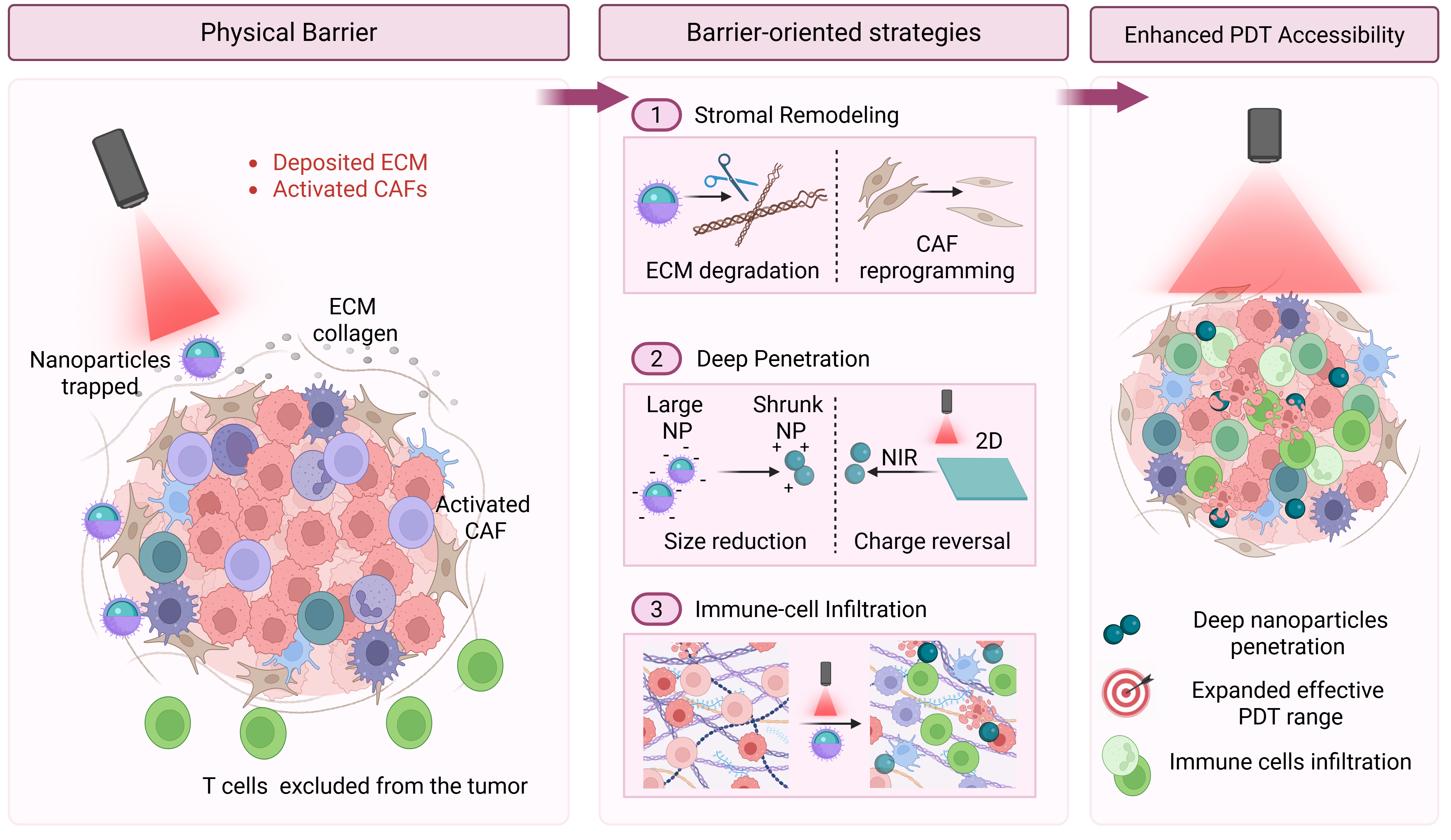
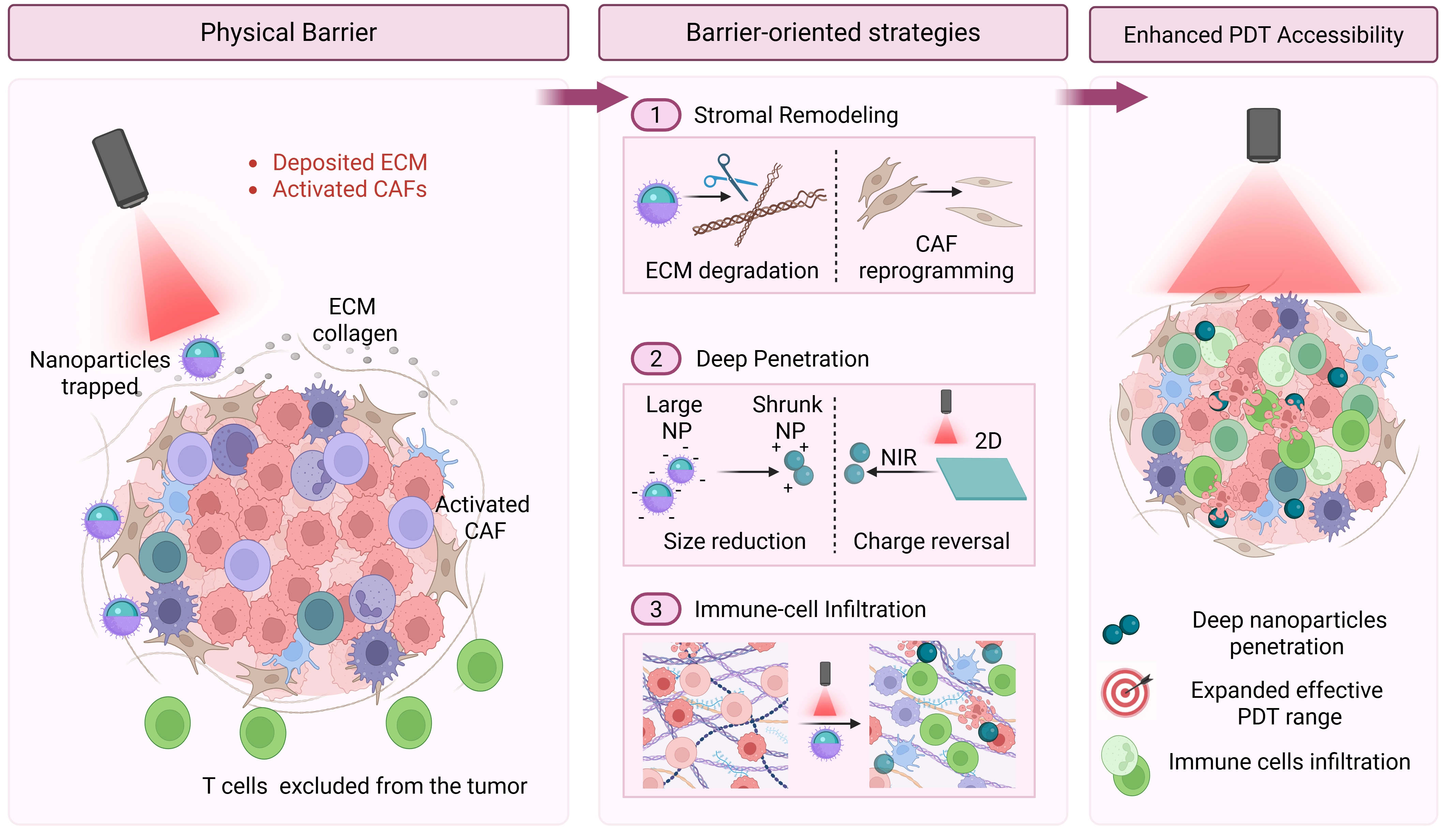
Scheme 3. Barrier-oriented strategies for improving PDT accessibility. Dense ECM and activated CAFs limit both photosensitizer penetration and immune-cell entry. Stromal remodeling, CAF regulation, and adaptive carriers help expand the effective PDT region and improve immune-cell infiltration. Created in BioRender. Xue, L. (2026) https://BioRender.com/vt53ufj. PDT: photodynamic therapy; ECM: extracellular matrix; CAFs: cancer-associated fibroblasts; NP: nanoparticle; NIR: near-infrared.
4. Immunosuppressive Barrier: Reprogramming the Immune Microenvironment
Even after ROS efficacy is restored and PSs reach deeper tumor regions, PDT-induced immune activation can still be extinguished by the immunosuppressive network within the TME. This barrier is not a collection of isolated suppressive cell types, but an interacting immunosuppressive network in which TAMs, Tregs, MDSCs, immune checkpoints, and dysfunctional antigen-presenting cells cooperate to neutralize ICD-derived signals[38,39]. M2-like TAMs secrete immunosuppressive cytokines such as IL-10 and TGF-β, directly limiting effector cell activation[40,41]. Tregs suppress CD8+ T cell function through IL-2 deprivation and contact-dependent inhibition[42]. MDSCs further restrict T cell proliferation through arginase activity, reactive nitrogen/oxygen species, and impaired DC function[43]. These suppressive components are spatially and functionally intertwined, meaning that PDT-induced antigen release and DAMP exposure may fail to progress into effective T cell priming and systemic immune propagation. Therefore, strategies targeting the immunosuppressive barrier must do more than induce ICD; they should reshape the immune context in which ICD signals are received, amplified, and transmitted.
4.1 Relieving immunosuppression
TAM reprogramming. TAMs are the most abundant and phenotypically plastic immune cell population within the TME, and their M2-like to M1-like phenotypic reversal is crucial for breaking post-PDT immunosuppression. Qu et al. designed a pure drug self-assembled nanoprodrug (PARE NPs) featuring covalent conjugation of a PS with a Toll-like receptor 7/8 (TLR7/8) agonist (R848). This design enabled esterase-triggered release of R848 to drive M1 polarization of TAMs concurrent with PDT-mediated tumor killing, ensuring that PDT-induced ICD signals and immune activation occurred within the same spatiotemporal window and preventing suppressive TAMs from neutralizing initial immune signals[44]. Zhao et al. approached the problem from the perspective of endoplasmic reticulum (ER) stress modulation, constructing an adaptively transformable nanoplatform (G@MDHP) whose outer shell carried PSs into tumor cells to amplify ER stress levels and induce ICD, while simultaneously reprogramming TAM polarization to achieve dual-pronged immune remodeling[45]. Chang et al. designed spatiotemporally controlled nanomicelles (IR825@HRG) that precisely matched TAM reprogramming with PDT-ICD within a coordinated temporal window, illustrating the principle of temporal synergy[46].
Tregs functional attenuation. As a core suppressive population that contributes to impaired effector T-cell activity after PDT, Tregs are a critical intervention target. Shanmugam et al. developed a 2,240 nm NIR-IV PDT system to investigate the unique immunomodulatory effects of ultra-long-wavelength PDT. The study found that this PDT regimen selectively weakened Treg-mediated suppression and sensitized tumors to anti-OX40 treatment. This finding suggests that specific PDT parameters may themselves reshape local immune suppression and improve responsiveness to immune stimulation[47].
MDSCs functional suppression. MDSCs form a suppressive axis independent of TAMs and Tregs and are another key target for restoring post-PDT immune function. Chen et al. combined Ce6 derivative-mediated PDT with stimulator of interferon genes activation and LAG3 blockade to relieve MDSC-associated suppression after PDT. This combination reduced nitric oxide and arginase-related suppressive activity, thereby supporting post-PDT T-cell proliferation and expansion[48].
PD-L1 degradation and relief of adaptive resistance. Treatment-induced PD-L1 expression can weaken T-cell activity and limit the persistence of PDT-induced immune responses. Liu et al. developed a PD-L1-targeted photodegradation chimera, PPA-VPF, by conjugating verteporfin to a PD-L1-binding peptide. Upon irradiation, singlet oxygen generated near PD-L1 promoted its degradation, whereas singlet oxygen generated away from the binding site induced ICD. This design combined PDT-mediated tumor-cell damage with local relief of checkpoint-mediated suppression. In immune-cold tumor models, PPA-VPF enhanced antitumor immunity and inhibited both primary and distant tumor growth[49].
4.2 Promoting antigen presentation and effector activation
DC maturation and antigen presentation. Xue et al. constructed a pure drug self-assembled nanoplatform (PDR NPs) that integrated a PS, doxorubicin, and a TLR7 agonist. By simultaneously activating the PDT, cyclic GMP-AMP synthase-stimulator of interferon genes (cGAS-STING), and TLR pathways, this platform achieved markedly enhanced DC maturation rates[50]. Wang et al. developed a dual-wavelength NIR light-activated upconversion nanodevice (PCpG/UCNP@mSiO2-RB-HA) that separately triggered ROS generation and CpG adjuvant release, achieving spatiotemporal matching of antigen and adjuvant at the DC level[51]. Xiao et al. designed nanodrugs co-loaded with Ce6 and doxycycline (CD-NDs) that restored surface expression of major histocompatibility complex class I (MHC-I) through autophagy inhibition and prevented tumor cell self-clearance of DAMPs. This approach enabled complete transmission of PDT-induced antigen signals to DCs and improved subsequent T-cell recognition[52].
Effector cell activation and systemic immune propagation. Beyond antigen presentation, several studies further enhanced propagation of PDT-induced immune responses toward effector cell activation. Liu et al. demonstrated that PDT-induced DNA damage upregulated tumor surface MICA/B ligands through the ATM/SMAD1 signaling pathway, engaging natural killer cell NKG2D receptors to activate their cytotoxic function and providing an alternative killing pathway for MHC-I-deficient immune escape[53]. Chen et al. utilized a manganese-coordinated nanoplatform (Mn-MC NPs) in which Mn2+ agonism of the cGAS-STING pathway enhanced type I interferon secretion and promoted distal DC maturation, facilitating propagation of local PDT-induced immune signals into systemic immune responses[54].
4.3 Current limitations and mechanistic considerations
The immunosuppressive barrier is difficult to remodel because its components often compensate for one another. TAMs, Tregs, MDSCs, dysfunctional DCs, and checkpoint pathways do not act as independent targets. Suppressing one population may produce a short-term immune response, but other suppressive mechanisms can emerge and limit the durability of the effect. PDT-induced inflammation itself may also create selective pressure for adaptive resistance, such as PD-L1 upregulation, recruitment of suppressive myeloid cells, or increased production of anti-inflammatory cytokines.
Another limitation is that immune remodeling is often judged by a small number of endpoint markers. Increased CD8+ T-cell infiltration, reduced M2-like TAM markers, or elevated inflammatory cytokines suggest immune activation, but they do not necessarily prove that the immune response is systemic or durable. More convincing evidence would include antigen-specific T-cell priming, DC activation in draining lymph nodes, memory T-cell formation, suppression of distant tumors, and protection after tumor rechallenge. At the same time, stronger immune activation is not always better, because excessive stimulation may increase inflammatory toxicity. Future designs therefore need to balance immune activation with immune control, rather than simply maximizing pro-inflammatory signals.
In summary, overcoming the immunosuppressive barrier requires more than removing one suppressive cell type. The key is to create an immune context in which PDT-induced ICD signals can be recognized, amplified, and transmitted. Barrier-oriented strategies, including smart nanoplatforms, can help coordinate tumor-cell killing with antigen presentation and immune activation within the same therapeutic window (Scheme 4). Only under these conditions can local PDT-induced immune activation progress toward systemic antitumor immunity.
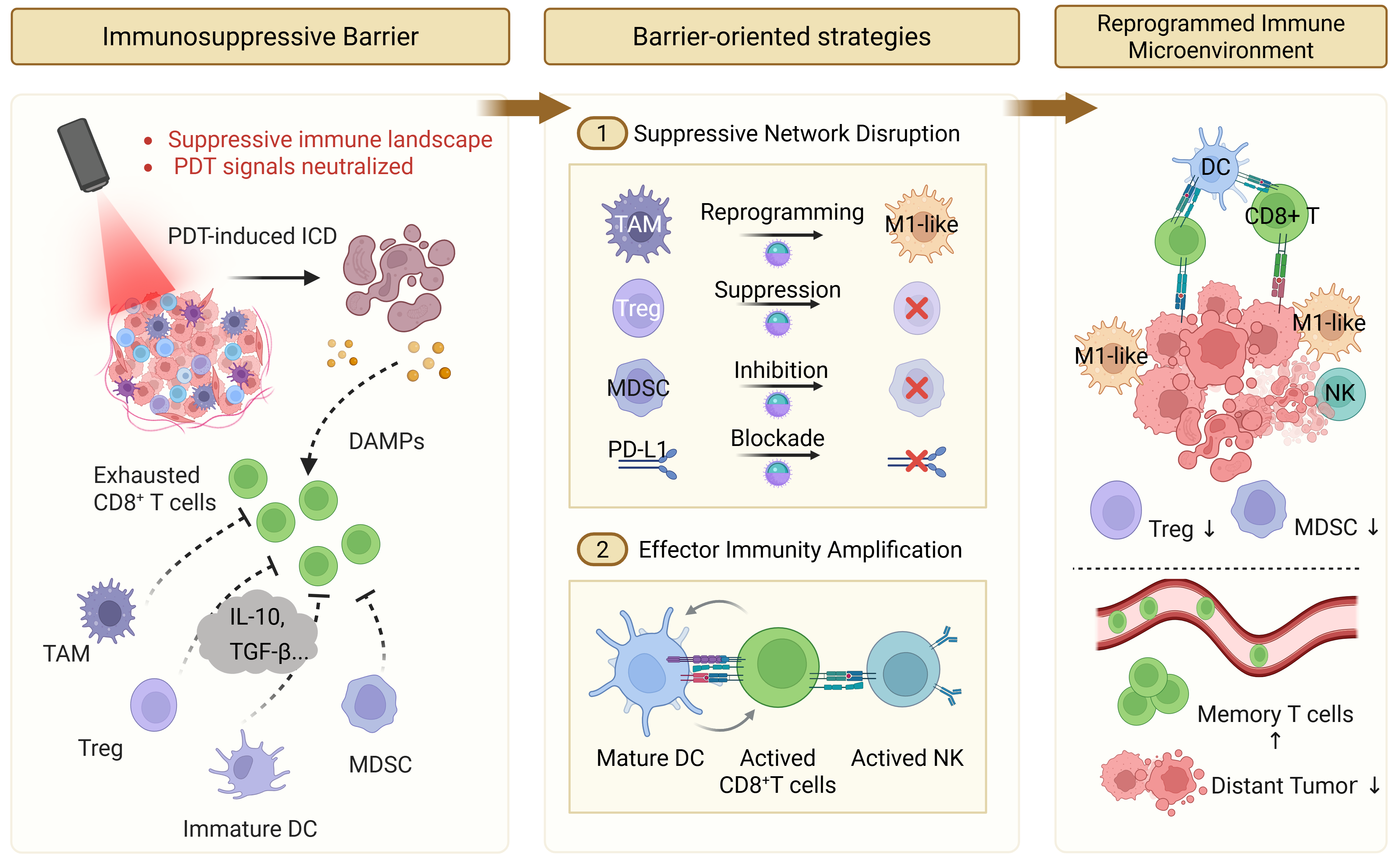
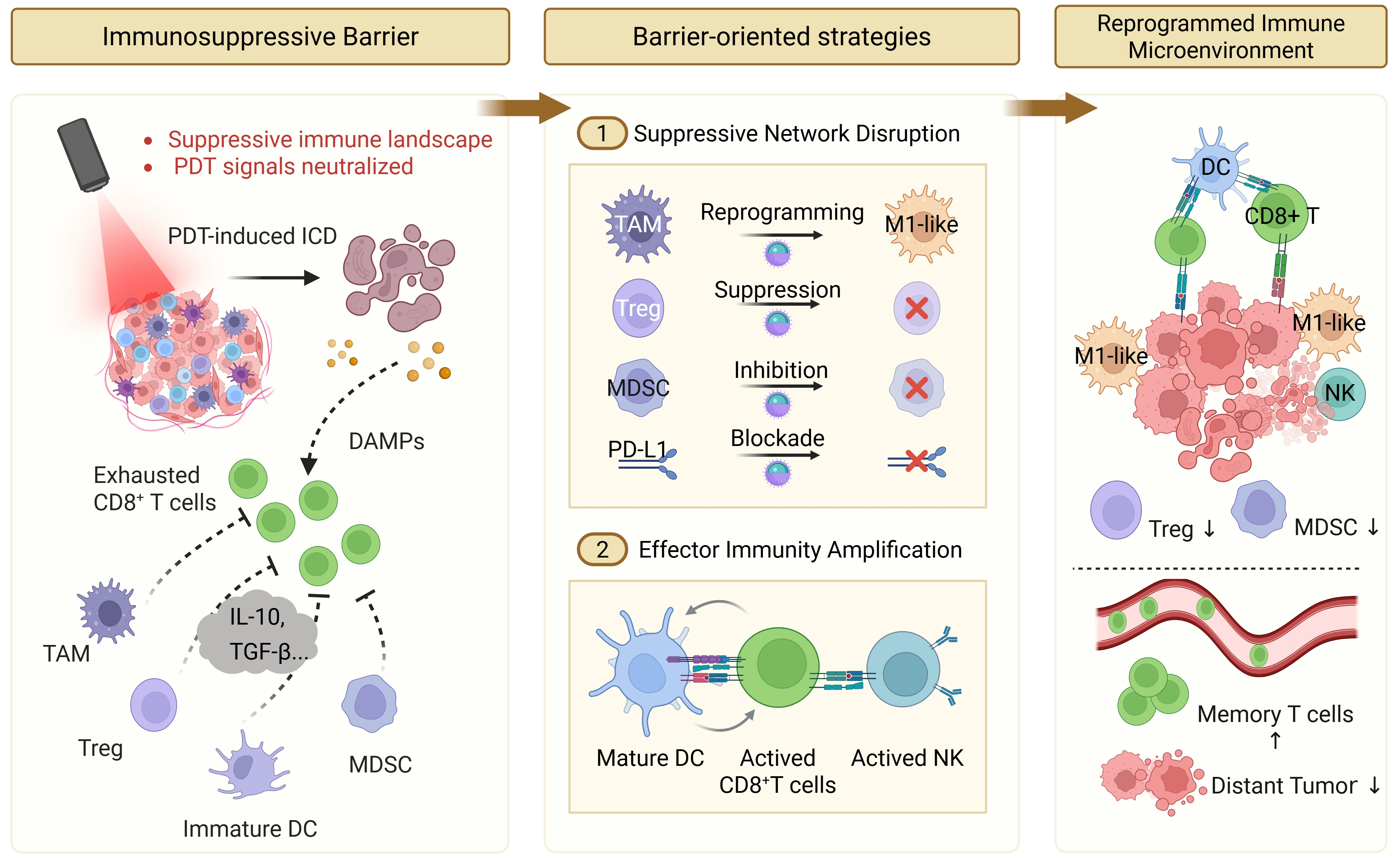
Scheme 4. Barrier-oriented strategies for reprogramming the immunosuppressive microenvironment. Relieving TAM-, Treg-, MDSC-, and checkpoint-mediated suppression can improve antigen presentation and effector-cell activation after PDT. Created in BioRender. Xue, L. (2026) https://BioRender.com/cn00bnb. TAM: tumor-associated macrophage; MDSC: myeloid-derived suppressor cell; PDT: photodynamic therapy; ICD: immunogenic cell death; DAMPs: damage-associated molecular patterns; IL-10: interleukin-10; TGF: transforming growth factor; DC: dendritic cell; PD-L1: programmed death-ligand 1; NK: natural killer; Treg: regulatory T cell.
5. From Localized PDT to Systemic Antitumor Immunity
The preceding sections have discussed how biochemical, physical, and immunosuppressive barriers interrupt different steps of PDT-immunotherapy (Table 1). These barriers ultimately determine whether local PDT-induced damage can be converted into systemic antitumor immunity. This conversion is not an automatic consequence of tumor ablation. It requires a coordinated sequence of immunological events, including ICD induction, antigen uptake by DCs, antigen transport to tumor-draining lymph nodes, T-cell priming and expansion, systemic trafficking of effector cells, and the establishment of immunological memory. Interruption of any step may explain why many PDT-based strategies suppress irradiated tumors but fail to control distant lesions or prevent recurrence.
Table 1. Summary of TME barriers, their interruption points in the PDT-immune cascade, and representative barrier-oriented strategies.
| Barrier Type | Interruption point in the PDT-immune cascade | Representative barrier-oriented strategies | Relevance to systemic antitumor immunity |
| Biochemical Barrier(Hypoxia & high reductive environment) | Signal Generation Blockade: Restricts effective ROS accumulation, hindering the initiation of PDT-induced ICD. | In situ catalytic O2 generation; reduced O2 consumption; type I PDT; GSH depletion; antioxidant enzyme inhibition. | Enhances ROS generation and oxidative stress, promotes DAMP release and antigen exposure. |
| Physical Barrier(Dense ECM & CAFs) | Spatial Propagation Restriction: Impedes deep photosensitizer delivery and immune cell infiltration, confining PDT effects to the tumor periphery. | ECM remodeling; CAF regulation; adaptive size/charge transformation; light-actuated penetration; extended activation strategies. | Expands the effective PDT region, improves intratumoral drug distribution and T-cell accessibility. |
| Immunosuppressive Barrier(TAMs, Tregs, MDSCs, etc.) | Signal Neutralization: Neutralizes immune activation signals induced by PDT. | TAM reprogramming; STING activation; immune checkpoint modulation; NK cell activation; Treg/MDSC attenuation. | Promotes DC maturation and T-cell activation, enhances the abscopal effect and establishment of immune memory. |
TME: tumor microenvironment; PDT: photodynamic therapy; ROS: reactive oxygen species; ICD: immunogenic cell death; GSH: glutathione; DAMP: damage-associated molecular pattern; ECM: extracellular matrix; CAFs: cancer-associated fibroblasts; TAM: tumor-associated macrophage; MDSCs: myeloid-derived suppressor cells; STING: stimulator of interferon genes; NK: natural killer; Treg: regulatory T cell.
The first step is the induction of sufficiently immunogenic tumor cell damage. When PDT-generated ROS reaches the threshold required for ICD, dying tumor cells expose or release DAMPs, including calreticulin, HMGB1, and adenosine triphosphate (ATP), together with tumor-associated antigens[55,56]. These signals provide the basis for subsequent antigen presentation and T-cell activation. However, the magnitude and spatial distribution of ICD are strongly shaped by the three barriers discussed above. If the biochemical barrier restricts ROS accumulation, ICD may be incomplete. If the physical barrier confines PSs to perivascular or peripheral tumor regions, antigen release may be spatially limited and may not reflect intratumoral heterogeneity. If local immunosuppression persists, antigen uptake and presentation may occur under tolerogenic conditions. Therefore, local ICD is necessary but not sufficient for systemic immunity; its downstream effect depends on whether the TME allows ICD-derived signals to be captured and amplified. This requirement is also reflected in recent work on photoactivated nanovaccines, in which light-responsive nanomaterials are used to coordinate antigen release, adjuvant activation, and antigen presentation. These studies support the view that photoinduced tumor damage must be coupled to organized immune priming before it can contribute to systemic antitumor immunity[57].
After PDT-induced ICD, DCs must take up tumor antigens and migrate to tumor-draining lymph nodes, where cross-presentation and cross-priming of CD8+ T cells occur. This step in the tumor-draining lymph node is essential because it links local tumor damage to systemic cellular immunity. Productive priming requires not only antigen availability, but also DC maturation, costimulatory signaling, and an inflammatory cytokine environment that supports expansion of tumor antigen-specific CD8+ T cells. Conversely, dysfunctional DCs, suppressive myeloid cells, Tregs, or checkpoint signaling can limit T-cell priming even when antigen release at the tumor site is substantial. Tumor-draining lymph nodes should therefore be treated as a critical site of immune priming rather than a passive downstream compartment.
Once primed, tumor antigen-specific effector T cells can enter systemic circulation and infiltrate both irradiated and non-irradiated lesions. This provides the immunological basis for the abscopal effect. In this context, distant tumor suppression should be viewed as a functional consequence of successful antigen presentation, T-cell priming, and effector-cell trafficking, rather than as a direct extension of local phototoxicity. Durable immune control further requires the generation of memory T-cell populations. Effector-to-memory transition supports long-term immune surveillance and may provide protection against tumor rechallenge. Therefore, durable systemic antitumor immunity depends not only on the acute inflammatory response after PDT, but also on whether PDT-induced antigenic stimulation leads to sustained tumor-reactive T-cell responses.
A major limitation in current PDT-immunotherapy studies is that systemic immunity is often inferred from local immune markers. Increased CD8+ T-cell infiltration in the treated tumor, elevated inflammatory cytokines, or reduced immunosuppressive cell populations indicate local immune activation, but they do not necessarily demonstrate systemic or durable antitumor immunity. More rigorous evidence would include suppression of non-irradiated tumors, antigen-specific CD8+ T-cell responses, DC activation in tumor-draining lymph nodes, memory T-cell formation, immune remodeling in spleen or lymph nodes, and protection in tumor rechallenge models. These endpoints are particularly important for distinguishing transient immune stimulation from durable systemic immunity.
Overall, the systemic outcome of PDT-immunotherapy depends on whether the three TME barriers are relieved in a way that preserves the continuity of the immune cascade. The biochemical barrier determines whether PDT produces sufficient ICD-inducing oxidative stress; the physical barrier determines whether this damage is spatially broad enough to support antigen release and immune-cell access; and the immunosuppressive barrier determines whether antigen presentation and effector responses are amplified or suppressed. Only when these steps are connected can local PDT progress toward abscopal tumor control and long-term immune memory.
6. Conclusions and Perspectives
PDT can initiate strong local oxidative damage, but this does not automatically lead to durable systemic antitumor immunity. The outcome depends on whether ROS accumulation reaches the threshold required for ICD, whether PSs, light, oxygen, and immune cells are spatially matched within the tumor, and whether antigen presentation and T-cell activation can persist in an immunosuppressive environment. In this review, we have organized these obstacles into three interconnected layers: the biochemical barrier, the physical barrier, and the immunosuppressive barrier. This organization helps explain why many PDT-based treatments produce marked local tumor damage but only limited abscopal effects or long-term immune protection.
Future PDT-immunotherapy strategies should therefore be judged by whether they maintain the continuity of the PDT-immune cascade. Improving PS delivery alone is unlikely to be sufficient if hypoxia and antioxidant buffering still prevent ROS accumulation. Enhancing ROS production may also be insufficient if dense stroma restricts antigen-presenting cells or effector T cells. Similarly, immune checkpoint blockade may have limited benefit if PDT fails to generate enough antigenic and danger signals in the first place. The central question is not how many barriers a system claims to target, but whether each intervention supports the next step: ROS generation, ICD induction, dendritic-cell activation, tumor-draining lymph node priming, systemic T-cell trafficking, and memory formation. This favors simpler systems whose individual contributions can be tested. Many current nanoplatforms combine oxygen delivery, GSH depletion, stromal remodeling, PS transport, immune agonists, and checkpoint modulation within one formulation. Such designs can be valuable for proof-of-concept studies, but they often make it difficult to determine which component is responsible for the therapeutic effect. Each added function may also increase formulation heterogeneity, manufacturing difficulty, safety uncertainty, and regulatory burden. A more practical direction is to start from the limiting step in a given tumor model and retain only the modules needed to relieve that bottleneck. In this sense, simplified formulation and mechanism-driven design should be considered together. Clear attribution of each component’s contribution will also be important for translation.
Conventional PDT already has a clinical basis, but its success is closely tied to lesion accessibility, illumination route, PS behavior, and local disease setting[58]. Recent clinical studies follow the same logic. For example, interim results from the ongoing phase 3 ENLIGHTED study have evaluated padeliporfin vascular-targeted PDT for low-grade upper tract urothelial cancer, with endoscopic light delivery incorporated into the treatment procedure[59]. In cervical high-grade squamous intraepithelial lesions, a multicenter randomized controlled trial showed that PDT improved histological regression and HPV clearance compared with placebo control, providing randomized evidence for PDT in light-accessible premalignant disease[60]. Together, these studies show that PDT translation is not determined only by PS potency. It also depends on whether drug accumulation, lesion accessibility, light delivery, PS-light matching, and local toxicity can be controlled in the same clinical setting. This clinical boundary also explains the relevance of comparing PDT with sonodynamic therapy. SDT benefits from deeper ultrasound penetration and is attractive for deep-seated lesions, whereas PDT offers more direct spatial control, more established PS-light matching, and a clearer photochemical link between ROS generation and ICD[58,61,62]. The main limitation of PDT remains optical access, which has motivated the development of X-ray-activated and internally activated PDT as extended activation strategies for less accessible lesions.
For PDT-immunotherapy, regression of the illuminated tumor should not be the only criterion for efficacy. Future studies should combine pharmacokinetic, dosimetric, and safety data with immune readouts beyond the treated lesion, including tumor-draining lymph node activation, antigen-specific CD8+ T-cell responses, non-irradiated tumor control, and memory or rechallenge assays. Such evidence is necessary to support claims of durable systemic antitumor immunity.
Overall, overcoming the three-layered TME barriers requires more than adding functions to PDT systems. The more important task is to identify where the PDT-immune cascade fails and to intervene at that point with a controllable and interpretable design. Systems that generate sufficient ROS, preserve ICD-derived signals, permit immune-cell entry, and support systemic T-cell responses are more likely to convert local phototoxicity into durable antitumor immunity. Future work should prioritize systems that are mechanistically attributable, manufacturable, and able to link local PDT with verified systemic immune responses.
Acknowledgements
The authors used Gemini 3.1 and ChatGPT 5.5 for language editing and the preliminary organization of conceptual illustrations. All literature selection, scientific interpretation, and final manuscript content were independently reviewed and approved by the authors. The authors take full responsibility for the integrity, originality, and accuracy of the work.
Authors contribution
Pan Y: Conceptualization, investigation, writing-original draft.
Xue X: Funding acquisition, writing-review & editing.
Conflicts of interest
Xiangdong Xue is a Junior Executive Editor of BME Horizon. Yuqing Pan declares no conflicts of interest.
Ethical approval
Not applicable.
Consent to participate
Not applicable.
Consent for publication
Not applicable.
Availability of data and materials
Not applicable.
Funding
The work was supported by the National Natural Science Foundation of China (Grant Nos 32571603 and 82172084).
Copyright
© The Author(s) 2026.
References
-
4. Kroemer G, Galassi C, Zitvogel L, Galluzzi L. Immunogenic cell stress and death. Nat Immunol. 2022;23(4):487-500.[DOI]
-
8. Bejarano L, Jordāo MJC, Joyce JA. Therapeutic targeting of the tumor microenvironment. Cancer Discov. 2021;11(4):933-959.[DOI]
-
10. Mitchell MJ, Billingsley MM, Haley RM, Wechsler ME, Peppas NA, Langer R. Engineering precision nanoparticles for drug delivery. Nat Rev Drug Discov. 2021;20(2):101-124.[DOI]
-
16. Qu H, Chen H, Cheng W, Pan Y, Duan Z, Wang Y, et al. Charge-reversible crosslinked nanoparticle for pro-apoptotic peptide delivery and synergistic photodynamic cancer therapy. Nano Res. 2023;16(12):13267-13282.[DOI]
-
17. Li J, Pang E, Debnath S, Hong J, Qiu A, Kim D, et al. Self-immolative activatable nanoassembly toward immuno-photodynamic therapy in TME. Adv Funct Mater. 2026;36(14):e13250.[DOI]
-
18. Huang X, Wang L, Guan Q, Chen L, Qi H, Wang H, et al. Self-amplifying nanomedicine reprograms redox metabolism to trigger immunogenic ferroptosis in colon cancer: Multiomics identifies AMPD3 as a novel regulator. Biomaterials. 2026;333:124196.[DOI]
-
19. Tang N, Tong F, Wu P, Xie X, Wang L, Yu M, et al. Glutathione/cysteine dual-consuming prodrug nanoassemblies for potentiated gas-photodynamic therapy. Adv Funct Mater. 2025;35(37):2503604.[DOI]
-
26. Yang C, McAndrews KM, Kalluri R. Clinical and therapeutic relevance of cancer-associated fibroblasts. Nat Rev Clin Oncol. 2021;18(12):792-804.[DOI]
-
38. DeNardo DG, Ruffell B. Macrophages as regulators of tumour immunity and immunotherapy. Nat Rev Immunol. 2019;19(6):369-382.[DOI]
-
43. Li K, Shi H, Zhang B, Ou X, Ma Q, Chen Y, et al. Myeloid-derived suppressor cells as immunosuppressive regulators and therapeutic targets in cancer. Sig Transduct Target Ther. 2021;6:362.[DOI]
-
47. Shanmugam M, Chiang CS, Hwang KC. 2240 nm NIR-IV photodynamic therapy can reverse ineffective anti-OX40 cancer immunotherapy to become effective. ACS Nano. 2025;19(41):36129-36147.[DOI]
-
57. Ding Q, Chen S, Hua S, Yoo J, Yoon C, Li Z, et al. Photoactivated nanovaccines. Chem Soc Rev. 2025;54(21):9807-9848.[DOI]
-
58. Cai Y, Chai T, Nguyen W, Liu J, Xiao E, Ran X, et al. Phototherapy in cancer treatment: Strategies and challenges. Sig Transduct Target Ther. 2025;10:115.[DOI]
-
59. Margulis V, Kaufman RP, Marcq G, Shvero A, Shore ND, Psutka SP, et al. ENLIGHTED phase 3 study: Interim results of efficacy and safety of padeliporfin vascular targeted photodynamic therapy (VTP) in the treatment of low-grade upper tract urothelial cancer (LG UTUC). J Clin Oncol. 2025;43(17):LBA4513.[DOI]
Copyright
© The Author(s) 2026. This is an Open Access article licensed under a Creative Commons Attribution 4.0 International License (https://creativecommons.org/licenses/by/4.0/), which permits unrestricted use, sharing, adaptation, distribution and reproduction in any medium or format, for any purpose, even commercially, as long as you give appropriate credit to the original author(s) and the source, provide a link to the Creative Commons license, and indicate if changes were made.
Publisher’s Note
Science Exploration remains a neutral stance on jurisdictional claims in published
maps
and institutional affiliations. The views expressed in this article are solely those
of
the author(s) and do not reflect the opinions of the Editors or the publisher.
Share And Cite


Science Exploration Style
Pan Y, Xue X. Overcoming three-layered tumor microenvironmental barriers to photodynamic therapy-induced systemic antitumor immunity. BME Horiz. 2026;4:202624. https://doi.org/10.70401/bmeh.2026.0032
Tips
Copy completed.
Submit a Manuscript
Author Instructions
Cite this Article
Article Metrics
0
View
0
Download
Cited
Article Updates

- Abstract
- Keywords
- 1. Introduction
- 2. Biochemical Barrier: Restoring PDT-ROS Efficacy
- 3. Physical Barrier: Enhancing PDT Accessibility
- 4. Immunosuppressive Barrier: Reprogramming the Immune Microenvironment
- 5. From Localized PDT to Systemic Antitumor Immunity
- 6. Conclusions and Perspectives
- Acknowledgements
- Authors contribution
- Conflicts of interest
- Ethical approval
- Consent to participate
- Consent for publication
- Availability of data and materials
- Funding
- References
- Copyright
 Get citation
Science Exploration Style

Pan Y, Xue X. Overcoming three-layered tumor microenvironmental barriers to photodynamic therapy-induced systemic antitumor immunity. BME Horiz. 2026;4:202624. https://doi.org/10.70401/bmeh.2026.0032
copy
Share Link
copy