Targeting the biofilm microenvironment: Advances in stimuli-responsive antibacterial coating strategies for implant-associated infections

*Correspondence to:
Wei Wang, Department of Orthopedic Surgery, The First Affiliated Hospital of Soochow University, Suzhou 215006, Jiangsu, China.
E-mail: gkww1995@163.com
Dechun Geng, Department of Orthopedic Surgery, The First Affiliated Hospital of Soochow University, Suzhou 215006, Jiangsu, China. E-mail: szgengdc@163.com
Jun Zhou, Department of Orthopedic Surgery, The First Affiliated Hospital of Soochow University, Suzhou 215006, Jiangsu, China. E-mail: royf1@163.com
Dechun Geng, Department of Orthopedic Surgery, The First Affiliated Hospital of Soochow University, Suzhou 215006, Jiangsu, China. E-mail: szgengdc@163.com
Jun Zhou, Department of Orthopedic Surgery, The First Affiliated Hospital of Soochow University, Suzhou 215006, Jiangsu, China. E-mail: royf1@163.com
BME Horiz. 2026;4:202618. 10.70401/bmeh.2026.0033
Received: March 31, 2026Accepted: August 24, 2026Published: August 24, 2026
Abstract
As the global population ages, the prevalence of age-related conditions such as osteoporosis continues to increase, leading to greater use of orthopedic implants. However, implant-associated infections can cause severe complications, including loss of implant function, limb sequelae, and increased mortality. Biofilm formation is a major contributor to persistent implant-associated infection. Although biofilm formation and treatment strategies are increasingly understood, conventional approaches remain limited by delayed action, drug resistance, and off-target effects. Intelligent stimuli-responsive materials that react to pathological microenvironments or externally applied stimuli therefore have considerable potential for implant-related applications. This review first outlines the mechanisms of biofilm formation and current treatment strategies, with emphasis on their advantages and limitations. We then summarize advances in stimuli-responsive antibiofilm materials in orthopedics and related fields. Finally, we discuss the remaining obstacles that must be addressed before implantable stimuli-responsive systems can effectively target the biofilm microenvironment and progress toward clinical translation.
Keywords
Stimuli-responsive materials, antibacterial, biofilms, implants, infection
1. Introduction
Biomedical implants, such as artificial joints and bone-repair materials, are widely used in orthopedic clinical practice and have markedly improved the treatment of many diseases. However, as the demand for implants grows, implant-associated infections have become increasingly common. Implant-associated infections remain a major clinical challenge in fracture fixation and joint replacement procedures[1]. Bone loss and mucosal inflammation triggered by implant-associated infections often culminate in clinical failure[2]. Beyond the severe psychological toll on patients, these complications impose substantial financial burdens on families and healthcare providers[3]. Unlike topical or temporary antimicrobial materials, implantable strategies must navigate a complex “race for the surface”, in which rapid host-cell integration is paramount to prevent microbial colonization at a permanent, immunologically privileged site.
Biofilms formed by bacterial adhesion on implants often compromise material longevity. This process can escalate into severe local infections or implant loss, seriously endangering patient survival[4]. Acting as a protective shield for bacteria, biofilms resist the lethal effects of external antibiotics and aggravate the wound by inducing cell death, inhibiting cell proliferation, and delaying wound healing[5]. Consequently, biofilm formation impairs host immune function and complicates drug therapy[6,7]. Currently, clinical management of implant-associated infections typically involves antibiotic treatment, debridement of lesions, and implant replacement. Conventional antibiotics generally lack lesion specificity and therefore damage healthy tissue. Moreover, high-dose antibiotic therapy may not be administered quickly enough during drug release, leading to incomplete eradication of the infection, which may result in recurrence, metastasis, or sepsis[5].
Given that conventional antibacterial treatments cannot fully resolve these problems, there is an urgent need for multifunctional biomaterials that respond to environmental changes to exert antibacterial effects and promote wound healing. The design of intelligent, stimuli-responsive biomaterials has accordingly become an active research focus. For orthopedic applications, these materials must satisfy stringent mechanical constraints, maintaining structural integrity under physiological loading while ensuring that antimicrobial coatings do not compromise the implant’s fatigue resistance or osseointegration potential. These materials can respond in real time to specific environmental factors, playing a therapeutic role, enhancing treatment efficacy and reducing side effects[8]. However, current research lacks a systematic classification and detailed account of stimuli-responsive anti-infective biomaterials. This review will introduce the formation mechanisms and treatment strategies of implant-associated biofilm infections. We specifically delineate how implant-associated strategies differ from general antimicrobial development by addressing the need for long-term biostability and the clinical imperative to avoid systemic toxicity and secondary surgical interventions. Importantly, we will categorize stimulus-responsive materials based on internal and external stimuli and introduce their application (Figure 1). Throughout, we place primary emphasis on antibacterial systems built directly on implant surfaces. Injectable local-delivery systems and non-implant antibacterial platforms are discussed more briefly and chiefly to illustrate stimuli-responsive mechanisms that are transferable to implant coatings. Unlike previous reviews that primarily catalogue responsive materials by stimulus type, this review integrates stimulus classification with a four-stage framework of sensing, activation, bacterial killing, and tissue regeneration, while further evaluating each strategy according to implant relevance, experimental validation, and translational feasibility. Finally, we address the remaining obstacles and limitations that need to be overcome to render stimulus-responsive implants more effective in controlling bacterial infections and biofilm formation.
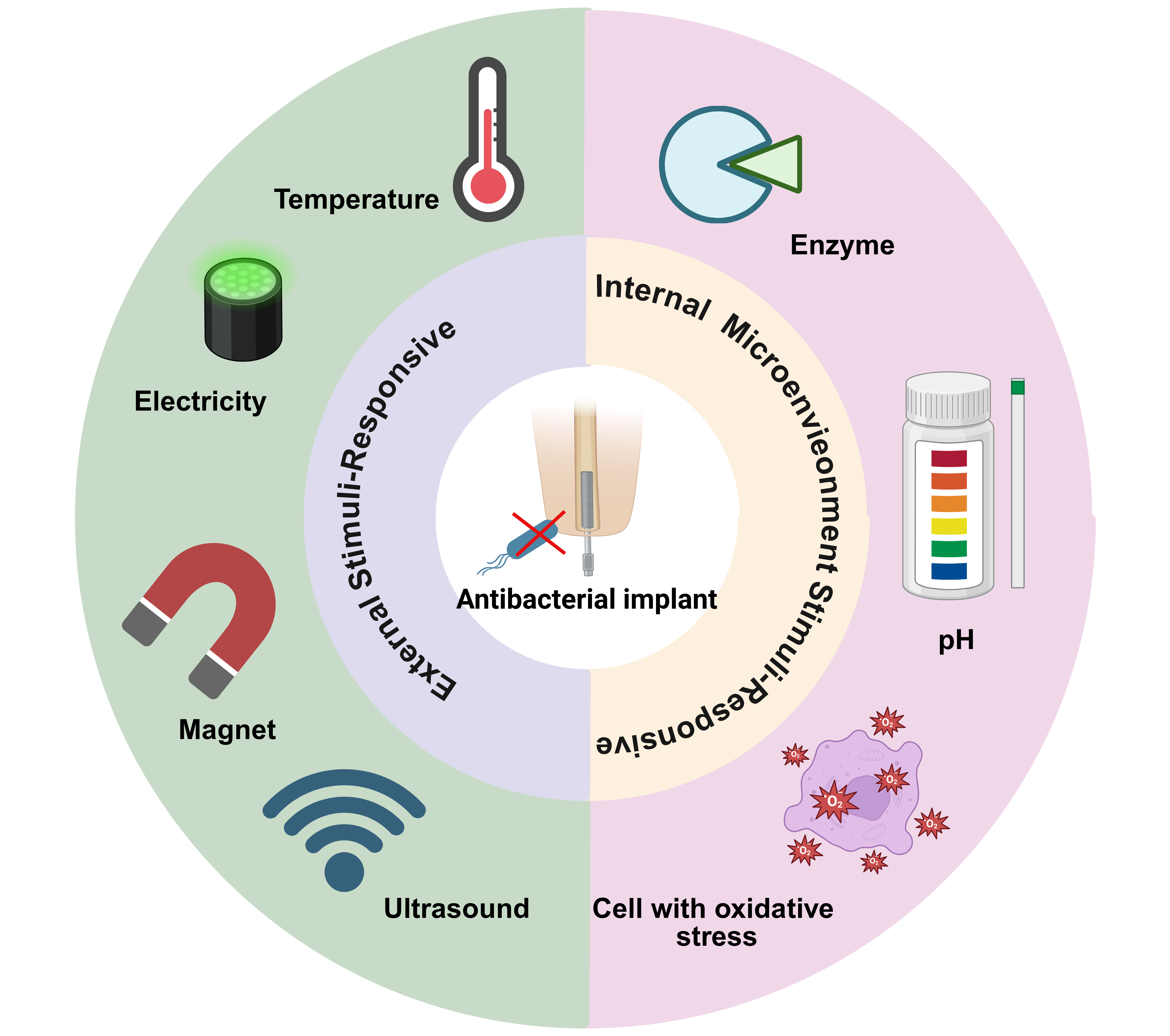
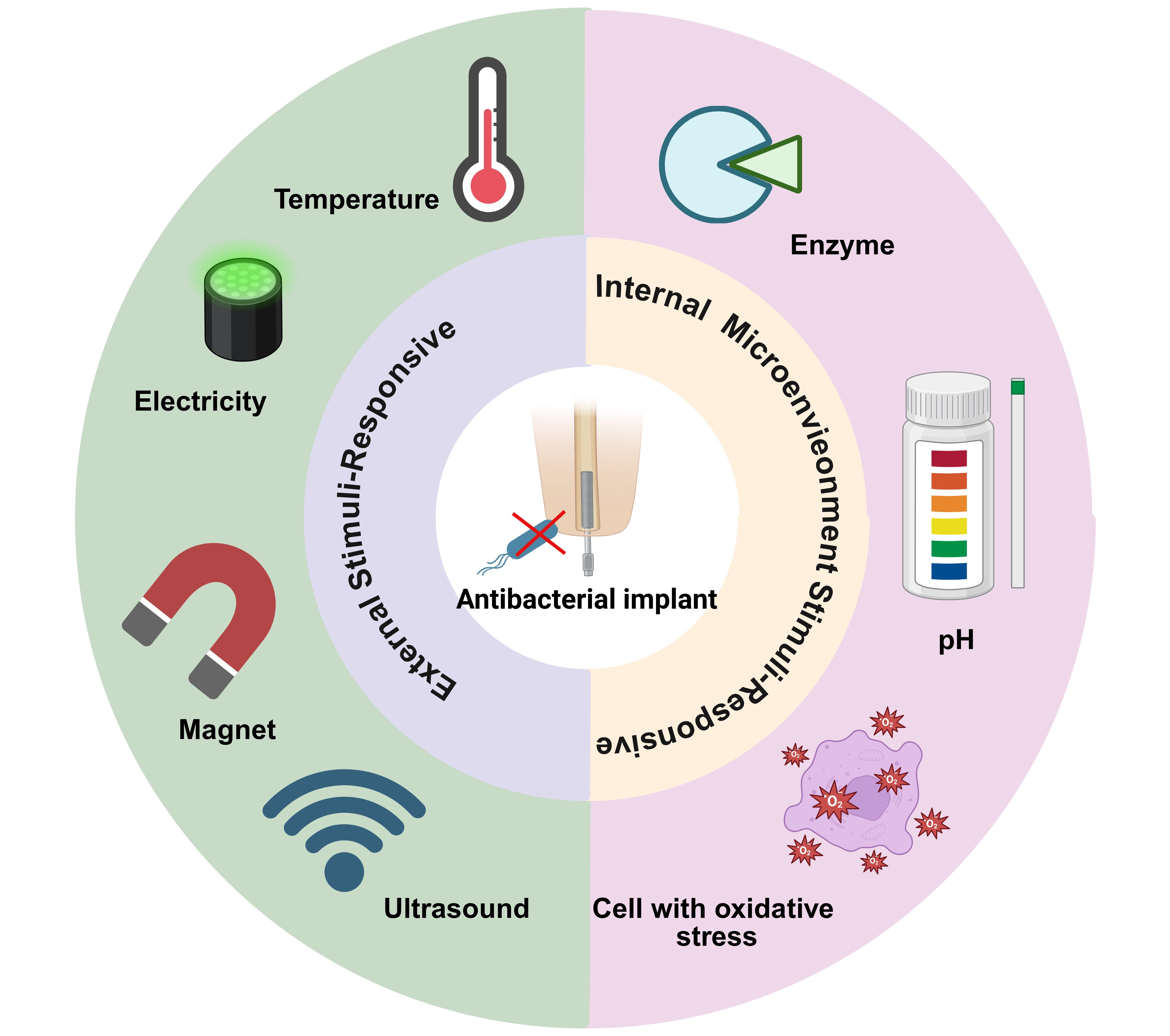
Figure 1. Scheme summarizing different strategies in the design and fabrication of versatile biomaterials with antibacterial functions. To better realize an antibacterial effect, these materials can respond to external stimuli like temperature, electricity, magnet, ultrasound, and internal microenvironment stimuli like enzyme, pH and oxidative stress.
2. Current Therapeutic Strategies for Biofilm Formation and Its Characteristics
2.1 Mechanisms of biofilm formation
Biofilms are organized microbial communities encased in a self-secreted matrix of extracellular polymeric substances (EPS). Their sessile nature confers markedly greater antimicrobial tolerance than that of planktonic cells, complicating infection management. In clinical orthopedics, “surface competition” occurs between host cells and microbes: rapid host tissue integration is essential, because otherwise bacterial colonization overwhelms the site, leading to robust biofilm development and possible surgical failure[9].
Biofilm formation begins with bacterial adhesion to the implant, followed by the development of microcolonies through bacterial aggregation and EPS production (Figure 2)[2]. As the microcolonies remodel and mature, larger colonies are formed, eventually resulting in bacterial dispersion into the planktonic state. During maturation, the microbes secrete EPS composed of eDNA, proteins, and various polysaccharides[2,10,11]. This polymeric scaffold encases the bacterial clusters and ensures stable attachment to the device surface[2,12]. Biofilm-associated infections are therefore notoriously difficult to eradicate and prone to dissemination. Because conventional antibiotics fail to clear these structures, the host frequently develops prolonged chronic inflammation[13].
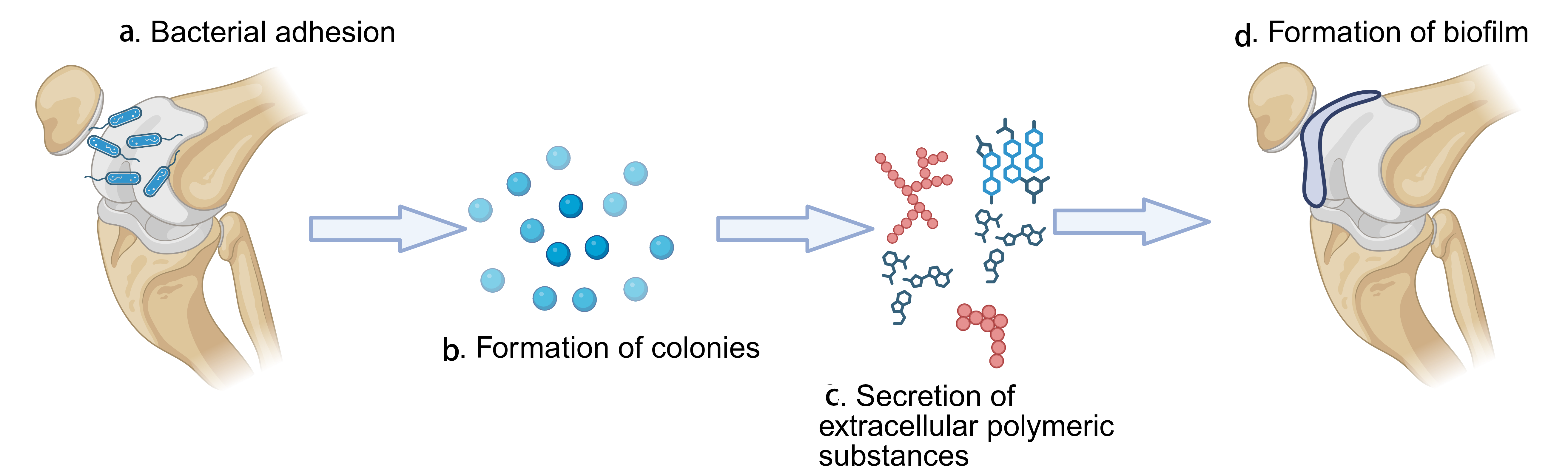
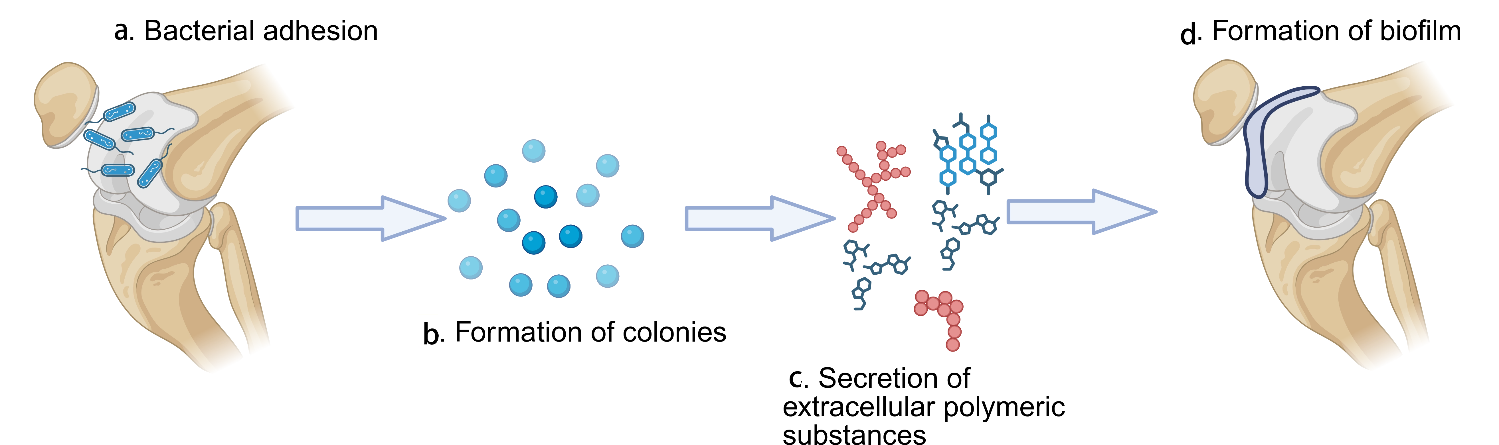
Figure 2. Mechanism of biofilm formation. (a) At the beginning, bacteria adhere to the surface of implant; (b) With the large aggregation of bacteria, microcolonies are formed; (c) When the formation and maturation of microcolonies, bacteria secrete lots of EPS; (d) With the formation of microcolonies and EPS, biofilms are formed on the surface of implant. EPS: extracellular polymeric substances.
2.2 Mechanisms of biofilm resistance
Biofilm formation on medical implants often increases antibiotic resistance through both intrinsic and acquired mechanisms. Intrinsic resistance arises from structural and functional traits, such as the absence of a suitable target for a given antibiotic, and varies between species because some carry resistance genes while others do not. Acquired resistance stems from several pathways: impaired drug entry leading to insufficient cellular levels, active efflux, and genomic alterations. Furthermore, bacteria may employ target site modifications or enzymatic degradation to neutralize antibiotics[14]. Biofilm formation on medical devices is a key contributor to drug tolerance. These structures impede antibiotic diffusion and harbor slow-growing pathogens that evade conventional antimicrobials[15]. Suci et al. observed that the fluoroquinolone ciprofloxacin penetrated Pseudomonas aeruginosa biofilms poorly[16], and Hoyle et al. reported similar findings for piperacillin[17]. This impaired penetration is largely attributable to polymeric compounds, particularly those that bind to and block cationic antibiotics, such as alginate[18]. Even when antibiotics diffuse through biofilms, they often fail to eradicate infection, indicating that additional resistance mechanisms are at play.
Biofilms display three forms of heterogeneity: spatial, response, and cellular[19]. Different regions grow at different rates, often because of nutrient depletion or the accumulation of inhibitory compounds. Antibiotics that target cell-wall synthesis are effective against growing bacteria but far less so against non-growing cells within the biofilm, reflecting a non-uniform bacterial response to treatment[20]. Huang et al. showed that, in Klebsiella pneumoniae and P. aeruginosa biofilms on stainless steel, bacterial respiration ceased near the biofilm-fluid interface but continued deeper within the biofilm[21]. Similarly, Korbar et al. observed cell elongation near the biofilm interface in Pseudomonas fluorescens biofilms treated with the fluoroquinolone fleroxacin[22].
In polymicrobial biofilms, different species can form mutualistic relationships that produce variable antibiotic responses and generally poor treatment outcomes[18]. Environmental factors, such as oxygen levels, pH, osmotic stress, and other conditions, also affect antibiotic efficacy. Furthermore, horizontal gene transfer between species, particularly via plasmid conjugation, spreads antibiotic resistance within the biofilm and further complicates treatment.
2.3 Therapeutic strategies about biofilms
Bacterial biofilms underlie a wide range of infections, and antibiotic therapy remains the most common treatment. For implant-associated biofilm infections, treatment typically requires both surgical debridement and prolonged antibiotic therapy[23]. Currently, clinical antimicrobial strategies for implants primarily rely on passive release systems, such as antibiotic-loaded bone cements (ALBCs) and silver-coated prostheses. Although these provide immediate local protection, they often exhibit a rapid initial burst release and lack long-term adaptability to the biofilm microenvironment. Combination antibiotic therapy is frequently more effective than a single agent. However, it is crucial to carefully manage both the dosage and duration of antibiotic treatment to optimize outcomes[24]. Antibiotics are generally more effective against early, immature biofilms than against mature biofilms, which are far harder to treat. Unfortunately, early-stage biofilms are difficult to diagnose, and their multiple tolerance mechanisms contribute to their resistance to antibiotics, resulting in chronic infections. As a biofilm matures, its antibiotic resistance increases in parallel with changes in its growth pattern[25].
Although numerous antibiofilm strategies have been investigated, no treatment specifically designed to target biofilms has yet received Food and Drug Administration (FDA) approval, and the clinical translation of these approaches remains limited[26]. Another promising approach is surface modification of biomaterials, for example, coating catheters, implants, and repair materials with surfactants, antibiotics, or antimicrobial peptides (AMPs) to prevent bacterial adhesion and biofilm formation[27]. These strategies have shown encouraging therapeutic effects.
Beyond surface modification, the development of novel implant materials with intrinsic antibacterial properties has emerged as a complementary approach. Metal alloys incorporating antibacterial elements, including Cu-, Ag-, and Zn-doped Ti alloys, confer broad-spectrum antimicrobial activity through sustained ion release that disrupts bacterial membrane integrity and inhibits key enzymatic functions, while preserving favorable mechanical properties[28,29]. Antibacterial ceramics such as silver-doped hydroxyapatite inhibit the growth of both Gram-positive and Gram-negative bacteria while maintaining osteoconductivity, making them well suited to bone-repair applications[30]. Antibacterial polymer implants, notably polyetheretherketone (PEEK) functionalized with silver nanoparticles (AgNPs) or AMPs integrated directly into the polymer matrix, provide an alternative for load-bearing scenarios owing to their bone-matched elastic modulus[31]. Biodegradable magnesium-based alloys possess inherent antibacterial potential through Mg2+ release and localized alkalization during degradation; moreover, their bioresorbability eliminates the need for secondary removal surgery, although rapid corrosion and hydrogen-gas evolution remain obstacles to widespread clinical adoption[32].
Despite these advances, important challenges persist. Conventional antibiotic therapy and broad-spectrum antibacterial agents can cause considerable toxicity, harm the patient, disrupt the host microbiome, and increase selective pressure on bacterial populations. Photodynamic therapy (PDT) shows promise against biofilm infections, particularly at wound sites, where photoactive agents kill bacteria under specific lighting conditions. However, PDT must be applied cautiously to avoid damaging healthy tissue, since laser exposure can affect both infected and normal tissue[33]. Electrical stimulation is also under preclinical investigation for implant-associated applications. Implant-related studies have shown that electrically stimulated conductive coatings can support osteogenic activity[34], whereas cathodic voltage-controlled stimulation, alone or combined with vancomycin (VAN), reduced the bacterial burden in rodent models of titanium implant-associated methicillin-resistant Staphylococcus aureus (MRSA) infection[35,36]. These findings support the preclinical potential of electrical stimulation for combined infection control and bone regeneration. However, its clinical efficacy and long-term safety remain unestablished.
Stimuli-responsive nanoparticles, activated by either endogenous or exogenous stimuli, offer great promise in combating biofilm infections. Infected tissues and biofilms typically present distinctive microenvironments, such as low pH, elevated enzyme levels, and high reactive oxygen species (ROS), that differ from normal tissues[37]. These differences can be exploited to trigger specific nanoparticle behaviors, including drug release, charge reversal, and changes in size[38]. This strategy enables nanoparticle activation only at the infection site, thereby improving drug bioavailability and minimizing side effects. Nanoparticles that respond to external stimuli, such as light, magnetic fields, electric fields, or ultrasound, offer the advantage of spatiotemporally controlled drug delivery, since external stimuli can be easily manipulated. Nevertheless, the design of stimuli-responsive antibacterial surfaces requires careful optimization to ensure they are both effective and biocompatible[38,39].
From the standpoint of infection control, antibiotic therapy remains the most widely used option, but the growing problem of bacterial resistance is difficult to overcome. Approaches like focal debridement and treatment modifications, such as grafting, often result in increased patient discomfort and higher medical costs. Additionally, several therapeutic methods remain under investigation (Table 1).
Table 1. The treatment strategies for bacterial biofilms.
| Treatment strategy | Composition | Applicable scope | Advantages | Disadvantages | Examples |
| Antibiotic Therapy | Combination of antibiotics, tailored dosage and duration | General implant-associated bacterial infections | Well-established, commonly used | Limited effectiveness against mature biofilms | [23-25] |
| Combination therapy can enhance effectiveness | Risk of resistance and toxicity | [23] | |||
| Surgical Debridement | Physical removal of infected tissue and biofilms | Implant-associated infections | Direct removal of infected tissue and reduction of the local biofilm burden | Invasive; may require additional surgery and prolonged antimicrobial therapy | [19,23] |
| Surface Modification of Biomaterials | Coating implants, catheters, or repair materials with surfactants, antibiotics, or antimicrobial peptides | Implant, catheter, and repair material applications | Prevents bacterial adhesion and biofilm formation | Requires biocompatible and durable coatings | [27,40] |
| Photodynamic Therapy | Photoactive agents activated by light exposure | Wound infections, localized biofilm infections | Non-invasive, targets biofilm directly with light | Risk of damaging healthy tissue, requires specific conditions | [41] |
| Bioelectric Therapy | Electrically stimulated conductive coatings or cathodic voltage-controlled stimulation | Preclinical in vitro and rodent implant models | Supports osteogenic activity and can reduce the bacterial burden on titanium implants when combined with VAN | Long-term safety, optimal stimulation parameters, and clinical efficacy remain unestablished | [34-36] |
| Stimuli-Responsive Nanoparticles | Nanoparticles activated by endogenous (e.g., pH) or exogenous (e.g., light, magnetic fields) stimuli | Biofilm-associated infections | Targeted activation at infection site, improves bioavailability | Complex design, need for further optimization, safety concerns | [37-39] |
| Novel Intrinsically Antibacterial Implant Materials | Metal alloys incorporating antibacterial elements (e.g., Cu-, Ag-, or Zn-doped Ti alloys, antibacterial stainless steel); antibacterial ceramics (e.g., Ag-doped hydroxyapatite); antibacterial polymers (e.g., antimicrobial-modified PEEK); biodegradable magnesium-based alloys | Orthopedic implants, dental implants, fracture fixation devices, joint prostheses | Intrinsic long-term antibacterial activity without reliance on external coatings; no risk of coating delamination; broad-spectrum efficacy; sustained ion release | Potential cytotoxicity at elevated ion concentrations; limited control over ion release kinetics; incorporation of antibacterial elements may compromise mechanical properties; complex regulatory approval pathways | [28,30-32] |
PEEK: polyetheretherketone; VAN: vancomycin.
3. External Stimuli-Responsive Antibacterial Implants
Figure 3 provides an overview of the external and internal stimuli exploited by these strategies and of the four functional stages, sensing, activation, killing, and regeneration, that they share.
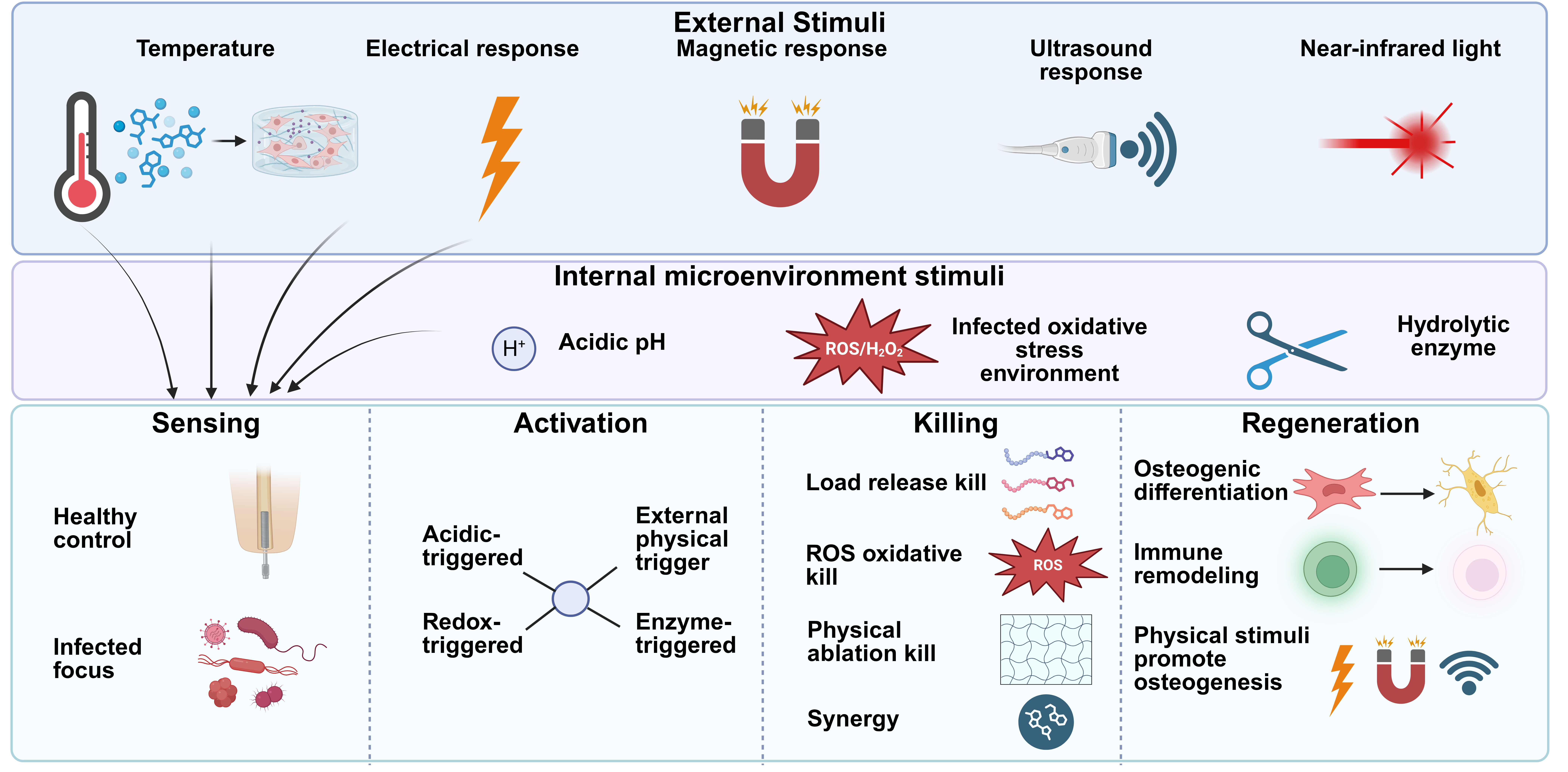
Figure 3. Overview of stimuli-responsive antibacterial strategies and their shared working principle. Top: representative external stimuli (temperature, electrical, magnetic, ultrasound, and near-infrared light) and internal microenvironmental stimuli (acidic pH, ROS/H2O2, and hydrolytic enzymes). Bottom: the four functional stages common to these strategies: sensing of the infection, responsive activation (acid-, redox-, enzyme-, or external physical trigger), bacterial killing (drug release, ROS-mediated oxidation, physical ablation, and their synergy), and tissue regeneration (osteogenic differentiation, immune remodeling, and physically promoted osteogenesis). ROS: reactive oxygen species.
3.1 Temperature responsive strategy
Temperature-responsive materials leverage the temperature differential within the human body to induce changes in their chemical state over time, facilitating functions such as bone therapy and the promotion of bone growth. These stimuli-responsive substances demonstrate nonlinear thermal sensitivity. Their solubility shifts abruptly at specific thresholds, such as the lower critical solution temperature (LCST) or upper critical solution temperature (UCST), transitioning between soluble and insoluble states[42].
Hydrogels, due to their reversible bonds, are prime candidates for temperature-responsive macrostructures. These hydrogels, known as temperature-responsive hydrogels (TRHs), contain hydrophobic groups (e.g., propyl, ethyl, methyl) that enable them to undergo changes in shape, size, and volume in response to physiological temperature fluctuations. At room temperature, TRHs are typically liquid or semisolid, undergoing a sol-gel transition when exposed to body temperature. This behavior allows liquid therapeutic compounds to be loaded into the hydrogel, which can then be easily applied and solidified.
One example is the thermosensitive, injectable nanocomposite hydrogel developed by Makvandi et al., composed of hyaluronic acid, β-tricalcium phosphate, and corn-silk-extract-derived AgNPs[43]. The hydrogel exhibits a gelation temperature close to body temperature, allowing it to be delivered as a liquid and to solidify in situ. The green-synthesized AgNPs confer broad-spectrum antibacterial activity against both Gram-positive (Bacillus subtilis, Staphylococcus aureus) and Gram-negative (Pseudomonas aeruginosa, Escherichia coli) bacteria without appreciable cytotoxicity, while the β-tricalcium phosphate component promotes the osteogenic differentiation of mesenchymal stem cells (MSCs). The translational value of this dual antibacterial-osteogenic strategy is supported in vivo by a related thermosensitive chitosan/quaternized-chitosan/nano-hydroxyapatite hydrogel, which reduced S. aureus infection and enhanced bone regeneration in a rabbit infected radial-defect model over 8-12 weeks[44]. Such temperature-responsive systems thus illustrate how in situ gelation, sustained silver release, and osteoconductivity can jointly address infection control and bone repair, although for the silver formulation efficacy has so far been demonstrated only in vitro.
Another recent development involved the use of TRH-loaded coatings for bone tissue engineering to combat implant-associated infections. A highly temperature-sensitive hydrogel, chitosan-glycerin-hydroxypropyl methyl cellulose hydrogel (CGHH), was created by combining hydroxypropyl methylcellulose (HPMC), chitosan, and glycerol[45]. This hydrogel undergoes a sol-to-gel transition at 40 °C, releasing large amounts of glycerin, which activates the body’s innate immune system. This leads to the recruitment of M1 macrophages and other inflammatory cells to the site, effectively controlling bacterial infections. In addition, M2 macrophages are recruited to accelerate tissue healing and promote osteoblast differentiation[46]. Compared with the AgNP hydrogel described above, this hydrogel better mobilizes innate immunity, improving bacterial clearance at the infection site.
Additionally, a temperature-sensitive hydroxybutyl chitosan (HBC) hydrogel was designed by Wang et al. utilizing chitosan, an abundant biological polysaccharide known for its antibacterial and hemostatic properties[47]. They manufactured HCS-3,4-dihydroxyphenylalanine (DOPA) composite hydrogel by combining HBC, neutral chitosan solution, and dopamine in three different proportions[48]. When the weight ratio of chitosan to HBC was 1:3, a temperature-responsive hydrogel was produced. At body temperature (~ 37 °C), hydrogels with different dopamine concentrations showed correspondingly different antibacterial effects against S. aureus. Based on the mechanism of marine mussel adhesion, 3,4-DOPA was utilized to enhance the solid substrate adhesion ability of biomimetic materials. It has been reported that this material can not only increase adhesion, but also promote osteogenic differentiation of MSCs to some extent[49]. This thermosensitive hydrogel material not only has anti-infective effects, but also promotes bone proliferation and bone healing, which provides good prospects for future research.
In summary, temperature-responsive materials, particularly hydrogels, represent a versatile and innovative approach for addressing implant-associated infections and promoting tissue regeneration. These materials exploit the physiological temperature variations within the human body to undergo sol-gel transitions, enabling controlled drug release, antibacterial activity, and enhanced bone integration. TRHs exhibit unique properties such as shape and volume changes in response to temperature fluctuations, making them ideal for localized therapeutic delivery. By incorporating components like AgNPs, immunosuppressants, or immune-modulating agents, these hydrogels not only combat infections but also stimulate osteogenesis and tissue repair. Furthermore, their ability to recruit immune cells and promote osteoblast differentiation highlights their potential for improving clinical outcomes in bone-related therapies. Despite their promising applications, further research is needed to optimize their performance and ensure their safety and efficacy in clinical settings. Overall, temperature-responsive materials offer a promising avenue for advancing personalized and targeted treatments in orthopedic and regenerative medicine.
3.2 Electronic responsive strategy
An in vitro study demonstrated that electrical stimulation applied to an antibacterial polyaniline (PANI) coating on titanium promoted the proliferation and osteogenic differentiation of MC3T3 cells[34]. This process is facilitated by the upregulation of bone morphogenetic proteins, which activate the calcium-calmodulin pathway, transforming growth factors, and other cytokines[8]. Conductive polymers (CPs) have gained significant attention in orthopedic applications as charge carriers for electrical stimulation, owing to their biocompatibility, environmental stability, and ease of synthesis. However, a major challenge remains: preventing biofilm formation caused by infections on orthopedic implants when CPs are used.
Among the various conductive materials, CPs such as polypyrrole, PANI, and polythiophene have become key research focus areas. For example, Zhu et al. developed a PANI coating on biomedical Ti sheets, doped with Ag-loaded terminal carboxyl-polyamide amine dendrimers (PAMAM)[34]. The coating demonstrated superior bactericidal efficacy against E. coli and S. aureus. Additionally, it enhanced MC3T3 cell viability when combined with electrical intervention. When compared to conventional Ti implants, Ti coated with PANI showed enhanced osteogenesis with appropriate electrical stimulation, making this material highly promising for both bone regeneration and infection resistance.
In a similar vein, Nodzo et al. conducted mouse experiments to assess the effectiveness of cathode-voltage-controlled electrical stimulation combined with VAN in treating Ti implant-associated infections[35]. Mice with shoulder Ti implant-associated infections were subjected to 1.8V Ag/AgCl cathode-voltage electrical stimulation for about 1 hour, alongside VAN treatment. The combined therapy significantly outperformed either treatment alone in combating biofilm formation. This approach of combining electrical stimulation with antibiotics offers a promising strategy for preventing implant-associated infections. However, there are some limitations to the study: it was conducted in rodent models, which may not fully simulate all infection risks of osseointegrated prostheses. Future research could explore the effects of cathodic voltage-controlled electrical stimulation at transcutaneous sites to more closely mimic human clinical scenarios.
In summary, electrical stimulation has emerged as a promising therapeutic approach for enhancing bone regeneration and combating implant-associated infections. By promoting the proliferation and differentiation of MSCs and activating key signaling pathways, electrical stimulation facilitates new bone formation. CPs like polypyrrole and PANI are promising for orthopedics, offering excellent biocompatibility and electro-activity. Integrating these polymers with antimicrobials provides a synergistic effect on bone growth and infection control. Nonetheless, achieving clinical translation requires more sophisticated models to mimic human physiology and refine therapeutic strategies. Despite these limitations, the integration of electrical stimulation with CPs and antimicrobial strategies holds great promise for advancing orthopedic implant technologies and improving patient outcomes.
3.3 Magnetically responsive strategy
Magnetic field-based stimuli offer several advantages, primarily due to the diverse magnetic responses they elicit. These can include magnetic guidance under static magnetic fields and localized heating under alternating fields. Such systems provide unique flexibility in antimicrobial drug delivery, allowing precise control over release time, amount, and range.
Magnetic field-responsive drug delivery systems, often using superparamagnetic nanoparticles smaller than 15 nm (typically Fe3O4), are gaining significant attention. Fe3O4 nanoparticles are magnetized by external magnetic fields and often coated with biocompatible polymers to form nanoscale core-shell structures. Harris et al. developed a magnetically responsive local drug-delivery system by loading superparamagnetic Fe3O4 nanoparticles and VAN into chitosan microbeads cross-linked with polyethylene glycol dimethacrylate. In vitro, 30 min of magnetic stimulation temporarily increased the daily VAN elution rate by up to 45%, depending on the magnetic field parameters and cross-linker length. These results demonstrate proof-of-concept magnetically triggered antibiotic release, although efficacy in an in vivo implant-associated infection model has not yet been established[50].
3.4 Ultrasound-responsive strategy
As a non-invasive modality, sonodynamic therapy (SDT) utilizes low-intensity ultrasound to activate sonosensitizers. Unlike photoelectric therapies, SDT offers superior tissue penetration, enhanced targeting precision, and a more favorable safety profile[51]. Bone regeneration is markedly enhanced by ultrasound therapy, extending its utility past tumor resection. It promotes the expression of vascular endothelial growth factor A (VEGF-A) and chondrocyte proliferation, which in turn accelerates the mineralization and development of new bone tissue[8,52]. This technique has been applied in the field of infection and can be used clinically to treat bone defects and severe bacterial infections[8]. Usually in the treatment of SDT, a suitable set of ultrasound and sonosensitizer are required. Therapeutic ultrasound should be characterized using independent acoustic parameters rather than frequency alone. Frequency, acoustic pressure, intensity, duty cycle, pulse duration, and focusing geometry jointly determine the resulting thermal and mechanical effects. Therefore, high- and low-intensity ultrasound cannot be distinguished solely by a frequency threshold[53]. SDT involves the absorption of acoustic energy and subsequent relaxation of sonosensitizers between ground and high-energy states. This energy transfer generates ROS, a potent byproduct that serves to inhibit biofilm formation and persistence.
Pang et al. developed a smart nanoliposomes platform (MLP18), a bacteria-responsive nanoliposome for the targeted delivery of the sonosensitizer purpurin 18. The platform uses the bacterial maltodextrin transport pathway for infection-site targeting and promotes bacteria-responsive release and internalization of the sonosensitizer. Under ultrasound irradiation, MLP18 mediated the elimination of multidrug-resistant bacteria and resolved inflammation and abscesses in a mouse model of bacterial myositis. This non-implant study demonstrates the potential of bacteria-responsive SDT but does not establish efficacy against implant-associated biofilms[54].
In summary, external stimuli-responsive strategies based on temperature, electricity, magnetism, and ultrasound share a key advantage: an operator-applied trigger affords precise spatiotemporal control over the antibacterial action, and several also promote osteogenesis. However, their reliance on external equipment and intervention prevents them from responding autonomously to the dynamic course of an infection. An ideal implant should instead sense the infection itself. This directs attention to the biofilm microenvironment, where bacterial colonization generates elevated ROS, localized acidity, and overexpressed enzymes. These endogenous cues can act as intrinsic triggers for on-demand antibacterial release, as discussed in the following section on oxidative stress responsive, pH responsive, and enzyme responsive systems.
4. Internal Microenvironment Stimuli-Responsive Antibacterial Implants
The overall working principle of these microenvironment-responsive implants-associated infection sensing, responsive activation, bacterial killing, and subsequent tissue regeneration is summarized in Figure 4.
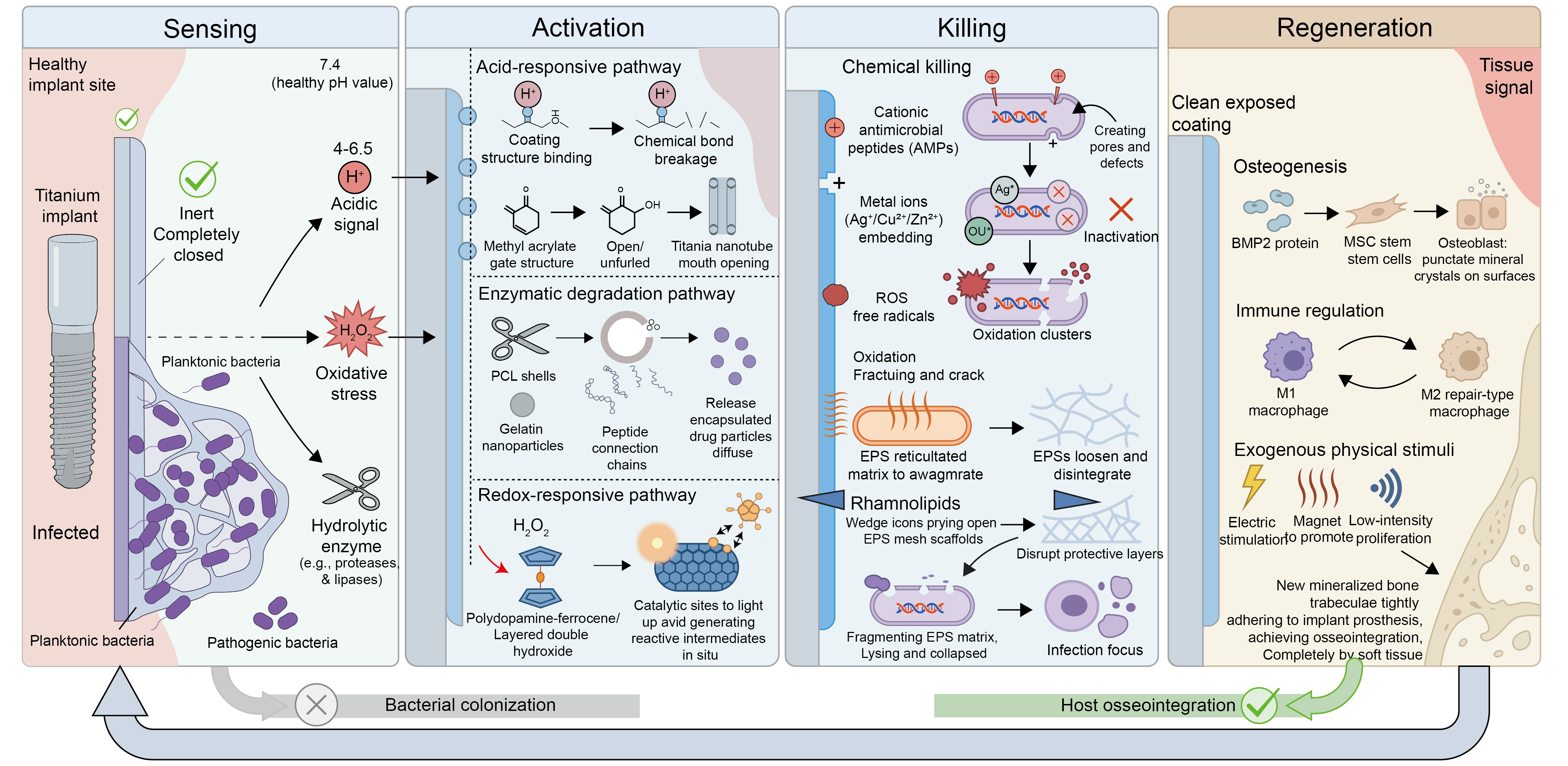
Figure 4. Mechanism of microenvironment-responsive antibacterial implants, showing the integrated sensing-activation-killing-regeneration cascade. Sensing: the coating remains inert at healthy pH (7.4) but detects the acidic (pH 4-6.5), oxidative (ROS/H2O2), and enzyme-rich signals of an infected focus. Activation: acid-, redox-, or enzyme-triggered pathways open the coating and release its payload. Killing: released AMPs, metal ions, and ROS, together with physical disruption of the EPS matrix, eradicate the bacteria; Regeneration: the cleared surface supports osteogenesis, M1-to-M2 macrophage remodeling, and physically stimulated bone formation, allowing host osseointegration to prevail over bacterial colonization; ROS: reactive oxygen species; EPS: extracellular polymeric substances; PCL: poly(ε-caprolactone); BMP2: bone morphogenetic protein 2; AMPs: antimicrobial peptides.
4.1 Oxidative species-responsive strategy
ROS are highly reactive oxygen-containing molecules, including peroxides, hydroxyl radicals, superoxides, singlet oxygen, and atomic oxygen. They are released in response to injury, infection, inflammation, and other physiological conditions. While moderate ROS levels exhibit strong antibacterial properties, excessive ROS can lead to mitochondrial and cellular damage, harming the body. Conversely, harnessing controlled ROS generation has proven effective in tissue regeneration. For instance, Zhang et al. demonstrated that nanodefective zinc oxide (NDZnO) can optimize skin regeneration following invasive dermatological procedures by modulating local oxidative stress, highlighting the therapeutic window of ROS-based materials[55]. However, overexpressed ROS can be harnessed as a trigger in stimuli-responsive materials designed to treat infections and promote bone healing. Many researchers have developed smart materials based on this principle.
Chitosan-based platforms can also exploit oxidative stress for antibacterial action. Zhang et al. constructed a chitosan/zinc-peroxide (CS@ZnO2) nanocomposite that self-decomposes in the slightly acidic biofilm microenvironment to release H2O2 and generate a burst of ROS, completely eliminating E. coli and S. aureus in vitro through oxidative membrane damage[56]. Complementarily, the elevated ROS at infection foci can serve as a release trigger rather than a bactericidal agent: a ROS-responsive nanoparticle cloaked in a bacteria-activated macrophage membrane selectively accumulated at S. aureus-infected sites and, upon sensing the local ROS, released VAN to eradicate the bacteria both in vitro and in vivo[57]. Together these designs illustrate the dual role of ROS in this strategy, as a directly bactericidal species and as an endogenous trigger for on-demand drug release, although ROS levels vary between individuals, complicating standardized material design.
In contrast to ROS-triggered drug delivery systems, some researchers have designed antibacterial coatings that leverage ROS’s inherent antimicrobial properties. For example, Song et al. functionalized Ti dioxide (TiO2) nanorods with a polydopamine-ferrocene (Ti-Nd-PDA-Fc) coating, which exhibits redox activity[58]. In the presence of implant-associated infections, the coating locally releases antibacterial ROS. Ti-Nd-PDA-Fc demonstrated strong antibacterial activity, achieving over 95% and 92% inhibition of Methicillin-resistant E.coli and S. aureus, respectively. Additionally, Ti-Nd-PDA-Fc showed good biocompatibility in pre-osteoblast MC3T3 E1 cell models, promoting cell adhesion and spreading. Wang et al. took a similar approach to develop scaffolds with selective tumor-killing and antibacterial effects. By using a hydrothermal treatment method, they created a composite scaffold with an internal microenvironment-responsive Ni-Ti LDH membrane. This membrane utilizes the overexpression of H2O2 in infected environments to release butyrate, which selectively inhibits bacterial infections and tumor growth while promoting osteogenesis[8]. This redox-active coating imbues implants with context-dependent antimicrobial activity, reducing the risk of bacterial resistance, unlike conventional antibiotic-releasing systems. However, ROS therapy remains nonspecific and can potentially damage normal cells.
4.2 Acidic environmental-responsive strategy
The pH of blood is regulated by the concentration of carbon dioxide, electrolytes, and weak acids, which are essential for maintaining acid-base balance. However, in certain pathological conditions, the pH can deviate from the physiological value of 7.4. This property has recently spurred interest in developing pH-responsive materials that release drugs in response to pH changes, particularly for antibacterial purposes[59]. Medical implant biofilms exhibit heterogeneous pH microenvironments. The accumulation of acidic metabolites generated by bacterial respiration and fermentation, together with limited metabolite clearance and host inflammatory responses, can lower the local pH. However, the magnitude and spatial distribution of this acidification depend on the bacterial species and implant site[60]. This enables pH-triggered antibiotic release systems to effectively target acidic infection environments, protecting antibiotics in circulation, minimizing exposure to healthy tissues, and enhancing localized drug delivery. Such systems ensure adequate antibiotic concentration at infection sites, facilitating bacterial eradication while preserving the activity of the drug and minimizing the development of bacterial resistance[60-62].
For eliminating bacteria and orchestrating the wound healing cascade, hydrogels with antimicrobial properties are highly effective. These materials actively intervene in the repair process by regulating moisture, absorbing excess fluids, and suppressing microbial proliferation through integrated therapeutic agents[63,64]. Nevertheless, these antimicrobial hydrogels exhibit limited efficacy in fully eliminating established biofilms. Furthermore, their application is often constrained by significant cytotoxicity toward healthy mammalian tissues[65], which calls for the emergence of pathological conditions-activated antimicrobial hydrogels. A smart antimicrobial hydrogel based on a designed octapeptide (IKFQFHFD) has been engineered to combat biofilms through pH-dependent structural transitions. Unlike traditional cationic hydrogels, this nanofiber network destabilizes in acidic conditions to activate its therapeutic payload. By incorporating photothermal agents and procollagen precursors, the integrated system achieves total MRSA removal while minimizing damage to healthy tissues. This pH-responsive delivery mechanism demonstrated preclinical potential in an MRSA-infected chronic-wound model. However, its applicability to implant coatings and implant-associated infections remains to be validated[66]. This is a wound-healing hydrogel rather than an implant coating, but its pH-gated activation exemplifies a principle applicable to implant surfaces. Hu et al. designed a hydrogel that responds to changes in pH. By using a supramolecular chemistry approach, they integrated guanosine and potassium ions to form a G-quadruplex structure, which was combined with aminoglycoside antibiotics to create a stable hydrogel. In this system, as the pH shifts from neutral to acidic during bacterial proliferation, the release of aminoglycosides is accelerated, demonstrating strong antibacterial activity against both Gram-negative bacteria like E. coli and P. aeruginosa and Gram-positive bacteria like S. aureus. This hydrogel not only responds to acidic environments but also to other stimuli like temperature, glucose, and peroxides, providing controlled drug release with low molecular weight, low toxicity, and ease of preparation[67]. Liu et al. developed a pH-responsive antibacterial hydrogel based on reversible catechol-boronate complexation between intrinsically bactericidal chlorinated catechol and phenylboronic acid[68]. Under acidic conditions, dissociation of the catechol-boronate complexes exposed the chlorinated catechol groups, thereby activating the antibacterial properties of the hydrogel. The hydrogel exhibited pH-dependent antibacterial activity against both Gram-positive and Gram-negative bacteria, including methicillin-resistant S. aureus. Conversely, catechol-boronate complex formation under alkaline conditions reduced the accessibility of the chlorinated catechol groups and attenuated both antibacterial activity and cytotoxicity. When the hydrogel was subsequently returned to an acidic environment, it recovered its antibacterial activity, demonstrating reversible pH-responsive behavior. However, this system was evaluated under simplified in vitro pH conditions and has not been validated as an orthopedic implant coating or in an implant-associated infection model[68].
Tao et al. constructed a pH-responsive multilayer coating on bone morphogenetic protein 2 (BMP2)-loaded TiO2 nanotubes. The multilayer film consisted of alginate dialdehyde-gentamicin and chitosan assembled through a layer-by-layer process. An acidic environment triggered gentamicin release and accelerated BMP2 release from the nanotubes. In vitro, the coating showed antibacterial activity against E. coli and S. aureus for up to 72 hours and promoted osteoblast differentiation, as indicated by increased alkaline phosphatase activity, mineralization, and osteogenic gene expression. These findings support its potential as a dual antibacterial and osteogenic Ti-based implant coating, although in vivo osseointegration was not evaluated[69].
Study-specific evidence supports pH-responsive drug release from silk-based implant coatings. Sang et al. applied a gentamicin-silk protein coating to titanium, polyethylene, and Al2O3. Exposure to pH 5.5 accelerated gentamicin release, and antibacterial activity was evaluated both in vitro and in vivo[70]. Zhou et al. constructed an AgNPs/gentamicin-loaded silk fibroin (SF) coating with an outer chitosan nanovalve on titanium. Acidification induced collapse of the chitosan layer and accelerated bactericide release, resulting in pH-dependent activity against S. aureus and inhibition of biofilm formation[71]. In a separate study, Yan et al. constructed an AgNPs-incorporated SF/gentamicin sulfate coating on porous PEEK. The release of Ag+ and gentamicin increased as pH decreased, while the coating showed antibacterial and osteogenic activity in preclinical experiments[72].
Chlorhexidine (CHX) and silver have attracted considerable interest as antimicrobial agents, and their combined use has demonstrated enhanced activity against bacterial and fungal biofilms[73,74]. Mesoporous silica nanoparticles provide a versatile platform for the co-delivery and controlled release of antimicrobial agents[75]. Based on this approach, Lu et al. developed CHX-loaded, silver-decorated mesoporous silica nanoparticles for the pH-responsive co-delivery of CHX and Ag+[74]. Acidic conditions accelerated the simultaneous release of both agents. In vitro, the system exhibited synergistic antibacterial activity against E. coli and S. aureus, while its effective antibacterial concentration showed relatively low cytotoxicity toward normal cells. However, the system was not evaluated in an implant-associated infection model[74]. Zhao et al. employed an electrostatic self-assembly strategy to integrate cationic CHX with negatively charged citrate-modified poly(2-((2-aminoethyl)carbamoyl)oxy)ethyl methacrylate (PAECOEMA), thereby constructing a pH-responsive nanocomposite[76]. Under acidic biofilm conditions, cleavage of the acid-labile citrate amide bonds promoted CHX release and produced antibacterial activity against cariogenic bacteria. However, this system was developed for dental applications, and its suitability for orthopedic implant-associated infections remains to be established[76]. Utilizing carboxyl group protonation is a key strategy for developing pH-sensitive delivery platforms. While AMPs provide powerful, broad-spectrum defense against bacteria, their susceptibility to enzymatic degradation limits clinical use. To facilitate controlled administration, Chen et al. synthesized Ti implants modified with Ti dioxide nanotubes (Ti-NTs) to host AMPs. This method was demonstrated using the HHC36 peptide sequence (KRWWWKWWRR) for triggered antibiotic delivery[77]. AMPs were encapsulated within TiO2 nanotubes (Ti-NTs), utilizing pH-responsive polymethacrylic acid (PMAA) as a molecular gate. At physiological pH 7.4, the expanded conformation of PMAA provides steric hindrance that seals the nanotubes. Conversely, the protonation of carboxyl groups in acidic infection sites (pH < 6) triggers the collapse of PMAA, opening the Ti-NTs for localized peptide delivery. This system demonstrated potent broad-spectrum efficacy against pathogens such as E. coli, S. aureus, P. aeruginosa, and MRSA.
Different from the conventional antibacterial strategy led by various types of antibiotics, good antibacterial effects can also be achieved by releasing high concentrations of H+ and H2O2 on the implant surface. Ding et al. designed and constructed a surface capable of global translation through self-propagation reactions in response to local and transient stimuli[78]. This material utilizes an autocatalytic mechanism where glucose boronate functional groups interact with immobilized glucose oxidase (GOx). Initial proton (H+) exposure triggers the hydrolytic release of glucose, which serves as a substrate for GOx to yield H2O2 and more H+. This positive feedback cycle rapidly transforms the material’s surface properties, creating a lethal environment rich in H+ and H2O2. Such a system exhibits potent antibacterial properties, with a marked inhibitory effect on E. coli.
Yang et al. developed Azo-QPS-C16, a pH-sensitive quaternary pyridinium compound with a long alkyl chain. Under neutral or mildly basic conditions, the molecules formed aggregates with reduced antibacterial activity. Acidification induced aggregate disassembly and increased the number of accessible antimicrobial sites, resulting in enhanced inhibition of acid-producing bacteria within a multispecies community. This dental proof-of-concept study demonstrates pH-selective antibacterial activity but does not establish efficacy in implant-associated infections[79].
In summary, pH-responsive materials have emerged as a highly effective strategy for targeted drug delivery and antibacterial applications, leveraging the acidic microenvironment characteristic of bacterial infections. These systems exploit the pH deviations caused by bacterial metabolic byproducts, such as acetic and lactic acids, to trigger the controlled release of antimicrobial agents, ensuring localized delivery while minimizing systemic exposure and toxicity. Various pH-responsive platforms, including hydrogels, chitosan-based systems, SF coatings, and mesoporous nanoparticles, have demonstrated significant potential in eradicating biofilms, promoting tissue regeneration, and enhancing therapeutic efficacy. Additionally, innovative approaches such as protonation-driven drug release and self-propagating surface reactions further expand the versatility of pH-responsive systems. By integrating biocompatibility, targeted delivery, and multifunctionality, these advanced materials offer promising solutions for combating implant-associated infections and improving clinical outcomes in wound healing and orthopedic applications.
4.3 Enzyme environmental-responsive strategy
Enzymes play a critical role in regulating various biological processes in vivo, and many orthopedic implant-associated infections are linked to the overexpression of specific enzymes. Manipulating these dysregulated enzymes offers a promising strategy for drug release, as they can initiate structural changes, disintegration, or cleavage of nanocarriers.
By recognizing enzymes secreted by bacteria, drugs can be precisely released to the infection site. Zhang et al. developed a surface modification technique for Ti implants, utilizing a peptide-based system that recognizes S. aureus-secreted serine proteinase-like proteases (SplB). To mitigate adverse effects and lower the probability of antimicrobial resistance, this platform utilizes an enzyme-triggered mechanism to release VAN. By ensuring that the antibiotic is only discharged in the presence of pathogenic stimuli, the system achieves precise, on-demand delivery at the infection interface[80]. To achieve targeted drug delivery at infection sites, Xiong et al. engineered a polymeric triple-layered nanogel (TLN) responsive to microbial enzymes. The mechanism relies on the poly(ε-caprolactone) (PECL) shell of the polymeric TLN, which breaks down in the presence of bacterial lipase, allowing for the precise release of the therapeutic payload only when infection is sensed[81]. To combat resistant infections, a gelatinase-sensitive Ru-Se@GNP-RBCM platform was engineered by Lin et al. The mechanism involves the breakdown of gelatin nanoparticles by bacteria-secreted enzymes, which subsequently liberates Ru-Se nanoparticles to exert their bactericidal effects. This responsive strategy was proven effective for treating MRSA infections in live animal subjects[82]. The polyion complex nanoparticles (PIC-NPs) presented by Insua et al. function via an enzyme-triggered mechanism. At infection sites where P. aeruginosa liberates elastase, the PIC-NPs are degraded, allowing the system to exert a concentrated and responsive antibacterial effect[83].
To combat the defense mechanisms of resistant bacteria, which rely on the hydrolytic activity of penicillinamidase (PGA) and β-lactamase (Bla), researchers are developing enzyme-triggered delivery vehicles. Such systems leverage the high local concentrations of these enzymes to release payloads like gentamicin directly within the biofilm. A key example is the work of Lee et al. who constructed polymer vesicles by incorporating PGA and Bla-sensitive moieties, allowing for precise drug discharge in response to bacterial enzyme secretion[84]. Functional monomers sensitive to PGA and Bla were synthesized and subsequently utilized in reversible addition-fragmentation chain transfer polymerization to develop poly(ethylene glycol) (PEG)-based amphiphilic block copolymers. These copolymers autonomously self-assemble into vesicles that maintain structural integrity in 20% fetal bovine serum (FBS). Upon exposure to either PGA or Bla, the system facilitates the rapid discharge of encapsulated antibiotics.
The integration of enzyme-responsive layer-by-layer@mesoporous silica nanoparticles-silver (LBL@MSN-Ag) onto Ti implants offers a synergistic approach to antibacterial therapy and bone healing. The poly-l-glutamic acid (PLGA) component of these nanoparticles is selectively broken down by S. aureus-secreted glutamyl endopeptidase. In vitro and in vivo results confirmed that this nanocoating provides an effective defense against microbes while actively fostering bone tissue formation throughout a 4-week observation period[85]. Shi et al. developed an infection-responsive guided tissue regeneration/guided bone regeneration (GTR/GBR) membrane based on electrospun polycaprolactone (PCL) nanofibers[86]. The PCL nanofiber mats were coated with polydopamine and subsequently functionalized to enable the covalent grafting of metronidazole (MNA) through ester linkages. These ester bonds could be selectively hydrolyzed by cholesterol esterase (CE), an enzyme secreted by macrophages accumulating at infection sites, thereby triggering MNA release. Increasing CE concentrations resulted in greater MNA release and enhanced antibacterial activity, indicating that the system could respond to changes in enzyme abundance associated with infection. The nanofiber membrane also exhibited favorable cytocompatibility. However, this system was developed as a GTR/GBR membrane, and its effectiveness in orthopedic implant-associated infection models remains to be established[86]. In conclusion, enzyme-responsive systems represent a highly promising approach for addressing orthopedic implant-associated infections and promoting bone regeneration. By leveraging the overexpression of specific enzymes associated with bacterial infections and antibiotic resistance, these systems enable precise, targeted drug release at infection sites. Enzyme-responsive nanocarriers and surface modifications can degrade or undergo structural changes in the presence of bacterial-secreted enzymes, ensuring localized and controlled delivery of antimicrobial agents. This strategy not only minimizes side effects and reduces the risk of bacterial resistance but also enhances therapeutic efficacy. Furthermore, enzyme-responsive systems have demonstrated potential in dual-function applications, combining antimicrobial effects with bone regeneration capabilities. As research progresses, these intelligent, enzyme-triggered delivery platforms hold great promise for advancing personalized and effective treatments for implant-associated infections and associated complications.
5. Multiple Stimuli-Responsive Antibacterial Implants
So far, we have discussed the use of single-stimulus materials for combating implant-associated infections and for drug delivery. Recently, scientists have begun to explore the development of combinatorial stimuli-responsive materials to better control the delivery and action of antibacterial agents and improve their accuracy.
A dual-responsive antibiotic delivery system was engineered by Chen et al. to enhance drug bioavailability at bacterial infection foci. By conjugating VAN as a bacteria-targeting ligand to poly(ethylene glycol)-PECL via pH-sensitive hydrazone bonds and encapsulating ciprofloxacin (CIP) within the hydrophobic core, they synthesized Van-hyd-PECL/Cip micelles. While the PEG layer ensures systemic stability, the acidic infection environment deshields the VAN ligand and enlarges the micelle, which in turn promotes lipase-mediated breakdown of the PCL core and release of the encapsulated ciprofloxacin. In a Pseudomonas aeruginosa-infected mouse lung model, this system improved survival and reduced lung bacterial burden and alveolar injury compared with free drugs[87]. As a systemic platform tested in a pulmonary model, this micelle is not implant-specific and is cited to illustrate pH/lipase dual-triggered release.
While PDT effectively disrupts biological functions via ROS production, most photosensitizers are excluded from biofilms by the electrostatic barrier of the EPS. Wang et al. developed an acid-responsive superporogen and photosensitizer, SiO2-P_chlorin e6 (Ce6)-IL. In the acidic biofilm microenvironment, protonation converted the material into positively charged SiO2-P_IL+, promoting its interaction with negatively charged EPS, rapid Ce6 release, and disruption of the biofilm barrier. In vitro and in vivo experiments demonstrated improved photodynamic antibacterial efficacy against MRSA biofilm infection[88]. However, this platform was evaluated in a non-implant infection model, and its effectiveness on implant surfaces remains to be established. In MRSA biofilms, overexpressed glutathione (GSH) can reduce ROS availability and thereby limit photodynamic antibacterial efficacy[89]. Hu et al. developed a surface charge-switchable supramolecular nanocarrier, α-CD-Ce6-nitric oxide (NO)-DA, by integrating GSH-sensitive α-cyclodextrin-conjugated NO and Ce6 prodrugs with a pH-sensitive PEG-based polypeptide copolymer through host-guest interactions. At the acidic biofilm pH, the surface charge changed from negative to positive, promoting penetration into MRSA biofilms. After penetration, overexpressed GSH triggered NO release and reduced the biofilm GSH level. NO also reacted with ROS to generate reactive nitrogen species (RNS), further enhancing photodynamic antibacterial efficacy. The system improved MRSA biofilm eradication in vitro and in vivo while causing limited damage to healthy tissues[89]. However, it was evaluated as a non-implant nanocarrier and its applicability to implant-associated infections remains to be established. More recently, Guo et al. constructed an aggregation-induced emission photosensitizer by functionalizing tetraphenylethylene with guanylhydrazone moieties. The guanylhydrazone groups confer intrinsic membrane-targeting bactericidal activity, while the tetraphenylethylene core generates ROS under light. This dual action eradicated antibiotic-resistant bacteria and biofilms, suppressed MRSA, and accelerated wound healing[90]. Although demonstrated as a wound-healing agent rather than an implant coating, its combination of an intrinsic bactericidal moiety with light-triggered ROS generation exemplifies a design potentially transferable to photofunctional implant surfaces.
Preclinical studies indicate that photothermal therapy (PTT) and PDT can provide localized, light-controlled antibacterial activity and may enhance biofilm eradication when combined with other antimicrobial mechanisms. However, their efficacy and safety depend on light penetration, irradiation parameters, therapeutic-agent dose, and the infection model[41,88,90,91]. Yang et al. developed lipase-responsive PGL-DPP-FLU nanoparticles by co-loading diketopyrrolopyrrole (DPP) and fluconazole (FLU) into a poly(ethylene glycol)-poly(ε-caprolactone) (PGL) polymeric carrier. Lipases secreted by C. albicans accelerated the release of FLU. Under laser irradiation, DPP generated ROS and heat, increasing the susceptibility of azole-resistant C. albicans to FLU. The combined photodynamic, photothermal, and pharmacological treatment showed antifungal activity against C. albicans biofilms and wound infection in preclinical models[91]. However, this system was not evaluated in an implant-associated infection model.
TiO2-based sonosensitizers can generate ROS under ultrasound through ultrasound-induced charge-carrier processes, providing a potential strategy for treating deep-seated infections[92]. Su et al. created an oxygen-deficient, sulfur-doped Ti surface, Ti-S-TiO2-x, with combined sonodynamic and photothermal activity. Under 15 min of combined near-infrared light and ultrasound treatment, the modified implant achieved 99.995% antibacterial efficiency against S. aureus in vitro. In an infected bone-implant model, the combined treatment reduced the bacterial burden and improved osseointegration. The modified surface also retained its structure and antibacterial properties after immersion in water for 6 months[93].
Magnetic fields can be combined with PDT to provide magnetic targeting and optical monitoring. Sun et al. developed multifunctional Fe3O4-silane@Ce6/C6 nanoparticles by co-loading chlorin e6 and coumarin 6 into a Fe3O4-silane core-shell structure. Ce6 mediated antibacterial photodynamic activity, whereas C6 enabled real-time fluorescence monitoring. Magnetic targeting was provided by the superparamagnetic Fe3O4 component. In vitro, light-activated treatment reduced the colony-forming units of S. sanguinis, P. gingivalis, and F. nucleatum biofilms by approximately 4-5 orders of magnitude[94]. However, the system was evaluated only in periodontal biofilm models and has not been validated for implant-associated infections.
Magnetic nanoparticles exposed to an alternating magnetic field (AMF) can generate localized heat and disrupt bacterial biofilms. Wang et al. developed nitrosothiol-functionalized CoFe2O4@MnFe2O4 nanoparticles for magnetically triggered treatment of implant-associated infection. Under an AMF, magnetic hyperthermia dispersed the dense biofilm and facilitated nanoparticle penetration. The heat-sensitive nitrosothiol groups subsequently released NO within the biofilm, producing a combined magnetothermal and antibacterial effect. The system reduced infection in a rat model involving S. aureus-colonized PEEK implants and also modulated macrophage-associated innate immune responses[95]. However, further investigation is needed to define the safe thermal window and prevent damage to surrounding tissues. Magnetic heating may also influence osteogenesis, although its effects depend strongly on temperature and exposure frequency. Ota et al. combined a hydroxyapatite scaffold with ferucarbotran, a clinically used superparamagnetic iron oxide material, and applied alternating-magnetic-field-induced heating at 45 °C for 15 min. Weekly heat stimulation increased new bone formation in rat and rabbit bone-defect models and enhanced alkaline phosphatase expression in MC3T3 cells. However, more frequent heating did not further improve osteogenesis and may reduce bone formation, indicating the importance of controlling the thermal dose[96]. The mechanisms underlying heat-enhanced osteogenesis and the safe exposure window require further investigation. Lu et al. designed a mesoporous BG/CS porous scaffold filled with magnetic SrFe12O19 nanoparticles to treat bone deficiencies caused by tumors[97]. By stimulating the BMP-2/Smad/Runx pathway, the magnetic field created by the scaffold increased osteogenic-related gene expression and bone formation. Under near-infrared irradiation, the SrFe12O19 nanoparticles also increased the local temperature for tumor ablation. This strategy was evaluated for bone tumor treatment and regeneration rather than implant-associated infection.
Recent advancements utilize an AMF to generate a localized magnetothermal effect at metal surfaces. Munaweera et al. developed ciprofloxacin-loaded temperature-sensitive liposomes for use with alternating-magnetic-field-induced heating of infected metal surfaces. The localized heating disrupted the biofilm and triggered ciprofloxacin release from the liposomes. In vitro, the combined treatment reduced P. aeruginosa biofilm bacteria on metal washers by approximately 3 log units[98]. This proof-of-concept study supports magnetothermally triggered antibiotic delivery but has not yet established efficacy in an in vivo implant-associated infection model. Yu et al. developed a hierarchical co-delivery system based on MSNs to address incomplete pathogen eradication caused by insufficient drug loading. The platform integrates large-pore MSNs and magnetic core-shell structures, stabilized by β-cyclodextrin and cucurbituril supramolecular gates. The synergistic action of pathogen presence and AMF heating triggers rapid release of melittin and ofloxacin. Compared with monotherapies, this responsive platform eliminated biofilm biomass and prevented secondary infections in an in vivo implantation model without inducing systemic toxicity[99].
Direct comparison among these strategies is limited by heterogeneous antibacterial endpoints and experimental models. Results from short-term in vitro assays cannot be equated with mature-biofilm eradication in load-bearing implant models. Implant coatings offer greater orthopedic relevance but require durable adhesion, wear resistance, long-term biocompatibility, and preserved osseointegration. Endogenous stimuli enable simpler treatment workflows, whereas external stimuli provide greater spatiotemporal control but require suitable activation devices and tissue penetration. Although multi-stimulus systems may enhance efficacy, their manufacturing complexity, scalability, and regulatory burden remain important translational barriers. The principal characteristics, validation models, advantages, and limitations of the different stimuli-responsive strategies are summarized in Table 2.
Table 2. Comparison of different stimuli-responsive strategies.
| Strategy name | Synthetic materials/Methods | Antibacterial mechanism | Biofilm/bacterial model | In vivo validation | Advantages | Disadvantages | System type | Refs. |
| Temperature-Responsive | Thermosensitive HA/β-TCP/corn-silk AgNP hydrogel (silver example); chitosan/QCS/β-GP/nHA hydrogel (in vivo example) | Near-body-temp gelation; sustained Ag+/cationic release → broad-spectrum membrane disruption | B. subtilis, S. aureus (G+); P. aeruginosa, E. coli (G-) | Makvandi AgNP hydrogel: in vitro (MSC osteogenic differentiation + broad-spectrum antibacterial). Related thermosensitive chitosan/QCS-nHA hydrogel: in vivo rabbit S. aureus-infected radial defect, reduced infection and enhanced bone regeneration at 8-12 wk | Near-body-temp in situ gelation; broad-spectrum antibacterial; promotes MSC osteogenesis | Requires optimization for clinical safety and efficacy. | Local delivery | [43,44] |
| CGHH hydrogel (HPMC, chitosan, glycerol) | 40 °C sol-gel releases glycerol/glycine → M1/M2 macrophage immune clearance | Bacterial infections (strain NR) | In vivo: mouse subcutaneous infection; infection-triggered gelation gave effective in vivo antibacterial activity | Triggers innate immunity (M1/M2 macrophage recruitment), sol-gel transition at 40 °C. | Limited in vivo validation; rodent models may not fully mimic human physiology. | Implant coating | [45] | |
| HCS-DOPA hydrogel (HBC, dopamine-modified chitosan) | Thermogelation; DOPA-mediated adhesion + contact antibacterial | S. aureus | In vitro only: antibacterial vs S. aureus + hemostasis; no infection animal model | Adhesion enhancement via DOPA, osteogenic differentiation of MSCs. | Dopamine concentration affects antibacterial efficacy variability. | Local delivery | [48] | |
| Electron-Responsive | PANI-Ti coating (PANI doped with Ag-PAMAM dendrimers) | Electrically assisted Ag+ release + contact killing | E. coli, S. aureus | In vitro only: bactericidal vs E. coli/S. aureus; enhanced MC3T3 viability under electrical stimulation | Enhanced osteogenesis under electrical stimulation, broad-spectrum antibacterial. | Risk of biofilm formation on CPs. | Implant coating | [35] |
| Cathodic voltage-controlled stimulation + VAN | Cathodic-voltage electrochemical effect + antibiotic synergy | MRSA | In vivo: mouse shoulder Ti-implant MRSA infections; 1.8 V cathodic stimulation + VAN reduced bacterial burden vs either alone | Synergistic reduction of biofilm formation. | Limited to rodent models; transcutaneous effects need validation. | Implant coating | [37] | |
| Magnetically Responsive | Fe3O4-loaded chitosan microspheres (crosslinked with PEGDMA) | Magnetothermal-triggered VAN release | Musculoskeletal (strain NR) | In vitro only: magnetothermal VAN release; primary study states in vivo pending | Localized drug release via magnetic heating, targets musculoskeletal infections. | Uncertain effects of MNP heating on drug stability; in vivo validation needed. | Local delivery | [50] |
| SrFe12O19/MB chitosan scaffold | Osteogenesis + photothermal tumor ablation | None (no bacterial model) | In vivo: rat bone-defect/tumor model; magnetic field promoted osteogenesis, NIR photothermal ablated tumor; antibacterial not tested | Magnetic field-enhanced osteogenesis via BMP-2/Smad pathway; photothermal tumor ablation. | Mechanisms of thermal osteogenesis unclear. | Implant coating | [97] | |
| Ultrasound-Responsive | MLP18 nanoliposomes (maltohexaose-cholesterol, PLA2-responsive) | PLA2-triggered sonosensitizer release → US-generated ROS (SDT) | MRSA (broad-spectrum) | In vivo: mouse MRSA myositis; ultrasound-activated SDT eradicated abscess | Enzyme-triggered sonosensitizer release, effective against MRSA/ESBL biofilms. | Antibacterial effect reduced by PLA2 inhibitors. | Non-implant platform | [54] |
| ROS-Responsive | CS@ZnO2 chitosan nanocomposite (ROS-generating); ROS-responsive macrophage-membrane VAN NP (ROS-triggered release) | (i) Acidic milieu self-decomposes ZnO2 → H2O2 → ROS burst → oxidative killing; (ii) infection ROS triggers on-demand VAN release | E. coli, S. aureus (CS@ ZnO2); S. aureus (membrane NP) | In vitro: CS@ZnO2 eliminated E. coli/S. aureus at 24 hours. In vivo: ROS-responsive membrane NP targeted S. aureus-infected sites in mice, released VAN to eradicate bacteria | Chitosan self-activated ROS + ROS-triggered antibiotic release; in vivo-validated (B) | CS@ZnO2 in vitro/planktonic; B is non-implant (wound) | Local delivery / Non-implant | [56,57] |
| Ti-Nd-PDA-Fc coating | Redox-active local ROS generation → oxidative damage | MRSA (> 95%), E. coli (92%) | In vitro only: > 90% inhibition of MRSA/E. coli; MC3T3 cytocompatibility | Redox-active ROS release, > 90% bacterial inhibition, promotes cell adhesion. | Nonspecific ROS damage to healthy cells. | Implant coating | [58] | |
| pH-Responsive | IKFQFHFD peptide hydrogel | pH structural transition + photothermal; AMP activation | MRSA | In vivo: MRSA-infected diabetic wound model; pH-triggered release eradicated biofilm and rescued stalled wound healing | Acidic pH-triggered antimicrobial peptide release, low toxicity to normal tissues. | Limited efficacy against mature biofilms. | Non-implant platform | [66] |
| AgNPs/gentamicin-loaded SF coating with a chitosan nanovalve on Ti | Acid-induced chitosan collapse accelerates Ag+ and gentamicin release | S. aureus | In vitro: pH-dependent antibacterial activity and inhibition of biofilm formation | Combines responsive release of two bactericidal agents with osteogenic potential | In vivo implant validation and long-term coating stability remain required | Implant coating | [71] | |
| Enzyme-Responsive | SplB-responsive VAN release system | SplB protease cleaves peptide →VAN release | S. aureus | In vitro only: S. aureus SplB-triggered VAN release | Infection-specific drug release, reduces resistance risk. | Limited to SplB-secreting pathogens (e.g., S. aureus). | Implant coating | [80] |
| PGA/Bla-responsive polymer vesicles | Penicillin acylase/Bla cleave sensitive units → antibiotic release | Strain-selective | In vitro only: enzyme-triggered antibiotic release; stable in 20% FBS | Targets antibiotic-resistant bacteria via enzyme-triggered release. | Stability in serum needs improvement. | Non-implant platform | [84] | |
| Multi-Stimuli-Responsive | Van-hyd-PECL/Cip micelles (pH/lipase-responsive) | pH deshielding + lipase degradation of PCL → release of CIP; VAN serves as targeting ligand | Pulmonary infection (P. aeruginosa) | In vivo: P. aeruginosa-infected mouse lung; improved survival, reduced lung bacterial burden and alveolar injury | Enhanced bacterial targeting, prolonged drug circulation, restores alveolar structure. | Requires validation in chronic infection models. | Non-implant platform | [87] |
| SiO2-PCe6-IL nanoparticles (pH/light-responsive) | pH-responsive accumulation + PDT (Ce6 → ROS) | MRSA biofilms | In vivo: mouse MRSA biofilm infections; markedly enhanced PDT efficacy | Rapid photosensitizer accumulation in biofilms, effective against Gram-positive infections. | Limited efficacy for Gram-negative bacteria. | Non-implant platform | [88] | |
| MSNLP/PEICD-MagNP@MSNA (pathogen/AMF/heat-responsive) | Pathogen + AMF heat triggers release of melittin + ofloxacin | Pathogenic biofilm (strain NR) | In vivo: mouse implantation model; eliminated biofilm biomass, prevented secondary infection, no systemic toxicity | Dual drug release (melittin + ofloxacin), eradicates biofilms without mammalian toxicity. | Complex synthesis process. | Non-implant platform | [99] |
AgNPs: silver nanoparticles; MSC: mesenchymal stem cell; HPMC: hydroxypropyl methylcellulose; DOPA: 3,4-dihydroxyphenylalanine; HBC: hydroxybutyl chitosan; PANI: polyaniline; PAMAM: polyamidoamine; MRSA: methicillin-resistant Staphylococcus aureus; MNP: magnetic nanoparticle; BMP2: bone morphogenetic protein 2; PLA2: phospholipase A2; US: ultrasound; ROS: reactive oxygen species; SDT: sonodynamic therapy; ESBL: extended-spectrum β-lactamase; AMP: antimicrobial peptide; SplB: serine proteinase-like proteases; PGA: penicillinamidase; Bla: β-lactamase; FBS: fetal bovine serum; PECL: Poly(ε-caprolactone); PCL: poly(ε-caprolactone); CIP: ciprofloxacin; VAN: vancomycin; Ce6, chlorin e6; PDT: photodynamic therapy; AMF: alternating magnetic field; CGHH: chitosan-glycerin-hydroxypropyl methyl cellulose hydrogel; PEGDMA: polyethylene glycol dimethacrylate; NIR: near-infrared; MSNLP: mesoporous silica nanoparticles with large pores; PEICD: β-cyclodextrin-modified polyethylenimine; MagNP: magnetic nanoparticle; CPs: conductive polymers; .
6. Conclusion and Perspectives
Stimuli-responsive nanoplatforms, capable of reacting to both endogenous and exogenous triggers, have become a cornerstone of advanced antibacterial research. By leveraging the unique microenvironmental signatures of infected sites, such as localized acidity, enzymatic overexpression, and elevated H2O2 levels, these carriers facilitate on-demand drug release and enhanced bioaccumulation. While pH-responsive systems are the most documented, the inherent biological variability among different bacterial strains and infection stages presents significant design challenges. Conversely, exogenous stimuli-responsive nanoplatforms, particularly those triggered by light, US, or AMF, offer superior spatiotemporal regulation. Integrating photothermal agents with thermoresponsive matrices enables potent synergistic therapies, while ultrasound (US) and magnetism provide the necessary penetration depth for treating deep-seated internal infections.
Although stimuli-responsive nanoplatforms have been intensively studied, most current strategies remain at the preclinical or proof-of-concept stage, and their clinical translation remains challenging. From an engineering perspective, clinical feasibility depends not only on antibacterial efficacy but also on coating thickness, interfacial bonding strength, resistance to delamination and wear, and retention of antibacterial and osteogenic functions over time. However, these parameters were inconsistently reported across the reviewed studies, limiting direct comparison and reliable assessment of long-term implant performance. Future studies should therefore report standardized coating thickness, quantitative adhesion or bonding strength, wear resistance under physiological loading, and time-dependent functional retention after prolonged immersion or repeated stimulation. Three issues in particular must be resolved before these materials can reach the clinic: their translational potential relative to systems already in use, their biocompatibility, and their realistic clinical prospects.
To enter the clinic, these platforms must be benchmarked against the passive systems already in use, such as ALBCs s and silver-coated prostheses, whose efficacy is offset by uncontrolled burst release and inconsistent in vivo performance[30]. The principal value of a stimuli-responsive coating is to convert this burst release into an infection-triggered, on-demand release, thereby lowering the total drug load and systemic exposure[8]. Among the strategies reviewed, endogenous pH-responsive implant coatings and ultrasound-responsive implant surfaces currently appear to have the greatest practical translation potential. pH-responsive coatings exploit a broadly relevant pathological cue without requiring external activation hardware, whereas ultrasound-responsive systems can potentially leverage clinically established equipment and provide non-invasive spatiotemporal control. By contrast, platforms involving multicomponent nanostructures, narrowly pathogen-specific triggers, or bespoke external devices face greater challenges in manufacturing reproducibility, sterilization, cost, regulatory evaluation, and long-term safety. Beyond regulatory considerations, clinical adoption also demands robust coating stability, as the antibacterial layer must adhere firmly to the substrate and resist delamination and wear during press-fit implantation and cyclic physiological loading while retaining its trigger sensitivity over the implant’s service life. In this regard, Bai et al. systematically dissected the synergistic interactions among phenyl, amino, carboxyl, and hydroxyl groups that govern the interfacial adhesion of amyloid-inspired polymers, offering rational design rules for constructing mechanically stable, substrate-independent coatings, an essential prerequisite for durable antibacterial layers on implant surfaces[100]. It further requires compatibility with terminal sterilization, since autoclaving, gamma irradiation, or ethylene oxide can denature the peptides, enzymes, and polymers or cleave the responsive linkers on which these systems depend, making sterilization-tolerant chemistries or validated low-temperature processing essential. Finally, translation hinges on reproducible, scalable, and ideally GMP-compatible manufacturing that maintains batch-to-batch consistency and conformal coverage of complex implant geometries at acceptable cost.
Biocompatibility remains the decisive gate for clinical adoption. Many antibacterial moieties discussed here, including silver and copper ions, ROS, and cationic agents, possess a narrow therapeutic window in which the bactericidal concentration approaches the cytotoxic one[28]. For a permanent orthopedic implant this concern extends beyond acute cytotoxicity to the long-term effects of sustained ion release and degradation byproducts, namely local accumulation, hemolysis, chronic inflammation, and impaired osseointegration[32]. Future evaluation should therefore move beyond short-term antibacterial efficacy and adopt standardized, long-term assessments of hemocompatibility, immune response, and bone integration under physiological loading, ideally in large-animal models that approximate the human bone-implant interface more closely than the rodent studies dominating the current literature.
Looking ahead, three concrete directions deserve priority. The first is the development of antibacterial-osteogenic implants. Rather than merely co-loading the two functions, future designs should program their delivery in time, with a rapid antibacterial phase during the early "race for the surface" followed by a sustained osteogenic phase, using spatially graded coatings and antibacterial doses kept below the threshold that impairs osteoblast activity, as foreshadowed by dual-function systems such as titania nanotubes (TNTs)-BMP2-LBLg[69] and LBL@MSN-Ag[85]. A second direction is the design of autonomous responsive coatings, whereby the field should move from single-shot triggers toward closed-loop coatings that sense, respond, and reset for repeated activation over the implant’s service life, ideally coupled with a built-in diagnostic readout of bacterial burden, building on self-propagating surfaces[78] and bacteria-gated nanovalves[77]. The third direction concerns on-demand drug release systems, in which release should be quantitatively tuned to the local bacterial burden rather than being all-or-nothing, so as to maintain bactericidal levels while avoiding the sub-inhibitory concentrations that drive resistance, with orthogonal multi-trigger designs enabling programmed combination regimens[8]. Integrating these directions into a single platform that couples diagnostic sensing with autonomous, quantitatively dosed release represents the most credible path to a clinically ready antibacterial implant.
In spite of this, there is little doubt that stimuli-responsive nanomaterials are among the most promising choices for combating bacterial resistance.
Authors contribution
Xia H: Conceptualization, supervision, writing - original draft.
Wei X, Jin C: Conceptualization, investigation.
Jin T, Rao Z: Investigation.
Chen M: Investigation, formal analysis.
Wang W, Geng D: Writing-review & editing.
Zhou J: Supervision.
Conflicts of interest
Wei Wang is a member of the Youth Editorial Board of BME Horizon. The other authors declare no conflicts of interest.
Ethical approval
Not applicable.
Consent to participate
Not applicable.
Consent for publication
Not applicable.
Availability of data and materials
Not applicable.
Funding
This study was funded by the Innovation Center Project of Orthopedic Surgery in Jiangsu Province (CXZX202209), Basic Research Program of Jiangsu (BK20251794), the Special Project of Diagnosis and Treatment Technology for Key Clinical Diseases in Suzhou (LCZX202302), the Key Project of “Strengthening Health through Science and Education” in Suzhou (ZDXM2024001), China Postdoctoral Science Foundation (2026M792386) and the Bo Xi Cultivation Youth Fund Project (BXQN2025001).
Copyright
© The Author(s) 2026.
References
-
3. Dhaliwal JS, Abd Rahman NA, Ming LC, Dhaliwal SKS, Knights J, Albuquerque RF. Microbial biofilm decontamination on dental implant surfaces: A mini review. Front Cell Infect Microbiol. 2021;11:736186.[DOI]
-
4. Kadirvelu L, Sivaramalingam SS, Jothivel D, Chithiraiselvan DD, Karaiyagowder Govindarajan D, Kandaswamy K. A review on antimicrobial strategies in mitigating biofilm-associated infections on medical implants. Curr Res Microb Sci. 2024;6:100231.[DOI]
-
9. Zhou Y, Yang Y, Liu R, Zhou Q, Lu H, Zhang W. Research progress of polydopamine hydrogel in the prevention and treatment of oral diseases. Int J Nanomedicine. 2023;18:2623-2645.[DOI]
-
12. Liu J, Li A, Li Y, Li J, Geng X, Wan J, et al. Identification and characterization of a novel GAPDH-derived antimicrobial peptide from jellyfish. J Pept Sci. 2025;31(4):e70011.[DOI]
-
15. Vanamala K, Tatiparti K, Bhise K, Sau S, Scheetz MH, Rybak MJ, et al. Novel approaches for the treatment of methicillin-resistant Staphylococcus aureus: Using nanoparticles to overcome multidrug resistance. Drug Discov Today. 2021;26(1):31-43.[DOI]
-
19. García-Gareta E, Davidson C, Levin A, Coathup MJ, Blunn GW. Biofilm formation in total hip arthroplasty: Prevention and treatment. RSC Adv. 2016;6(83):80244-80261.[DOI]
-
28. Mahmoudi P, Akbarpour MR, Lakeh HB, Fengjuan J, Hadidi MR, Akhavan B. Antibacterial Ti-Cu implants: A critical review on mechanisms of action. Mater Today Bio. 2022;17:100447.[DOI]
-
30. Funao H, Nagai S, Sasaki A, Hoshikawa T, Tsuji T, Okada Y, et al. A novel hydroxyapatite film coated with ionic silver via inositol hexaphosphate chelation prevents implant-associated infection. Sci Rep. 2016;6:23238.[DOI]
-
33. Li Y, Miao Y, Yang L, Zhao Y, Wu K, Lu Z, et al. Recent advances in the development and antimicrobial applications of metal-phenolic networks. Adv Sci. 2022;9(27):2202684.[DOI]
-
35. Ehrensberger MT, Tobias ME, Nodzo SR, Hansen LA, Luke-Marshall NR, Cole RF, et al. Cathodic voltage-controlled electrical stimulation of titanium implants as treatment for methicillin-resistant Staphylococcus aureus periprosthetic infections. Biomaterials. 2015;41:97-105.[DOI]
-
36. Nodzo SR, Tobias M, Ahn R, Hansen L, Luke-Marshall NR, Howard C, et al. Cathodic voltage-controlled electrical stimulation plus prolonged vancomycin reduce bacterial burden of a titanium implant-associated infection in a rodent model. Clin Orthop Relat Res. 2016;474(7):1668-1675.
-
40. Qamer S, Che-Hamzah F, Misni N, Joseph NMS, Al-Haj NA, Amin-Nordin S. Deploying a novel approach to prepare silver nanoparticle Bellamya bengalensis extract conjugate coating on orthopedic implant biomaterial discs to prevent potential biofilm formation. Antibiotics (Basel). 2023;12(9):1403.
-
47. Wang R, Song X, Xiang T, Liu Q, Su B, Zhao W, et al. Mussel-inspired chitosan-polyurethane coatings for improving the antifouling and antibacterial properties of polyethersulfone membranes. Carbohydr Polym. 2017;168:310-319.[DOI]
-
48. Zhang X, Sun GH, Tian MP, Wang YN, Qu CC, Cheng XJ, et al. Mussel-inspired antibacterial polydopamine/chitosan/temperature-responsive hydrogels for rapid hemostasis. Int J Biol Macromol. 2019;138:321-333.[DOI]
-
49. Zhang S, Xu K, Ali Darabi M, Yuan Q, Xing M. Mussel-inspired alginate gel promoting the osteogenic differentiation of mesenchymal stem cells and anti-infection. Mater Sci Eng C Mater Biol Appl. 2016;69:496-504.[DOI]
-
51. Shan M, Wang X, Wang Z, Xu C, Li L, Li W, et al. NIR-II aggregation-induced emission sonosensitizer for pyroptosis induction in bladder cancer. Bioact Mater. 2026;61:229-242.[DOI]
-
55. Zhang XD, Teng C, Bai X, Baum B, Clifton M, Bay S. Optimizing skin regeneration following invasive dermatological procedures with ND-ZnO: Real-world case studies. Regen Med Rep. 2025;2(4):137-142.[DOI]
-
59. Li H, Zhang Z, Liu J, Wang H. Antioxidant scaffolds for enhanced bone regeneration: Recent advances and challenges. BioMedical Eng OnLine. 2025;24(1):41.[DOI]
-
61. Zhou L, Li H, Luo G. Consensus on the diagnosis and treatment of adult necrotizing fasciitis (2025 edition). Burns Trauma. 2025;13:tkaf031.[DOI]
-
64. Wang Z, Sun L, Wang W, Wang Z, Shi G, Dai H, et al. A double-network porous hydrogel based on high internal phase emulsions as a vehicle for potassium sucrose octasulfate delivery accelerates diabetic wound healing. Regen Biomater. 2024;11:rbae024.[DOI]
-
65. Zumbuehl A, Ferreira L, Kuhn D, Astashkina A, Long L, Yeo Y, et al. Antifungal hydrogels. Proc Natl Acad Sci U S A. 2007;104(32):12994-12998.[DOI]
-
69. Tao B, Deng Y, Song L, Ma W, Qian Y, Lin C, et al. BMP2-loaded titania nanotubes coating with pH-responsive multilayers for bacterial infections inhibition and osteogenic activity improvement. Colloids Surf B Biointerfaces. 2019;177:242-252.[DOI]
-
73. Monteiro DR, Silva S, Negri M, Gorup LF, de Camargo ER, Oliveira R, et al. Antifungal activity of silver nanoparticles in combination with nystatin and chlorhexidine digluconate against Candida albicans and Candida glabrata biofilms. Mycoses. 2013;56(6):672-680.[DOI]
-
84. Li Y, Liu G, Wang X, Hu J, Liu S. Enzyme-responsive polymeric vesicles for bacterial-strain-selective delivery of antimicrobial agents. Angew Chem Int Ed. 2016;55(5):1760-1764.[DOI]
-
86. Shi R, Ye J, Li W, Zhang J, Li J, Wu C, et al. Infection-responsive electrospun nanofiber mat for antibacterial guided tissue regeneration membrane. Mater Sci Eng C Mater Biol Appl. 2019;100:523-534.[DOI]
-
90. Guo S, Chen Z, He Y, Xu H, Zhao Y, Yang Y, et al. Synthesis of guanylhydrazone functionalized tetraphenylethylene for combating antibiotic resistance and biofilms. Chem Eng J. 2025;525:169722.[DOI]
-
100. Bai JW, Liu W, Wen B, Lei ZL, Li C, Ren H, et al. Synergistic functional group interactions for stable interfacial adhesion: Insights from amyloid-inspired polymers. Chin J Polym Sci. 2025;43(7):1096-1104.[DOI]
Copyright
© The Author(s) 2026. This is an Open Access article licensed under a Creative Commons Attribution 4.0 International License (https://creativecommons.org/licenses/by/4.0/), which permits unrestricted use, sharing, adaptation, distribution and reproduction in any medium or format, for any purpose, even commercially, as long as you give appropriate credit to the original author(s) and the source, provide a link to the Creative Commons license, and indicate if changes were made.
Publisher’s Note
Science Exploration remains a neutral stance on jurisdictional claims in published
maps
and institutional affiliations. The views expressed in this article are solely those
of
the author(s) and do not reflect the opinions of the Editors or the publisher.
Share And Cite


Science Exploration Style
Xia H, Wei X, Jin C, Jin T, Chen M, Rao Z, et al. Targeting the biofilm microenvironment: Advances in stimuli-responsive antibacterial coating strategies for implant-associated infections. BME Horiz. 2026;4:202618. https://doi.org/10.70401/bmeh.2026.0033
Tips
Copy completed.
Submit a Manuscript
Author Instructions
Cite this Article
Article Metrics
0
View
0
Download
Cited
Article Updates

- Abstract
- Keywords
- 1. Introduction
- 2. Current Therapeutic Strategies for Biofilm Formation and Its Characteristics
- 3. External Stimuli-Responsive Antibacterial Implants
- 4. Internal Microenvironment Stimuli-Responsive Antibacterial Implants
- 5. Multiple Stimuli-Responsive Antibacterial Implants
- 6. Conclusion and Perspectives
- Authors contribution
- Conflicts of interest
- Ethical approval
- Consent to participate
- Consent for publication
- Availability of data and materials
- Funding
- References
- Copyright
 Get citation
Science Exploration Style

Xia H, Wei X, Jin C, Jin T, Chen M, Rao Z, et al. Targeting the biofilm microenvironment: Advances in stimuli-responsive antibacterial coating strategies for implant-associated infections. BME Horiz. 2026;4:202618. https://doi.org/10.70401/bmeh.2026.0033
copy
Share Link
copy