Multistructured biomimetic nerve guidance conduits for peripheral nerve regeneration: Materials, architectures, and functionalization strategies

*Correspondence to:
Xiaopei Wu, School of Future Medical Science and Technology, Institute WUT-AMU, State Key Laboratory of Advanced Glass Materials, Wuhan University of Technology, Wuhan 430070, Hubei, China.
E-mail: wxp06134@whut.edu.cn
Peihu Xu, School of Future Medical Science and Technology, Institute WUT-AMU, State Key Laboratory of Advanced Glass Materials, Wuhan University of Technology, Wuhan 430070, Hubei, China. E-mail: whutxph68@126.com
Haixing Xu, School of Future Medical Science and Technology, Institute WUT-AMU, State Key Laboratory of Advanced Glass Materials, Wuhan University of Technology, Wuhan 430070, Hubei, China; Hubei Key Laboratory of Embryonic Stem Cell Research, Hubei University of Medicine, Shiyan 442000, Hubei, China; Hubei Provincial Clinical Research Center for Umbilical Cord Blood Hematopoietic Stem Cells, Taihe Hospital, Hubei University of Medicine, Shiyan 442000, Hubei, China. E-mail: xhx040328@whut.edu.cn
Peihu Xu, School of Future Medical Science and Technology, Institute WUT-AMU, State Key Laboratory of Advanced Glass Materials, Wuhan University of Technology, Wuhan 430070, Hubei, China. E-mail: whutxph68@126.com
Haixing Xu, School of Future Medical Science and Technology, Institute WUT-AMU, State Key Laboratory of Advanced Glass Materials, Wuhan University of Technology, Wuhan 430070, Hubei, China; Hubei Key Laboratory of Embryonic Stem Cell Research, Hubei University of Medicine, Shiyan 442000, Hubei, China; Hubei Provincial Clinical Research Center for Umbilical Cord Blood Hematopoietic Stem Cells, Taihe Hospital, Hubei University of Medicine, Shiyan 442000, Hubei, China. E-mail: xhx040328@whut.edu.cn
BME Horiz. 2026;4:202623. 10.70401/bmeh.2026.0034
Received: June 09, 2026Accepted: August 31, 2026Published: August 31, 2026
Abstract
Autologous nerve grafting remains the clinical benchmark for the repair of peripheral nerve injury (PNI), but its widespread application is limited by restricted donor availability, donor-site morbidity, and functional mismatch. Allogeneic nerve grafting introduces additional concerns, including immune rejection and potential disease transmission. Accordingly, tissue-engineered nerve guidance conduits (NGCs) have emerged as promising alternatives for peripheral nerve repair. This review systematically summarizes recent advances in the design and fabrication of high-performance NGCs. We first discuss material-selection principles related to biodegradability, mechanical performance, and cell-adhesive properties, with emphasis on synthetic polymers such as polycaprolactone and natural biomaterials such as silk fibroin and chitosan. We then compare major fabrication strategies, including mold casting, freeze-drying, 3D printing, and electrospinning, highlighting their respective capabilities and limitations in controlling conduit geometry, porosity, micro/nanotopography, and directional guidance. Finally, we summarize functionalization strategies for remodeling the local regenerative microenvironment, including electrical functionalization, anti-inflammatory and antioxidant modifications, and pro-vascularization approaches. Compared with conventional single-layer conduits, multistructured biomimetic NGCs offer greater potential to integrate mechanical support, topographical guidance, electrical regulation, immunomodulation, and vascular support within a single regenerative platform, thereby providing a promising framework for the development and clinical translation of next-generation multifunctional NGCs.
Keywords
Peripheral nerve regeneration, nerve guidance conduit, electrospinning, microenvironment remodeling
1. Introduction
The nervous system is anatomically divided into two major components: the central nervous system (CNS) and the peripheral nervous system (PNS)[1,2]. The CNS, consisting of the brain and spinal cord, serves as the central information-processing unit by integrating afferent sensory inputs and coordinating efferent motor responses through complex neuronal and glial networks. In contrast, the PNS comprises nerves extending from the spinal cord and brainstem, functioning as a communication bridge that transmits central commands for sensory perception and motor execution[3-6]. Functionally, the PNS can be divided into the somatic and autonomic nervous systems. The somatic nervous system comprises sensory neurons involved in the perception of external stimuli and motor neurons that control voluntary movement, whereas the autonomic nervous system connects the CNS with peripheral organs to regulate visceral functions and maintain physiological homeostasis[7,8].
Peripheral nerve injury (PNI) remains a major global clinical challenge, imposing a substantial burden worldwide[9,10]. Beyond acute tissue damage, PNI can cause long-lasting impairment of quality of life, functional independence, and socioeconomic productivity. It is commonly caused by physical trauma, including motor vehicle accidents, lacerations, gunshot wounds, and falls, which disrupt neural continuity and lead to sensory deficits or motor paralysis[11,12]. These impairments are often not transient and may progress to chronic or permanent functional disability. Depending on the type of mechanical force, PNI is generally classified into three major injury patterns[13-15]: stretching, tearing, and compression (Figure 1). Stretch-induced injury is particularly common in high-impact traffic accidents and sports trauma, whereas tearing and compression occur less frequently but can still cause severe neurological dysfunction[16-20].
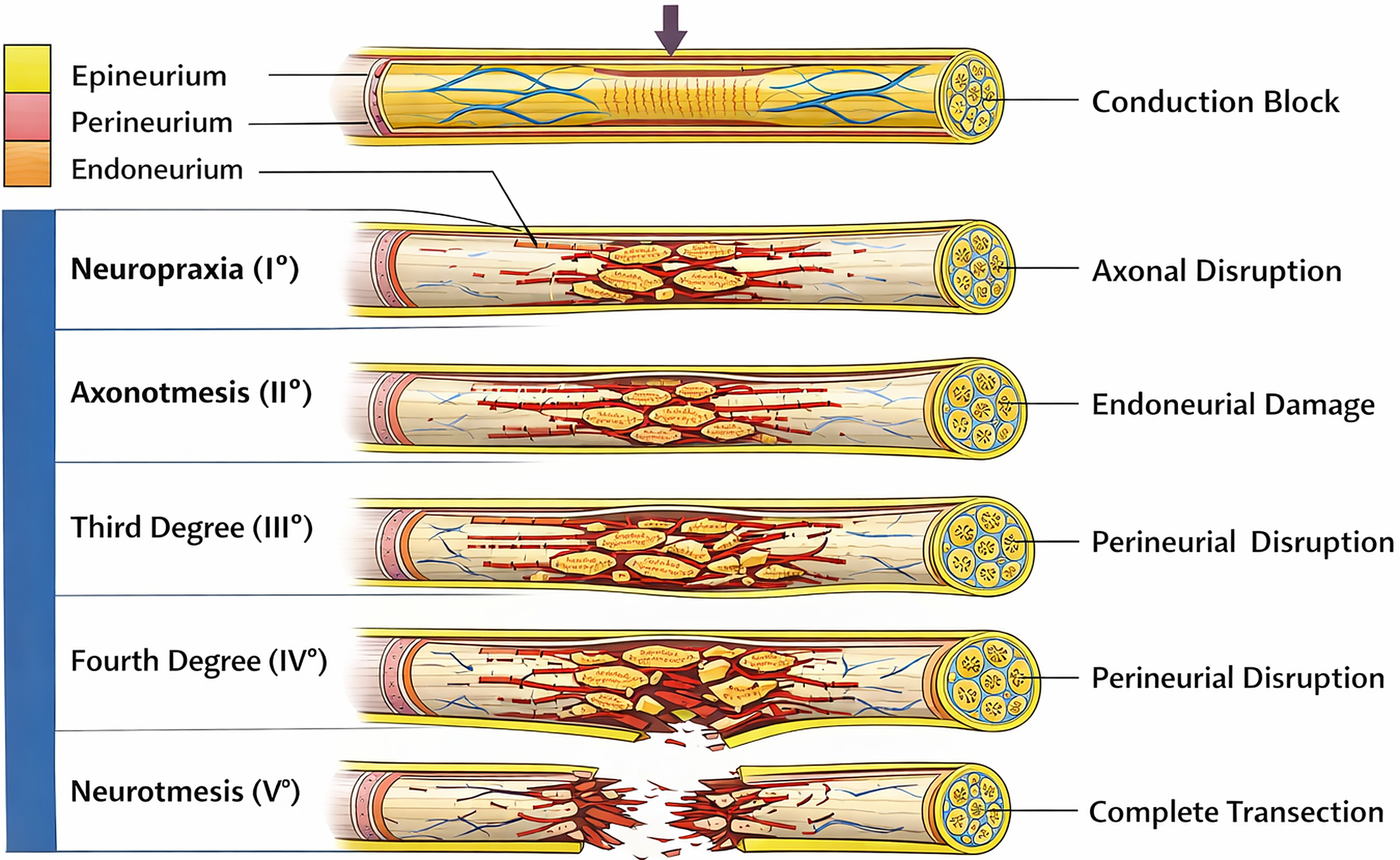
Figure 1. Schematic illustration of peripheral nerve injury grading according to the Seddon-Sunderland classification, showing progressive structural damage from conduction block to complete transection. Reproduced from reference[21]. CC BY 4.0.
Peripheral nerve regeneration is a highly coordinated multicellular process. After injury, local SCs proliferate and undergo phenotypic reprogramming to form longitudinal bands of Büngner[22-24], which provide essential topographical tracks for distal axonal elongation. In clinical practice, however, long-distance defects, fibrotic scar formation, persistent inflammation, and insufficient neurotrophic support collectively hinder this process, often resulting in incomplete axonal regrowth and poor functional recovery. Although autologous nerve grafting remains the clinical benchmark[25-29], its application is limited by restricted donor availability, donor-site functional loss, and secondary surgical morbidity. Allogeneic grafting also faces substantial translational barriers, including immune rejection and potential cross-infection.
To overcome these limitations, tissue-engineered nerve guidance conduits (NGCs) have been widely explored as alternative strategies[30-32]. By mimicking native fascicular architecture, providing supportive three-dimensional channels, modulating local biochemical cues, and delivering bioactive factors, NGCs offer a controllable platform for targeted axonal guidance. Recent progress in this field has shifted the design paradigm from simple single-layer hollow conduits toward multistructured biomimetic systems[33]. The key concept of next-generation NGCs is an integrated, multidimensional strategy that combines ordered directional topographies and interconnected porous matrices with electroactive components and sustained factor-release systems[34-37]. Such a strategy can synergistically address major in vivo challenges, including mismatched degradation and regeneration rates, mechanical collapse, and early inflammatory cascades, thereby supporting high-fidelity peripheral nerve regeneration.
2. Material Systems for the Fabrication of Nerve Guidance Conduits
The choice of fabrication materials is critical for determining the biocompatibility, biodegradability, mechanical performance, and regenerative efficacy of NGCs. An ideal material should not only bridge the nerve gap but also recapitulate key features of the native neural extracellular matrix. Beyond providing passive structural support, biomaterials can also serve as instructive microenvironments that regulate cell adhesion, survival, migration, and differentiation. By mimicking extracellular matrix cues and presenting appropriate biochemical and biophysical signals, biomaterial scaffolds may further support neural regeneration and tissue integration[38]. Current material systems used for conduit construction mainly include synthetic polymers, natural biomaterials, and functionalized composites, whose structural characteristics and modification strategies are summarized in Figure 2.
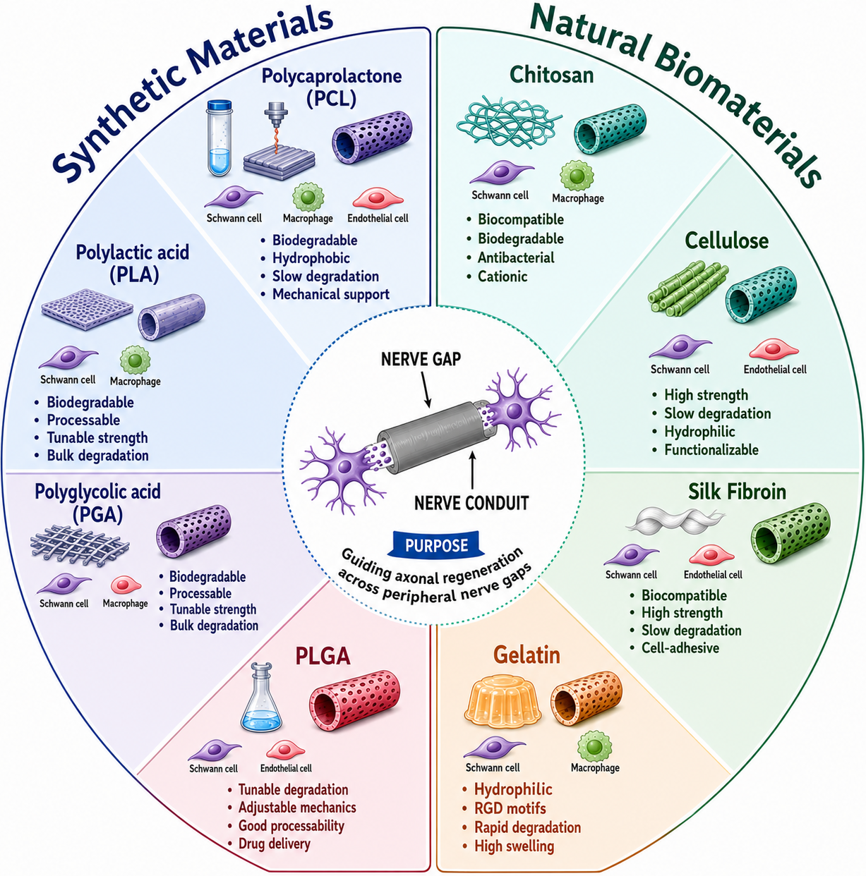
Figure 2. Classification and schematic representation of representative synthetic and natural biomaterials used for nerve guidance conduit fabrication. Created by the authors using Microsoft PowerPoint.
2.1 Synthetic polymer materials
Polycaprolactone (PCL): PCL has attracted considerable attention because of its slow degradation kinetics, favorable processability, and robust mechanical properties. Its good biocompatibility makes it a promising biodegradable candidate for NGC fabrication[39,40]. In particular, PCL can provide sufficient mechanical support during the early post-injury stage and maintain structural integrity over an extended period, making it suitable for repairing extensive or long-distance nerve defects. PCL primarily degrades through progressive hydrolytic cleavage, and its byproducts can be metabolized and eliminated by the host, thereby minimizing systemic toxicity and bioaccumulation[41,42].
Polylactic acid (PLA): PLA is a classical aliphatic polyester-based biodegradable polymer. It ultimately degrades in vivo into non-toxic metabolites and exhibits favorable biocompatibility, making it a commonly used candidate material for constructing NGCs[43,44]. However, its practical application often requires structural or compositional modification to reduce adverse changes in the local microenvironment caused by bulk degradation.
Polyglycolic acid (PGA): PGA can provide initial mechanical support and undergoes in vivo degradation, thereby assisting the regenerative process. Because of its rapid degradation and good biocompatibility[45], PGA was widely used in early NGC studies, where it was often braided or woven into tubular scaffolds to mimic perineurial architecture and guide directional axonal regeneration. The degradation behavior of PGA should be considered when it is used for nerve guidance conduits. Previous studies have shown that PGA-based materials are biodegradable[46]. In a rat sciatic nerve defect model, PGA nerve conduits were found to completely collapse at 12 weeks after implantation, suggesting that their structural stability may become insufficient during a prolonged regeneration period[47]. On the other hand, PGA-collagen conduits have also been reported to support peripheral nerve regeneration[48]. These findings indicate that the degradation rate of PGA needs to be balanced with the structural support required during nerve repair.
Poly(lactic-co-glycolic acid) (PLGA): To overcome the respective limitations of PLA and PGA, copolymerization of lactic acid (LA) and glycolic acid (GA) has been widely used to synthesize PLGA[49-52]. By adjusting the molar ratio of the two monomers, the degradation rate and mechanical properties of PLGA can be tuned over a broad range. This tunability enables better coordination between the duration of structural support and the biological requirements of nerve regeneration.
In summary, the synthetic polymer systems shown on the left side of Figure 2 offer clear advantages in mechanical support, structural stability, and processability. However, their standalone use remains limited. Synthetic polymers used for NGC fabrication differ considerably in their degradation behavior, mechanical properties, and biological performance. PCL provides favorable biocompatibility, processability, and long-term mechanical support[39,40], whereas PLA and PGA are biodegradable polyester materials that have been widely investigated for nerve conduit fabrication[43-48]. In addition, the degradation rate and mechanical properties of PLGA can be adjusted by varying its copolymer composition[49-52]. These characteristics should be considered when selecting and modifying synthetic polymers for peripheral nerve repair.
Accordingly, current NGC design often combines polymer selection with structural engineering and surface modification to improve both mechanical support and biological performance. For example, PCL or PLGA can be used as a mechanical backbone to fabricate aligned nanofibrous conduits via electrospinning. Subsequent surface modification with polydopamine (PDA), deposition of conductive polypyrrole (PPy) nanoparticles, or loading of anti-inflammatory molecules can further improve bioactivity. This integrated strategy maintains long-term physical support while providing electrical signal-transduction pathways for Schwann cells (SCs) migration and axonal extension, and it can also modulate macrophage polarization to suppress inflammation and accelerate peripheral nerve regeneration.
2.2 Natural biomaterials
Natural biomaterials have garnered extensive attention in NGC research owing to their excellent biocompatibility, inherent biodegradability, and cytophilic bioactivity. They not only provide an ideal biochemical microenvironment for nerve regeneration but also effectively promote the adhesion, proliferation, migration, and axonal extension of SCs and neurons.
Silk fibroin (SF): Extracted from natural silkworm silk (such as Bombyx mori) or spider silk, SF is a fibrous, semi-crystalline structural protein composed of light and heavy chains linked by disulfide bonds[53,54]. Glycine, alanine, and serine constitute the primary amino acid components of its heavy chain. The high content of glycine significantly facilitates the tight packing of stable β-sheet nanocrystals within the SF structure[55,56]. In vitro and in vivo studies have confirmed that SF can be smoothly degraded by endogenous enzymes without inducing any overt inflammatory responses or immunogenicity. The application of SF in NGCs primarily benefits from its outstanding mechanical strength and relatively slow enzymatic degradation rate, allowing it to provide long-term physical support for nerve regeneration[57,58]. Furthermore, fabricating SF into nanofibers via electrospinning can highly efficiently promote neural cell adhesion and growth.
Gelatin: As a hydrolytic denaturation product of collagen, gelatin retains the native arginine-glycine-aspartic acid (RGD) cell-adhesion sequences[59-61]. Despite its excellent hydrophilicity and cell-recognition properties, gelatin exhibits a high propensity for water absorption, swelling, and rapid enzymatic erosion in vivo. Consequently, it often necessitates blending with degradable polyesters (such as PCL) or undergoing cross-linking modification prior to use[62-64].
Chitosan: Chitosan is a linear aminopolysaccharide obtained through the deacetylation of natural chitin, consisting of glucosamine and N-acetylglucosamine units. It possesses a unique combination of excellent biodegradability, antibacterial activity, and biocompatibility. When utilized as an NGC material, the abundant free amine functional groups on its surface can engage in specific electrostatic interactions with receptors on neural cell membranes, thereby significantly enhancing cell adhesiveness[63,64]. Various forms of chitosan nerve grafts have shown promising potential for peripheral nerve repair. In vivo studies in rats have demonstrated beneficial effects on nerve regeneration[65], whereas in vitro studies have reported favorable Schwann-cell responses to chitosan-based scaffolds[66]. Recent studies have further explored functionalized chitosan-based NGCs, including aligned PLGA/chitosan nanofibers incorporating bFGF and ROS-scavenging functionality[67] and oriented chitosan scaffolds with sustained NO release[68]. However, its relatively low mechanical strength and poor suture resistance limit its standalone clinical application, frequently requiring blending or compositing with other materials.
Cellulose: As the most abundant natural polymer on Earth, cellulose is linearly arranged by D-glucose units linked via β-1,4-glycosidic bonds. Its derivatives, such as bacterial cellulose and nanocellulose, possess exceptional mechanical strength and an ultra-fine three-dimensional nanonetwork structure that highly mimics the native extracellular matrix[69]. The abundant hydroxyl groups on its surface are extremely amenable to neurotrophic factor grafting or functionalization with conductive materials. Given its inherent lack of specific bioactivity and slow in vivo degradation, cellulose is typically blended with other highly degradable materials, such as chitosan or silk fibroin.
2.3 Composite and emerging material systems
Conventional synthetic polymers and natural biomaterials possess complementary advantages but also distinct limitations in constructing NGCs. Synthetic polymers generally provide favorable mechanical stability, structural integrity, and processability, whereas natural biomaterials offer superior cytocompatibility, hydrophilicity, and extracellular matrix (ECM)-mimicking bioactivity. However, neither category alone fully satisfies the complex requirements of peripheral nerve regeneration. Synthetic polymers often lack intrinsic biological recognition sites, whereas natural biomaterials may suffer from insufficient mechanical strength, excessive swelling, or unstable degradation behavior.
Composite material systems have therefore emerged as important platforms for integrating the complementary strengths of different materials. In these systems, synthetic polymers can provide mechanically robust structural support, while natural biomaterials improve surface hydrophilicity, cell affinity, and ECM-like biochemical signaling. For example, ultrafine fibrous PCL/collagen/human umbilical cord serum (hUCS) conduits have demonstrated enhanced bioactivity compared with pure PCL conduits, illustrating how compositional integration can simultaneously improve structural and biological performance[70]. Similarly, Wang et al. combined aligned poly(L-lactide-co-ε-caprolactone) (PLCL) nanofibers with gelatin methacryloyl (GelMA) hydrogel and multifunctional surface modification, which promoted SC alignment and differentiation while enhancing axonal regeneration, remyelination, and angiogenesis in vivo[71]. Rodriguez-Sanchez et al. further developed 3D-printed PCL/GelMA conduits with controlled fibroblast growth factor 2 (FGF-2) release, demonstrating that the integration of structural support with sustained biochemical signaling can improve peripheral nerve regeneration[72]. Together, these studies illustrate that the rationale for composite design is shifting from simply compensating for the limitations of individual materials toward the coordinated integration of multiple regenerative functions.
Emerging material systems further extend this concept from structural complementation toward multifunctional regulation. Conductive composites can provide electroactive cues, biomimetic matrix-based systems can enhance biological signaling, and bioactive factor-loaded systems can regulate specific aspects of the regenerative microenvironment. However, increasing compositional and functional complexity also introduces important trade-offs. The incorporation of multiple components may complicate fabrication, affect degradation behavior and long-term stability, increase batch-to-batch variability, and create additional challenges for sterilization, standardization, and eventual clinical translation. Therefore, the optimal NGC design should not simply maximize the number of functional components, but rather achieve a rational balance among structural integrity, biological activity, manufacturability, and translational feasibility.
Table 1 and Table 2 summarize representative synthetic/natural materials and composite/emerging systems, respectively. Together, these materials provide a critical foundation for next-generation multifunctional biomimetic NGCs by integrating mechanical support with biological, electrical, immunomodulatory, antioxidant, and pro-vascularization functions. Nevertheless, material composition alone is insufficient to achieve optimal regenerative outcomes. Controlled fabrication strategies, including electrospinning, molding, freeze-drying, and 3D bioprinting, remain essential for translating these materials into conduits with tailored macroscopic geometry, interconnected porous structures, aligned micro/nanotopographies, and spatially organized functional cues.
Table 1. Representative synthetic and natural biomaterials for nerve guidance conduit fabrication.
| Material category | Representative materials | Advantages | Limitations | Ref |
| Synthetic polymer | PCL | Good processability, slow degradation, strong mechanical support, long-term stability. | Hydrophobic, lacks cell-recognition motifs, relatively slow degradation. | [73-77] |
| Synthetic polymer | PLA | Biocompatible, tunable mechanical strength, suitable for electrospinning/3D printing. | Bulk degradation may generate acidic products; brittle and hydrophobic without modification. | [78-82] |
| Synthetic polymer | PGA | Rapid biodegradation, good initial mechanical support, clinically applied. | May degrade too quickly for long-gap defects; acidic byproducts; poor suturability. | [83,84] |
| Synthetic polymer | PLGA | Adjustable degradation rate, good processability, suitable for drug/factor delivery. | Acidic byproducts may cause local inflammation; hydrophobic, limits early cell adhesion. | [85-90] |
| Natural biomaterial | SF | Biocompatible, strong, slow enzymatic degradation, supports neural cell adhesion. | Processing affects crystallinity, degradation, and reproducibility. | [91-94] |
| Natural biomaterial | Gelatin | Good biocompatibility and cell affinity; easy to process and functionalize. | Rapid swelling and enzymatic degradation; weak mechanical strength if unmodified. | [95-103] |
| Natural biomaterial | Chitosan | Biodegradable, antibacterial, cytocompatible, supports neural cell adhesion. | Low mechanical strength and poor suturability. | [104-109] |
| Natural biomaterial | Cellulose/bacterial cellulose | High mechanical strength, porous structure, hydroxyl groups for functionalization. | Limited intrinsic neurobioactivity; slow degradation, usually needs blending. | [110-114] |
| Natural biomaterial | Collagen | Excellent cytocompatibility and ECM-mimicking bioactivity. | Weak mechanical strength, fast degradation, batch variability, potential immunogenicity. | [115-119] |
| Natural biomaterial | Alginate | Mild gelation, biocompatible, suitable for cell/factor encapsulation. | Lacks intrinsic cell adhesion, low mechanical strength, difficult degradation control. | [120-125] |
| Natural biomaterial | HA | Highly hydrophilic, anti-inflammatory, easy chemical modification. | Poor mechanical strength, rapid degradation if not crosslinked. | [126-127] |
PCL: polycaprolactone; HA: hyaluronic acid; ECM: extracellular matrix; SF: silk fibroin; PLGA: poly(lactic-co-glycolic acid); PGA: polyglycolic acid; PLA: polylactic acid.
Table 2. Representative composite and emerging material systems for nerve guidance conduit fabrication.
| Material category | Representative materials | Advantages | Limitations | Ref |
| Synthetic/natural composites | PCL/gelatin, PCL/collagen, PCL/SF, PLGA/chitosan, SF/chitosan, PCL/cellulose | Balance mechanical support, biodegradation, cell adhesion, and ECM-mimicking bioactivity. | Component ratio, phase separation, reproducibility, and sterilization require careful optimization. | [128-131] |
| Conductive composites | PPy, PEDOT: PSS, GO, rGO, CNT-containing conduits | Promote SC alignment, neurite extension, and electrical signal transduction. | Potential cytotoxicity, poor biodegradability of some conductive fillers, and unstable long-term conductivity. | [132-136] |
| Bioactive factor-loaded composites | NGF, VEGF, bFGF, exosomes, microRNAs, antioxidants, bioactive ions | Provide sustained biochemical cues, promote angiogenesis, immunomodulation, antioxidant defense, and axonal regeneration. | Release kinetics are difficult to control; bioactivity may decrease during fabrication or storage. | [137-139] |
| Biomimetic matrix-based systems | dECM | Strong biomimetic bioactivity; promotes SC adhesion, axonal extension, and angiogenesis. | Source variability, possible immunogenicity if decellularization is incomplete, weak mechanical strength alone, and standardization challenges. | [140-141] |
| Flexible synthetic polymers | PU | Good elasticity, flexibility, and fatigue resistance; suitable for mechanically compliant conduits. | Biodegradability depends on formulation; limited intrinsic bioactivity. | [142-143] |
| Biodegradable synthetic polyesters | PPDO/PDS | Good flexibility, biodegradability, and surgical handling; reinforces natural biomaterials. | Limited cell-recognition ability and possible acidic degradation products. | [144] |
| Bio-based biodegradable polyesters | PHAs | Good mechanical properties, natural-source biodegradable, potential long-term support. | Brittleness, slow degradation, processing difficulty may limit standalone use. | [145] |
PPy: polypyrrole; PEDOT:PSS: poly(3,4-ethylenedioxythiophene):poly(styrene sulfonate); GO: graphene oxide; rGO: reduced graphene oxide; CNT: carbon nanotube; NGF: nerve growth factor; VEGF: vascular endothelial growth factor; bFGF: basic fibroblast growth factor; dECM: decellularized extracellular matrix; PU: polyurethane; PPDO/PDS: poly(p-dioxanone); PHAs: polyhydroxyalkanoates; CNPs: cerium oxide nanoparticles; PCL: polycaprolactone; SF: silk fibroin; PLGA: poly(lactic-co-glycolic acid); SC: Schwann cell; ECM: extracellular matrix.
3. Fabrication Methods of NGCs
The fabrication method has a direct influence on the structural and mechanical characteristics of NGCs. Different techniques can produce marked differences in conduit porosity, pore organization, fiber diameter and alignment, mechanical strength, and internal architecture. Molding and freeze-drying are relatively simple approaches for producing tubular and porous structures, although precise control over pore morphology is limited. In contrast, 3D printing enables greater control over complex geometries and multichannel architectures, whereas electrospinning is particularly suitable for generating micro- and nanofibrous structures with controlled fiber orientation. These structural differences can further affect nutrient transport, mechanical support, SCs migration, and directional axonal growth. Therefore, the choice of fabrication method should be determined by both the structural requirements of the conduit and the biological demands of peripheral nerve regeneration.
Molding method: As a conventional technique characterized by straightforward operation, this method involves injecting a polymer solution into a specific mold, followed by low-temperature freezing and solidification, and subsequent freeze-drying to remove the solvent to yield the conduit. Although this approach can achieve high overall porosity, it suffers from inherent defects, including non-uniform pore distribution and irregular microstructures, which may adversely affect the homogeneous ingrowth of neural cells[146]. To provide a clearer visual summary, the key fabrication steps and structural features of molding-derived NGCs are presented in Figure 3.
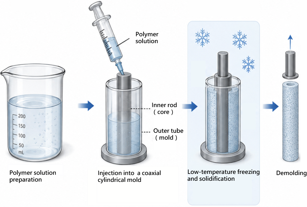
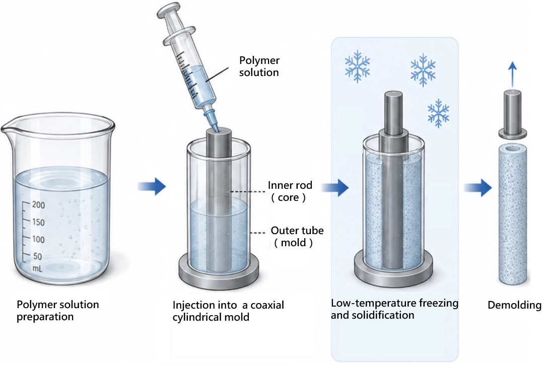
Figure 3. Schematic illustration of the molding process for fabricating NGCs. Created in BioRender. Xu, H. (2026) https://BioRender.com/mki4lv4. NGCs: nerve guidance conduits.
Freeze-drying method: Under low-pressure and low-temperature conditions, the solvent bypasses the liquid phase to directly complete a solid-to-gas sublimation transition, thereby constructing a porous network structure with high porosity within the polymer system[147,148]. While this provides an expansive space for nutrient permeation and cell ingrowth, the resulting pore morphologies and spatial distributions are relatively irregular, often making it difficult to effectively guide the directional growth of neural cells. To overcome the limitations inherent in standalone conventional techniques, contemporary research frequently coordinates molding with freeze-drying technologies to construct composite, functionalized NGCs that possess both precise macroscopic configurations and highly interconnected microscopic pores. For instance, an enzymatic cross-linking system can be utilized to thoroughly blend a SF solution with electroactive poly(3,4-ethylenedioxythiophene) (PEDOT) nanoparticles, which is then injected into a coaxial cylindrical mold for rapid gelation and shaping at 37 °C. Subsequently, liquid nitrogen low-temperature freezing is applied to assist demolding, followed by ethanol immersion to highly efficiently induce the structural transition of silk fibroin from an amorphous state into stable β-sheet crystals. Finally, upon removing moisture via freeze-drying, a conductive biocomposite nerve guidance conduit with a homogeneous structure and highly controllable morphology is obtained (Figure 4).
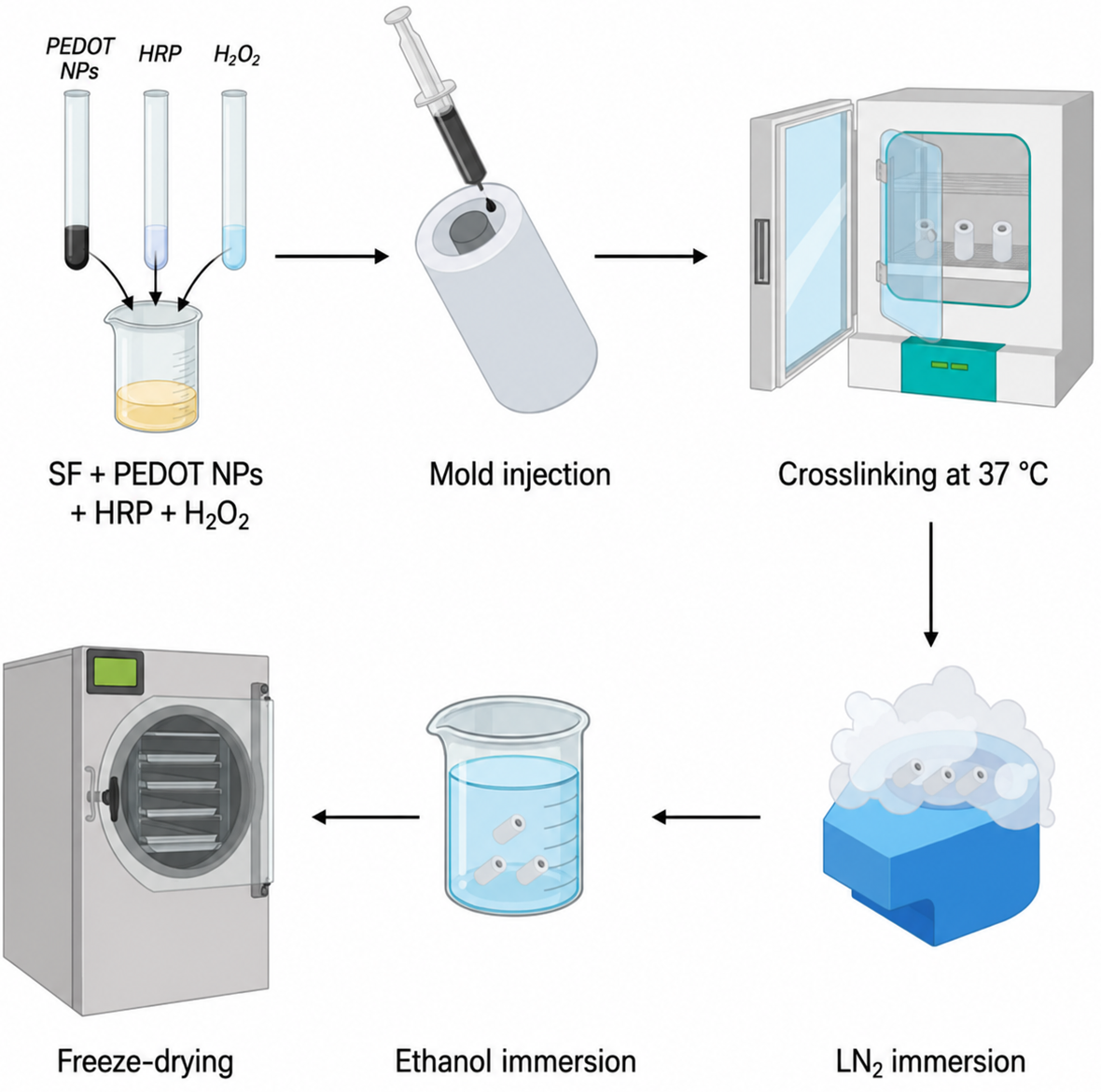
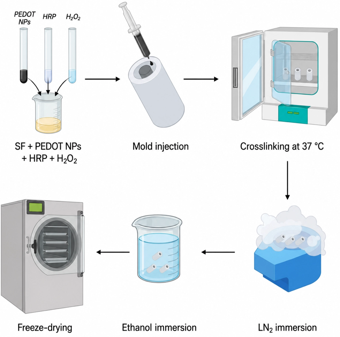
Figure 4. Schematic illustration of the fabrication process of the silk fibroin/PEDOT nanoparticle conductive composite scaffold. Reproduced from reference[149]. CC BY 4.0. PEDOT: poly(3,4-ethylenedioxythiophene); SF: silk fibroin; NPs: nanoparticles; HRP: horseradish peroxidase.
3D printing: 3D printing technologies for fabricating NGCs are predominantly categorized into light-based and extrusion-based modalities[150-152]. Light-based printing utilizes specific wavelengths of light to crosslink photocurable bioinks in a layer-by-layer manner[153]. Owing to its superior microscale resolution, this approach enables the precise construction of biomimetic, multichannel honeycomb architectures featuring highly aligned microchannels (e.g., 200 μm), thereby providing exceptional spatial cues to guide directional axonal regeneration[154,155]. Conversely, extrusion-based printing relies on mechanical or pneumatic pressure to dispense shear-thinning fluid inks through a micro-nozzle for shape fixation. While it accommodates a broader spectrum of biomaterials, its structural fidelity remains inherently constrained by the nozzle diameter, presenting a critical bottleneck for fabricating intricate microscale structures. To circumvent this limitation, Maeng et al.[156] integrated light-based printing with a tape-casting process, employing a high-viscosity GelMA/polyethylene glycol diacrylate (PEGDA) composite hydrogel as the bioink. By incorporating a light absorber to finely tune the optical path length, they effectively mitigated light scattering and subsequent overcuring. This precision-control strategy yielded high-fidelity, biomimetic honeycomb conduits with perfectly aligned 200 µm channels, achieving a high degree of anatomical mimicry of native peripheral nerve fascicles (Figure 5).
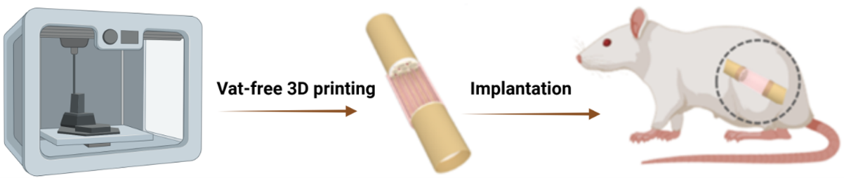
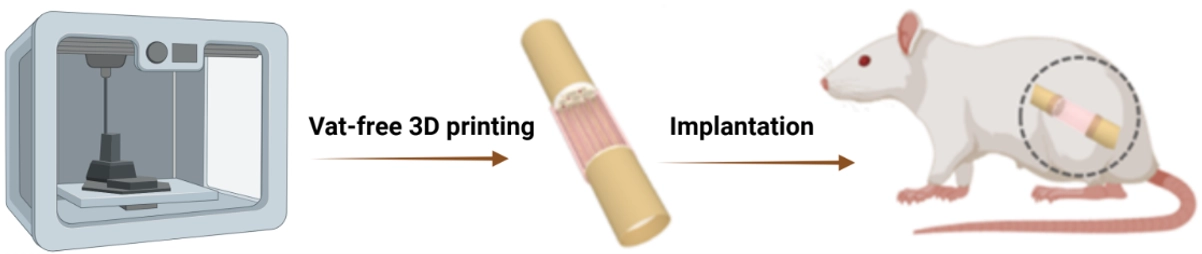
Figure 5. Schematic illustration of the fabrication and implantation of a 3D-printed nerve guidance conduit. Created in BioRender. Xu, H. (2026) https://BioRender.com/j088ft7.
Electrospinning method: Among the various NGC fabrication techniques, electrospinning has emerged as the most widely used method due to its unique capability to produce nanofibers. By applying a high voltage, the polymer solution is stretched into continuous nanoscale fibers, which are collected to form a nerve guidance conduit with a nanoscale architecture (Figure 6). The distinct advantage of electrospinning lies in its ability to create highly microscopic fibrous structures that mimic the native ECM, providing an ideal substrate for cell adhesion, proliferation, and migration. Moreover, electrospinning allows control over fiber diameter, porosity, and mechanical properties through the formulation of the spinning solution, meeting the requirements for different nerve injury repairs. These features make electrospinning one of the most promising technologies for contemporary nerve guidance conduit fabrication.
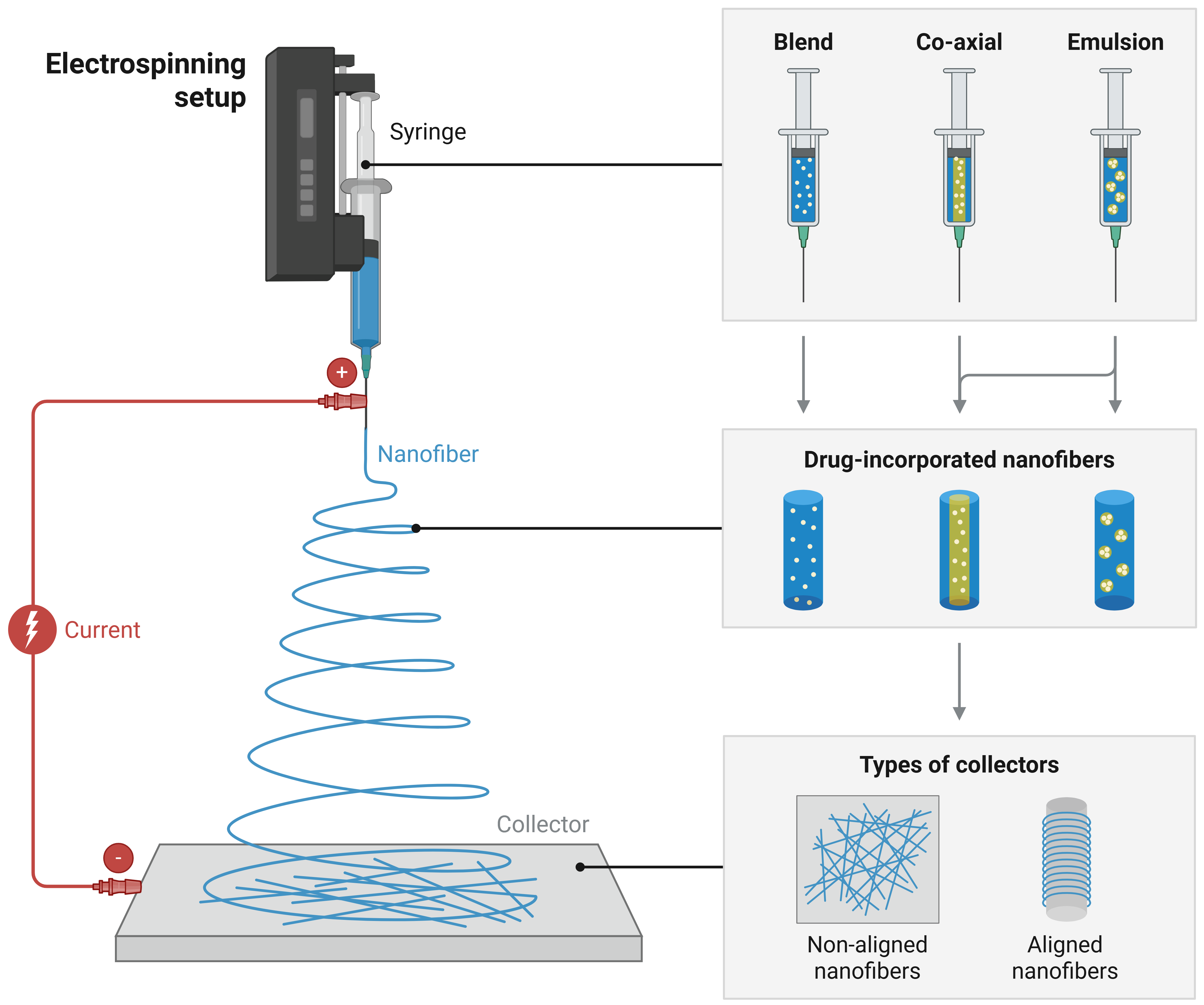
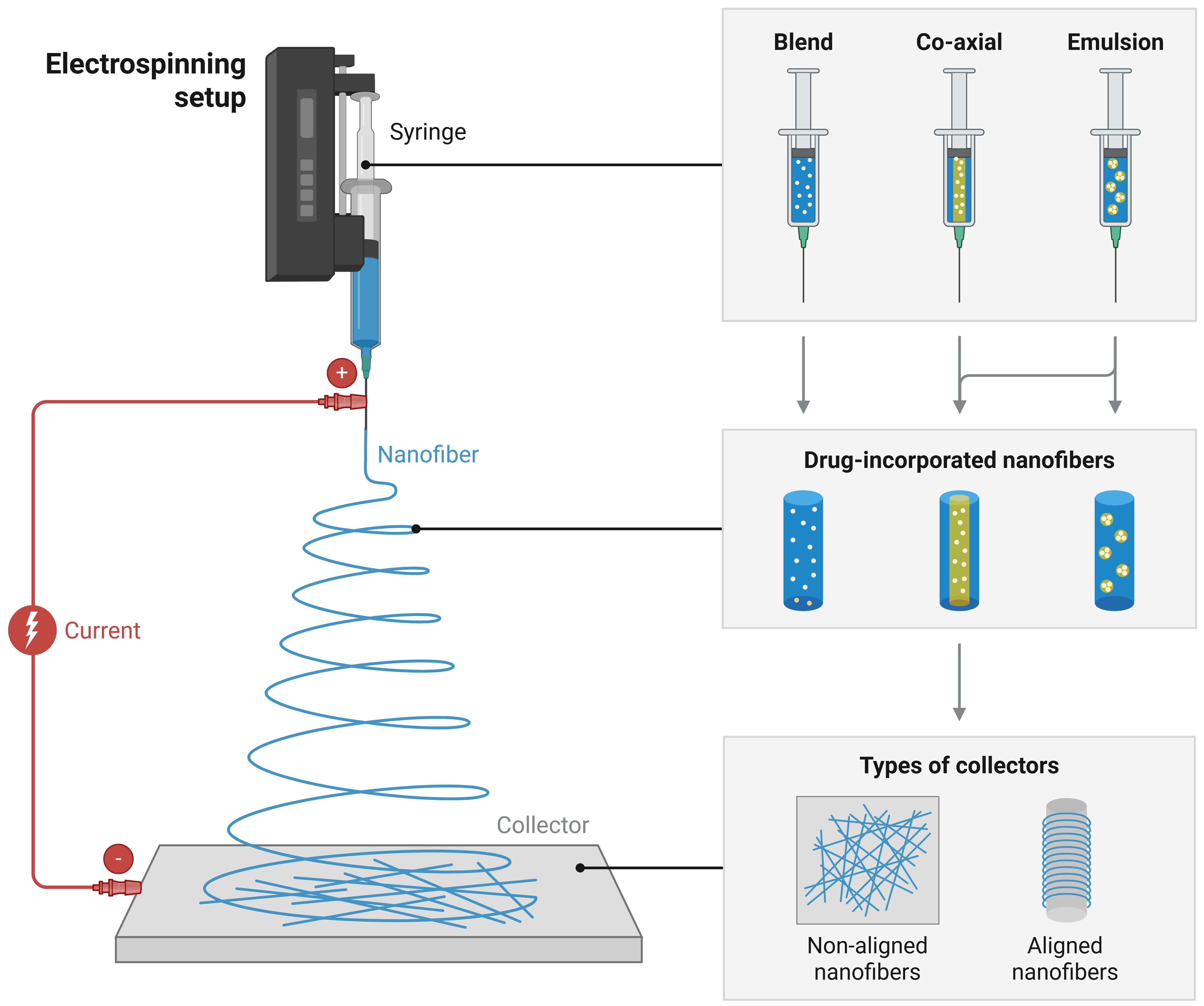
Figure 6. Schematic illustration of the electrospinning process for nerve guidance conduit fabrication. Created in BioRender. Xu, H. (2026) https://BioRender.com/nz9zf08.
A comparative summary of the major fabrication methods for NGCs, including their respective advantages, limitations, and representative references, is provided in Table 3. This table is intended to offer a concise yet comprehensive overview of each method, facilitating a clear understanding of how different fabrication strategies, such as molding, freeze-drying, electrospinning, and 3D bioprinting, affect conduit architecture, porosity, micro/nanotopography, mechanical properties, and potential for functionalization. Readers can use this summary to quickly assess the suitability of each method for specific design requirements and experimental objectives in peripheral nerve repair.
Table 3. Comparative analysis of various fabrication methodologies for NGCs.
| Fabrication Method | Advantages | Disadvantages | Ref |
| Mold Casting | • Simple and low-cost • Suitable for mass replication • Good control of macroscopic shape | • Limited interconnected microporosity • Dense conduit walls may hinder nutrient diffusion | [157,158] |
| Freeze-Drying | • Mild processing conditions • High porosity • Facilitates nutrient and oxygen transport | • Limited macroscopic shape control • Poor pore-size uniformity • Weak mechanical stability | [159,160] |
| 3D Printing (Additive Manufacturing) | • Enables complex geometries • Precise control of material composition and gradients • Good control of internal architecture | • High cost for high-resolution printing • Relatively slow printing speed • Limited nanoscale resolution | [161-164] |
| Electrospinning | • Produces ECM-like nanofibers • High specific surface area • Supports SC adhesion and alignment • Enables oriented fiber structures | • May involve organic solvents • Residual-solvent concerns require appropriate post-treatment | [165-168] |
NGCs: nerve guidance conduits; ECM: extracellular matrix; SC: Schwann cell.
4. Multifunctional Synergistic Regulation Strategies for Nerve Guidance Conduits
4.1 Electrical functionalization
Next-generation NGCs integrate electrical functionalization, anti-inflammatory/antioxidant regulation, and pro-vascularization strategies to remodel the local regenerative microenvironment (Figure 7). Electrical functionalization provides electroactive and topographical cues to guide SCs migration and axonal extension[169,170]. Anti-inflammatory/antioxidant regulation and pro-vascularization strategies are discussed separately in Section 4.2 and Section 4.3, respectively, where representative material systems and their biological effects are summarized. Together, these strategies provide a framework for multifunctional NGC design.
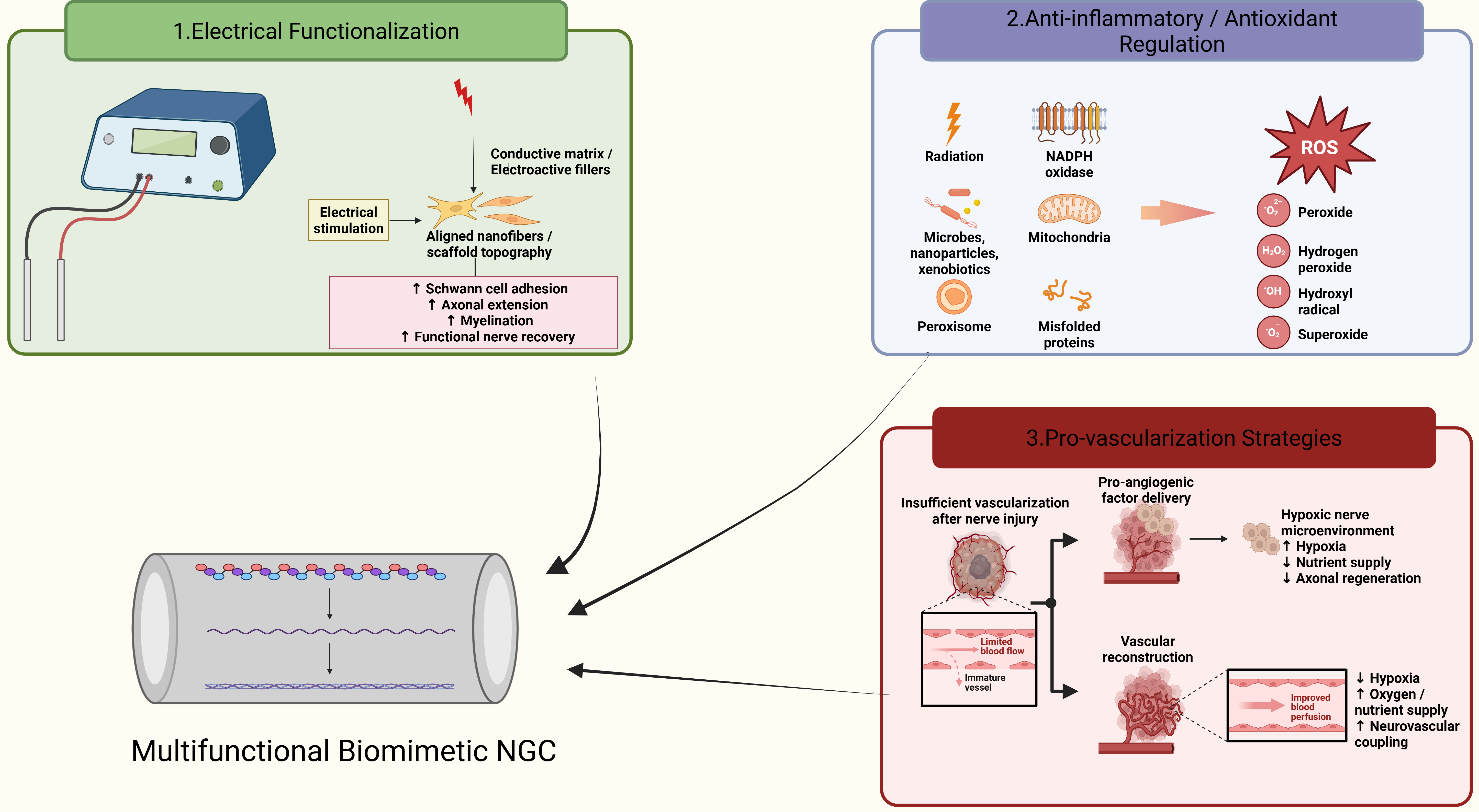
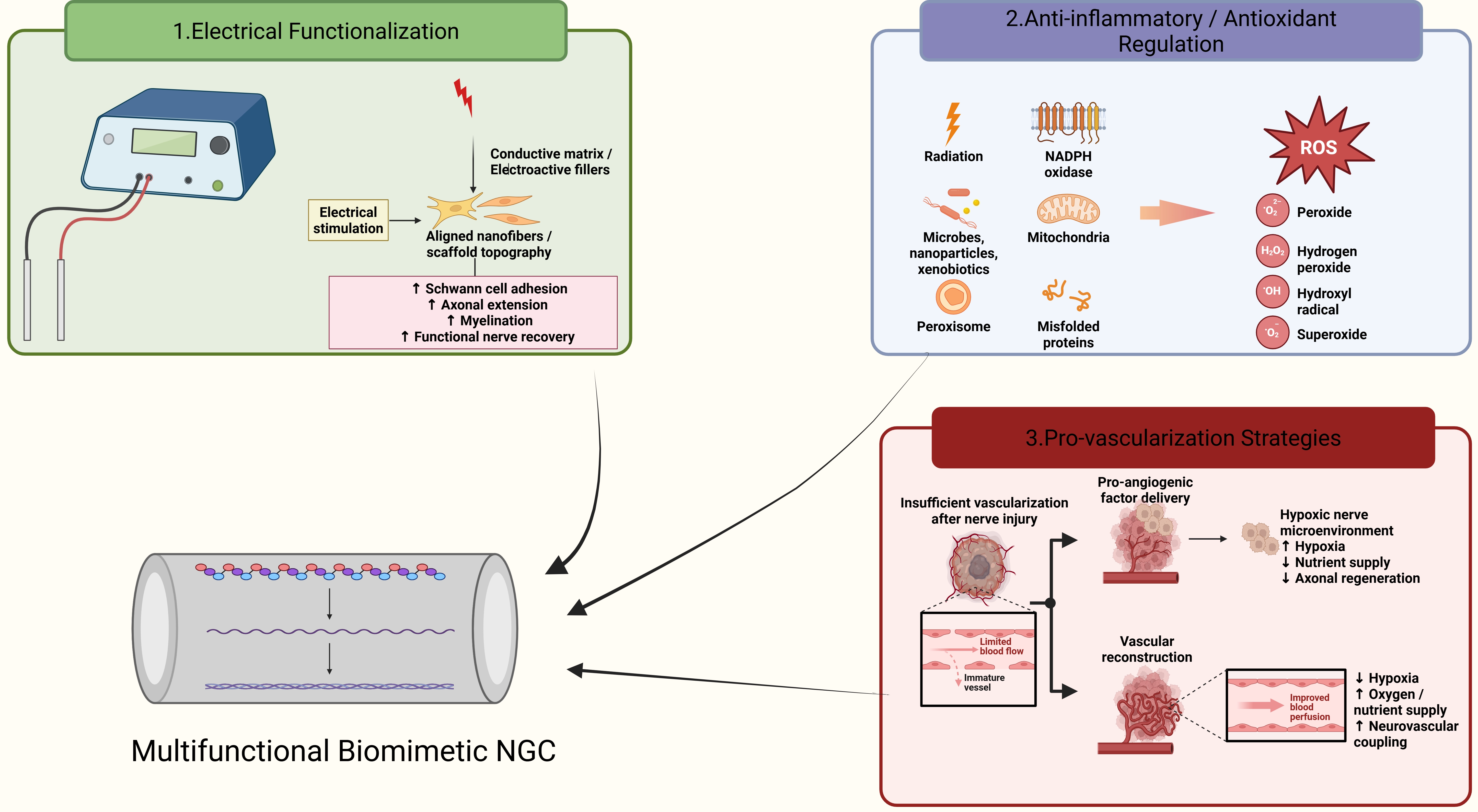
Figure 7. Schematic illustration of multifunctional synergistic regulation strategies for biomimetic NGCs, including electrical functionalization, anti-inflammatory/antioxidant regulation, and pro-vascularization approaches. Created in BioRender. Xu, H. (2026) https://BioRender.com/6qb3z1i. NGCs: nerve guidance conduits; ROS: reactive oxygen species.
The inherent high sensitivity of the PNS to electrical signals makes the fabrication of biomimetic functionalized NGCs a pivotal strategy for reshaping damaged electrophysiological microenvironments and directionally guiding the growth of axons and SCs[171-175]. To achieve stable electronic/ionic conduction and precisely match the physiological electrical demands of injured nerves, researchers have made significant breakthroughs in conductive matrix development and high-precision fabrication. For instance, Geiger et al.[176] utilized spinning technology to successfully fabricate mechanically robust, pure poly(3,4-ethylenedioxythiophene):poly(styrene sulfonate) (PEDOT:PSS) microfiber conduits with precisely controllable fiber diameters ranging from 1.2 to 3 μm. Concurrently, Lu et al.[177] constructed uniform PPy coatings on the surface of natural SF nanofibers via electrospinning combined with in situ chemical polymerization. This approach achieved fine-tuning of the conduit conductivity within the range of 10-5 to 10-3 S/cm, thereby significantly enhancing SC attachment and proliferation.
Building upon these advancements, integrating highly conductive pathways with micro-topographical geometries endows conduits with dual biomimetic functionalities: spatial “contact guidance” and “localized electric field regulation”. Song et al.[178] ingeniously exploited the electrostatic interaction between negatively charged hydroxyethyl cellulose (HEC) and positively charged PEDOT to achieve continuous polymerization, processing the matrix into a helical conduit filled with aligned nanofibers (PLCL/HEC-PEDOT). This design not only substantially enhanced the electrochemical stability of the material, but its unique helically aligned topographical cues also significantly upregulated the expression of neural-specific proteins.
This combined strategy of “topographical guidance and electrical functionalization” has been further sophisticated in multilayered composite scaffolds and exogenous stimulation combination therapies[179-184]. For instance, the “dual-regulation” biomimetic scaffold proposed by Liu et al.[185] integrated an aligned nanofibrous inner layer with a conductive hydrogel outer layer to synergistically induce cell proliferation and directional migration. Similarly, Qi et al.[186] constructed bilayer NGCs comprising an inner layer of aligned conductive PEDOT:PSS/chitosan (CS) fibers and a high-strength hydrogel outer layer. Combined with clinical percutaneous electrical stimulation therapy, they demonstrated that this aligned conductive network could efficiently and targetingly transmit exogenous electrical signals to the lesion site, achieving a robust integration of active electrical stimulation and passive topographical guidance.
To bypass the constraints of conventional external power supplies or transcutaneous devices, intelligent piezoelectric wireless stimulation systems that spontaneously generate bioelectricity from tissue deformation are emerging as the frontier of next-generation electroactive NGC design. Moezzi et al.[187] explored the feasibility of using PLA/ZnO composite nanofibrous scaffolds, which possess optimized self-generating electrical output performance, as wireless electrical stimulators to promote PC12 cell growth. To overcome the bottleneck where self-generated electrical signals suffer from limited propagation within insulating polymer matrices, Bian et al.[188] reported a promising “dual electroactivity” synergistic strategy. By introducing highly conductive reduced graphene oxide (rGO) into a CS/poly (L-lactic acid) (PLLA) piezoelectric matrix, rGO served as an efficient electrical signal transmission network that perfectly synergized with the mechanical-to-electrical conversion functionality of PLLA. This integration endowed the composite conduit (CGP) with outstanding antibacterial activity, tensile strength, and the capacity for autonomous electrical regulation of the regenerative microenvironment.
In addition to electronic conductive materials, ionic conductive hydrogels have also been explored for peripheral nerve repair. Electrical conduction in these hydrogels mainly depends on the migration of mobile ions within hydrated polymer networks, which is closer to the ionic conduction behavior of biological tissues. Krishnadoss et al. developed a GelMA-based hydrogel containing a bio-ionic liquid, providing a soft and hydrated conductive interface[189]. More directly, Xu et al. constructed a zwitterionic conductive hydrogel-based nerve guidance conduit and demonstrated its ability to support SCs growth, neurite extension, and peripheral nerve regeneration in a rat sciatic nerve defect model[190]. These studies suggest that ionic conductive hydrogels may provide an alternative to conventional electronic conductive systems. However, their long-term stability, mechanical properties, and in vivo safety still require further investigation.
4.2 Anti-inflammatory and antioxidant modifications
Following PNI, oxidative stress, persistent inflammation, and impaired vascular support can create an unfavorable microenvironment that limits axonal regeneration and functional recovery[191,192]. Consequently, the development of multifunctional composite NGCs endowed with antioxidant and immunomodulatory capabilities has attracted widespread attention. Studies indicate that incorporating polyphenols or biometabolites with inherent antioxidant and anti-inflammatory activities can effectively suppress the early cascade of inflammatory responses. For example, Fang et al.[193] embedded chlorogenic acid (CGA)-loaded GelMA microspheres into PCL conduits, successfully improving the local immune microenvironment via the natural antioxidant bioactivity of the polyphenol. Bi et al.[194] constructed topographically aligned PCL/GelMA composite conduits to synergistically deliver the macrophage-specific metabolite 4-octyl itaconate. Their work demonstrated that this system effectively blocked tumor necrosis factor alpha (TNF-α)-induced nuclear factor kappa B (NF-κB) pathway activation, thereby exerting potent immunomodulatory and anti-inflammatory effects.
Crucially, the organic integration of antioxidant and anti-inflammatory microenvironmental regulation with electroactive signal transduction has become the core strategy for multifunctional conduit design in recent years. Song et al.[195] injected a luteolin (Lut)-loaded conductive hydrogel into nanofibrous conduits, leveraging the exceptional anti-inflammatory and antioxidant properties of luteolin to successfully induce macrophage polarization toward the pro-healing M2 phenotype, which provided a robust microenvironmental guarantee for sciatic nerve defect repair in rats. In another study, the same team achieved a similar synergistic remodeling of electrical conduction and the anti-inflammatory niche by introducing simvastatin (SIM) alongside a conductive PEDOT component[196]. Additionally, the electroactive dual-drug composite conduit developed by Li et al.[197] exhibited significant anti-inflammatory activity and a potent capacity to promote SC adhesion and differentiation through the sustained release of emodin (RH) and nerve growth factor (NGF). Meanwhile, the L-Zein composite conduit reported by Xu et al.[198] co-encapsulated melatonin (MT) and graphene oxide (GO) to balance stable electrical conduction with excellent free radical scavenging (antioxidant) capabilities, effectively suppressing inflammatory responses mediated by RAW264.7 macrophages.
Beyond solid active components, the monolithic delivery of gaseous signaling molecules offers new paradigms for antioxidant therapies. Addressing the microenvironmental imbalance induced by oxidative stress and inflammation, Huo et al.[199] respectively proposed intelligent delivery schemes for the sustained and orderly release of nitric oxide (NO). These included a temperature-sensitive hydrogel-based NO sustained-release nanoparticle system and a tannin acid/selenamine-catalyzed coating-based NO-releasing conduit. While continuously reducing the levels of pro-inflammatory cytokines (such as TNF-α and interleukin-6 (IL-6)) and mitigating oxidative stress damage, these systems pleiotropically enhanced localized microvascularization and neural axonal regeneration.
4.3 Pro-vascularization strategies
The successful regeneration of PNIs is a complex physiological process that relies heavily on energy and oxygen supply. Given that vascular networks are intricately interwoven within nerve bundles, neural regeneration is inherently coupled with angiogenesis[200-204]. Traditional artificial NGCs often suffer from a severe lack of early blood supply, which frequently leads to the stagnation of long-distance nerve regeneration. To break through this bottleneck, constructing engineered multifunctional NGCs with dual pro-vascularization and neurogenerative properties has emerged as a prominent research hotspot in tissue engineering[205,206].
First, by developing spatio-temporally remodeled delivery systems, the co-encapsulation or gradient-controlled release of conventional organic growth factors (e.g., vascular endothelial growth factor (VEGF)/basic fibroblast growth factor (bFGF)) or natural drug monomers with pro-vascularization activities within the conduit matrix can precisely induce the directional migration and lumen formation of endothelial cells (ECs). Beyond conventional organic components, the modulation of EC behaviors using inorganic functional ions (Cu2+, Co2+, and Mg2+) with unique chemical stabilities is gaining substantial traction. A Cu-doped mesoporous bioactive glass (Cu-MBG)-based system has been reported to promote angiogenic responses in endothelial cells. In this study, Cu-MBG enhanced human umbilical vein endothelial cell (HUVEC) migration and tube formation and increased the expression of angiogenesis-related genes, including hypoxia-inducible factor alpha (HIF-1α), angiogenin (ANG), endothelial nitric oxide synthase (eNOS), and kinase insert domain receptor (KDR)[207].
Furthermore, mimicking endogenous hypoxic responses or utilizing gaseous small molecules for microenvironmental remodeling can further optimize the vascularization process. For instance, introducing photo-responsive composite systems[208], such as novel zinc-citrate metal-organic frameworks (Zn-CA MOFs), to artificially control the co-release of hydrogen sulfide (H2S) and Zn2+, can timely and effectively scavenge free radicals, regulate inflammatory responses, and restore mitochondrial energy metabolism, thereby reversing the microenvironmental imbalance caused by insufficient blood supply. Concurrently, emerging delivery strategies utilizing small-molecule/nucleic acid non-biological agents have exhibited immense potential for synergistic therapy. Specifically, the continuous release of secreted frizzled-related protein-2 (SFRP2) from polydopamine (PDA)-modified methacrylated gelatin (GelMA) hydrogel nanofibers can enhance the lumen formation of HUVECs via calcium-dependent calcineurin/NFATc3 pathways[209]. In the miR-29a@Cu-MBG-NH₂ conduit system, sustained release of miR-29a@Cu-MBG effectively delivered miR-29a into PC12 cells, leading to increased expression of the nerve regeneration-associated genes GAP-43 and NF-200 and enhanced neurite outgrowth[207]. Separately, Cu-MBG promoted HUVEC migration and tube formation and increased the expression of angiogenesis-related genes, including HIF-1α, ANG, eNOS, and KDR[207].
From a structural perspective, porous and multichannel NGCs can provide both molecular exchange and directional guidance. Porous NGC walls permit the transport of nutrients and molecular signals and can influence the behavior of Schwann cells and vascular endothelial cells[210]. In a murine sciatic nerve transection model, Schimelman et al. designed a multichannel hydrogel conduit with regularly spaced micropores oriented orthogonally to the axon-guidance channels to investigate extraneural vascular infiltration and nerve regeneration[161]. These findings, together with evidence supporting the importance of vascularization in peripheral nerve repair[205,206], suggest that coordinated control of pore architecture, axonal guidance channels, and vascular access may provide a more supportive regenerative microenvironment.
In summary, the multi-in-one integration of pro-vascularization strategies with conductive, anti-inflammatory, and antioxidant properties represents the cutting-edge development of next-generation, high-quality nerve repair conduits.
5. Conclusions and Perspectives
PNI remains a formidable clinical challenge due to the complex interplay of axonal regeneration, SC migration, remyelination, inflammatory modulation, and vascular reconstruction. In this review, we summarized recent advances in multifunctional biomimetic NGCs, emphasizing the integration of synthetic and natural biomaterials, advanced fabrication techniques, and microenvironmental functionalization strategies. Synthetic and natural biomaterials provide complementary mechanical and biological properties, and their combination has become an important strategy for balancing structural support, degradation behavior, and bioactivity.
Advanced manufacturing approaches, including electrospinning, molding, freeze-drying, and 3D printing, enable precise control over macro- and micro-architectures, including aligned nanofibers, multichannel structures, and porous scaffolds, which collectively provide topographical guidance and structural support. Beyond structural design, multifunctional strategies integrating electrical functionalization, anti-inflammatory and antioxidant regulation, and pro-vascularization interventions synergistically remodel the local regenerative microenvironment. Electrical cues facilitate directional axonal growth and SCs alignment, antioxidant and immunomodulatory components reduce oxidative stress and promote pro-regenerative macrophage polarization, and vascularization strategies enhance endothelial migration, angiogenesis, and nutrient/oxygen supply. However, increasing the number of functional components does not necessarily lead to better regenerative outcomes, and the complexity of NGC design should be balanced against its biological requirements and practical feasibility.
Despite these advances, several challenges still need to be addressed before advanced NGCs can move toward clinical application. The degradation rate of the conduit should match the course of nerve regeneration while maintaining sufficient mechanical stability and long-term biocompatibility. More complex and multicomponent designs may also make large-scale manufacturing, batch-to-batch reproducibility, sterilization, and storage more difficult. In addition, much of the current evidence is still derived from in vitro experiments and small-animal models. Further validation in large-animal models and clinically relevant nerve defects is therefore necessary. Standardized evaluation methods and appropriate regulatory requirements will also be important for translating promising experimental designs into clinically applicable products.
Future studies should consider regenerative performance and translational feasibility together. Intelligent and patient-specific NGCs integrating multiscale architectures, aligned nanofibers, and gradient functionalization may provide more precise regulation of electrical, immune, oxidative, and vascular microenvironments. Stem cell-derived matrices, exosomes, gene-regulatory molecules, and self-powered bioelectronic systems may further expand the functional potential of NGCs. Nevertheless, future designs should remain reproducible, manufacturable, safe, and suitable for long-term implantation. The continued integration of biomaterials science, advanced fabrication, neurobiology, immunology, vascular biology, and bioelectronics may ultimately help bridge the gap between experimental nerve conduits and practical clinical applications.
Acknowledgements
During the preparation of this manuscript, the authors used ChatGPT (OpenAI) solely for language polishing and improving readability. The authors reviewed and edited the output and take full responsibility for the content of the manuscript.
Authors contribution
Zhao L: Conceptualization, data curation, writing-original draft.
Shi H: Conceptualization, writing-review & editing.
Sheng Y, Li J: Investigation.
Zhang Q: Conceptualization, validation.
Liu T: Conceptualization.
Wu X: Supervision, data curation, validation.
Xu P: Conceptualization, resources, supervision.
Xu H: Conceptualization, funding acquisition, resources, supervision, writing-review & editing.
Conflicts of interest
Xiaopei Wu is an Editorial Board member of BME Horizon. The other authors declare no conflicts of interest.
Ethical approval
Not applicable.
Consent to participate
Not applicable.
Consent for publication
Not applicable.
Availability of data and materials
Not applicable.
Funding
This project was supported by the Innovation project of Wuhan Science and Technology Bureau (Grant No. 2023020201020519) and the Natural Science Foundation of Hubei Province, China (Grant Nos. 2023AFB1046 and 2019CFB407), National key clinical specialty discipline construction program, Hubei Provincial Clinical Research Center for Breast Cancer, Wuhan Clinical Research Center for Breast Cancer (Grant No. HBCHBCC-D03), Project of Hubei Cancer Hospital (Grant No. 2024HBCHYN10), Knowledge Innovation Program of Wuhan-Basic Research (Grant No. 20221j0034), and National innovation and entrepreneurship training program for college students (Grant Nos. 202615004 and 202615005).
Copyright
© The Author(s) 2026.
References
-
2. Fang X, Guo H, Yu F, Zhang W, Li Q, Bai S, et al. Biomimetic core-sheath GelMA/PCL nanofibers for enhanced peripheral nerve regeneration. Polymers. 2026;18(10):1241.[DOI]
-
8. Gutruf P, Owens RM, Pappa AM, Rogers JA. Neuroengineering and bioelectronics for advanced healthcare materials. Adv Healthcare Mater. 2024;13(24):2402328.[DOI]
-
9. Wu F, Huang Z, Yang H, Huang H. Research progress of platelet-rich plasma in promoting peripheral nerve repair. Neurogenetics. 2025;26(1):85.[DOI]
-
14. Yu Y, Jin B, Chen J, Lou C, Guo J, Yang C, et al. Nerve-on-a-chip derived biomimicking microfibers for peripheral nerve regeneration. Adv Sci. 2023;10(20):2207536.[DOI]
-
15. Yu L, Bennett CJ, Lin CH, Yan S, Yang J. Scaffold design considerations for peripheral nerve regeneration. J Neural Eng. 2024;21(4):041001.[DOI]
-
17. Johnson JM, Lama C, Singh M, Piana LE, Gil JA. Electrical stimulation in peripheral nerve injuries: A review of current literature. Hand Surg Rehabil. 2025;44(6):102525.[DOI]
-
18. Kennemer A, Sonti A, Herring EZ, Kwiecien G, Kao D, Jack MM. Evaluation and grading of peripheral nerve injuries. Oper Tech Orthop. 2025;35(1):101170.[DOI]
-
20. Harhaus L, Dengler NF, Schwerdtfeger K, Stolle A. The treatment of peripheral nerve injuries. Dtsch Arztebl Int. 2024;121(16):534.[DOI]
-
21. Rodríguez-Aceves CA, Socolovsky M, Schravesande-de la Macorra S, Robrla-Costales J, Martínez-Benia F, Domínguez-Paez M. Current surgical management of traumatic peripheral nerve injuries. Brain Spine. 2026;6:106119.[DOI]
-
22. Yu W, Zhang X, Gu M, Zhang Y, Zhang X, Wang J, et al. A Janus Mg2+ and H2 pump conduit for enhanced peripheral nerve regeneration. Adv Funct Mater. 2025;35(46):2424040.[DOI]
-
24. Hasani SJ, Mohammadi R, Jafarbeglou A, Abbasi F, Tehrani AA, Afshar A, et al. A review of new developments in peripheral nerve regeneration. Biochem Biophys Rep. 2026;45:102514.[DOI]
-
26. Bai H, Zhang S, Yang H, Wang J, Chen H, Li J, et al. Advanced nerve regeneration enabled by neural conformal electronic stimulators enhancing mitochondrial transport. Bioact Mater. 2024;39:287-301.[DOI]
-
28. Yu T, Xu Y, Ahmad MA, Javed R, Hagiwara H, Tian X. Exosomes as a promising therapeutic strategy for peripheral nerve injury. Curr Neuropharmacol. 2021;19(12):2141-2151.[DOI]
-
30. Zhang D, Shang X, Wu Y, Li Y, Zhang Y, Han X, et al. Multi-scale oriented PLCL/CNT@a conduit filled with dual-sided micropatterned filaments enhanced peripheral nerve regeneration. Adv Funct Mater. 2026;36(40):e28219.[DOI]
-
37. Liu Y, Jiang G, Gao B, Zhao T, Li L, Chen X, et al. Polydopamine-coated electroconductive nanofibrous scaffold with sustained NGF release for peripheral nerve regeneration. Biomater Adv. 2026;180:214556.[DOI]
-
38. Shafiq M, Jung Y, Kim SH. Insight on stem cell preconditioning and instructive biomaterials to enhance cell adhesion, retention, and engraftment for tissue repair. Biomaterials. 2016;90:85-115.[DOI]
-
39. Zhan L, Wang X, Lv Y, Deng J, Nan L, Ke Q, et al. Anisotropic single-layer graphene/nanodiamond loaded PCL conduits provide biophysical cues to manipulate nerve biomechanics and bioelectric function in the restoration of nerve microenvironment. Adv Funct Mater. 2025;35(26):2419411.[DOI]
-
40. Ikegami Y, Shafiq M, Aishima S, Ijima H. Heparin/growth factors-immobilized aligned electrospun nanofibers promote nerve regeneration in polycaprolactone/gelatin-based nerve guidance conduits. Adv Fiber Mater. 2023;5(2):554-573.[DOI]
-
45. Zhang J, Zhang Y, Jiang YK, Li JA, Wei WF, Shi MP, et al. The effect of poly(lactic-co-glycolic acid) conduit loading insulin-like growth factor 1 modified by a collagen-binding domain on peripheral nerve injury in rats. J Biomed Mater Res. 2022;110(9):2100-2109.[DOI]
-
46. Tang X, Ji X, Zhang R, Xu N, Feng Y, Pan L. Preparation and properties of a green material: A seawater degradable polyglycolic acid (PGA)/chitosan (CS) antibacterial composite material. Int J Biol Macromol. 2025;318:144993.[DOI]
-
52. Ai J, Salehi M, Shojaie S, Jazdani T, Ehterami A, Zamani S, et al. Sciatic nerve regeneration in rat model with PLGA-MWCNT conduit loaded by fibrin hydrogel containing nanolycopene and schwann cells. J Biomed Mater Res. 2025;113(9):e35643.[DOI]
-
53. Thompson MJ, Frampton JP. Bioengineered models of nerve regeneration. Adv Mater Technol. 2025;10(20):e02062.[DOI]
-
54. Han G, Fu T, Xiang X, Wei P, Huang L, Wang N, et al. SF-based electro-responsive conductive composite NGCs promote peripheral nerve regeneration by inhibiting ferroptosis in Schwann cells. Chem Eng J. 2026;535:175581.[DOI]
-
56. Kaewpirom S, Teangtam W, Monthatong M, Boonsang S. Modification of silk fibroin film structure: From silkworm diet to material properties. Int J Biol Macromol. 2025;317:144855.[DOI]
-
57. He X, Wang R, Liu X, Peng R, Zhou B, Wang L, et al. Customizable silk fibroin-based hydrogel fibrous scaffold for on-demand multifaceted tissue repair. ACS Nano. 2025;19(22):20841-20862.[DOI]
-
58. Chen Y, Zhang Z, Zhong Z, Lu K, Li B, Jin Y, et al. Development of a multifunctional eggshell membrane-supported silk conduit for enhanced repair of peripheral nerve injuries. Compos Part B Eng. 2025;294:112152.[DOI]
-
59. Vares P, Dehghan MM, Bastami F, Biazar E, Shamloo N, Heidari Keshel S, et al. Effects of platelet-rich fibrin/collagen membrane on sciatic nerve regeneration. J Craniofacial Surg. 2021;32(2):794-798.[DOI]
-
60. Tusnim J, Budharaju K, Grasman JM. Fabrication of ECM protein coated hollow collagen channels to study peripheral nerve regeneration. Sci Rep. 2024;14:16096.[DOI]
-
68. Zhao W, Chen Y, Wu X, Dong X, Dai H. Construction of a sustainably NO-releasing RGDC grafted chitosan oriented peripheral nerve scaffold. Mater Des. 2025;257:114498.[DOI]
-
69. Qiao D, Yang N, Shi L, Chen L, Gu J, Peng X, et al. Interface modifiable bacterial cellulose hydrogel based nerve guidance conduit effectively rehabilitate sciatic nerve injured rat. Biomacromolecules. 2025;26(11):7434-7446.[DOI]
-
71. Wang P, You J, Liu G, Wang Q, Zhang L, Lu X, et al. The combination of aligned PDA-Fe@PLCL conduit with aligned GelMA hydrogel promotes peripheral nerve regeneration. Adv Healthc Mater. 2025;14(4):2403370.[DOI]
-
74. Xiao D, Pan X, Jiao E, Yuan K, Li K, Li Y, et al. A novel nerve guide conduit applied with conductive polymer PEDOT: PSS for repair of long sciatic nerve gap. Polym Polym Compos. 2026;34:09673911261454110.[DOI]
-
75. Demirtas S, Yildiran G, Cicek G, Eren F, Akdeniz H, Celik ZE, et al. BDNF-enriched Wharton’s jelly-derived secretome combined with 3D biodegradable chitosan-PCL conduits enhances peripheral nerve regeneration in a rat model. Microsurgery. 2026;46(3):e70199.[DOI]
-
76. Liu H, Li X, Wu C, Luo J, Li Z, Dai F. Layer-by-layer degrading sericin nerve guidance conduit for peripheral nerve injury repair. Chem Eng J. 2026;539:177194.[DOI]
-
77. Sun X, Liu M, Ruhan A, Xiao Y, An Z, Bai R, et al. Protective effect of an oriented PCL electrospun membrane loaded with red ginseng polysaccharides and magnetic nanoparticles against nerve injury of mice. Int J Biol Macromol. 2025;310:143222.[DOI]
-
79. Wang D, He W, Dong C, Xiong Z, Zhao R, Deng X, et al. Reconstruction of joint-associated peripheral nerve defects using novel piezoelectric spiral-structured poly(lactic acid) fiber conduits: Regeneration efficacy and mechanistic insights. Adv Funct Mater. 2025;35(50):e10552.[DOI]
-
81. Fuster-Gómez S, Navarro-Páez F, Gisbert-Roca F, Monleón-Pradas M. Biofunctionalization of plasma-treated polylactic acid microfibers with silk fibroin for peripheral nerve repair. Int J Biol Macromol. 2026;354:151329.[DOI]
-
87. Lackington WA, Kočí Z, Alekseeva T, Hibbitts AJ, Kneafsey SL, Chen G, et al. Controlling the dose-dependent, synergistic and temporal effects of NGF and GDNF by encapsulation in PLGA microparticles for use in nerve guidance conduits for the repair of large peripheral nerve defects. J Control Release. 2019;304:51-64.
-
89. Xu TM, Chu HY, Li M, Talifu Z, Ke H, Pan YZ, et al. Establishment of FK506-enriched PLGA nanomaterial neural conduit produced by electrospinning for the repair of long-distance peripheral nerve injury. J Nanomater. 2022;2022:3530620.[DOI]
-
90. Yalikun A, Wang S, Li L, Liu K, Hamiti Y, Yusufu A. GDNF-loaded composite microspheres enhanced porous-oriented conduits for peripheral nerve regeneration. Chem Eng J. 2025;519:165574.[DOI]
-
93. Wu Y, Chen D, Zhu L, Tong Y, Tian C, Lv J, et al. Injectable rhNGF-functionalized silk hydrogels derived from transgenic silkworms accelerate sciatic nerve regeneration in rats. Mater Des. 2026;267:116287.[DOI]
-
96. Zhong W, Ai Z, Li X, Zhu B, Chen X, Peng Y. An inflammation-responsive hydrogel based on gelatin, loaded with chrysanthemum-derived exosomes, enhances nerve injury repair and mitigates neuropathic pain through the modulation of macrophage polarization. Int J Biol Macromol. 2026;354:151013.
-
98. Lotfi R, Dolatyar B, Zandi N, Tamjid E, Pourjavadi A, Simchi A. Electrically conductive and photocurable MXene-modulated hydrogel conduits for peripheral nerve regeneration: In vitro and in vivo studies. Biomater Adv. 2025;170:214197.[DOI]
-
105. Yao X, Xue T, Chen B, Zhou X, Ji Y, Gao Z, et al. Advances in biomaterial-based tissue engineering for peripheral nerve injury repair. Bioact Mater. 2025;46:150-172.[DOI]
-
107. Sun L, Wang W, Kang H, Xu Y, Luo P, Liang X, et al. Eel-inspired self-powered hydrogel nerve conduit: A fully degradable scaffold for peripheral nerve repair. Adv Mater. 2026;38(26):e16645.[DOI]
-
108. Mutlu Özçınar B, Üstündağ CB, Çakır R. Fabrication of taurine-containing poly(vinyl alcohol)/chitosan electrospun mat: Characterization and biological activity on neuroblastoma cells as promising neural tissue scaffold. ChemistrySelect. 2025;10(21):e01728.[DOI]
-
109. Li K, Ye D, An Z, Xu J, Sun X, Liu M, et al. Nerve tissue regeneration based on magnetic and conductive bifunctional hydrogel scaffold. Mater Today Commun. 2024;39:109120.[DOI]
-
110. Zhao F, Liu G, Guan Y, Li J, Wang T, Zhao J, et al. An electromechanical converted bacterial cellulose based composite film for repairing peripheral nerve injury through mimicking physiological electrical signal. Adv Fiber Mater. 2025;7(6):1929-1948.[DOI]
-
112. Altun E, Aydogdu MO, Togay SO, Sengil AZ, Ekren N, Haskoylu ME, et al. Bioinspired scaffold induced regeneration of neural tissue. Eur Polym J. 2019;114:98-108.[DOI]
-
114. Yasinoglu N, Temizsoy M, Akdeniz Z, Cilingir-Kaya OT, Unal S, Sehirli US, et al. The effect of VEGF-containing bacterial cellulose/chitosan graft on regeneration in rat sciatic nerve injury. Injury. 2025;56(12):112813.[DOI]
-
115. Fan N, Zhang A, Yan J, Ding H, Song D, Yang H, et al. Cod-derived collagen peptides with nerve-regeneration function enable sustained release through 4-Arm PEG hydrogel for peripheral nerve repair. Mater Des. 2025;259:114931.[DOI]
-
117. Sananpanich K, Wiwattanapanich K, Daranarong D, Namhongsa M, Jringjit W, Pleumsamran A, et al. Low loading of collagen in electrospun polyester nerve conduits for repairing segmental nerve defect: An experimental study using the tibial nerve in rats with multiple measurements. ACS Omega. 2025;10(18):18470-18479.
-
122. Lin Y, Guo Q, Liu Q, Wang W, Lv A, Zhang L, et al. An injectable responsive exosome-releasing hydrogel based on sodium alginate restores motor and bladder function by alleviating the injury microenvironment and facilitating distal nerve repair. Int J Biol Macromol. 2025;304(Pt 1):140819.
-
124. Cai Y, Chen Y, Li H, Wang Y, Zhang G, Liang J, et al. Fabrication of GDNF-Gel/HA-Mg nerve conduit and its role in repairing peripheral nerve defects. Mater Today Bio. 2025;32:101764.[DOI]
-
125. Delavar F, Mohseni M, Jahandideh A, Khajehmohammadi M, Najmoddin N. Piezoelectric bilayer fibrous conduit with gellan/curcumin encapsulated alginate infilling for promotion of sciatic nerve regeneration in the rat models. Int J Biol Macromol. 2025;286:137833.[DOI]
-
128. Gong B, Jin B, Qin J, Sun Y, Du W, Zhou X, et al. Synergistic integration of immune regulation and bioactive guidance cues in multi-channel nanofibrous nerve guidance conduits for accelerated peripheral nerve regeneration. Adv Fiber Mater. 2025;7(4):1244-1259.[DOI]
-
129. Shariati Najafabadi S, Karizmeh MS, Rafienia M, Kazemi M, Dastjerdi HA, Bahramian H, et al. Bioactive aligned PCL/CQD/Moringa nanofiber conduits accelerate peripheral nerve regeneration: In vitro and in vivo breakthroughs in sciatic nerve repair. Biomater Sci. 2025;13(18):5071-5095.
-
131. Fakhr MJ, Farsani ME, Fath-Bayati L, Seyedebrahimi R, Sadrjahani M, Kavakebian F, et al. Development and evaluation of BDNF-loaded PCL/PVA two-layer nerve guidance conduit with enhanced biomechanical and biological properties for peripheral nerve regeneration. Heliyon. 2025;11(4):e42792.[DOI]
-
134. Cai Y, Wang P, Li Y, Tang TW, Zhang L, Shu H, et al. Triple-cue-guided multichannel hydrogel conduit to synergistically enhance peripheral nerve repair. ACS Nano. 2025;19(24):22163-22178.[DOI]
-
135. Simsek I, Unal S, Cekic E, Dogan E, Kirazli O, Harman F. Mesenchymal stem cell-engrafted bacterial cellulose and graphene oxide scaffolds enhance peripheral nerve repair in a rat model. Macro Materials & Eng. 2025;310(11):e00165.[DOI]
-
136. Zhao H, Bai X, Wu B. Ultrasonic neural regulation over two-dimensional graphene analog biomaterials: Enhanced PC12 cell differentiation under diverse ultrasound. Ultrasound Med Biol. 2025;51:S242.[DOI]
-
137. Ali S, Sun M, Khan MN, Qiang F. Advances in sciatic nerve regeneration: A review of contemporary techniques. Regen Ther. 2025;29:563-574.[DOI]
-
139. Ma B, Zhang F, Su J, Gu B, Duan L, Zhang W, et al. SOX5 overexpression in Schwann cell combined with chitosan-based conduit promotes peripheral nerve regeneration. J Peripher Nerv Syst. 2026;31:e70120.[DOI]
-
144. Wu S, Qi Y, Shi W, Kuss M, Chen S, Duan B. Electrospun conductive nanofiber yarns for accelerating mesenchymal stem cells differentiation and maturation into Schwann cell-like cells under a combination of electrical stimulation and chemical induction. Acta Biomater. 2022;139:91-104.
-
146. Lanier ST, Hill JR, Dy CJ, Brogan DM. Evolving techniques in peripheral nerve regeneration. J Hand Surg. 2021;46(8):695-701.[DOI]
-
147. Ogawa S, Miyawaki S, Teranishi Y, Umekawa M, Tsuchiya T, Sato D, et al. Peripheral nerve regeneration: From molecular mechanisms to biomaterial design strategies. Mater Des. 2026;264:115779.[DOI]
-
149. Escobar A, Serafin A, Carvalho MR, Culebras M, Cantarero A, Beaucamp A, et al. Electroconductive poly(3,4-ethylenedioxythiophene) (PEDOT) nanoparticle-loaded silk fibroin biocomposite conduits for peripheral nerve regeneration. Adv Compos Hybrid Mater. 2023;6(3):118.[DOI]
-
152. Oktay B, Ciftci F, Abdulazez IF, Bingol AB, Kose A, Erarslan A, et al. Controlled drug release and electroconductive performance of 3D printed scaffolds for neural tissue regeneration. J Mater Sci Mater Med. 2026;37(1):78.[DOI]
-
156. Maeng WY, Lee Y, Chen SH, Kim KS, Sung D, Tseng WL, et al. 3D printed biodegradable hydrogel-based multichannel nerve conduits mimicking peripheral nerve fascicules. Mater Today Bio. 2025;31:101514.[DOI]
-
157. Sarker M, Naghieh S, McInnes AD, Schreyer DJ, Chen X. Strategic design and fabrication of nerve guidance conduits for peripheral nerve regeneration. Biotechnol J. 2018;13(7):e1700635. [DOI:10.1002/biot.201700635 PMID:29396994] HYPERLINK "https://pubmed.ncbi.nlm.nih.gov/29396994/" \o "自助复核"
-
159. Xiang R, Chen J, Dang J, Zhu H, Ran Z, Dong Y, et al. Electrospun silk fibroin/polylactic acid conduit filled with proangiogenic carboxylated silk fibroin/chitosan hydrogel facilitates peripheral nerve regeneration. Int J Polym Mater Polym Biomater. 2025;74(5):377-390.[DOI]
-
160. Kim J, Park J, Choe G, Jeong SI, Kim HS, Lee JY. A gelatin/alginate double network hydrogel nerve guidance conduit fabricated by a chemical-free gamma radiation for peripheral nerve regeneration. Adv Healthc Mater. 2024;13(20):2400142.[DOI]
-
161. Schimelman J, Berry DB, Johnson S, Shi ZR, Brown S, Nguyen QT, et al. 3D printed nerve guidance conduit for biologics-free nerve regeneration and vascular integration. Bioeng Transl Med. 2025;10(6):e70057.[DOI]
-
162. Harley-Troxell M, Pedersen A, Newby S, Christoph E, Stephenson S, Masi T, et al. 3D-printed poly (lactic-co-glycolic acid) and graphene oxide nerve guidance conduit with mesenchymal stem cells for effective axon regeneration in a rat sciatic nerve defect model. Int J Nanomedicine. 2025;20:3201-3217.[DOI]
-
163. Zennifer A, Chellappan DR, Chinnaswamy P, Subramanian A, Sundaramurthi D, Sethuraman S. Efficacy of 3D printed anatomically equivalent thermoplastic polyurethane guide conduits in promoting the regeneration of critical-sized peripheral nerve defects. Biofabrication. 2024;16(4):045015.
-
166. Wan Y, Zhang J, Luo Y, Zhou T, Wu H. Preparation and degradation of chitosan-poly(p-dioxanone)/silk fibroin porous conduits. Polym Degrad Stab. 2015;119:46-55.[DOI]
-
167. Ji D, Lin Y, Guo X, Ramasubramanian B, Wang R, Radacsi N, et al. Electrospinning of nanofibres. Nat Rev Methods Prim. 2024;4:1.[DOI]
-
168. Chew SY, Mi R, Hoke A, Leong KW. The effect of the alignment of electrospun fibrous scaffolds on Schwann cell maturation. Biomaterials. 2008;29(6):653-661.[DOI]
-
170. Yang Y, Yin X, Wang H, Qiu W, Li L, Li F, et al. Engineering a wirelessly self-powered and electroconductive scaffold to promote peripheral nerve regeneration. Nano Energy. 2023;107:108145.[DOI]
-
172. Zhang J, Wang Y, Wei C, Zhu Y, Wei X, Chen S, et al. Conductive hydrogels: Bridging bioelectrical signal regulation and multitissue regenerative repair in tissue engineering. Sustain Mater Technol. 2026;48:e02023.[DOI]
-
175. Tan MH, Xu XH, Yuan TJ, Hou X, Wang J, Jiang ZH, et al. Self-powered smart patch promotes skin nerve regeneration and sensation restoration by delivering biological-electrical signals in program. Biomaterials. 2022;283:121413.[DOI]
-
176. Geiger M, Frank J, Schmitz F, Paul R, Rauer SB, Koulchitsky S, et al. Microfluidic spinning of PEDOT: PSS microfibers for nerve guidance conduits. Adv Mater Technol. 2025;10(12):2401996.[DOI]
-
177. Lu N, Wang Y, Song G, Zhang Z, Li P, Wang J, et al. Fabrication of the conductive polypyrrole/silk fibroin nanofibrous mats and its biodegradable activity. Bull Mater Sci. 2025;48(1):32.[DOI]
-
178. Song J, Wu S, Liao C, Yuan Z, Yu X, Shang P, et al. Spiral-structured electrospun conductive conduits filled with aligned nanofibers for peripheral nerve regeneration. Chem Eng J. 2025;508:160899.[DOI]
-
179. Li X, Heng BC, Yu H, Yuan M, Bai Y, Wu K, et al. Electroactive stimulation coupled with mechanotransduction inhibition enhance neuroregeneration. Mater Today. 2026;93:103198.[DOI]
-
181. Wang E, Huang J, Shan Y, Luo L, Ren Y, Wen X, et al. Bioabsorbable Si-Mg galvanic cells in flexible scaffolds for symbiotic electrical stimulation to promote nerve regeneration. npj Flex Electron. 2026;10:68.[DOI]
-
183. Tan R, Zhang K, Si Y, Huang K, Jia W, Li M, et al. Multi-functional self-powered nanofiber conduit for peripheral nerve regeneration and muscle atrophy prevention. J Nanobiotechnology. 2026;24(1):384.[DOI]
-
184. Tabatabai TS, Alizadeh M, Atashi A, Ehterami A, Farahani MK, Kolarijani NR, et al. Multifunctional electrospun nerve conduits enriched with stem cells and bioactive molecules for sciatic nerve regeneration. Dev Neurobiol. 2026;86(3):e70022.[DOI]
-
185. Liu J, Lin Z, Wu H, Zhang J, Wang F, Wang L, et al. Dual-regulation biomimetic composite nerve scaffold with oriented structure and conductive function for skin peripheral nerve injury repair. Colloids Surf B Biointerfaces. 2025;253:114768.[DOI]
-
186. Qi T, Wang X, Zhang J, Gou M, He C. Percutaneous electrical stimulation combined with conductive nerve guidance conduits for peripheral nerve regeneration. Chem Eng J. 2025;511:162124.[DOI]
-
187. Moezzi M, Moghaddam MK, Rahimzadeh J, Ranjbar-Mohammadi M, Barez F. Investigation of PLA/ZnO nanofibers for piezoelectric and nerve regeneration applications. Sens Actuators A Phys. 2025;386:116340.[DOI]
-
188. Bian T, Jiang Y, Cao J, Wu W, Zhang L, Yang Y. Fabrication of piezoelectric/conductive composite nerve conduits for peripheral nerve regeneration. Colloids Surf B Biointerfaces. 2025;250:114544.[DOI]
-
189. Krishnadoss V, Kanjilal B, Masoumi A, Banerjee A, Dehzangi I, Pezhouman A, et al. Programmable bio-ionic liquid functionalized hydrogels for in situ 3D bioprinting of electronics at the tissue interface. Mater Today Adv. 2023;17:100352.[DOI]
-
194. Bi D, Huang Y, He L, Jiang J, Chen W, Xu K, et al. Electrowritten topographical conduit integrated with 4-octyl itaconate for peripheral nerve immunomodulation and regeneration. Acta Biomater. 2026;210:280-295.[DOI]
-
195. Song J, Liao C, Yuan Z, Yu X, Cui J, Ding Y, et al. Electrically conductive and anti-inflammatory nerve conduits based on chitosan/hydroxyethyl cellulose hydrogel for enhanced peripheral nerve regeneration. Carbohydr Polym. 2025;368:124178.[DOI]
-
197. Li K, Wu X, Xiong P, He H, Xia Z, Zhou Y, et al. Electroactive dual-drug hydrogel-PLA conduit for nerve regeneration. J Biomed Mater Res B Appl Biomater. 2026;114(4):e70066.[DOI]
-
198. Xu R, He H, Deng H, Dong Y, Wu X, Xia Z, et al. Study of conductive nerve conduits for anti-inflammatory and antioxidant effects. RSC Adv. 2025;15(18):14136-14151.[DOI]
-
201. Shi S, Yu X, Ou X, Zheng C, Xie F, Huang Y. Extracellular vesicles in peripheral nerve regeneration: From biology to therapeutic engineering. Int J Nanomedicine. 2025;20:11941-11957.[DOI]
-
207. Wei Y, Wang H, Zhu Y, Zhan G, Zhang L, Gao H, et al. A multifunctional hydrogel conduit loaded with miR-29a@Cu-MBG-NH2 for potential applications in peripheral nerve repair. ACS Appl Polym Mater. 2025;7(12):8101-8114.[DOI]
-
208. Huo Y, Tan X, Dong X, Liang X, Liu K, Zhang H, et al. Photo-responsive H2S composite system regulates the nerve regeneration microenvironment through multiple pathways. Adv Mater. 2025;37(13):2413992.[DOI]
-
209. Zhang L, Qiu P, Shen S, Zhang C, Qin J, Xu Z, et al. Polydopamine-modified hydrogel nanofibers for sustained SFRP2 release: Synergistic promotion of angiogenesis and nerve regeneration. NPG Asia Mater. 2025;17(1):29.[DOI]
Copyright
© The Author(s) 2026. This is an Open Access article licensed under a Creative Commons Attribution 4.0 International License (https://creativecommons.org/licenses/by/4.0/), which permits unrestricted use, sharing, adaptation, distribution and reproduction in any medium or format, for any purpose, even commercially, as long as you give appropriate credit to the original author(s) and the source, provide a link to the Creative Commons license, and indicate if changes were made.
Publisher’s Note
Science Exploration remains a neutral stance on jurisdictional claims in published
maps
and institutional affiliations. The views expressed in this article are solely those
of
the author(s) and do not reflect the opinions of the Editors or the publisher.
Share And Cite


Science Exploration Style
Zhao L, Shi H, Sheng Y, Zhang Q, Li J, Liu T, et al. Multistructured biomimetic nerve guidance conduits for peripheral nerve regeneration: Materials, architectures, and functionalization strategies. BME Horiz. 2026;4:202623. https://doi.org/10.70401/bmeh.2026.0034
Tips
Copy completed.
Submit a Manuscript
Author Instructions
Cite this Article
Article Metrics
0
View
0
Download
Cited
Article Updates

- Abstract
- Keywords
- 1. Introduction
- 2. Material Systems for the Fabrication of Nerve Guidance Conduits
- 3. Fabrication Methods of NGCs
- 4. Multifunctional Synergistic Regulation Strategies for Nerve Guidance Conduits
- 5. Conclusions and Perspectives
- Acknowledgements
- Authors contribution
- Conflicts of interest
- Ethical approval
- Consent to participate
- Consent for publication
- Availability of data and materials
- Funding
- References
- Copyright
 Get citation
Science Exploration Style

Zhao L, Shi H, Sheng Y, Zhang Q, Li J, Liu T, et al. Multistructured biomimetic nerve guidance conduits for peripheral nerve regeneration: Materials, architectures, and functionalization strategies. BME Horiz. 2026;4:202623. https://doi.org/10.70401/bmeh.2026.0034
copy
Share Link
copy