Nanomedicine-mediated ferroptosis regulation in myocardial injury: Emerging opportunities and translational challenges

Yuzhu Qi
1,2,#

,
Xiwen Zheng
1,2,#
,
Kai Wang
1,*
*Correspondence to:
Kai Wang, Department of Medical Laboratory, Central China Subcenter of National Center for Cardiovascular Diseases, Henan Cardiovascular Disease Center, Fuwai Central-China Cardiovascular Hospital, Central China Fuwai Hospital of Zhengzhou University, Zhengzhou 450046, Henan, China.
E-mail: wangkai@nccdhz.org.cn
BME Horiz. 2027;5:202637. 10.70401/bmeh.2026.0038
Received: August 24, 2026Accepted: October 21, 2026Published: October 22, 2026
Abstract
Ferroptosis is a dependent and controlled form of cell death, which is characterized by lipid peroxidation and impaired antioxidant defense, and has become an important mechanism of myocardial damage. It promotes myocardial ischemia-reperfusion injury (MIRI), chemotherapy-induced cardiotoxicity, septic cardiomyopathy, and pathological remodeling, making it a potential therapeutic target. A nanozyme is a nanomaterial with enzyme mimicking catalytic activity, which provides a multifunctional method for oxidative stress regulation, iron metabolism, lipid peroxidation, and inflammation. In this mini-review, we summarize the participation of ferroptosis in myocardial lesions and its main treatment goals, as well as the recent use of nanozymes to reduce cardiac inflammation, myocardial cell death, fibrosis, and adverse remodeling. Although some studies provide direct evidence of ferroptosis regulation, the nanozyme-mediated heart protection mechanism has not been fully defined. Problems such as cardiac targeting, biosafety, pharmacokinetics, biodegradability, and clinical verification also limit translation. Subsequently, the development of nanozymes that are targeted to the heart and mechanically determined can promote clinical research on the regulation of ferroptosis during myocardial lesions.
Keywords
Ferroptosis, nanomedicine, myocardial injury, cardioprotection, oxidative stress
1. Introduction
Myocardial injury is a common pathological consequence of acute myocardial infarction, myocardial ischemia-reperfusion injury (MIRI), chemotherapy-induced cardiotoxicity, septic cardiomyopathy, and chronic heart failure[1-5]. Despite remarkable advances in cardiovascular medicine, it remains a major contributor to cardiovascular morbidity and mortality worldwide. Current therapeutic strategies, including timely coronary reperfusion and guideline-directed pharmacological therapies, have substantially improved clinical outcomes, but effective approaches for directly limiting cardiomyocyte death and subsequent adverse remodeling remain limited. Consequently, irreversible myocardial loss and adverse ventricular remodeling continue to compromise long-term cardiac function, underscoring the need for novel cardioprotective strategies[6,7].
Cardiomyocyte death in myocardial injury is mediated by multiple regulated cell death (RCD) pathways, including apoptosis, necroptosis, pyroptosis, and ferroptosis, which may occur concurrently and interact through shared upstream signals such as oxidative stress, mitochondrial dysfunction, calcium dysregulation, and inflammation. Therefore, ferroptosis should not be considered an isolated or universally dominant mechanism of myocardial injury. Rather, its therapeutic relevance arises from its distinct dependence on iron metabolism and phospholipid peroxidation, which directly links cellular iron availability, membrane lipid oxidation, and antioxidant capacity. This feature is particularly relevant to myocardial injury, in which ischemia-reperfusion, mitochondrial dysfunction, and excessive oxidative stress can promote iron accumulation and lipid peroxidation. Experimental studies have further shown that pharmacological or genetic modulation of ferroptosis-related pathways, including the solute carrier family 7 member 11 (SLC7A11)/glutathione (GSH)/glutathione peroxidase 4 (GPX4) axis, can attenuate cardiomyocyte injury and improve cardiac outcomes in MIRI models[8,9]. Thus, the rationale for targeting ferroptosis is not that it replaces other forms of RCD, but that it provides a therapeutically actionable interface between iron homeostasis, lipid peroxidation, and antioxidant defense. This mechanistic interface is also particularly compatible with nanozyme-based intervention, because nanozymes can be engineered to modulate redox reactions, iron-related processes, and lipid oxidative damage within a single platform.
However, the clinical application of ferroptosis-targeting therapies remains limited by poor bioavailability, insufficient cardiac specificity, and potential systemic toxicity. Nanozymes have emerged as promising therapeutic platforms because of their intrinsic enzyme-like catalytic activities and multifunctional antioxidant properties. By scavenging ROS, regulating iron homeostasis, and suppressing lipid peroxidation, nanozymes can simultaneously modulate ferroptosis, oxidative stress, and inflammatory responses[10,11]. In this review, we summarize recent advances in ferroptosis regulation during myocardial injury, discuss key therapeutic targets involved in this process, and highlight the emerging potential of nanozyme-based strategies for reducing inflammatory injury, inhibiting cardiomyocyte death, and preventing adverse cardiac remodeling.
2. Ferroptosis in Myocardial Injury
2.1 Molecular basis of ferroptosis
Ferroptosis is a unique form of RCD driven by iron-dependent lipid peroxidation. Its occurrence is mainly determined by three interconnected processes: dysregulated iron metabolism, excessive lipid peroxidation, and impaired antioxidant defense (Figure 1). Intracellular iron overload accelerates ROS generation through the Fenton reaction, while oxidation of polyunsaturated fatty acid (PUFA)-containing phospholipids ultimately disrupts membrane integrity. In addition to the canonical GPX4 pathway, regulators such as ferroptosis suppressor protein 1 (FSP1), nuclear factor erythroid 2-related factor 2 (Nrf2), and acyl-CoA synthetase long-chain family member 4 (ACSL4) further modulate cellular sensitivity to ferroptosis[12,13].
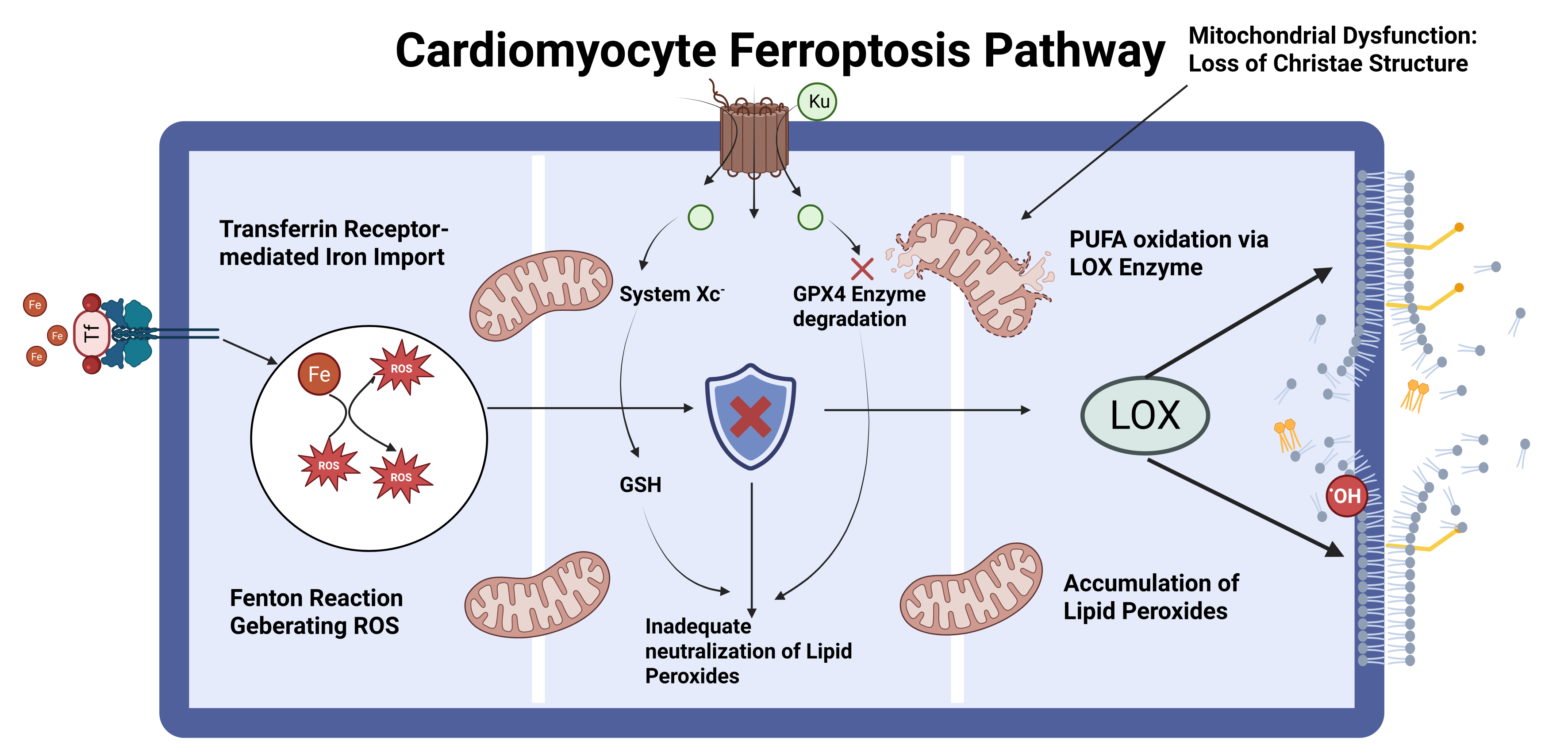
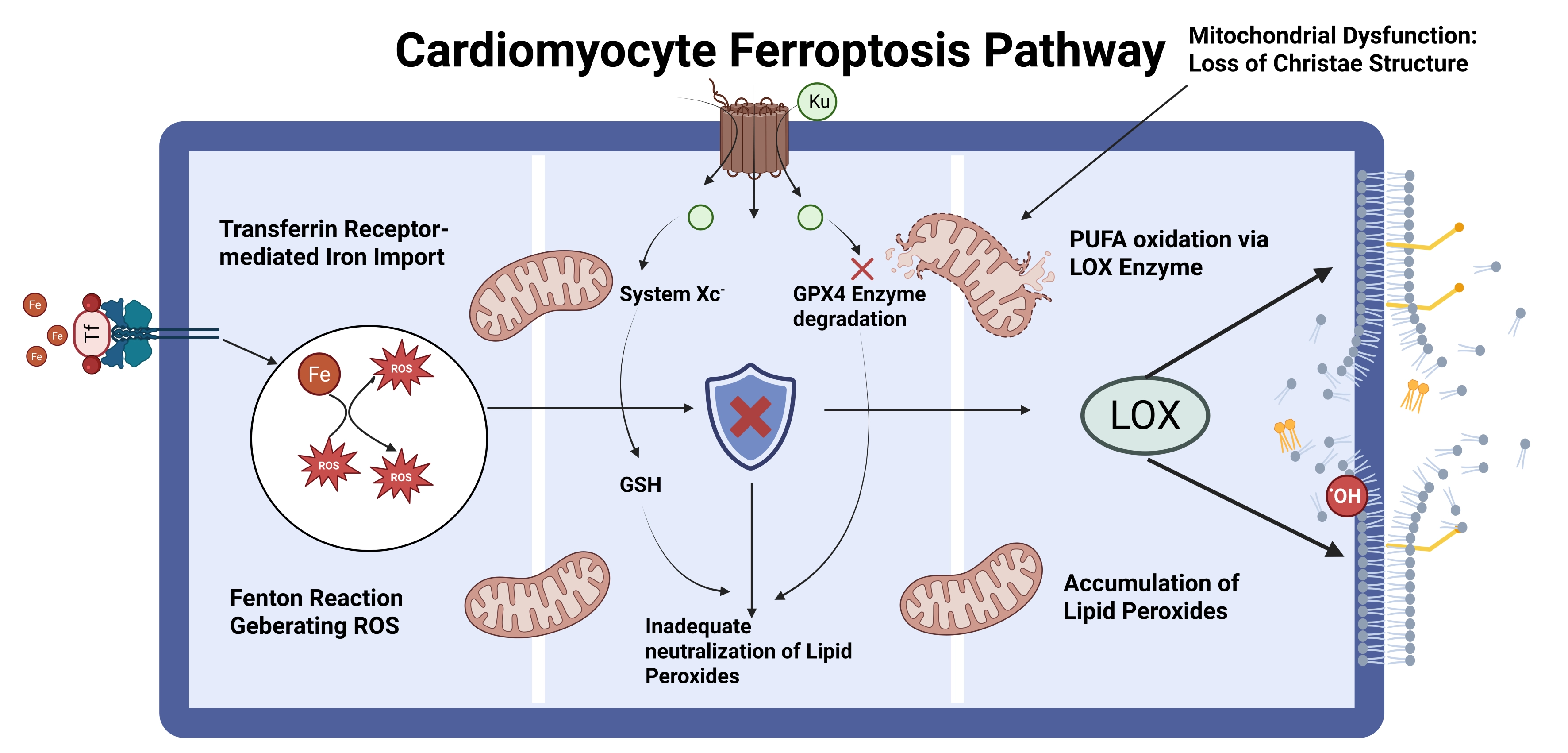
Figure 1. Molecular mechanisms and therapeutic targets of ferroptosis in myocardial injury. Schematic depicting the three interconnected axes driving ferroptosis: iron metabolism dysregulation, lipid peroxidation, and antioxidant defense failure, and their convergence on cardiomyocyte death. LOX: lipoxygenase; GSH: glutathione; ROS: reactive oxygen species; PUFA: polyunsaturated fatty acid.
2.2 Evidence linking ferroptosis to myocardial injury
Accumulating evidence suggests that ferroptosis is involved in multiple forms of myocardial injury, although the strength of evidence varies across disease contexts. Among these conditions, MIRI has been one of the most extensively investigated settings. Reperfusion following ischemia induces profound oxidative stress and metabolic disturbance, which can promote iron-dependent lipid peroxidation and ferroptotic cardiomyocyte death. Experimental studies have shown that pharmacological or targeted inhibition of ferroptosis can alleviate myocardial injury and improve cardiac function, supporting ferroptosis as an important component of MIRI pathogenesis[8,14,15].
Ferroptosis has also been increasingly implicated in doxorubicin (DOX)-induced cardiotoxicity, an important limitation of anthracycline-based chemotherapy. DOX can disrupt mitochondrial iron utilization and antioxidant defenses, thereby promoting lipid peroxidation and ferroptotic cardiomyocyte death. Mechanistic and experimental studies have demonstrated that inhibition of ferroptosis can attenuate DOX-induced cardiac injury, supporting ferroptosis as a potential therapeutic target for chemotherapy-associated cardiotoxicity[16-18].
Emerging evidence further links ferroptosis to sepsis-associated myocardial injury. The inflammatory and oxidative environment accompanying sepsis can disturb iron and redox homeostasis in cardiomyocytes, thereby facilitating ferroptotic injury. Recent experimental studies have shown that modulation of ferroptosis-related pathways can attenuate myocardial injury in septic models, while a recent prospective clinical study also identified alterations in ferroptosis-related biomarkers in patients with sepsis-associated myocardial injury[19-21].
Beyond acute myocardial injury, ferroptosis may also participate in cardiac remodeling and heart failure. Persistent ferroptosis has been associated with adverse cardiac remodeling after myocardial infarction, while recent evidence suggests that ferroptotic signaling is also involved in several experimental models of heart failure and cardiomyopathy[22,23]. However, compared with MIRI and acute cardiotoxicity, the causal contribution of ferroptosis to chronic cardiac dysfunction remains less firmly established and warrants further investigation.
Collectively, these findings indicate that ferroptosis may represent a convergent pathological process across diverse forms of myocardial injury, providing a rationale for investigating ferroptosis-directed interventions as a potential cardioprotective strategy.
2.3 Therapeutic targets for ferroptosis intervention in myocardial injury
The identification of ferroptosis as a contributor to myocardial injury has prompted increasing interest in strategies aimed at restoring the balance between iron availability, lipid peroxidation, and endogenous antioxidant defenses. Current therapeutic approaches can be broadly categorized according to these three interconnected axes, providing multiple potential entry points for ferroptosis intervention.
Targeting iron metabolism represents one of the most direct approaches to limiting ferroptosis. Excess intracellular iron expands the labile iron pool and facilitates oxidative reactions that promote lipid peroxidation. Accordingly, iron chelators such as deferoxamine and ciclopirox have been investigated as potential ferroptosis-targeting interventions[24,25]. In parallel, regulation of iron uptake, storage, export, and NCOA4-dependent ferritinophagy provides additional opportunities for therapeutic intervention[26]. Recent cardiovascular research has further highlighted the importance of the transferrin receptor 1 (TFR1)/ferroportin (FPN)/ferritin axis in controlling ferroptotic susceptibility in cardiac disease[27].
The System Xc-/GSH/GPX4 axis constitutes another central therapeutic target. System Xc- maintains intracellular cysteine availability for GSH synthesis, while GPX4 uses GSH to reduce membrane lipid hydroperoxides and thereby constitutes a major defense against ferroptosis[9,28]. Accordingly, preservation or restoration of SLC7A11, GSH, and GPX4 activity may reinforce endogenous resistance to ferroptotic injury. The Nrf2 pathway represents an important upstream regulator of this defense system by coordinating antioxidant responses and regulating genes involved in GSH metabolism, GPX4 expression, and iron homeostasis[17].
Lipid metabolism provides a further opportunity for therapeutic intervention. ACSL4 promotes the incorporation of polyunsaturated fatty acids into membrane phospholipids, thereby increasing the availability of substrates susceptible to peroxidation. In experimental DOX-induced cardiotoxicity, inhibition of the ACSL4/FTH1-dependent ferroptotic pathway alleviated myocardial injury and cardiac dysfunction, providing direct evidence that this axis may represent a therapeutically relevant target in cardiac disease[29]. Other regulators of lipid peroxidation, including LPCAT3 and lipoxygenases, may also influence ferroptotic susceptibility[30].
Beyond the canonical GPX4-dependent defense system, several alternative antioxidant pathways provide additional opportunities for ferroptosis intervention. FSP1 maintains a reduced coenzyme Q (CoQ) pool and limits lipid radical propagation independently of GPX4[31]. Similarly, dihydroorotate dehydrogenase (DHODH) protects against mitochondrial lipid peroxidation through a CoQ-dependent mechanism and has emerged as an important mitochondrial ferroptosis-suppressing pathway[32]. Notably, recent experimental evidence has directly implicated the voltage-dependent anion-selective channels (VDAC3)/DHODH axis in sepsis-induced myocardial injury, further supporting the relevance of mitochondrial ferroptosis regulation to cardiac disease[19].
Taken together, these results indicate that ferroptosis is controlled by an interconnected network, not by a single molecular switch. Several entry points for nanozyme-based interventions are therefore available, as summarized in Figure 1. This multifaceted regulation provides a conceptual basis for the potential use of nanozymes in myocardial injury associated with ferroptosis.
3. Nanozyme-Mediated Regulation of Ferroptosis in Myocardial Injury
Nanozymes used in myocardial injury differ considerably in their composition, catalytic activity, delivery strategy, and degree of evidence for ferroptosis regulation. Rather than treating these studies as a single group, their current evidence can be considered from three related aspects: the material and its catalytic function, the strategy used to deliver it to the heart, and the extent to which the observed cardioprotection can be attributed to ferroptosis.
3.1 Material composition and catalytic activity
Ceria-based nanozymes are among the better-characterized systems in this field. CeO2 can provide antioxidant activity through its reversible Ce3+/Ce4+ redox chemistry, and its combination with other therapeutic components has been used to broaden its biological effects. In sepsis-induced cardiac injury, CeCH, a ceria nanozyme coordinated with curcumin, reduced ROS accumulation and inhibited ferroptosis in cardiomyocytes, while also attenuating inflammatory responses and improving cardiac function. The study therefore provides direct evidence connecting a Ce-based nanozyme with ferroptosis-related cardiac protection[33]. A different CeO2-based system, D/Ce@mPDA-C/P, incorporated the iron chelator dexrazoxane (DXZ) into a ceria-containing carrier. In a mouse MIRI model, the system reduced oxidative stress and ferroptosis, increased GPX4 expression, and improved cardiac function[34]. Here, the ferroptosis-related effect results from the combination of antioxidant activity and iron chelation rather than from the intrinsic catalytic activity of CeO2 alone.
Pt-containing nanozymes have mainly been investigated for their efficient ROS-scavenging properties. Pt-N-C single-atom nanozymes showed multiple enzyme-mimicking activities and effectively reduced ROS and apoptosis after MIRI, with good biocompatibility[35]. Similarly, an ultrasmall PtIr nanozyme displayed SOD- and CAT-like activities and reduced oxidative stress, apoptosis, inflammation, and fibrosis in myocardial infarction models[36]. These studies support the cardioprotective potential of Pt-based catalysis, but neither study established ferroptosis as the primary mechanism. A newer Pt-doped ceria nanozyme further increased cardiomyocyte uptake and enhanced multi-enzyme-mimetic activity, leading to reduced ROS and improved cardiac function after MIRI[37]. Taken together, Pt-containing systems illustrate how catalytic optimization can strengthen antioxidant effects, while their specific contribution to ferroptosis remains to be established.
Manganese-based nanozymes have also been explored mainly as antioxidant and anti-remodeling agents. MnO2@TA possesses SOD- and CAT-like activities, and modification with tannic acid increases its affinity for the collagen- and elastin-rich extracellular matrix of the heart. In a mouse myocardial infarction model, MnO2@TA showed greater cardiac retention, reduced ROS accumulation and fibroblast activation, and attenuated cardiac fibrosis while improving cardiac function[38]. These findings support its role in redox control and post-myocardial infarction remodeling, but do not provide direct evidence that its anti-fibrotic effect is mediated by ferroptosis.
Prussian blue nanozymes provide another example of broad redox regulation. PBNz@PSC exhibits SOD-, CAT-, and peroxidase-like activities, with enhanced SOD-like activity after polysaccharide modification. The formulation passively accumulated in injured myocardium and reduced oxidative and inflammatory injury in MIRI mice; pharmacokinetic and safety assessments were also included[39]. However, the reported effects were primarily associated with ROS clearance, inflammation, and apoptosis rather than ferroptosis. It is therefore more appropriate to regard this study as evidence for antioxidant cardioprotection than as direct evidence of ferroptosis inhibition.
Ruthenium-based nanozymes provide a more recent example in which redox regulation is closely linked to ferroptosis-related changes. A conducting polymer-stabilized Ruzyme showed SOD- and CAT-like activities together with nitrogen-radical scavenging capacity. In septic myocardial injury, treatment reduced iron accumulation and lipid peroxidation, accompanied by attenuation of ferroptosis-related cardiac damage[40]. This study is particularly relevant to the present review because it connects the catalytic activity of the nanozyme with two core features of ferroptosis: iron dysregulation and lipid peroxidation-rather than relying on ROS measurements alone.
Single-atom nanozymes should be viewed as a structural category rather than a separate metal class. The Pt-N-C and Pt-doped CeO2 systems show how atomically dispersed catalytic centers can be used to modify ROS-scavenging activity and cellular uptake[35,37]. Their biological effects, however, still depend on the specific metal, support, catalytic reaction, and targeting design. Importantly, increased GPX4 expression after nanozyme treatment should not be interpreted as evidence that the nanozyme itself has GPX-like catalytic activity. In the current cardiac literature, SOD-like and CAT-like activities are much better established, whereas direct evidence for GPX-like nanozyme catalysis in ferroptosis-related myocardial injury remains limited.
3.2 Cardiac-targeting strategies
The therapeutic effect of a nanozyme depends not only on its catalytic activity but also on where it accumulates after administration. Several strategies have therefore been developed to increase exposure within injured myocardium or to further restrict delivery to cardiomyocytes and mitochondria.
Fe-Cur@TA uses the affinity of tannic acid for cardiac tissue to improve myocardial retention. Compared with Fe-Cur, Fe-Cur@TA showed approximately tenfold greater cardiac retention and uptake, and its therapeutic effects were evaluated in both mice and beagle dogs with myocardial infarction[41]. MnO2@TA adopts a different approach: tannic acid increases its interaction with the collagen- and elastin-rich cardiac extracellular matrix, resulting in greater cardiac retention and uptake after myocardial infarction[38]. These two studies illustrate tissue-level targeting without requiring a specific cardiomyocyte-targeting ligand.
More selective delivery has been achieved with peptide-based and hierarchical targeting systems. D/Ce@mPDA-C/P incorporates a cardiac homing peptide together with triphenylphosphine, enabling sequential targeting of injured myocardium and mitochondria. The system also carries DXZ, allowing iron chelation and antioxidant treatment to be combined within the same platform[34]. Genetically engineered ferritin nanocages provide another strategy, incorporating modules for recognition of ischemia-reperfusion-injured cardiomyocytes, lysosomal escape, and mitochondrial targeting. The resulting imFTn-Ru nanozyme generated NO within the targeted cells and reduced mitochondrial oxidative stress in MIRI models[42].
Targeting can also be combined with therapeutic cargo delivery. A hollow CeO2/Au nanozyme carrying miR-486 was modified with a cardiac-targeting peptide and designed to facilitate lysosomal escape. In MIRI mice, miR-486@CeO2/Au-pep accumulated in the heart, improved intracellular miRNA delivery, and reduced adverse ventricular remodeling[43]. These studies suggest that the next generation of cardiac nanozymes will need to address not only how much catalytic activity can be achieved, but also where and when that activity is exerted.
3.3 From redox regulation to ferroptosis control
The available studies can be further distinguished according to their relationship with the three major regulatory axes of ferroptosis: iron metabolism, lipid peroxidation, and antioxidant defense. The strongest evidence is currently concentrated in a small number of studies rather than being consistent across all nanozyme platforms.
For iron metabolism, D/Ce@mPDA-C/P combines CeO2-mediated redox regulation with DXZ-mediated iron chelation. Its effects on iron-related ferroptosis were examined together with changes in GPX4 and cardiac injury after MIRI[34]. The Ruzyme study similarly linked treatment to reduced iron accumulation and lipid peroxidation in septic myocardial injury[40]. These findings support iron handling as a tractable target for nanozyme-based intervention.
Lipid peroxidation provides a second point of convergence. Limiting ROS can reduce the oxidative pressure that drives membrane lipid oxidation, but ROS scavenging alone does not demonstrate ferroptosis inhibition. This distinction is evident in Pt-N-C, PtIr, MnO2@TA, and PBNz@PSC, where reduced ROS and improved cardiac outcomes were demonstrated without sufficient ferroptosis-specific validation[35,36,38,39]. By contrast, CeCH directly inhibited ferroptotic injury in cardiomyocytes, while the D/Ce@mPDA-C/P and Ruzyme studies examined iron and/or lipid-peroxidation changes alongside ferroptosis-related outcomes[33,34,40].
The third axis is the endogenous antioxidant system, particularly SLC7A11/GSH/GPX4. In D/Ce@mPDA-C/P-treated MIRI mice, increased GPX4 expression accompanied the suppression of ferroptosis[34]. CeCH also reduced RSL3-induced ferroptotic injury, supporting an interaction between nanozyme treatment and the cellular ferroptosis-defense system[33]. These findings should nevertheless be interpreted as modulation of cellular antioxidant defense rather than evidence of intrinsic GPX-like activity.
Taken together, the current literature does not support treating all cardiac nanozymes as ferroptosis inhibitors. A useful distinction is between direct evidence, in which ferroptosis is experimentally manipulated or specifically assessed alongside cardiac outcomes; supportive evidence, in which iron accumulation, lipid peroxidation, or GPX4-related changes are observed but causal dependence remains uncertain; and indirect evidence, in which nanozymes improve cardiac injury mainly through ROS scavenging, anti-inflammatory, anti-apoptotic, or anti-fibrotic effects. This distinction is important because the cardioprotective effects of a nanozyme may persist even when ferroptosis is not the dominant mechanism.
The material classes, catalytic activities, targeting strategies, and corresponding levels of ferroptosis evidence are summarized in Figure 2.
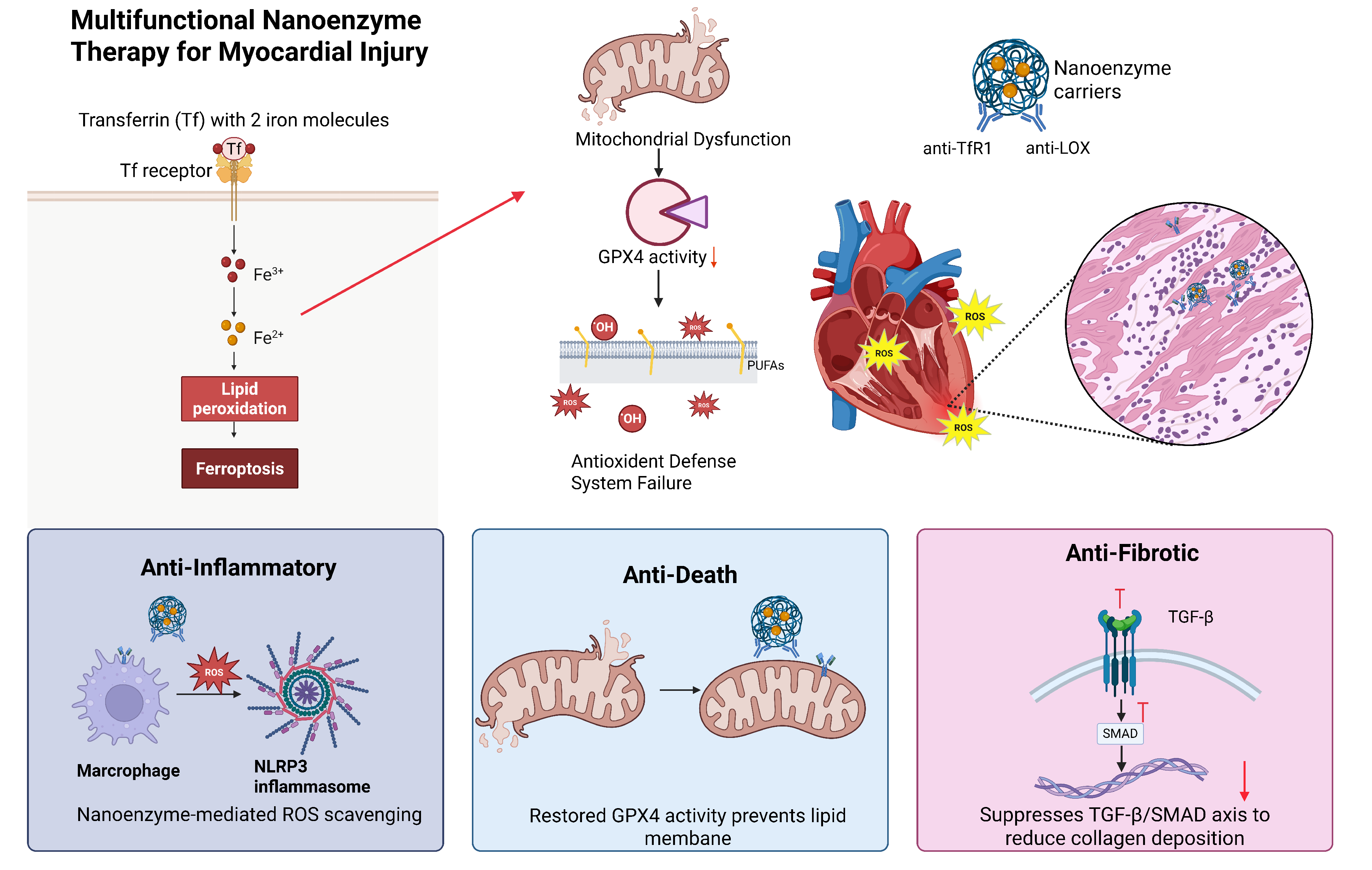
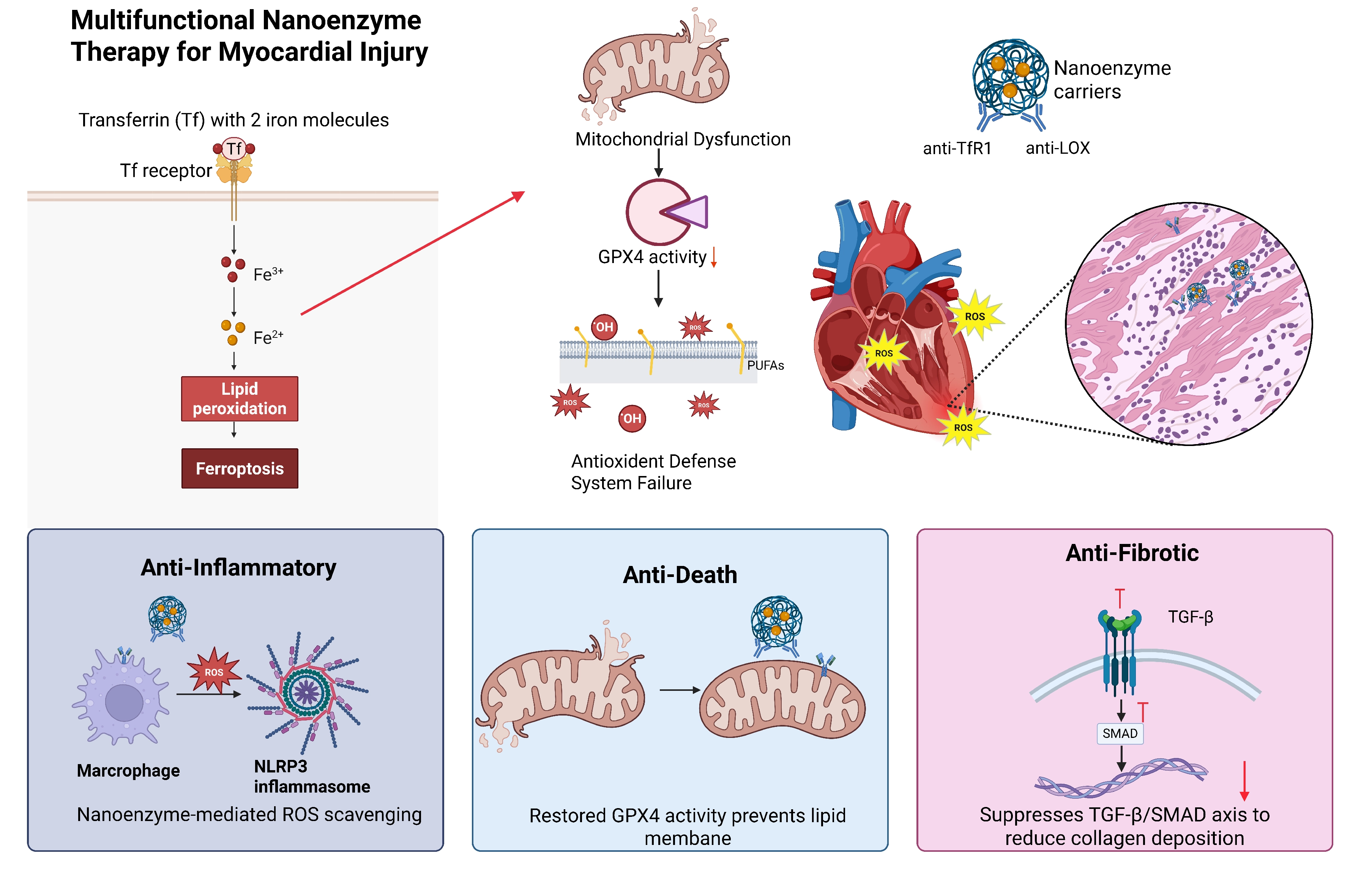
Figure 2. Nanozyme-mediated ferroptosis regulation confers multitargeted cardioprotection in myocardial injury. Cardiac-targeted nanozymes with enzyme-mimetic activities modulate iron metabolism, lipid peroxidation, and antioxidant defense, thereby suppressing cardiomyocyte ferroptosis and exerting anti-inflammatory, anti-death, and anti-fibrotic effects via ROS scavenging, GPX4 restoration, and TGF-β/SMAD inhibition. TFR1: transferrin receptor 1; ROS: reactive oxygen species; GPX4: glutathione peroxidase 4; PUFA: polyunsaturated fatty acid; LOX: lipoxygenase; NLRP3: NLR family pyrin domain containing 3; TGF-β: transforming growth factor-β; SMAD: small mothers against decapentaplegic.
4. Clinical Perspectives
The recent development of cardiac-targeted and multifunctional nanozymes suggests that the field is moving beyond simple ROS scavenging toward more precise therapeutic design. However, successful translation will depend not only on therapeutic efficacy but also on cardiac targeting, pharmacokinetics, biosafety, biodegradability, and scalable manufacturing. These considerations are particularly important because myocardial injury is heterogeneous across ischemia-reperfusion, infarction, sepsis, and chronic remodeling.
4.1 Cardiac targeting and delivery
Cardiac targeting is a major determinant of the translational potential of nanozyme-based therapies. Although nanozymes can be engineered with antioxidant, anti-inflammatory, and drug-delivery functions, systemic administration may limit their effective exposure in injured myocardium. Future designs may incorporate tissue-affinity ligands, cell-targeting peptides, or microenvironment-responsive components to improve cardiac localization and spatial control of catalytic activity[41].
Targeting can also be extended to the subcellular level. A mitochondria-targeting CeO2 nanozyme system equipped with a cardiac-homing peptide and triphenylphosphine achieved hierarchical targeting of injured myocardium and mitochondria, while sustained release of the iron chelator dexrazoxane enhanced therapeutic effects in a mouse model of MIRI[34]. Similarly, a genetically engineered ferritin-based nanozyme incorporating modules for injured cardiomyocyte targeting, lysosomal escape, and mitochondrial targeting was developed specifically to overcome intracellular delivery barriers during MIRI[42].
These findings indicate that future nanozyme design should prioritize not only catalytic activity but also the route, tissue, and subcellular compartment in which catalytic activity is required. Importantly, however, enhanced accumulation in experimental models does not necessarily predict human biodistribution. Differences in cardiac blood flow, tissue permeability, immune clearance, particle-protein interactions, and disease-associated heterogeneity may substantially alter nanoparticle exposure after systemic administration. Thus, quantitative pharmacokinetic and biodistribution studies in large-animal models will be important before clinical translation.
4.2 Pharmacokinetics, safety and biodegradability
Pharmacokinetics and long-term safety remain major barriers to clinical translation. The catalytic properties that make nanozymes attractive therapeutics can also complicate their behavior in vivo, because catalytic activity depends on particle composition, size, surface chemistry, local pH, substrate availability, and interactions with proteins and cells. In addition, prolonged exposure may result in tissue accumulation or altered catalytic activity after degradation. These issues are particularly relevant for metal- and metal-oxide-based nanozymes, for which short-term biocompatibility cannot be considered sufficient evidence of long-term safety.
Several recent cardiac studies have reported favorable short-term safety profiles. For example, the MnO2@TA nanozyme showed no apparent adverse effects in mice while improving cardiac function and reducing post-infarction fibrosis[38]. The Pt-N-C single-atom nanozyme also showed good biocompatibility in experimental MIRI models[35]. However, these findings mainly establish tolerability over the experimental observation period rather than long-term clinical safety.
Accordingly, future studies should systematically evaluate biodistribution, clearance, degradation products, repeated-dose toxicity, immunogenicity, and organ-specific effects. Pharmacokinetic characterization should also be integrated with therapeutic efficacy rather than treated as a separate endpoint, because prolonged circulation may increase cardiac exposure but may simultaneously increase hepatic or splenic accumulation. For nanozymes containing metals or metal oxides, determining whether degradation alters catalytic activity or releases potentially toxic ions will be particularly important. These considerations favor the development of materials with predictable degradation and clearance profiles rather than simply maximizing catalytic activity.
4.3 From proof-of-concept to clinically relevant therapeutic design
Beyond efficacy in small-animal models, clinically relevant nanozyme development will require more rigorous validation of therapeutic reproducibility and manufacturing feasibility. Most current cardiac nanozyme studies remain based on proof-of-concept experiments in rodents, in which treatment is typically initiated under controlled experimental conditions. Clinically, however, MIRI is heterogeneous in timing, severity, comorbidity, and reperfusion status. Therapeutic performance should therefore be evaluated across clinically relevant treatment windows and in models that more closely reproduce human cardiovascular physiology.
The translational value of several recent studies is strengthened by attempts to address these issues. For example, Fe-Cur@TA was evaluated in both mice and beagle dogs, providing evidence beyond a single rodent model[41]. A separate nanozyme platform used a cardiac-targeting peptide to deliver miR-486 together with ceria-gold catalytic components and demonstrated cardiac accumulation, enhanced intracellular delivery, and reduced adverse ventricular remodeling after MIRI[43]. These studies illustrate a broader transition from simple antioxidant nanoparticles toward multifunctional systems that combine targeting, catalytic activity, and therapeutic cargo delivery.
However, increasing structural complexity may also complicate quality control, batch-to-batch reproducibility, pharmacokinetic characterization, and regulatory evaluation. Future development should therefore balance multifunctionality against formulation complexity and prioritize platforms with well-defined composition, reproducible catalytic properties, scalable synthesis, and predictable in vivo fate. Establishing standardized assays for catalytic activity, pharmacokinetics, biodistribution, toxicity, and therapeutic efficacy will be essential for comparing nanozyme platforms across laboratories and advancing the most robust candidates toward clinical evaluation.
4.4 A more clinically realistic strategy for ferroptosis modulation
For ferroptosis-oriented nanozyme therapy, clinical translation may ultimately require controlled modulation rather than indiscriminate suppression of oxidative reactions. Iron metabolism and lipid redox signaling are essential for normal cardiomyocyte function and tissue repair, whereas excessive iron accumulation and lipid peroxidation contribute to pathological myocardial injury. Nanozymes with spatially restricted activity, injury-responsive activation, or coordinated delivery of ferroptosis-modulating agents may therefore offer greater therapeutic control than constitutively active systemic antioxidants. Such strategies could enable nanozymes to act preferentially within injured myocardium while minimizing interference with physiological redox signaling in healthy tissues.
Future studies should consequently define therapeutic windows for ferroptosis modulation, identify biomarkers that distinguish ferroptosis-dominant injury from other forms of cardiomyocyte death, and evaluate whether targeted nanozyme treatment provides benefits beyond conventional antioxidant therapy. These questions will be particularly important for determining which patients and stages of myocardial injury are most likely to benefit from ferroptosis-directed nanomedicine.
5. Conclusions
Ferroptosis represents a therapeutically relevant mechanism in myocardial injury, particularly through the interplay of iron dysregulation, lipid peroxidation, and impaired antioxidant defense. Nanozymes provide a versatile platform for modulating these processes while simultaneously addressing oxidative stress, inflammation, and adverse cardiac remodeling. However, the current evidence does not support considering all cardioprotective nanozymes as direct ferroptosis inhibitors; in many studies, ferroptosis-specific mechanisms remain incompletely defined. Future work should therefore prioritize rigorous mechanistic validation, precise cardiac and subcellular targeting, long-term safety assessment, pharmacokinetic characterization, and large-animal evaluation. Establishing reproducible manufacturing and clinically relevant biomarkers will also be essential for determining whether nanozyme-based ferroptosis modulation can ultimately be translated into effective therapies for myocardial injury.
Acknowledgements
The authors declare that the generative AI tool GPT-5.6 Luna was used solely for language polishing during the preparation of this manuscript. All research content, including the study design, data analysis, scientific interpretation, figures, tables, and conclusions, was generated and verified by the authors and was not generated using AI tools. The authors reviewed, revised, and approved the final manuscript and take full responsibility for its content.
Authors contribution
Qi Y: Conceptualization, investigation, visualization, writing-original draft, Writing-review & editing.
Zheng X: Investigation, writing-review & editing.
Wang K: Conceptualization, supervision, writing-original draft, writing-review & editing, funding acquisition.
All authors have read and approved the final version of the manuscript.
Conflicts of interest
Kai Wang is a Youth Editorial Board Member of BME Horizon. The other authors declare no conflicts of interest.
Ethical approval
Not applicable.
Consent to participate
Not applicable.
Consent for publication
Not applicable.
Availability of data and materials
Not applicable.
Funding
This work was supported by the National Natural Science Foundation of China (82600550), Key Scientific Research Project of Henan Provincial Institutions of Higher Education (27AQ310004), Joint project of Henan Medical Science and Technology Research Program (LHGJ20260117), Independent Project of Central China Subcenter of National Center for Cardiovascular Diseases (2025-FZX05) and Excellent Talents Project of Fuwai Central China Cardiovascular Hospital (FWKY250005).
Copyright
© The Author(s) 2026.
References
-
7. Byrne RA, Rossello X, Coughlan JJ, Barbato E, Berry C, Chieffo A, et al. 2023 ESC Guidelines for the management of acute coronary syndromes: Developed by the task force on the management of acute coronary syndromes of the European Society of Cardiology (ESC). Eur Heart J. 2023;44(38):3720-3826.[DOI]
-
10. Zhang T, Deng W, Deng Y, Liu Y, Xiao S, Luo Y, et al. Mechanisms of ferroptosis regulating oxidative stress and energy metabolism in myocardial ischemia-reperfusion injury and a novel perspective of natural plant active ingredients for its treatment. Biomed Pharmacother. 2023;165:114706.
-
15. Yin J, Zheng X, Zhao Y, Shen X, Cheng T, Shao X, et al. Investigating the therapeutic effects of ferroptosis on myocardial ischemia-reperfusion injury using a dual-locking mitochondrial targeting strategy. Angew Chem Int Ed. 2024;63(21):e202402537.[DOI]
-
18. Wang J, Yi H, Li J, Yang Y, Sun G, Xue Y, et al. P62-autophagic pathway degrades SLC7A11 to regulate ferroptosis in doxorubicin-induced cardiotoxicity. Life Sci. 2024;356:122981.[DOI]
-
19. Wang J, Zhang F, Tan C, Wang N, Xia X, Ye Y, et al. The VDAC3/DHODH axis ameliorates sepsis-induced myocardial injury by regulating ferroptosis. Front Biosci. 2025;30(6):39559.[DOI]
-
20. Wang X, Tian Q, Li M, Liu Y. METTL5 triggers the ferroptosis of cardiomyocytes in sepsis-induced myocardial injury. Free Radic Biol Med. 2025;237:1-10.[DOI]
-
21. Chen D, Xie H, Huang P, Wang R, Xie Y. Ferroptosis and sepsis-induced myocardial injury: A prospective clinical study. BMC Infect Dis. 2026;26(1):1750.[DOI]
-
22. Shen C, Wei Y, Kang W, Wang Q, Li G, Chen X, et al. Persistent ferroptosis modulates cardiac remodeling and M2 macrophage polarization, which can be mitigated by astaxanthin during myocardial infarction recovery. Cardiovasc Toxicol. 2025;25(1):58-73.[DOI]
-
23. Karampinos KI, Farmakis D, Gurung R, Foo RSY, Filippatos GS, Cokkinos DV. Ferroptosis in heart failure: From molecular insights to therapeutic implications. Cardiovasc Res. 2026;122(3):313-335.[DOI]
-
25. Pratt DA, Dixon SJ. A guide to using small-molecule ferroptosis inhibitors. Nat Struct Mol Biol. 2025;32(10):1848-1851.[DOI]
-
27. Yu P, Ling J, Chen F, Xu Z, Hu K, Liu F, et al. Dapagliflozin ameliorates myocardial ischemia/reperfusion injury by modulating EGFR signaling and targeting NCOA4-mediated ferritinophagy. Life Sci. 2026;391:124254.[DOI]
-
33. Jiang C, Shi Q, Yang J, Ren H, Zhang L, Chen S, et al. Ceria nanozyme coordination with curcumin for treatment of sepsis-induced cardiac injury by inhibiting ferroptosis and inflammation. J Adv Res. 2024;63:159-170.[DOI]
-
37. Pu A, Sim WS, Ji Y, Kurian AG, Lee JH, Van Anh Bui T, et al. Single-atom Pt-doped ceria nanozymes mitigate myocardial ischemia reperfusion injury via cardiomyocyte-targeted uptake and suppression of reactive oxygen species. Bioact Mater. 2025;53:366-385.[DOI]
-
40. Wu T, Liu Y, Wang W, Sun R, Wang Y, Pan J, et al. Conducting polymer-stabilized nanozymes alleviate sepsis-induced myocardial injury by inhibiting iron accumulation and lipid peroxidation. Nat Commun. 2026;17(1):3874.[DOI]
-
43. Chen X, Chen H, Zhu L, Li Q, Sun P, Spanos M, et al. Cascade nanozyme delivering miRNA to ischemic heart to alleviate myocardial ischemia-reperfusion injury. Small. 2025;21(43):2502778.[DOI]
Copyright
© The Author(s) 2026. This is an Open Access article licensed under a Creative Commons Attribution 4.0 International License (https://creativecommons.org/licenses/by/4.0/), which permits unrestricted use, sharing, adaptation, distribution and reproduction in any medium or format, for any purpose, even commercially, as long as you give appropriate credit to the original author(s) and the source, provide a link to the Creative Commons license, and indicate if changes were made.
Publisher’s Note
Science Exploration remains a neutral stance on jurisdictional claims in published
maps
and institutional affiliations. The views expressed in this article are solely those
of
the author(s) and do not reflect the opinions of the Editors or the publisher.
Share And Cite


Science Exploration Style
Qi Y, Zheng X, Wang K. Nanomedicine-mediated ferroptosis regulation in myocardial injury: Emerging opportunities and translational challenges. BME Horiz. 2027;5:202637. https://doi.org/10.70401/bmeh.2026.0038
Tips
Copy completed.
Submit a Manuscript
Author Instructions
Cite this Article
Article Metrics
0
View
0
Download
Cited
Article Updates

- Abstract
- Keywords
- 1. Introduction
- 2. Ferroptosis in Myocardial Injury
- 3. Nanozyme-Mediated Regulation of Ferroptosis in Myocardial Injury
- 4. Clinical Perspectives
- 5. Conclusions
- Acknowledgements
- Authors contribution
- Conflicts of interest
- Ethical approval
- Consent to participate
- Consent for publication
- Availability of data and materials
- Funding
- References
- Copyright
 Get citation
Science Exploration Style

Qi Y, Zheng X, Wang K. Nanomedicine-mediated ferroptosis regulation in myocardial injury: Emerging opportunities and translational challenges. BME Horiz. 2027;5:202637. https://doi.org/10.70401/bmeh.2026.0038
copy
Share Link
copy