Mitochondria-regulating biomaterials for periodontitis therapy: From dysfunction mechanisms to tissue regeneration

Xuan Yan
1,2,3

,
Xiaojing Hu
1,3,*
,
Junchao Wei
1,2,3,*
*Correspondence to:
Junchao Wei, School of Stomatology, Jiangxi Medical College, Nanchang University, Nanchang 330006, Jiangxi, China.
E-mail: weijunchao@ncu.edu.cn
Xiaojing Hu, School of Stomatology, Jiangxi Medical College, Nanchang University, Nanchang 330006, Jiangxi, China; Jiangxi Province Clinical Research Center for Oral Disease, Nanchang 330006, Jiangxi, China. E-mail: DentistHXJ@163.com
Xiaojing Hu, School of Stomatology, Jiangxi Medical College, Nanchang University, Nanchang 330006, Jiangxi, China; Jiangxi Province Clinical Research Center for Oral Disease, Nanchang 330006, Jiangxi, China. E-mail: DentistHXJ@163.com
BME Horiz. 2027;5:202631. 10.70401/bmeh.2026.0039
Received: July 18, 2026Accepted: October 23, 2026Published: October 27, 2026
Abstract
Periodontitis, a bacteria-induced chronic inflammatory disease, is characterized by progressive destruction of periodontal tissues, thereby severely affecting aesthetics, mastication, and quality of life. A critical mechanism of periodontitis is mitochondrial dysfunction; therefore, various mitochondria-regulating biomaterials have been designed to restore mitochondrial homeostasis and further enhance antibacterial defense, modulate immune responses, and promote osteogenesis and angiogenesis. In this review, the pathogenic mechanisms derived from mitochondrial dysfunction of various cell types and their effect on periodontal regeneration are comprehensively summarized. Furthermore, mitochondria-regulating biomaterials for periodontitis treatment along with the challenges and future perspectives for clinical translation are discussed.
Keywords
Mitochondria-regulating biomaterials, mitochondrial dysfunction, periodontitis, periodontal tissue regeneration
1. Introduction
Periodontitis is a chronic inflammatory disease induced by plaque biofilms, characterized by progressive destruction of tooth-supporting tissues, including the gingiva, periodontal ligament, cementum and alveolar bone[1-4]. As the sixth most prevalent chronic inflammatory disease worldwide, it affects approximately 10-15% of the global population and remains a leading cause of tooth loss in adults[5-7]. Clinical manifestations include gingival bleeding, periodontal pocket formation, alveolar bone resorption, and eventual tooth loosening or loss[8-10]. Moreover, periodontitis is closely associated with various systemic diseases, including type 2 diabetes, cardiovascular disease, Alzheimer’s disease, and rheumatoid arthritis[11-14]. Current standard treatments, including mechanical debridement, antibiotic therapy, and surgical intervention, are limited by incomplete bacterial eradication, increasing antibiotic resistance, and insufficient capacity to promote periodontal tissue regeneration[15-18]. Consequently, innovative therapeutic strategies that effectively regenerate periodontal tissue are urgently needed.
Mitochondrial dysfunction is a pivotal pathogenic driver of periodontitis. Under physiological conditions, mitochondria generate adenosine triphosphate (ATP) via oxidative phosphorylation (OXPHOS), which is essential for maintaining periodontal tissue homeostasis, including cell differentiation, mineralization, and regeneration[19,20]. In periodontitis, multiple periodontal cell types exhibit mitochondrial abnormalities, including decreased membrane potential and elevated reactive oxygen species (ROS) levels, which impair mitochondrial quality control and perpetuate inflammation[20,21]. Specifically, inflammatory stimuli may trigger calcium overload, excessive mitochondrial reactive oxygen species (mtROS) production, and mitochondrial DNA (mtDNA) release, thereby driving the activation of NOD-like receptor family pyrin domain containing 3 (NLRP3) inflammasome, a key event in the periodontal inflammatory cascade[22]. Simultaneously, inflammation-induced glycolytic metabolism reduces ATP production and further elevates ROS levels[23], leading to oxidative stress and ultimately irreversible periodontal tissue damage[23,24].
Mitochondrial dysfunction inhibits periodontal repair while accelerating disease progression. For example, mitochondrial dysfunction undermines antibacterial defense by impairing the bactericidal activity of neutrophils and macrophages[25,26]. Additionally, mitochondrial dysfunction drives macrophages toward a pro-inflammatory M1 phenotype while suppressing the reparative M2 phenotype, thereby perpetuating inflammation[27]. During osteogenesis, mitochondria are indispensable for the osteogenic differentiation of stem cells as they coordinate oxidative stress and autophagy to transduce osteogenic signaling pathways[28,29]; however, excessive ROS may disrupt redox homeostasis, triggering osteoblast ferroptosis and impairing bone formation[30]. Furthermore, mitochondrial dysfunction hinders endothelial cell (EC) proliferation, migration and tube formation, thereby impairing angiogenesis and tissue repair[31,32]. Therefore, it is necessary to design functional biomaterials that modulate mitochondrial function to realize periodontal tissue regeneration.
To date, various mitochondria-regulating biomaterials have been developed for periodontitis therapy (Figure 1). In this review, the fundamental characteristics of mitochondrial dysfunction in periodontitis and its impact on key physiological processes are outlined. Subsequently, recent advances in mitochondria-regulating biomaterials for periodontitis therapy are highlighted. Finally, current challenges and future perspectives regarding clinical translation are discussed. This review provides a brief overview of mitochondria-regulating biomaterials for periodontitis therapy, aiming to offer a basic reference for researchers entering this field.
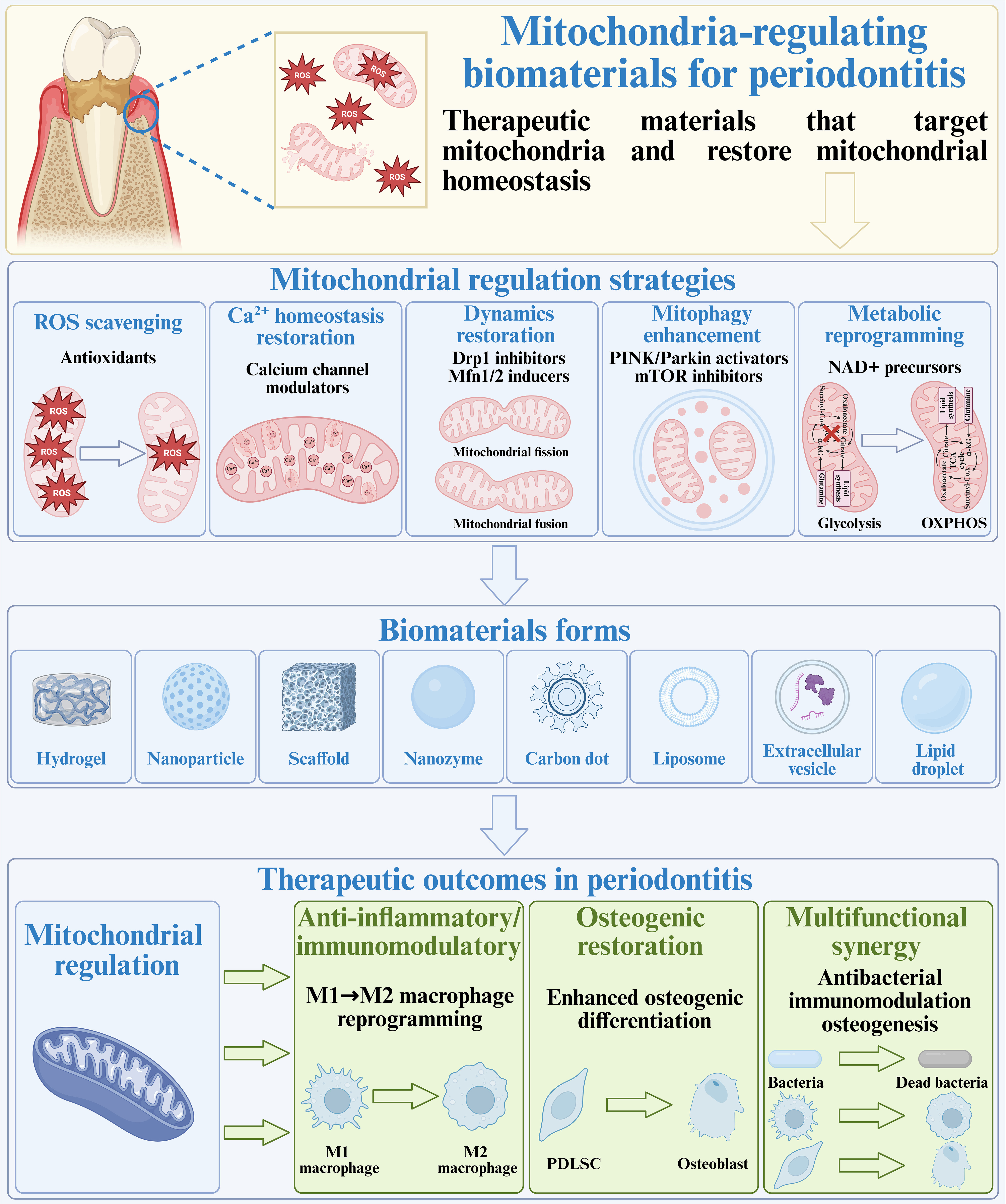
Figure 1. Design and therapeutic mechanisms of mitochondria-regulating biomaterials for periodontitis. Created in BioRender. Dwad, D. (2026) https://BioRender.com/fs7akdh. PDLSC: periodontal ligament stem cell; ROS: reactive oxygen species; OXPHOS: oxidative phosphorylation; mTOR: mammalian target of rapamycin; NAD: nicotinamide adenine dinucleotide.
2. Mitochondrial Dysfunction in Periodontitis
Pathogens such as Porphyromonas gingivalis (P. gingivalis) induce mitochondrial dysfunction, perturb metabolic homeostasis, and promote ROS production, thereby exacerbating periodontal inflammation and impairing tissue repair[33]. In this section, mitochondrial dysfunction of major cell types is introduced (Figure 2).
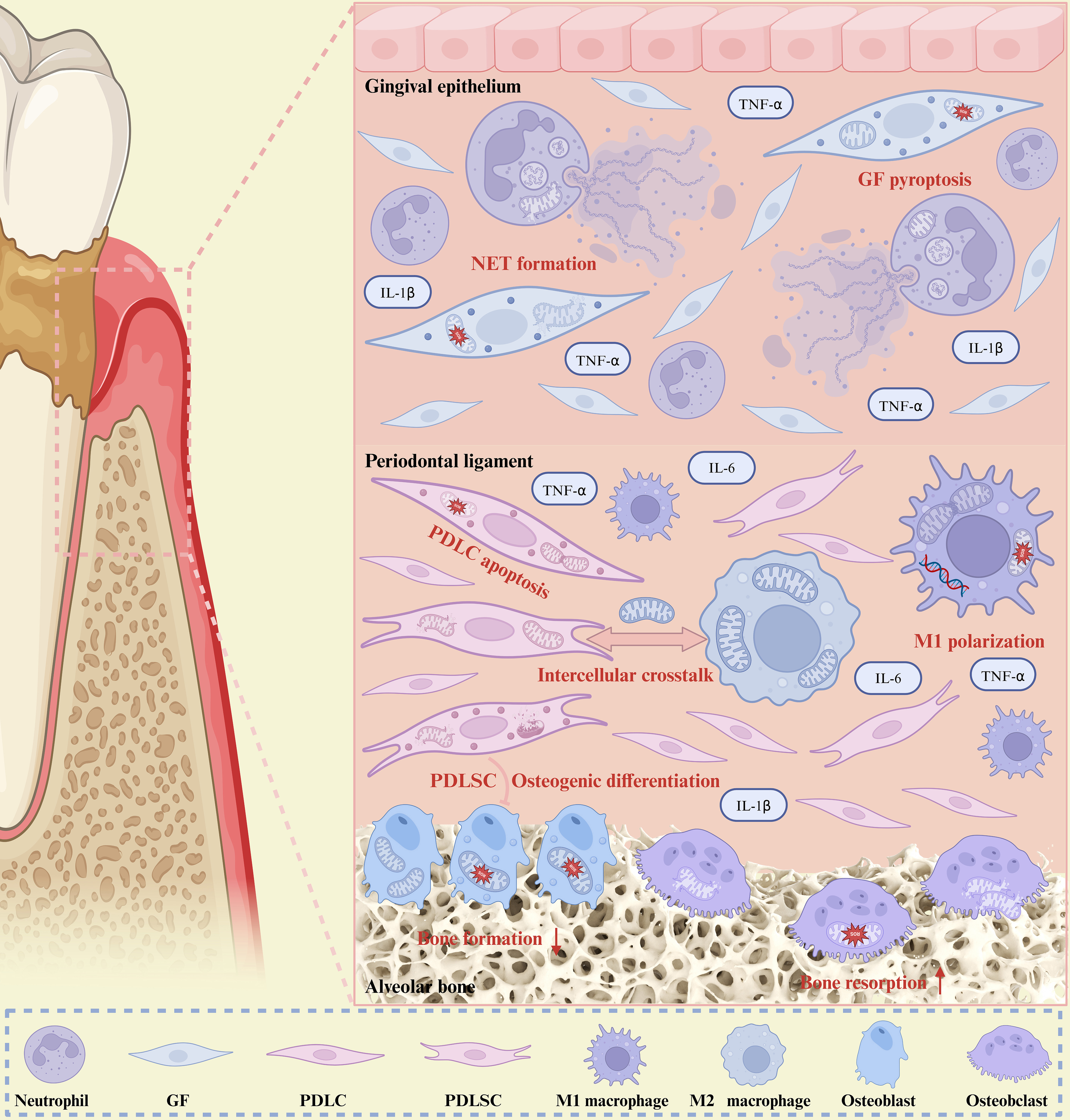
Figure 2. Cell-type-specific mitochondrial dysfunction and intercellular crosstalk in periodontitis. Created in BioRender. Dwad, D. (2026) https://BioRender.com/rtzmsgr. GF: gingival fibroblast; PDLC: periodontal ligament cell; PDLSC: periodontal ligament stem cell; NET: neutrophil extracellular trap; TNF-α: tumor necrosis factor-α; IL-1β: interleukin-1β.
2.1 Immune cells
Periodontal biofilms release virulence factors that induce metabolic reprogramming in local immune cells, driving sustained production of pro-inflammatory cytokines and ROS that lead to gingival tissue degradation and alveolar bone resorption[34]. Within this inflammatory milieu, mitochondrial dysfunction directly shapes the behavior of immune cells, particularly macrophages and neutrophils.
Macrophages, as a central component of innate immunity, play a critical role in periodontitis, where their polarization drives its pathogenesis[35]. Mitochondrial metabolic state shapes macrophage polarization, with M1 macrophages relying on enhanced glycolysis alongside impaired tricarboxylic acid (TCA) cycle and OXPHOS for rapid pro-inflammatory responses, whereas M2 macrophages depend on an intact TCA cycle and enhanced OXPHOS for anti-inflammatory cytokine production[36-38]. Besides, mitochondrial quality control also determines macrophage fate. Enhanced mitophagy promotes polarization towards M2 macrophages and attenuates periodontitis[39], while excessive Drp1 activation drives mitochondrial fragmentation and membrane potential loss in the P. gingivalis-induced periodontitis macrophage model, with Drp1-HK1 binding promoting excessive mitochondrial permeability transition pore (mPTP) opening and releasing mtDNA, mtROS, and other toxic substances into the cytoplasm, thereby activating the NLRP3 inflammasome to induce inflammatory damage[40].
Neutrophils, the most abundant innate immune cells in periodontal tissue, are essential for maintaining periodontal immune homeostasis[41]. In periodontitis, mitochondrial dysfunction in neutrophils manifests as excessive mtROS production, which promotes the release of mitochondrial neutrophil extracellular traps (NETs)[42]. Such dysfunction may further impair migration ability and delay inflammation resolution, collectively aggravating tissue damage[43].
2.2 Gingival fibroblasts
Gingival fibroblasts (GFs) are the predominant cells in gingival connective tissue, serving as sentinel cells for innate immune responses and major regulators of extracellular matrix (ECM) remodeling[44]. In periodontitis, GFs exhibit aberrant mitochondrial structure and function compared with periodontally healthy individuals[45]. Mechanistically, lipopolysaccharide (LPS) stimulation inhibits mitophagy in GFs, thereby activating the NLRP3 inflammasome and mediating pyroptosis[46]. In parallel, LPS derived from P. gingivalis elevates ROS levels and promotes the opening of mPTP by suppressing pyruvate dehydrogenase kinase 2 (PDK2) expression, leading to mtDNA release[47], which activates the cyclic GMP-AMP synthase (cGAS)-stimulator of interferon genes (STING) pathway and PANoptosis[48].
2.3 Periodontal ligament cells and stem cells
Periodontal ligament cells (PDLCs), also known as periodontal ligament fibroblasts (PDLFs), are the most abundant cells in the periodontal ligament and play a pivotal role in periodontitis progression via inflammation, tissue repair, and periodontal regeneration[20]. Excessive ROS induces mitochondrial dysfunction in PDLCs, characterized by elevated mtROS, reduced mitochondrial membrane potential and ATP levels, and increased Drp1-related mitochondrial fission, ultimately leading to apoptosis of PDLCs[49,50].
Periodontal ligament stem cells (PDLSCs), the main mesenchymal stem cells (MSCs) in the periodontal ligament, play a critical role in immune regulation and can differentiate into various cell types to repair damaged periodontal tissue[51]. In periodontitis, impaired mitophagy leads to the accumulation of damaged mitochondria, thereby compromising the osteogenic capacity of PDLSCs[52]. In parallel, periodontal inflammation induces mitochondrial dysfunction that impairs cementoblastic differentiation[53] and reduces ATP production and activates apoptotic signaling, ultimately diminishing the regenerative capacity of PDLSCs[54].
2.4 Bone-related cells
Alveolar bone resorption is a central pathological hallmark of periodontitis, fundamentally reflecting an imbalance between bone formation and bone resorption[55]. Mitochondrial dysfunction impairs osteoblast-mediated bone formation while enhancing osteoclast-mediated bone resorption, collectively driving alveolar bone loss.
Osteoblasts are the principal bone-forming cells residing in the alveolar bone[56]. In periodontitis, excessive ROS, resulting from mitochondrial dysfunction, inhibit the differentiation and mineralization of osteoblasts by suppressing the Wnt/β-catenin, mitogen-activated protein kinase (MAPK), and PI3K/Akt pathways[57], and mtROS-induced mitochondrial impairment ultimately leads to osteoblast death[58].
Osteoclasts serve as the key effector cells of alveolar bone resorption in periodontitis[59]. PTEN-induced kinase 1 (PINK1)-mediated mitophagy regulates osteoclast formation, differentiation, and function during alveolar bone resorption in periodontal disease[60]. In periodontitis, alveolar bone destruction is accompanied by decreased PINK1 expression and an increase in osteoclast number[61]. Consistently, PINK1 deficiency significantly increases the number of tartrate resistant acid phosphatase (TRAP) positive osteoclasts and exacerbated alveolar bone loss[62]. Besides, ROS overproduction promotes osteoclast differentiation by activating transient receptor potential melastatin 2 (TRPM2), eventually leading to bone degradation in periodontitis[63].
2.5 Intercellular crosstalk and synergistic effects
Mitochondrial dysfunction in periodontitis is multicellular and involves intercellular crosstalk among immune cells, stem cells, and bone cells. For example, in periodontitis, mitochondria-rich extracellular vesicles (EVs) derived from inflammatory macrophages disrupt mitochondrial homeostasis in bone marrow mesenchymal stem cells (BMSCs) by upregulating lipocalin 2 (LCN2), which induces OMA1 degradation and OPA1 accumulation, ultimately inhibiting osteogenesis and exacerbating alveolar bone resorption[21], demonstrating that mitochondrial stress can be transmitted between cell types, impairing the function of target cells and compromising tissue repair. Mitochondrial dysfunction in periodontitis is a transmissible pathological signal rather than a cell-intrinsic defect, which propagates through intercellular crosstalk to form a synergistic network that amplifies tissue destruction.
Moreover, mitochondrial repair in one cell type may produce synergistic therapeutic effects by influencing others. For instance, transplanting M2 macrophage-derived mitochondria into PDLSCs under inflammatory conditions restored mitochondrial function, ultimately promoting osteogenic differentiation and periodontal bone regeneration[64]. Similarly, a sericin-based dual-module microsphere system (SeHA@EC) attenuated intracellular ROS levels, stabilized mitochondrial membrane potential in macrophages, thereby promoting M2 polarization and subsequently enhancing the osteogenic differentiation of PDLSCs[65]. These findings demonstrate that regulating mitochondrial function in one cell type can influence other cell types via intercellular crosstalk, thereby achieving synergistic therapeutic effects. Overall, mitochondria may serve as a shared therapeutic target across multiple cell types in periodontitis.
3. Impact of Mitochondrial Dysfunction on Key Physiological Processes
Mitochondria regulate multiple crucial physiological processes involved in periodontal tissue regeneration, including generating mtROS for antibacterial defense, orchestrating immune responses to resolve inflammation, supporting osteogenic differentiation, and promoting EC proliferation and migration for angiogenesis. However, mitochondrial dysfunction impairs these physiological processes, leading to persistent infection, unresolved inflammation, and compromised tissue regeneration.
3.1 Antibacterial defense
Mitochondria are critical for antibacterial functions of innate immune cells. Physiologically, mitochondria provide ATP to support macrophage phagocytosis[66], produce mtROS as direct antimicrobial agents[67], and sense bacterial metabolites such as lactate to trigger NETs release[68], thereby contributing to antibacterial defense.
However, periodontal pathogens like P. gingivalis hijack mitochondrial homeostasis to subvert the host antibacterial defenses. In neutrophils, P. gingivalis outer membrane vesicles (OMVs) stimulation increases glucose uptake and lipid droplet accumulation, reduces lactate release, and lowers mitochondrial mass and membrane potential, which in turn enhances P. gingivalis internalization and intracellular bacterial survival, reflecting an impaired extracellular antibacterial response[69]. Similarly, in macrophages, P. gingivalis outer membrane vesicles (OMVs) promote a metabolic shift toward glycolysis and disrupt mitochondrial function and the TCA cycle, thereby activating pro-inflammatory cytokine production, inflammasome signaling, and pyroptosis and ultimately weakening their antibacterial capacity[70]. P. gingivalis also elicits a mitochondrial stress response that drives mtDNA release, which in turn engages Z-DNA binding protein 1 (Zbp1) to amplify downstream PANoptosis signaling[71]. Together, these mechanisms establish mitochondrial dysfunction as a central node that impairs cellular bioenergetics and activates the cGAS-STING pathway or NLRP3 inflammasome, thereby promoting pro-inflammatory cytokine secretion and further compromising immune cell function[72]. Consequently, restoring mitochondrial homeostasis represents a promising strategy to rescue antibacterial capacity. Indeed, baicalin induces Drp1-dependent mitochondrial biogenesis, enhances mitochondrial membrane potential and ATP levels, and reduces mtROS production in macrophages, thereby promoting antibacterial responses during Staphylococcus aureus infection[73].
3.2 Anti-inflammation and immunomodulation
Mitochondria regulate immune responses and determine the activation, differentiation, and effector functions of immune cells[74-76]. Under physiological conditions, mitochondria maintain immune homeostasis. However, when mitochondrial function is impaired, immune homeostasis is disrupted and inflammation is exacerbated. Specifically, mitochondrial dysfunction drives a metabolic shift toward glycolysis, sustaining the pro-inflammatory M1 phenotype and impairing M1-to-M2 repolarization, thereby exacerbating periodontal inflammation[75,77,78]. Mitochondrial dysfunction also induces mtDNA release, which triggers excessive inflammation by activating the cGAS-STING pathway[79]. Mitochondrial dysfunction further triggers excessive mitochondrial fission, driving M1 macrophage polarization and aggravating periodontal inflammation[80]. Overall, mitochondrial dysfunction sustains a pro-inflammatory state in periodontal tissues, impairing inflammation resolution and tissue repair.
3.3 Osteogenesis
Mitochondria are the primary source of ATP and critically regulate stem cell fate during bone regeneration[81]. During osteogenic differentiation of BMSCs, mitochondrial metabolism shifts from glycolysis to OXPHOS to support ATP production for the high energy demands of osteogenesis[82,83]. In addition, mitochondrial dynamics regulate osteogenesis, with moderate fission promoting osteogenesis and enhanced fusion suppressing it[84].
However, mitochondrial dysfunction suppresses osteogenesis through distinct mechanisms across cell types. In BMSCs, mitochondrial dysfunction promotes cellular senescence and impairs osteogenesis via impaired mitophagy, energy metabolism disorders, and fusion–fission imbalance[85]. In MC3T3-E1 cells, mitochondrial dysfunction disrupts osteogenic differentiation via fusion-fission disorders[86]. In osteoblasts, mitochondrial dysfunction triggers cell death and functional impairment via elevated mtROS[58]. Therefore, restoring mitochondrial function represents a viable strategy to promote osteogenesis and accelerate periodontal healing.
3.4 Angiogenesis
Mitochondria supply ATP to support EC proliferation, migration, and survival and sense hypoxia via complex III to generate transient mtROS, which stabilizes HIF-1α and upregulates vascular endothelial growth factor (VEGF)[87,88], while also activating endothelial nitric oxide synthase (eNOS) to promote EC proliferation and angiogenesis[89].
However, mitochondrial dysfunction results in excessive mtROS production, dysregulated mitochondrial dynamics, and disrupted Ca2+ homeostasis. Sustained mtROS generation produces ONOO- and inactivates MnSOD via nitrotyrosine formation, reducing mitochondrial membrane potential and thereby inhibiting EC proliferation[89,90]. Concurrently, excessive mitochondrial fission reduces mitochondrial mass, further compromising EC proliferation[91], while dysregulated Ca2+ signaling additionally suppresses EC migration and angiogenesis[92]. Collectively, mitochondrial dysfunction impairs EC proliferation, migration, and angiogenesis, underscoring the necessity of restoring mitochondrial function to preserve endothelial homeostasis. For example, L-arginine protects endothelial cells from ROS insult by promoting PINK1/Parkin- and BCL2 and adenovirus E1B 19 kDa-interacting protein 3 (BNIP3)-dependent mitophagy[93].
4. Application of Mitochondria-Regulating Biomaterials in Periodontitis
Mitochondrial dysfunction underlies the initiation and progression of periodontitis, triggering excessive ROS production, NLRP3 inflammasome activation, and the release of pro-inflammatory cytokines while impairing ATP synthesis. Accordingly, various mitochondria-regulating biomaterials including anti-inflammatory and immunomodulatory, osteogenic, and multifunctional biomaterials have been engineered to restore mitochondrial homeostasis and promote periodontal tissue regeneration.
4.1 Anti-inflammatory and immunomodulatory mitochondria-regulating biomaterials
Mitochondrial dysfunction is increasingly recognized as a common upstream event of inflammatory cell activation, driving persistent inflammation and tissue destruction, and therefore, biomaterials that regulate mitochondrial function can suppress the activation of multiple inflammatory cells, ultimately promoting periodontal tissue regeneration. Table 1 summarizes representative biomaterials with anti-inflammatory and immunomodulatory functions.
Table 1. Anti-inflammatory and immunomodulatory mitochondria-regulating biomaterial for periodontitis therapy.
| Material | Action mechanism | Main therapeutic function | Ref. |
| MTP hydrogel | Scavenge mtROS and repair oxidatively damaged mitochondria | Promote bone regeneration | [97] |
| CoQ10 MLip/Gel | Inhibit oxidative stress and restore mitochondrial energy metabolism and | Alleviate periodontal inflammation | [24] |
| mitochondrial integrity | |||
| TC/pSeSe | Target mitochondria, restore redox homeostasis, and suppress mtDNA-cGAS-STING signaling | Alleviate periodontal inflammation and promote alveolar bone regeneration | [99] |
| TPP-SAT nanogel | Target mitochondria and alleviate mitochondrial oxidative stress | Restore M1/M2 balance, reduce inflammation and immune dysfunction, inhibit bone resorption, and accelerate periodontal tissue regeneration | [100] |
| FIS-CDs | Reduce intracellular ROS levels and maintain mitochondrial homeostasis | Alleviate periodontal inflammation and oxidative damage, suppress alveolar bone loss, and promote periodontal tissue regeneration | [101] |
| MT-CDs | Scavenge intracellular ROS and maintain mitochondrial homeostasis | Suppress inflammatory mediator production, inhibit alveolar bone loss, and promote tissue regeneration | [102] |
| GelMA@AH-12 hydrogel | Stabilize mitochondrial Ca2+ levels and curtail mtROS overproduction and mtDNA release | Alleviate inflammation and promote tissue repair | [22] |
| AA@GO hydrogel | Enhance mitophagy, lower intracellular ROS levels, and restore macrophage mitochondrial function | Promote M1-to-M2 repolarization, reduce inflammation, and promote periodontal tissue repair | [103] |
| Mo-BGC scaffolds | Reprogram macrophage metabolism | Promote M2 macrophage polarization and orchestrate bone, periodontal ligament, and cementum regeneration | [104] |
mtROS: mitochondrial reactive oxygen species; CoQ10: coenzyme Q10; MLip: mitochondria-targeted liposome; Gel: ROS-responsive hydrogel; AA@GO: hybrid hydrogel; mtDNA: mitochondrial DNA; ROS: reactive oxygen species; mtDNA-cGAS-STING: mitochondrial DNA-cyclic GMP-AMP synthase-stimulating factor; FIS-CDs: FIS-derived carbon dots; FIS: fisetin; MT-CDs: melatonin-derived carbon dots; TPP-SAT: enzyme-responsive nanogel; TC: triphenylphosphine chitosan; pSeSe: ROS-responsive diselenide-containing copolymer nanoparticle; MTP hydrogel: ROS-responsive hydrogel.
Nanomaterials have attracted considerable attention as mitochondria-regulating platforms owing to their nanoscale dimensions, tunable surface properties, and targeting potential. Antioxidant-loaded nanocarriers, typically liposomes or polymeric nanoparticles, encapsulate small-molecule antioxidants to avoid rapid metabolism and facilitate mitochondria-specific delivery, thereby scavenging mtROS and enhancing mitophagy, which in turn alleviates periodontal inflammation. Mitoquidone mesylate (MitoQ), a well-established mitochondria-specific antioxidant[94], is limited by its poor water dispersion and stability, and gastrointestinal side effects[95,96]. To overcome these drawbacks, MitoQ was encapsulated in poly (lactic-co-glycolic acid) (PLGA)-poly (ethylene glycol) (PEG) nanoparticles modified with targeted peptides (MPPT NPs) and subsequently integrated into a ROS-responsive hydrogel (MTP hydrogel) that targeted mitochondria, scavenged ROS, and repaired damaged mitochondria, resulting in a bone volume fraction (BV/TV) 1.5 times higher than previously reported values[97]. However, the long-term safety of exogenous antioxidants remains a concern. As an alternative, coenzyme Q10 (CoQ10), an endogenous antioxidant[98], was encapsulated into mitochondria-targeted liposomes (CoQ10 mitochondria-targeted liposome (MLip)) and further incorporated into an ROS-responsive hydrogel (CoQ10 MLip/ROS-responsive hydrogel (Gel)), which scavenged ROS, rejuvenated mitochondrial energy metabolism, and restored mitochondrial integrity, ultimately alleviating periodontal inflammation (Figure 3a)[24].
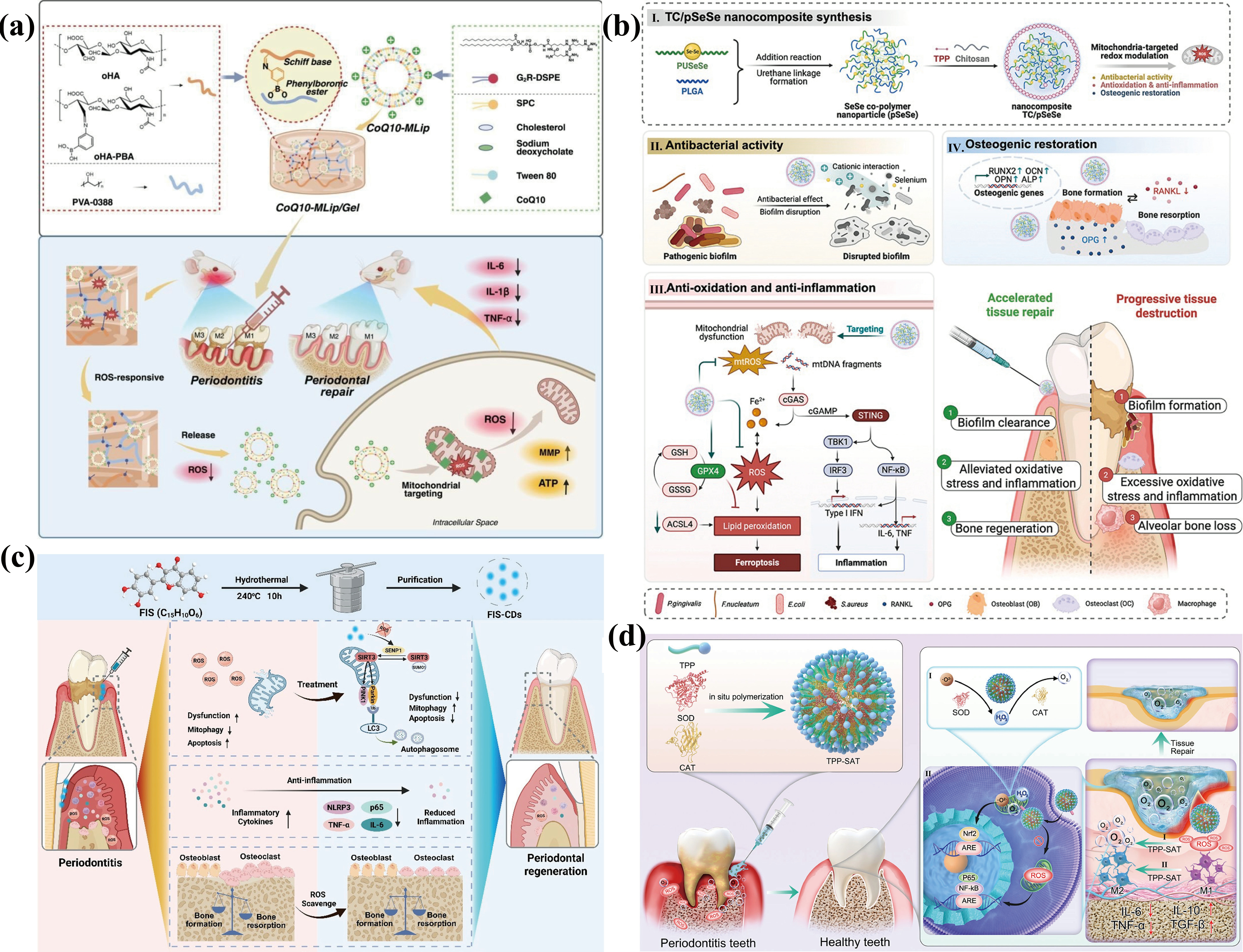
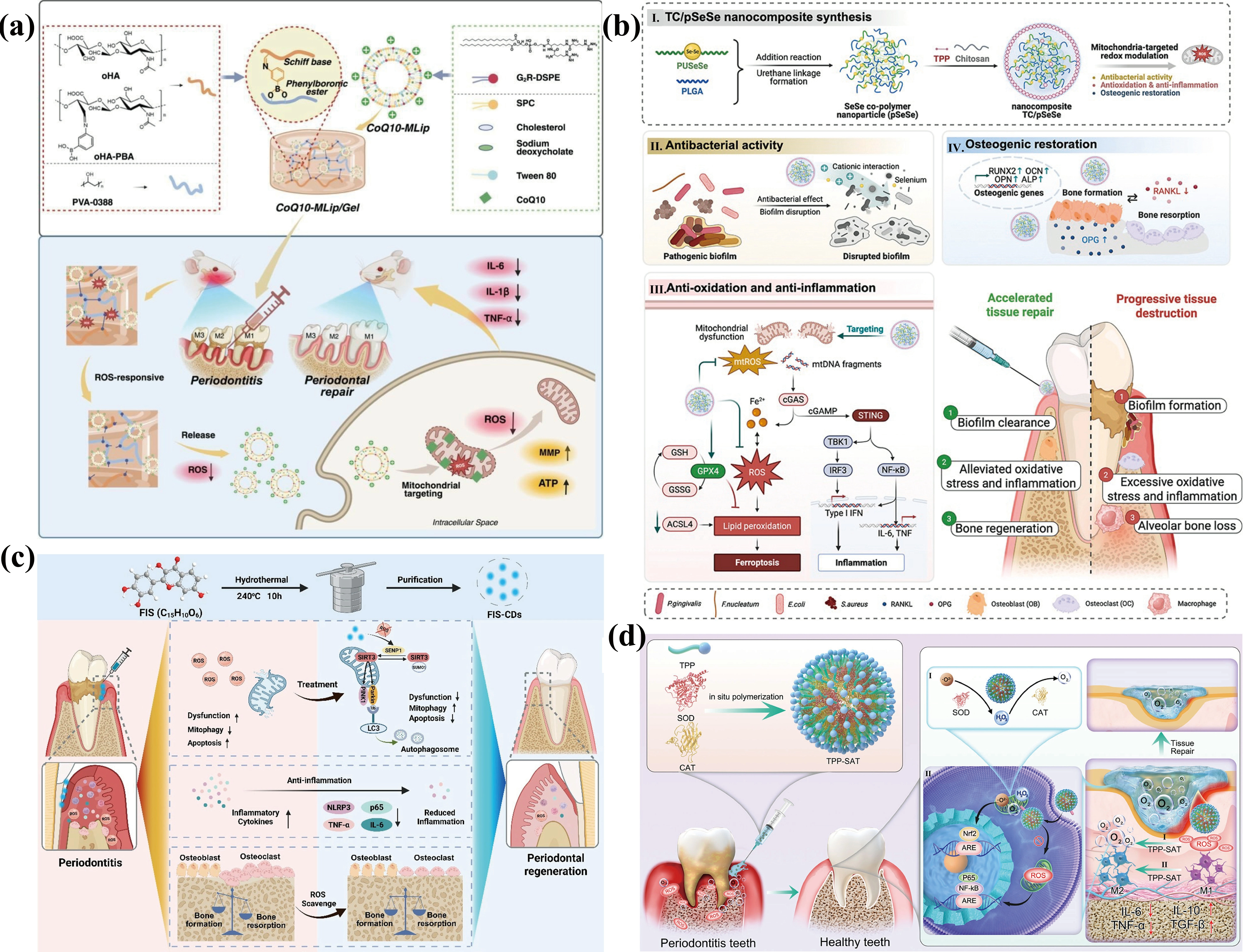
Figure 3. (a) Schematic illustration of the design of CoQ10-MLip/Gel and its mechanism diagram for the periodontal treatment. Reproduced from reference[24]. CC BY 4.0; (b) Schematic diagram of the design and mitochondria-centered redox modulation mechanism of TC/pSeSe nanocomposite for restoring alveolar bone homeostasis in periodontitis. Reproduced from reference[99]. CC BY 4.0; (c) Schematic illustration of the design of FIS-CDs and their anti-inflammatory and antioxidant mechanisms in periodontal diseases. Reproduced from reference[101]. CC BY 4.0; (d) Schematic diagram of synthesis and action mechanism of mitochondria-targeted TPP-SAT for periodontitis treatment. Reproduced from reference[100]. CC BY 4.0. CoQ10: coenzyme Q10; MLip: mitochondria-targeted liposome; Gel: ROS-responsive hydrogel; TC: triphenylphosphine chitosan; pSeSe: ROS-responsive diselenide-containing copolymer nanoparticle; FIS-CDs: FIS-derived carbon dots; FIS: fisetin; SOD: superoxide dismutase; CAT: catalase; ROS: reactive oxygen species; STING: stimulator of interferon genes; TPP: triphenylphosphine; cGAS: cyclic GMP-AMP synthase; ATP: adenosine triphosphate; TNF-α: tumor necrosis factor-α; IL-1β: interleukin-1β; SIRT3: SUMOylation of sirtuin 3; NLRP3: NOD-like receptor family pyrin domain containing 3; mtROS: mitochondrial reactive oxygen species; mtDNA: mitochondrial DNA; Nrf2: nuclear factor erythroid 2-related factor; NF-κB: nuclear factor-kappa B; PLGA: poly (lactic-co-glycolic acid); GPX4: glutathione peroxidase 4; GSH: glutathione; ACSL4: acyl-CoA synthetase long-chain family member 4; IFN: interferon; TGF-β: transforming growth factor-β; IRF3: interferon regulatory factor 3; cGAMP: cyclic guanosine monophosphate–adenosine monophosphate; RANKL: receptor activator of nuclear factor-κB ligand.
Stimuli-responsive nanomaterials scavenge ROS via responsive degradation and subsequent release of bioactive factors, thereby breaking the vicious cycle of ROS, inflammation and hypoxia in periodontitis. For example, a redox-responsive nanocomposite (TC/pSeSe) consisting of a ROS-responsive diselenide-containing copolymer nanoparticle (pSeSe) core and a triphenylphosphine (TPP)/chitosan (TC) coating was developed to achieve mitochondrial targeting, mucosal adhesion, and antibacterial functions (Figure 3b)[99]. Upon oxidative stress, diselenide bonds broke to release selenium, which scavenged ROS and suppressed mtDNA-mediated cGAS-STING signaling, alleviating periodontal inflammation and promoting alveolar bone regeneration. Extending this design, an enzyme-responsive nanogel (TPP-SAT) composed of TPP, superoxide dismutase (SOD) and catalase (CAT) was developed, which targeted mitochondria, converted ROS into O2 and relieved hypoxia, thereby rebalancing M1/M2 macrophages and inhibiting alveolar bone resorption (Figure 3d)[100].
Carbon dots (CDs) can scavenge ROS, activate multiple endogenous protective pathways and regulate mitophagy, thereby collectively preserving mitochondrial homeostasis and ameliorating periodontal tissue destruction. For example, fisetin (FIS)-derived carbon dots (FIS-CDs) reduced intracellular ROS and activated mitophagy by downregulating the SUMOylation of sirtuin 3 (SIRT3) SUMOylation, thereby attenuating periodontal inflammation, inhibiting alveolar bone loss, and promoting periodontal tissue regeneration (Figure 3c)[101]. Similarly, melatonin-derived carbon dots (MT-CDs) effectively scavenged intracellular ROS, maintained mitochondrial homeostasis and suppressed inflammatory cytokine production via the nuclear factor erythroid 2-related factor (Nrf2)/heme oxygenase-1 (HO-1) pathway[102].
Functional hydrogels serve as reservoirs for sustained release of therapeutic agents, thereby reshaping the periodontal microenvironment. Antibacterial hydrogels are designed to eliminate bacteria in periodontal pockets, thereby alleviating the inflammatory cascades. A photocrosslinked hydrogel loaded with antimicrobial peptide AH-12 inhibited key pathogens (Fusobacterium nucleatum, P. gingivalis, and Streptococcus gordonii), stabilized mitochondrial Ca2+ levels, reduced mtROS production and mtDNA release, thereby alleviating inflammation and promoting tissue repair[22]. Immunomodulatory hydrogels are intended to regulate the immune microenvironment to promote periodontal tissue regeneration. A hybrid hydrogel (AA@GO), composed of sodium alginate (SA), carboxymethyl chitosan (CCS), and graphene oxide (GO), promoted mitophagy, eliminated excess ROS, and improved mitochondrial function in macrophages via asiatic acid (AA) delivery, thereby exerting anti-inflammatory effects and enhancing tissue repair (Figure 4)[103]. Notably, functional hydrogels serve as drug delivery systems to exert therapeutic effects while influencing cellular behavior via their three-dimensional (3D) network and mechanical microenvironment.
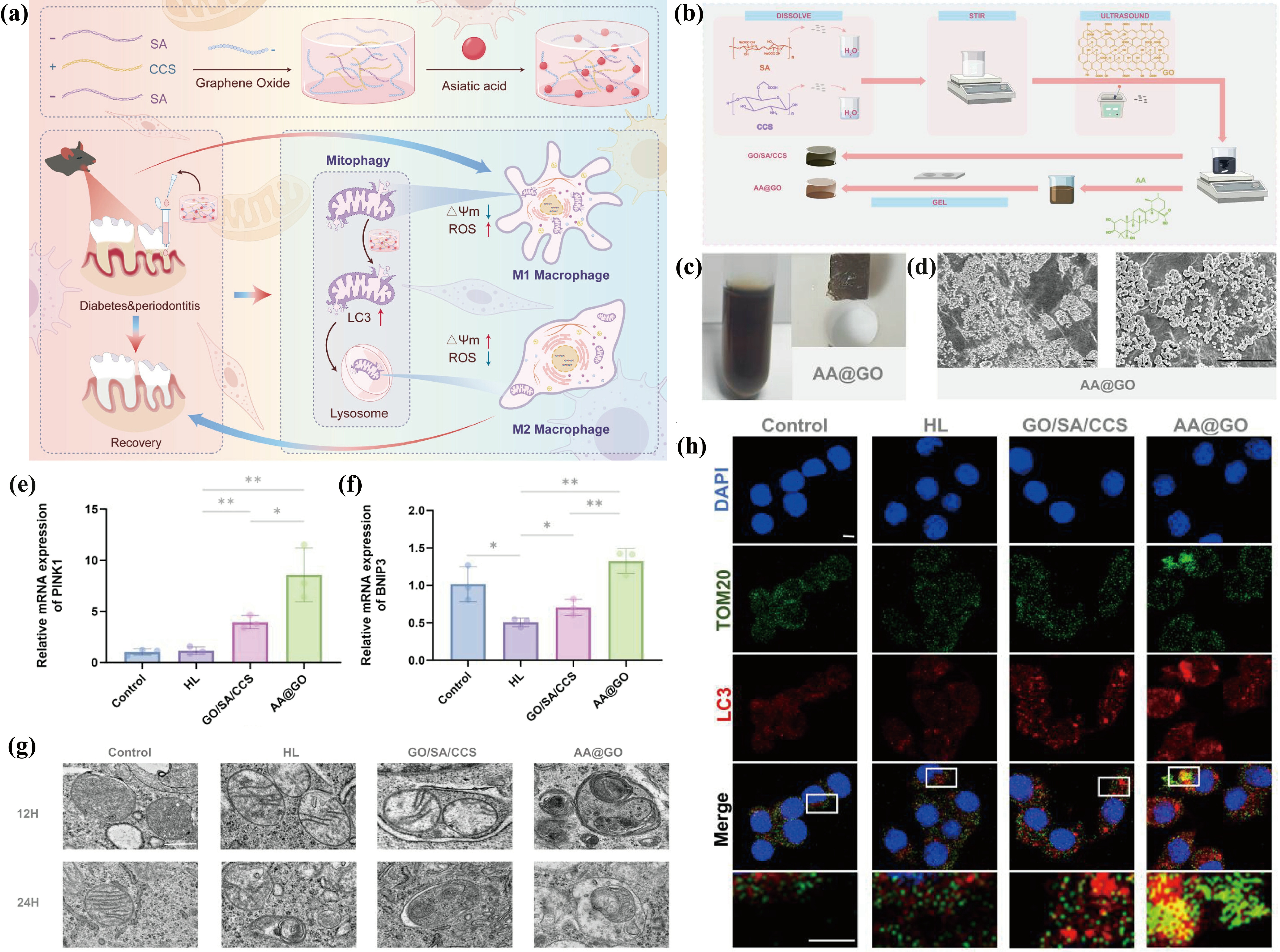
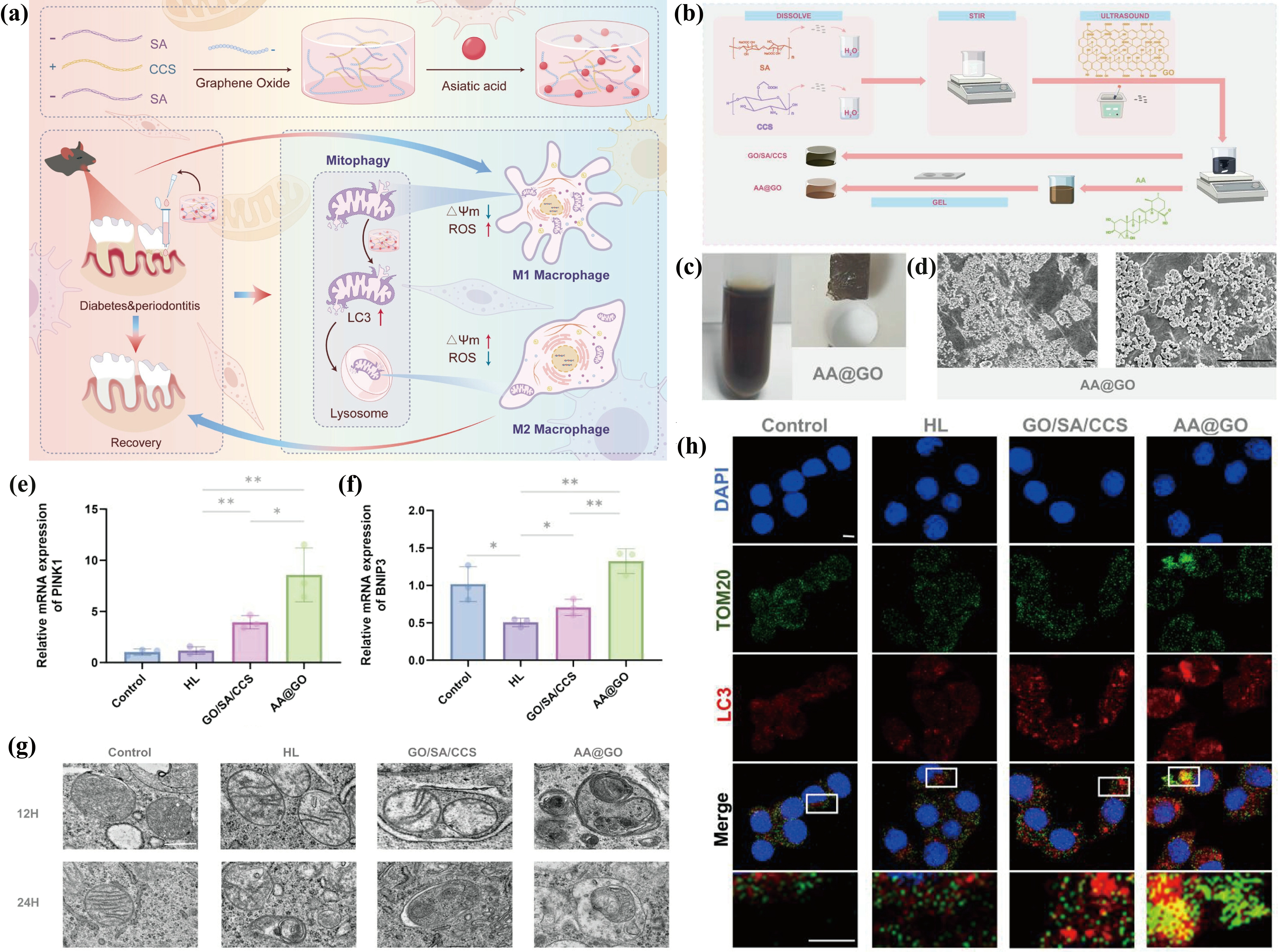
Figure 4. (a) Schematic diagram of AA@GO hydrogel for diabetic periodontitis via mitophagy-mediated M2 macrophage polarization; (b) Schematic diagram of AA@GO hydrogel synthesis; (c) Status comparison before and after AA@GO hydrogel gelatinization; (d) SEM images of AA@GO hydrogel; (e,f) Expression analysis of PINK1 and BNIP3 by qRT-PCR in RAW264.7 cells across groups; (g) TEM images indicated differences in mitophagy between groups; (h) Colocalization immunofluorescence images of the expression of LC3 and TOM2O in RAW264.7 cells. Reproduced from reference[103]. CC BY 4.0. SEM: scanning electron microscopy; TEM: transmission electron microscopy; AA@GO: hybrid hydrogel; PINK1: PTEN-induced kinase 1; BNIP3: BCL2 and adenovirus E1B 19 kDa-interacting protein 3; ROS: reactive oxygen species; CCS: carboxymethyl chitosan; SA: sodium alginate; GO: graphene oxide; Gel: ROS-responsive hydrogel; HL: high glucose+LPS; mtRNA: mitochondrial RNA; LC3: microtubule-associated protein 1 light chain 3.
For severe periodontitis with obvious bone defects, improving the periodontal microenvironment alone is insufficient to achieve tissue regeneration. Therefore, scaffolds with the capacity to guide cell migration and differentiation are indispensable. For example, 3D-printed molybdenum-containing bioactive glass ceramic (Mo-BGC) scaffolds induced M2 polarization by reprogramming macrophage metabolism from glycolysis to OXPHOS in vitro and orchestrated the regeneration of bone, periodontal ligament, and cementum in a canine critical-size periodontal defect model[104].
In summary, anti-inflammatory and immunomodulatory mitochondria-regulating biomaterials restore mitochondrial homeostasis to reprogram macrophage polarization and inhibit inflammatory signaling, thereby preventing periodontal tissue destruction while promoting regeneration. Future research should prioritize multi-target platforms that integrate complementary mechanisms.
4.2 Osteogenic mitochondria-regulating biomaterials
Progressive alveolar bone resorption is a core pathological feature of periodontitis and the primary cause of tooth loosening and tooth loss[105]. Under physiological conditions, mitochondria maintain bone homeostasis by regulating the balance between osteoblasts and osteoclasts, as well as the function of BMSCs[106]. However, in periodontitis, mitochondrial dysfunction suppresses bone formation while promoting bone resorption. Therefore, mitochondria-regulating biomaterials capable of restoring mitochondrial homeostasis to promote periodontal bone regeneration are of great importance and imperative. Herein, the representative biomaterials are summarized and shown in Table 2.
Table 2. Osteogenic mitochondria-regulating biomaterials for periodontitis therapy.
| Material | Action mechanism | Main therapeutic function | Ref. |
| GGC-Zn2+-RSV | Target mitochondria and alleviate mitochondrial dysfunction and oxidative stress | Promote PDLSCs osteogenic differentiation | [108] |
| RSV-Au@H | Improve oxidative stress status and repair dysfunctional mitochondria | Promote bone regeneration and tissue repair | [30] |
| RdAP | Increase the mitophagic flux, inhibit mitochondrial fission, and maintainmitochondrial homeostasis | Enhance the osteogenic capability of PDLSCs and prevent periodontal tissuedamage | [109] |
| MnO₂@UiO-66 | Enhance mitophagy and restore mitochondrial homeostasis | Restore osteogenic activity of periodontal ligament cells and alleviate inflammatory bone resorption | [110] |
| HMUiO-66-NH2 | Scavenge ROS | Enhance the osteogenic differentiation of BMSCs and promote bone repair | [111] |
PDLSCs: periodontal ligament stem cells; BMSCs: bone marrow mesenchymal stem cells; ROS: reactive oxygen species; RdAP: ginsenoside Rd (Rd)-loaded antioxidative polymersome; RSV-Au@H: light-responsive hydrogel; GGC-Zn2+-RSV: guanidine-grafted ethylene glycol chitosan (GGC) via zinc ion (Zn2+)-based metal coordination bonds to construct a mitochondria-targeted nanocarrier system.
Naturally derived small-molecule polyphenols scavenge mtROS and protect mitochondria, thereby alleviating inflammatory bone resorption and promoting bone regeneration. Resveratrol (RSV), a grape-derived polyphenol[107], suffers from low stability, poor aqueous solubility, limited bioavailability, and rapid metabolism. To overcome these limitations, RSV was crosslinked with guanidine-grafted ethylene glycol chitosan (GGC) via zinc ion (Zn2+)-based metal coordination bonds to construct a mitochondria-targeted nanocarrier system (GGC-Zn2+-RSV). GGC-Zn2+-RSV escaped lysosomal entrapment, alleviated mitochondrial dysfunction, and restored the osteogenic differentiation capacity of PDLSCs, ultimately repairing damaged periodontal tissue[108]. Rather than active targeting, another study encapsulated RSV in gold nanocages embedded in a near-infrared (NIR) light-responsive hydrogel (RSV-Au@H) to achieve on-demand RSV release, which effectively ameliorated oxidative stress, repaired mitochondrial dysfunction, and improved osteoblast mitochondrial morphology, thereby shortening the cementum–enamel junction-alveolar bone crest distance of the first molar by approximately 55% on the buccal side and 36% on the palatal side, and increasing BV/TV by approximately 48% after 4 weeks of treatment[30].
Synthetic polymeric vesicles scavenge ROS via surface chemical modification while releasing encapsulated therapeutic agents from their cores to activate mitophagy, preserving osteoblast function and promoting bone formation. Polymersomes, a typical subset of synthetic polymeric vesicles, exhibit this dual function. Specifically, a ginsenoside Rd (Rd)-loaded antioxidative polymersome (RdAP) was developed with a ROS-scavenging shell and a Rd-releasing core to activate mitophagy[109]. In a mouse periodontitis model, RdAP reduced oxidative DNA damage, increased mitophagic flux 2.5-fold, and maintained mitochondrial homeostasis, thereby increasing the bone mass by 1.37-fold.
Inorganic metal-based nanozymes scavenge ROS and generate O2 by mimicking SOD and CAT activities, thereby restoring mitochondrial homeostasis and promoting the osteogenic differentiation of PDLSCs. Manganese dioxide (MnO2) is a representative metal-based nanozyme with robust catalytic activity, but its clinical application is often constrained by aggregation and rapid clearance. Therefore, MnO₂ was incorporated into nanoscale mesoporous UiO-66 metal-organic frameworks (MOFs) to construct a cascade catalytic system (MnO2@UiO-66), which enhanced mitophagy via the SIRT1-FOXO3-BNIP3 signaling axis and restored mitochondrial homeostasis, ultimately alleviating inflammatory bone resorption[110].
MOFs combine their intrinsic redox activity for scavenging ROS with porous architectures for therapeutic molecule loading, activating osteogenic signaling pathways to promote alveolar bone regeneration. Among them, zirconium (Zr)-based mesoporous MOF nanoparticles HMUiO-66-NH2 scavenged ROS and enhanced the osteogenic differentiation of BMSCs via the Wnt and TGF-β signaling pathways, thereby facilitating bone repair[111].
In conclusion, mitochondria-regulating biomaterials preserve osteoblast function, suppress osteoclast activity, and promote alveolar bone regeneration, demonstrating significant potential for periodontal bone repair. Unlike traditional bone graft materials, mitochondria-regulating biomaterials restore bone metabolic imbalance by targeting mitochondria, offering well-defined mechanisms and sustained therapeutic effects.
4.3 Multifunctional mitochondria-regulating biomaterials
The pathogenesis of periodontitis involves multiple factors, including bacterial infection, immune dysfunction, and abnormal bone metabolism, which collectively contribute to periodontal tissue destruction[12]. Therefore, targeting a single factor is often insufficient to achieve satisfactory tissue repair. Since mitochondria regulate inflammation and bone metabolism, modulating mitochondrial function may simultaneously mitigate inflammation and enhance bone regeneration, positioning mitochondria as promising therapeutic targets. Accordingly, biomaterials that modulate mitochondrial function hold great potential for periodontitis treatment and can be categorized into biomimetic systems, hybrid composite systems, nanozyme catalytic systems, and smart responsive systems (Table 3).
Table 3. Multifunctional mitochondria-regulating biomaterials for periodontitis therapy.
| Material | Action mechanism | Main therapeutic function | Ref. |
| GA@LDs-CRAMP | Target mitochondria, scavenge ROS, and repair mitochondria | Inhibit inflammatory responses, restore immune homeostasis, and reduce alveolar bone loss | [112] |
| Ce-N-C@HA+CeO2-EV | Scavenged ROS | Induced M2 macrophage polarization and alleviate inflammatory microenvironment | [113] |
| GM/OHA-GZN&M hydrogel | Scavenge mtROS and restore mitochondrial function | Promote M2 macrophage polarization and alveolar bone regeneration and suppress the inflammatory response | [116] |
| SFD/CS/ZIF-8@QCT hydrogel | Enhance mitochondrial energy metabolism and reduce oxidative stress | Reduce P. gingivalis, reprogram macrophages to M2, and promote osteogenesis and angiogenesis | [117] |
| ACP@Z@C | Scavenge ROS and restore mitochondrial function | Inhibit periodontal pathogens, alleviate inflammation and oxidative stress, and restore alveolar bone height | [118] |
| MZT@S/PPP hydrogel | Scavenged mtROS and restored OXPHOS | Promote osteogenesis and angiogenesis, induce macrophage M2 polarization, inhibit alveolar bone loss, and facilitate new bone formation | [119] |
| MnO2@hPM nanozyme | Scavenge ROS, alleviate mitochondrial dysfunction, and reverse metabolic disorders | Ameliorate periodontal inflammation, reduce alveolar bone loss, and promote periodontal tissue regeneration. | [121] |
| Au@CeO2-DMF | Eliminate endogenous ROS and ameliorate mitochondrial dysfunction | Ameliorate immune dysregulation, alleviate ROS-induced tissue damage, and reestablish local periodontal homeostasis | [122] |
| PPTB NPs | Regulate mitochondrial Ca2+ levels | Inhibit inflammatory activation and restore bone homeostasis | [124] |
| J-CeM@Au | Scavenge excessive ROS | Eliminate periodontal pathogens, induce M1-to-M2 macrophage polarization, alleviate local inflammation, and promote periodontal regeneration | [125] |
ROS: reactive oxygen species; J-CeM@Au: Janus nanomotor; PPTB NPs: pH-responsive mitochondria-targeted nanocarriers; DMF: dimethyl fumarate; OXPHOS: oxidative phosphorylation; mtROS: mitochondrial reactive oxygen species; PPP: PLGA-PEG-PLGA; MZT@S: Mn-Zn-tannic acid@SS31; SFD/CS/ZIF-8@QCT hydrogel: injectable composite hydrogel; EV: extracellular vesicle; GZN: gradient-Zhang neuronet; GA@LDs-CRAMP: biomimetic lipid droplet system; CRAMP: cathelicidin-related antimicrobial peptide; LDs: lipid droplet; GA: gambogic acid; Ce-N-C@HA: hyaluronic acid (HA)-modified single-atom cerium nanozymes.
Biomimetic systems utilize natural structures including lipid droplets and EVs to enable precise mitochondria-targeted delivery and multifunctional integration. A biomimetic lipid droplet system (GA@LDs-CRAMP) was constructed by anchoring cathelicidin-related antimicrobial peptide (CRAMP) onto macrophage-derived LDs and encapsulating gambogic acid (GA) within the hydrophobic core. By targeting mitochondria, this system inhibited periodontal pathogens, reduced oxidative stress in gingival fibroblasts, and induced M2 macrophage polarization via the Nrf2 and nuclear factor-kappa B (NF-κB) pathways, thereby attenuating inflammation and alveolar bone loss (Figure 5a)[112]. In parallel, EVs have also been explored as biomimetic carriers. Lactobacillus reuteri-derived EVs loaded with ultra-small cerium oxide (CeO2) nanoparticles and hyaluronic acid (HA)-modified single-atom cerium nanozymes (Ce-N-C@HA) were combined to construct a dual-module cerium nanosystem, which effectively scavenged ROS and induced M2 macrophage polarization, thereby alleviating the inflammatory microenvironment[113]. Collectively, these biomimetic systems leverage natural carriers to deliver therapeutic agents specifically to mitochondria, achieving targeted intervention with minimal off-target effects.
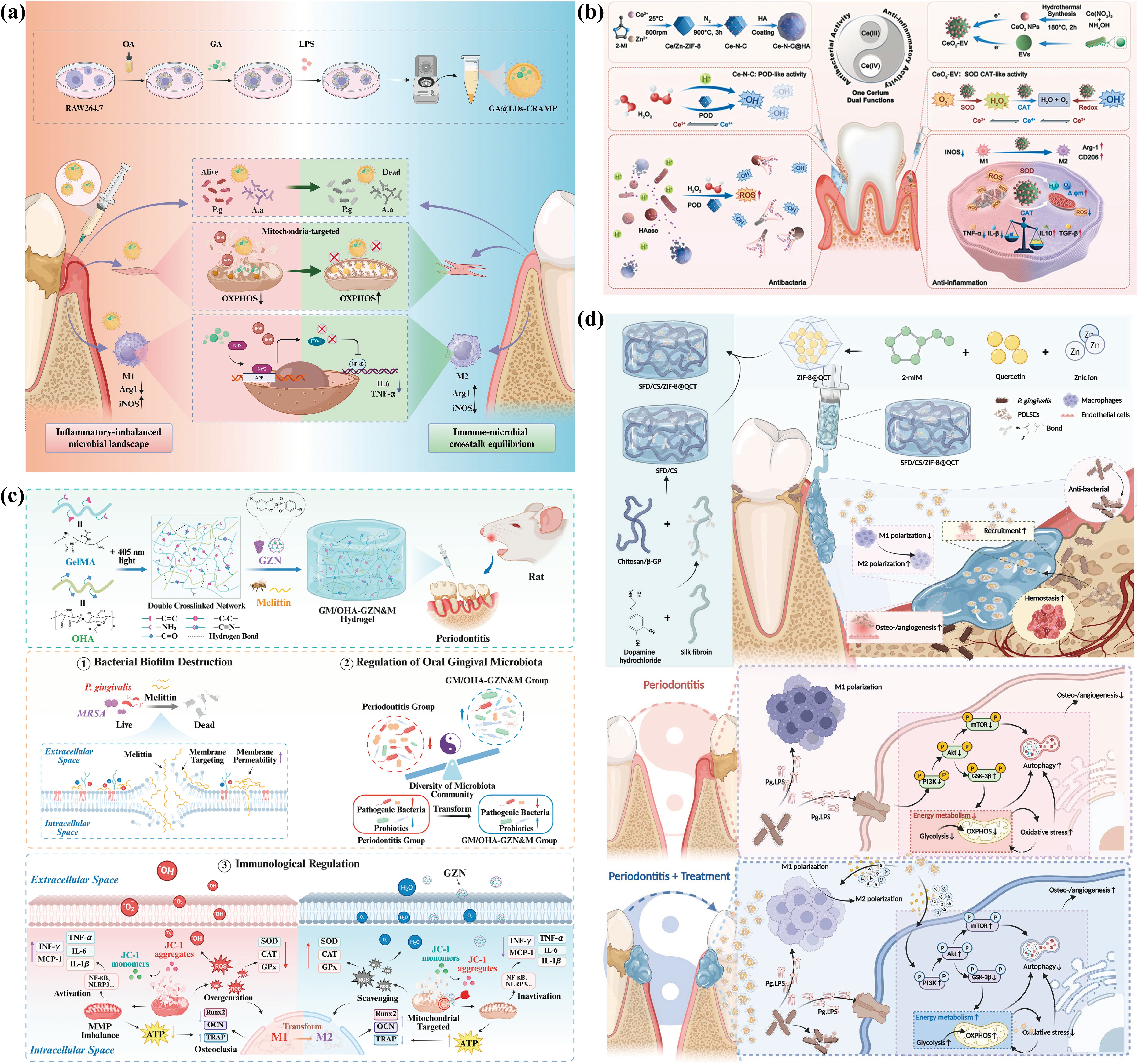
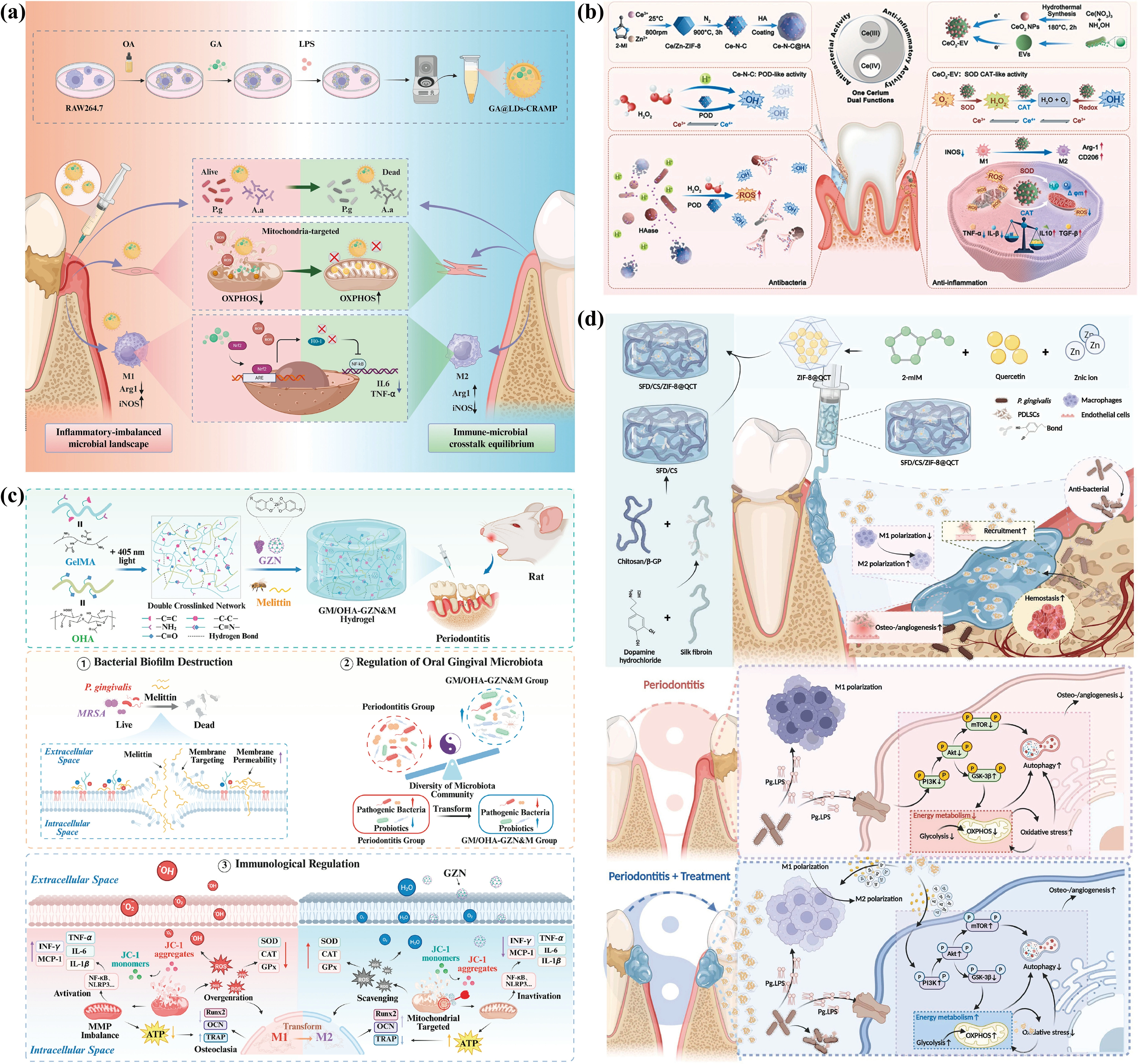
Figure 5. (a) Schematic diagram of GA@LDs-CRAMP preparation and promotion of periodontal tissue restoration. Reproduced from reference[112]. CC BY 4.0; (b) Schematic illustration of the construction and mechanism of Ce-N-C@HA and CeO2-EV for periodontitis recovery. Reproduced from reference[113]. CC BY 4.0; (c) Schematic diagram of GM/OHA-GZN&M hydrogel for periodontitis repair. Reproduced from reference[116]. CC BY 4.0; (d) Schematic illustration of the SFD/CS/ZIF-8@QCT hydrogel for regenerating alveolar bone in periodontitis. Reproduced from reference[117]. CC BY 4.0. LPS: lipopolysaccharide; OXPHOS: oxidative phosphorylation; TNF-α: tumor necrosis factor-α; IL-1β: interleukin-1β; mTOR: mammalian target of rapamycin; PDLSCs: periodontal ligament stem cells; NF-κB: nuclear factor-kappa B; SOD: superoxide dismutase; CAT: catalase; ATP: adenosine triphosphate; SFD/CS/ZIF-8@QCT: injectable composite hydrogel; ROS: reactive oxygen species; HA: hyaluronic acid; ZIF-8: zeolitic imidazolate framework-8; NLRP3: NOD-like receptor family pyrin domain containing 3; OCN: osteocalcin; INF-γ: interferon-γ; GZN: grape seed extract-zinc coordination nanoparticle; MCP-1: monocyte chemoattractant protein 1; MMP: mitochondrial membrane potential.
Hybrid composite systems integrate multiple functional components into a single platform via physical or chemical interactions to achieve combined antibacterial, anti-inflammatory, and osteogenic effects. Among diverse functional components, polyphenols represent a major class due to their intrinsic ability to bind mitochondrial outer membrane proteins and scavenge mtROS, making them attractive candidates for mitochondrial regulation[114]. Grape seed extract (GSE), a polyphenol-rich natural compound[115], was incorporated with GSE-Zn coordination nanoparticles and melittin into a mitochondria-targeting hydrogel (GM/OHA-GZN&M hydrogel) prepared by crosslinking modified gelatin (GelMA) and oxidized hyaluronic acid (OHA). The hydrogel eliminated bacteria and restored mitochondrial function by scavenging mtROS, thereby promoting M2 macrophage polarization and osteogenesis, ultimately leading to alveolar bone regeneration (Figure 5c)[116]. To further explore the therapeutic potential of polyphenols, an injectable composite hydrogel (SFD/CS/ZIF-8@QCT) was developed to co-deliver quercetin and zinc ions via pH-responsive zeolitic imidazolate framework-8 (ZIF-8). SFD/CS/ZIF-8@QCT eliminated P. gingivalis, restored energy metabolism, mitigated oxidative stress, and reversed excessive mitophagy via the PI3K/Akt signaling pathway, reprogrammed macrophages toward M2 polarization, enhanced osteogenesis and angiogenesis, and eventually facilitated periodontal alveolar bone regeneration (Figure 5d)[117]. Additionally, a polyphenol derivative, caffeic acid phenethyl ester (CAPE), was encapsulated within ZIF-8, which was incorporated into a phenylboronic acid (PBA)-modified carboxymethyl chitosan (CC) hydrogel (ACP@Z@C) for controlled release. The ACP@Z@C hydrogel exhibited antibacterial activity, scavenged ROS, alleviated inflammation, and restored mesenchymal stem cell (MSC) mitochondrial function by activating the SIRT1/p-AMPK/PGC-1α pathway, thereby promoting MSC osteogenic differentiation and alveolar bone regeneration (Figure 7a)[118]. Metal-polyphenol coordination further enhances the functionality of polyphenols by combining their ROS-scavenging capacity with the catalytic activity of metal ions. Tannic acid coordinates with Mn and Zn to form Mn-Zn-tannic acid@SS31 (MZT@S) nanozymes, which were loaded into a PLGA-PEG-PLGA (PPP) thermosensitive hydrogel (MZT@S/PPP). This system enhanced mitochondrial localization, promoted mtROS clearance, and restored OXPHOS, thereby promoting osteogenesis, angiogenesis, and M2 polarization, ultimately inhibiting bone resorption and stimulating bone formation in diabetic periodontitis (Figure 6)[119]. Overall, hybrid composite systems achieve multifunctionality via rational combination of diverse active components, offering a flexible and versatile platform for periodontitis therapy.
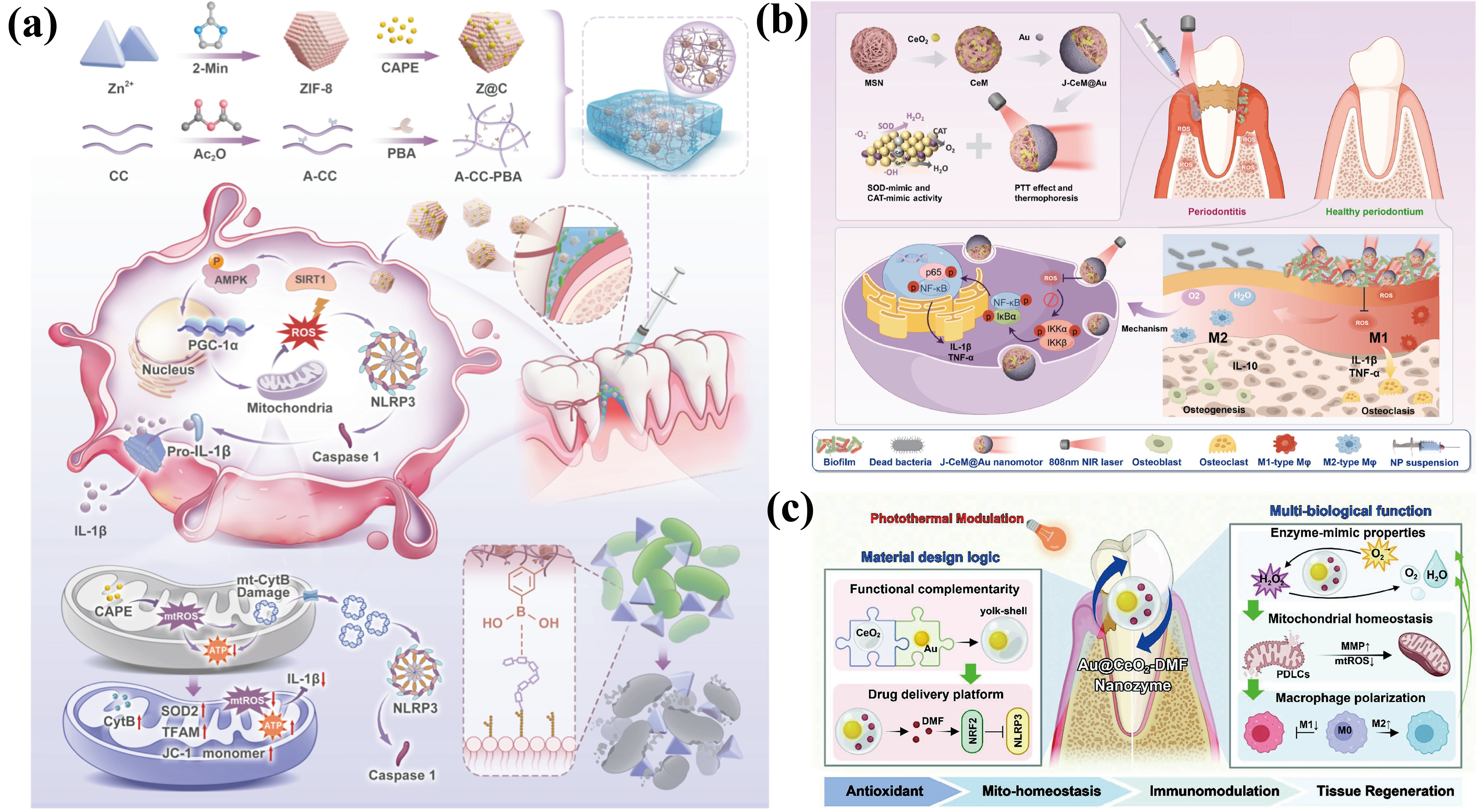
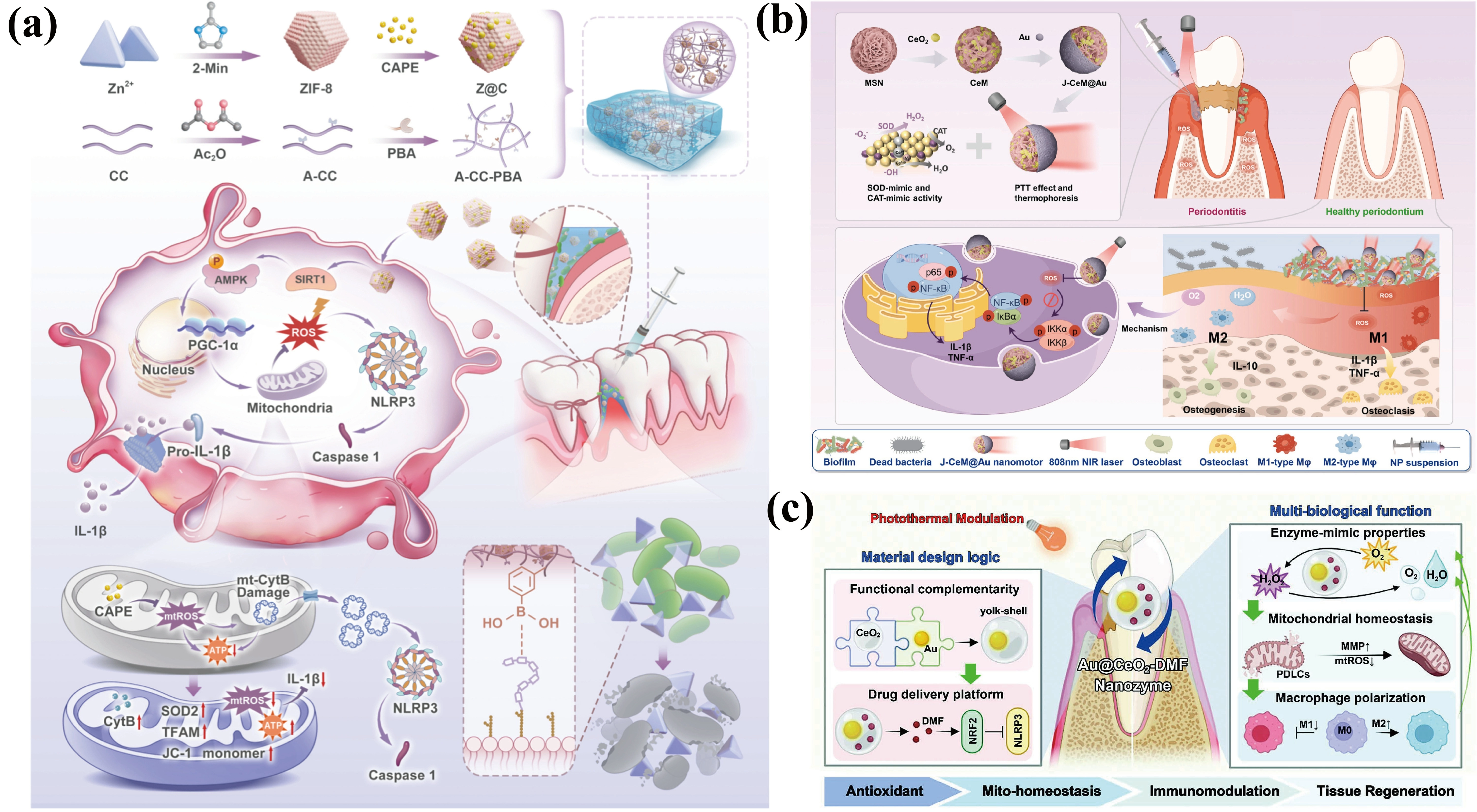
Figure 7. (a) Schematic diagram of ACP@Z@C hydrogel scavenging ROS to promote MSC osteogenesis and periodontal tissue regeneration. Reproduced from reference[118]. CC BY 4.0; (b) Schematic diagram of the synthesis process and therapeutic mechanism of J-CeM@Au for periodontitis treatment. Reproduced from reference[125]. CC BY 4.0; (c) Schematic diagram of material design of Au@CeO2-DMF and its triple-combination therapy for periodontitis through antioxidant effect, mitochondrial maintenance, and immunomodulatory mechanisms. Reproduced from reference[122]. CC BY 4.0. MSC: mesenchymal stem cell; ROS: reactive oxygen species; DMF: dimethyl fumarate; NLRP3: NOD-like receptor family pyrin domain containing 3; mtROS: mitochondrial reactive oxygen species; PDLCs: periodontal ligament cells; MMP: matrix metalloproteinase; TNF-α: tumor necrosis factor-α; IL-1β: interleukin-1β; NIR: near-infrared; ZIF-8: zeolitic imidazolate framework-8; SIRT1: SUMOylation of sirtuin 1; CAPE: caffeic acid phenethyl ester; ATP: adenosine triphosphate; PBA: phenylboronic acid; TFAM: mitochondrial transcription factor A; PGC-1α: peroxisome profiferator-activated receptor-γ coactivator 1α; MSN: mesoporous silica nanoparticle.
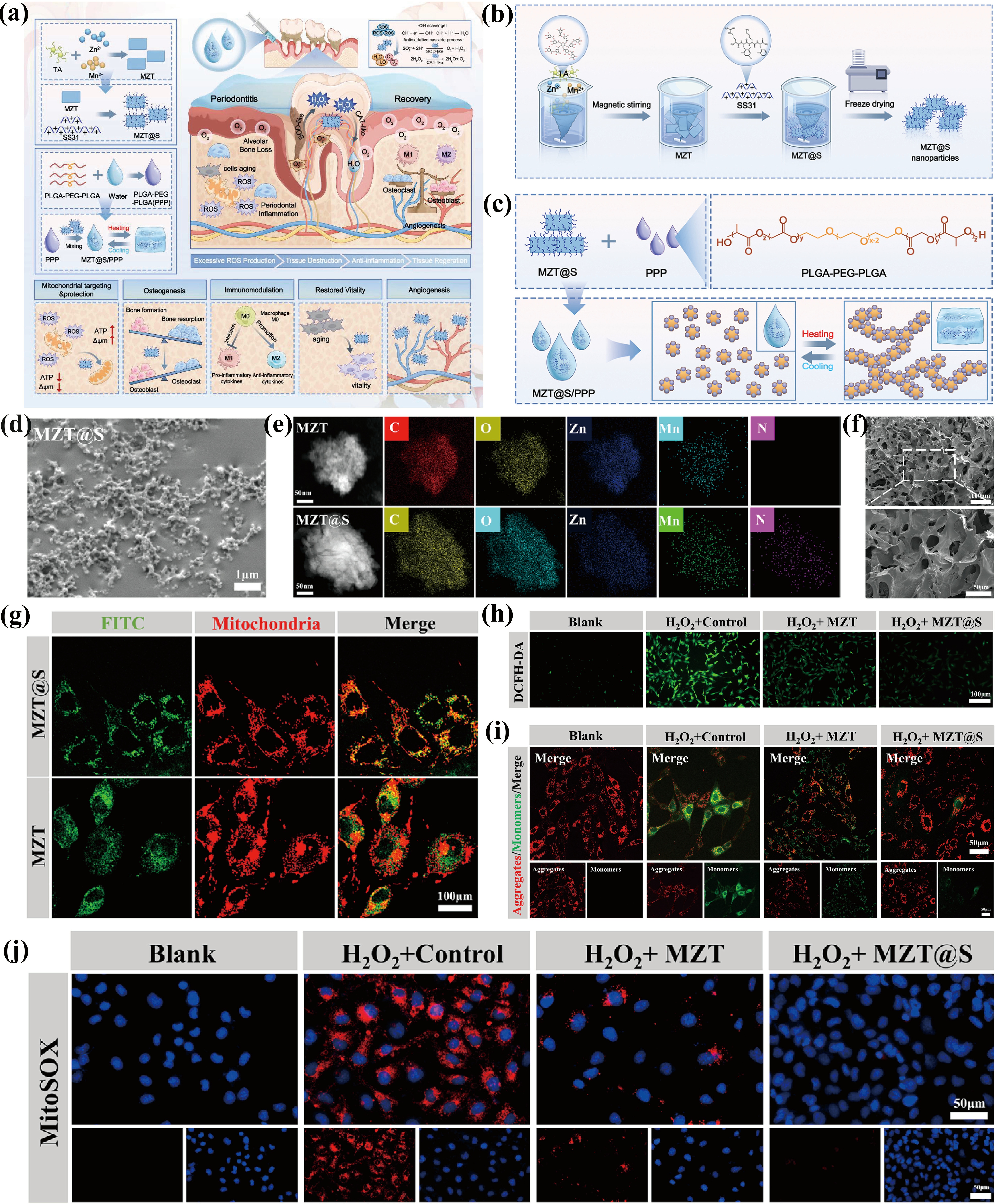
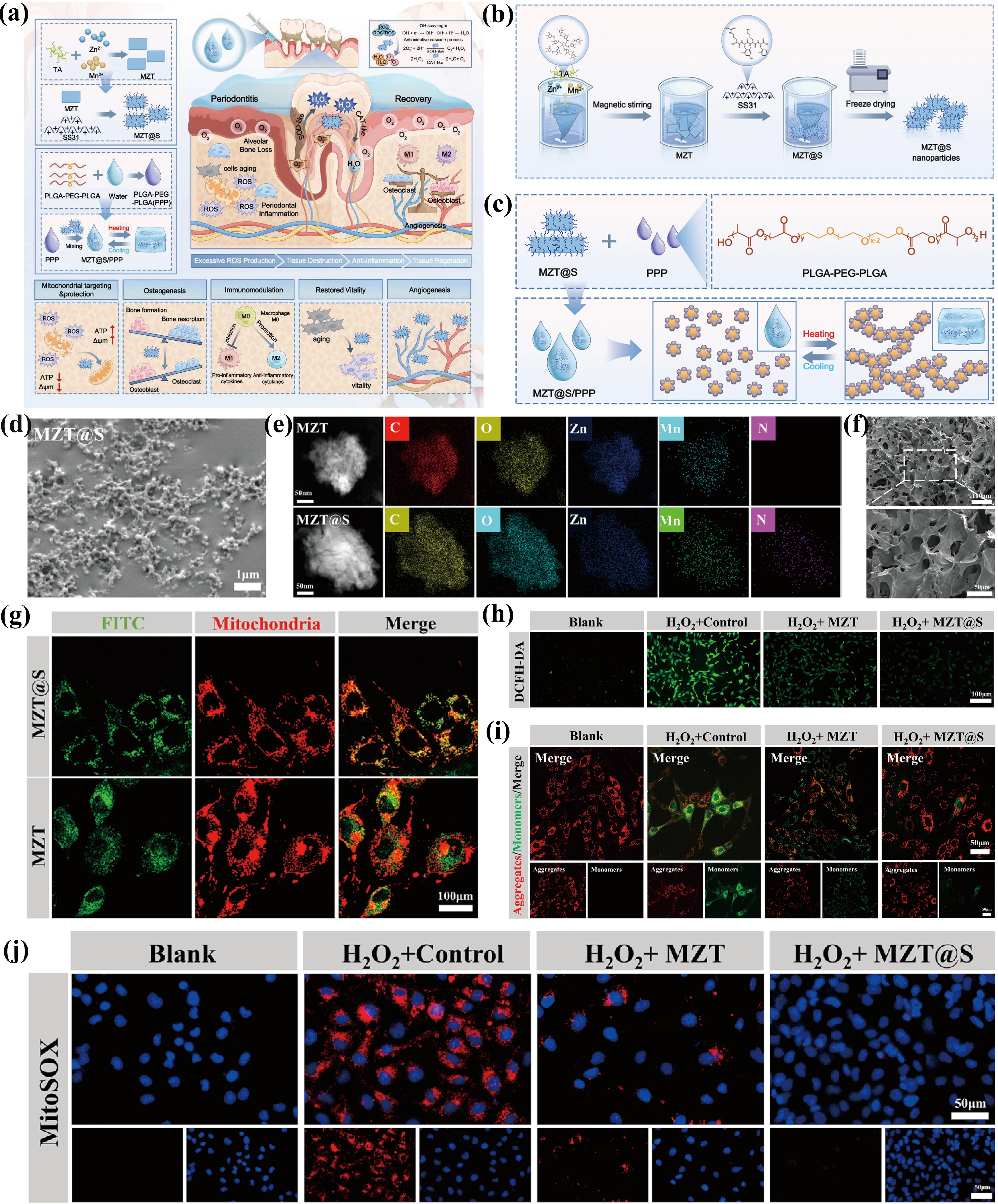
Figure 6. (a) Schematic illustration of MZT@S/PPP hydrogel preparation and its multifunctional effects during the progression of diabetes-associated periodontitis; (b) Schematic illustration of the synthesis of MZT@S; (c) Schematic illustration of the MZT@S/PPP hydrogel; (d) SEM image of MZT@S; (e) TEM images of MZT and MZT@S; (f) SEM image of MZT@S/PPP; (g) Mitochondrial localization of FITC-labeled MZT@S and MZT in BMSCs after 3 h incubation. Mitochondria were stained with MitoTracker Red; (h) Representative intracellular ROS staining; (i) JC-1 staining images showing mitochondrial membrane potential; (j) MitoSOX staining for mtROS levels. Reproduced from reference[119]. CC BY 4.0. PPP: PLGA-PEG-PLGA; MZT@S: Mn-Zn-tannic acid@SS31; SEM: scanning electron microscopy; TEM: transmission electron microscopy; ROS: reactive oxygen species; CAT: catalase; FITC: fluorescein isothiocyanate.
Nanozyme catalytic systems utilize metal nanoparticles to mimic natural antioxidant enzyme activities, thereby scavenging ROS, restoring mitochondrial function, alleviating periodontal inflammation, and preventing alveolar bone resorption. MnO2 nanoparticles, mimicking SOD and CAT[120], were encapsulated in hypoxia-educated PDLSC membranes (hPM) (MnO2@hPM) to scavenge ROS, reverse metabolic disorders, alleviate mitochondrial dysfunction and restore the osteogenic potential of PDLSCs under inflammatory conditions[121]. Similarly, a multifunctional yolk-shell nanozyme, Au@CeO2-dimethyl fumarate (DMF), ameliorated mitochondrial dysfunction and immune dysregulation and eliminated endogenous ROS in vitro and effectively reestablished local periodontal homeostasis and alleviated ROS-induced tissue damage in a rat periodontitis model (Figure 7c)[122]. These nanozymes operate through single or dual catalytic activities for ROS scavenging, whereas nanozyme cascade systems integrate multiple enzymatic reactions to simultaneously intervene at multiple nodes of mitochondrial oxidative stress, such as consuming upstream substrates and converting intermediate H2O2. For example, a cascade catalytic nanoplatform based on glucose oxidase (GOx) and CAT can consume excessive local glucose while simultaneously converting the generated H2O2 into O2[123]. Such cascade systems can cut off high-glucose-induced mitochondrial oxidative stress at its source and achieve deeper metabolic reprogramming, thereby breaking the self-amplifying cycle of ROS, inflammation, and hypoxia.
Smart responsive systems exploit the sensitivity of materials to microenvironmental stimuli to achieve spatiotemporally controllable drug release, enabling anti-inflammatory and pro-regenerative effects. In response to the acid microenvironment in periodontitis, pH-responsive mitochondria-targeted nanocarriers (PPTB NPs) loaded with calcium chelator BAPTA-AM were designed to release drugs under acidic conditions to chelate Ca2+ around and inside mitochondria, inhibit mPTPs opening, and suppress pro-inflammatory macrophage polarization[124]. In addition, a NIR light-responsive Janus nanomotor (J-CeM@Au) was developed by coating cerium dioxide-doped mesoporous silica (CeM) with gold nanoparticles (AuNP), which effectively eliminated periodontal pathogens, scavenged excessive ROS, and induced M1-to-M2 macrophage polarization via the NF-κB pathway, thereby alleviating local inflammation and promoting periodontal regeneration (Figure 7b)[125]. Both systems rely on a single stimulus, which may clear ROS too early and weaken the antibacterial ability, whereas periodontitis progresses through sequential phases of infection, oxidative stress, and inflammation. Therefore, sequential drug release is required, and one promising approach is to design nanoplatforms that respond to multiple triggers and spatiotemporally release each agent.
In conclusion, multifunctional mitochondria-targeting biomaterials integrate mitochondrial restoration with antibacterial, anti-inflammatory, and osteogenic functions, demonstrating significant potential for periodontitis therapy. Various platforms have been explored, including biomimetic systems, hybrid composite systems, nanozyme catalytic systems, and intelligent responsive systems, providing valuable design insights. Future efforts should further simplify material design, enhance biosafety, and establish comprehensive preclinical evaluation systems.
5. Challenges and Future Perspectives
Mitochondria-regulating biomaterials have shown encouraging therapeutic efficacy in preclinical models, with current materials achieving mitochondrial targeting via TPP, SS31, PDLSC membrane-mediated homologous targeting, and polyphenol-mediated mechanisms, enabling stimulus-responsive release under ROS, NIR, or pH conditions, and integrating antibacterial, anti-inflammatory, immunomodulatory, and osteogenic functions into a single platform. However, most evidence is derived from rodent models and in vitro assays, with limited evaluation of targeting specificity, long-term safety, and the evaluation of periodontal retention and degradation (Table 4), which hinders clinical translation.
Table 4. Comparative analysis of mitochondria-regulating biomaterials for periodontitis: advantages, limitations, evidence levels, and translational relevance.
| Category | Materials | Advantages | Limitations | Evidence levels | Translational relevance | Ref |
| Anti-inflammatory and immunomodulatory | MTP hydrogel | Sequential M1 macrophage and mitochondria targeting and ROS-responsive release | Dependence on tuftsin receptor expression and lack of biocompatibility and degradation data | Preclinical (in vitro + diabetic rat model) | Injectable hydrogel for periodontal pocket retention | [97] |
| CoQ10 MLip/Gel | Mitochondria-targeted delivery and ROS-responsive release | CoQ10 oxidation sensitivity and high loading requirement | Preclinical (in vitro + rat model) | Injectable and self-healing hydrogel for irregular pocket anatomy | [24] | |
| TC/pSeSe | TPP-mediated mitochondrial targeting, | Lack of long-term safety and | Preclinical (in vitro + rat model) | Mucoadhesive design under salivary flow | [99] | |
| ROS-responsive selenium release, and mucosal retention | stability evaluation | |||||
| TPP-SAT nanogel | Mitochondrial targeting | Unverified enzyme activity retention during storage | Preclinical (in vitro + rat model) | Periodontal injection, facile synthesis | [100] | |
| FIS-CDs | Enhanced water solubility | Lack of cell-specific or mitochondrial targeting and lack of local retention carrier | Preclinical (in vitro + rat model) | One-step synthesis, subgingival injection | [101] | |
| MT-CDs | Improved water solubility | Lack of cell-specific or mitochondrial targeting and lack of local retention carrier | Preclinical (in vitro + mouse model) | One-step synthesis, systemic administration | [102] | |
| GelMA@AH-12 hydrogel | Sustained AH-12 release | Unverified long-term peptide | Preclinical (in vitro + rat model) | Photocrosslinkable in situ gelation | [22] | |
| stability under proteolytic degradation | ||||||
| AA@GO hydrogel | Sustained and controlled AA delivery | Lack of cell-specific or mitochondrial targeting | Preclinical (in vitro + diabetic mouse model) | Injectable hydrogel for diabetic periodontitis | [103] | |
| Mo-BGC scaffolds | Sustained Mo release | Potential mitochondrial impairment at high Mo doses | Preclinical (in vitro + canine model | Large-animal model, multi-tissue regeneration | [104] | |
| Osteogenic | GGC-Zn2+-RSV | Guanidine-mediated lysosomal escape and mitochondrial targeting and zinc-coordinat ed RSV nanocarrier | Lack of cell-specific targeting | Preclinical (in vitro + rat model) | Simplified non-toxic synthesis | [108] |
| RSV-Au@H | NIR-responsive RSV release | Lack of mitochondrial targeting | Preclinical (in vitro + rat model) | NIR-assisted drug release | [30] | |
| RdAP | ROS-responsive polymersome | Lack of cell-specific or mitochondrial | Preclinical (in vitro + mouse | Reduced dosing frequency and drug dosage | [109] | |
| targeting | model) | |||||
| MnO2@UiO-66 | In-situ MnO2 coupling within mesoporous UiO-66 channels | Lack of targeting moiety | Preclinical (in vitro + rat model) | cost-effective; easy to use | [110] | |
| HMUiO-66-NH2 | Hierarchical mesoporous structure | Lack of cell-specific or mitochondrial targeting | Preclinical (in vitro + mouse model) | Drug-free therapy | [111] | |
| Multifunctional | GA@LDs-CRAMP | LPS-stimulated recruitment and mitochondria-targeted GA delivery | Source cell-dependent production | Preclinical (in vitro + mouse model) | Naturally derived carrier | [112] |
| Ce-N-C@HA+CeO2-EV | HAase-responsive release | Two-module injection | Preclinical (in vitro + mouse model) | Two-site administration | [113] | |
| GM/OHA-GZN&M hydrogel | Polyphenol-mediated mitochondrial targeting and dual-crosslinked hydrogel | Multi-compon ent formulation | Preclinical (in vitro + rat model) | Antibiotic-free therapy | [116] | |
| SFD/CS/ZIF-8@QCT hydrogel | Thermosensitive and adhesive hydrogel | Limited soft tissue regeneration validation | Preclinical (in vitro + rat model) | Rapid hemostasis after injection | [117] | |
| ACP@Z@C hydrogel | PBA-mediated bacterial recognition | Limited biofilm penetration | Preclinical (in vitro + rat model) | Injectable hydrogel for periodontal pocket | [118] | |
| MZT@S/PPP hydrogel | SS31-mediated mitochondrial targeting and thermosensitive hydrogel | No comparison with standard care | Preclinical (in vitro + rat model) | Injectable hydrogel for diabetic periodontitis | [119] | |
| MnO2@hPM nanozyme | PDLSC membrane-mediated homologous targeting | Membrane isolation complexity | Preclinical (in vitro + mouse and rat models) | Intravenous and local administration | [121] | |
| Au@CeO2-DMF | Yolk-shell structure for confined catalysis | Lack of targeting moiety | Preclinical (in vitro + rat model) | NIR-assisted drug release | [122] | |
| PPTB NPs | TPP mediated mitochondrial targeting | Limited local retention | Preclinical (in vitro + mouse model) | Local injection into periodontal pocket | [124] | |
| J-CeM@Au | Janus structure | Complex fabrication process | Preclinical (in vitro + rat model) | NIR-triggered photothermal therapy | [125] |
MTP hydrogel: ROS-responsive hydrogel; J-CeM@Au: Janus nanomotor; PPTB NPs: pH-responsive mitochondria-targeted nanocarriers; DMF: dimethyl fumarate; PDLSC: periodontal ligament stem cell; NIR: near-infrared; PPP: PLGA-PEG-PLGA; MZT@S: Mn-Zn-tannic acid@SS31; SFD/CS/ZIF-8@QCT hydrogel: injectable composite hydrogel; EV: extracellular vesicle; GA@LDs-CRAMP: biomimetic lipid droplet system; CRAMP: cathelicidin-related antimicrobial peptide; LDs: lipid droplet; GA: gambogic acid; ROS: reactive oxygen species; RSV: resveratrol; RdAP: Rd (Rd)-loaded antioxidative polymersome; RSV-Au@H: light-responsive hydrogel; CoQ10: coenzyme Q10; MLip: mitochondria-targeted liposome; Gel: ROS-responsive hydrogel; TPP: triphenylphosphine; LPS: lipopolysaccharide; GGC-Zn2+-RSV: guanidine-grafted ethylene glycol chitosan (GGC) via zinc ion (Zn2+)-based metal coordination bonds to construct a mitochondria-targeted nanocarrier system; Ce-N-C@HA: hyaluronic acid (HA)-modified single-atom cerium nanozymes; pSeSe: ROS-responsive diselenide-containing copolymer nanoparticle.
Therefore, several challenges still need to be addressed. First, the in vivo fate of mitochondrial-regulating materials remains unclear, as their retention in the periodontal pocket, cellular uptake, and mitochondrial targeting efficiency are rarely verified. Second, the pathological process of periodontitis is dynamic, and current biomaterials cannot precisely release therapeutic agents in appropriate amounts in response to disease progression. Third, the widely used animal models fail to recapitulate key features of human chronic periodontitis such as microbial ecological dysbiosis, immune memory, and tissue remodeling, rendering efficacy data unreliable for predicting long-term regenerative outcomes in complex clinical settings. Finally, the long-term safety of heavy-metal-based inorganic nanozymes and frameworks (e.g., Ce, Mn, Zr, Au) remains unclear, as degraded metal ions may accumulate in periodontal tissues or enter the bloodstream and deposit in organs such as the liver and spleen, and may interfere with autophagy-lysosomal pathways and disturb the mitophagy baseline in normal cells.
To address these challenges, several strategies can be adopted. First, fluorescent or radioactive labeling combined with live imaging, flow cytometry, and super-resolution microscopy can track material retention, cellular uptake, and mitochondrial targeting in real time. Second, multi-layered and multifunctional delivery systems, and three-dimensional scaffolds should be constructed to achieve the programmed release and spatiotemporal synergy of anti-inflammatory cytokines, osteogenic bioactive molecules, and therapeutic agents. Third, humanized mouse models and oral microbiome transplantation can better recapitulate human periodontal pathology and immune responses, while single-cell genomics can identify conserved and divergent features between animal models and human disease. Finally, for safety concerns such as metal ion accumulation and autophagy interference, systematic toxicological evaluation in large animal models is required.
In summary, mitochondria-regulating biomaterials stand at a pivotal juncture in periodontitis therapy, transitioning from proof-of-concept approaches toward functional repair strategies. The deep integration of materials science and biomedicine holds promise for addressing current bottlenecks, propelling the field toward a new developmental stage.
Authors contribution
Yan X: Investigation, writing-original draft.
Hu X: Conceptualization.
Wei J: Conceptualization, writing-review & editing.
Conflicts of interest
Junchao Wei is a Youth Editorial Board Member of BME Horizon. The other authors declare no conflicts of interest.
Ethical approval
Not applicable.
Consent to participate
Not applicable.
Consent for publication
Not applicable.
Availability of data and materials
Not applicable.
Funding
This work was supported by the financial support from the National Natural Science Foundation of China (Grant No. 52463017), Natural Science Foundation of Jiangxi Province (Grant No. 20242BAB26161).
Copyright
© The Author(s) 2026.
References
-
9. Li J, Hou M, Wang R, Li M, Chen J, Ge M, et al. Non-antibiotic lipid complex-in-thermogel strikes twice: Multimodal photosensitive antibacterial meets immunomodulation-boosted healing for periodontitis treatment. Mater Today Bio. 2026;37:102781.[DOI]
-
10. Ding Q, Teng Y, Zhu H, Jia Z, Xu S, Hu B, et al. Local periodontal injection, systemic heart repair: A dual-functional hydrogel for non-invasive concurrent treatment of periodontitis and myocardial infarction. Bioact Mater. 2026;62:17-33.[DOI]
-
12. Wang Q, Zhao Y, Han L, Li C, Wang Z, Zhang X, et al. A dual-action nucleoside-based supramolecular hydrogel combats periodontitis by disrupting the bacteria-osteoclast cascade. Adv Mater. 2026;38(24):e16181.[DOI]
-
13. Zhou T, Xie Y, Cheng A, Wu J, Yao Y, Yin Y, et al. Nanotherapeutics for microenvironment modulation and immune homeostasis in periodontitis treatment. Biomaterials. 2026;326:123713.[DOI]
-
14. Mutailifu D, Aini A, Maimaitiaili A. Integrated bioinformatics analysis and machine learning approach for the identification of immune-related genes in the diagnosis of aortic valve calcification with periodontitis. Biomed Technol. 2025;10:100087.[DOI]
-
15. Qin Y, Chen L, Ding Z, Zhang C, Cheng L, Tian W, et al. Covalent organic framework-based artificial antioxidases with π-electron delocalization and asymmetric coordination sites for superior inflammation inhibition and oral bone modulation. Adv Mater. 2026;38(5):e14915.[DOI]
-
16. Ou Y, Liu Y, Cai Q, Wan P, Gao G, Li X, et al. A sonosensitizer-cationic polypeptide conjugate potentiates sonodynamic therapy for mature biofilm eradication and periodontitis treatment. Adv Funct Mater. 2026;36(45):e75242.[DOI]
-
17. Wu Y, Liao J, Pu Y, Gong L, Liu X, Wu Y, et al. Mechanistic insights and therapeutic applications of metal-based nanomaterials in oral infectious diseases: Current advances and future perspectives. Biomaterials. 2026;324:123528.[DOI]
-
18. Han Z, Bao L, Yu Y, Zhao Y, Wang M, Sun Y, et al. Injectable short-fiber hydrogel with fatigue resistance and antibacterial properties for synergistic periodontitis therapy. Chem Eng J. 2025;520:166298.[DOI]
-
19. Miao MA, Wu XQ, Yuan Y, Wang YJ, Dai WT. Biochanin A as a potential agent in the disease therapy via mitochondria-mediated mechanisms. Biomed Pharmacother. 2025;190:118363.[DOI]
-
24. Liu Q, Cong R, Yu J, Zhang X, Tu J, Dong Y, et al. Ros-sensitive hydrogel system incorporating mitochondria-targeted liposomes for periodontitis treatment. Mater Des. 2025;257:114557.[DOI]
-
28. Xiang Y, Li Z, He X, Chu X, Gao C, Guo J, et al. Puerarin relives inflammation, bone destruction and facilitates osteogenic differentiation in periodontitis by enhancing mitochondrial autophagy via activating mitochondrial Mitofusin 2. Stem Cell Res Ther. 2025;16(1):218.[DOI]
-
30. Han M, Tang K, Chen Z. Preparation of NIR responsive resveratrol@Au nanocages using for regulating oxidative stress microenvironment of periodontitis. Chem Eng J. 2025;504:158774.[DOI]
-
33. Wadan AS, Moshref AS, Emam AM, Bakry YG, Khalil BO, Chaurasia A, et al. Mitochondrial dysfunction as a key player in aggravating periodontitis among diabetic patients: Review of the current scope of knowledge. Naunyn Schmiedebergs Arch Pharmacol. 2025;398(10):13173-13204.[DOI]
-
34. Zhai J, Yang X, Ren S, Wang Y, Chen J, Luo J, et al. Spatiotemporally programmable hydrogel enables NIR-triggered biofilm disruption and mitophagy-driven immunometabolic remodeling for periodontitis. Biomaterials. 2026;334:124281.[DOI]
-
41. Wu X, Lu J, Xiong Z, Huang J, Zhang J, Luo L. Melatonin alleviates periodontitis by suppressing neutrophil extracellular traps–dependent neutrophil recruitment. J Clin Periodontol. 2026;53(4):630-643.[DOI]
-
43. Wang Z, Saxena A, Yan W, Uriarte SM, Siqueira R, Li X. The impact of aging on neutrophil functions and the contribution to periodontitis. Int J Oral Sci. 2025;17(1):10.[DOI]
-
48. Cheng X, Cai Y, Geng Y, Zang X, Liu J, Luan Q. Mitochondrial calcium overload drives mtDNA-cGAS-STING activation via VDAC1 and MCU upregulation in periodontitis. Int J Mol Sci. 2026;27(10):4317.[DOI]
-
49. Shi L, Ji Y, Zhao S, Li H, Jiang Y, Mao J, et al. Crosstalk between reactive oxygen species and Dynamin-related protein 1 in periodontitis. Free Radic Biol Med. 2021;172:19-32.[DOI]
-
50. Chen Y, Ji Y, Jin X, Sun X, Zhang X, Chen Y, et al. Mitochondrial abnormalities are involved in periodontal ligament fibroblast apoptosis induced by oxidative stress. Biochem Biophys Res Commun. 2019;509(2):483-490.[DOI]
-
52. Cai Q, Quan Z, Yang X, Li C, Chen Y, Wei Y, et al. GATA-1-driven TOMM20-mediated mitophagy regulates PDLSCs osteogenesis and enhances periodontal bone regeneration. Biochem Biophys Res Commun. 2026;795:153121.[DOI]
-
53. Li X, Tian BM, Yin Y, Zhou H, Gan D, Liu F, et al. Melatonin ameliorates inflammation-induced mitochondrial dysfunction and cementoblastic differentiation in cells by regulating the METTL3/LINC01444/HSPD1 axis. Fundam Res. 2026;6(3):1948-1965.[DOI]
-
54. You J, Wu Q, Tang S, Kang J, Li D, Wang Q, et al. Microenvironment-heterogeneity-driven self-adaptive CuMo diatomic nanozyme hydrogel for NIR-II photoacoustic imaging-assisted periodontitis theranostics. Adv Funct Mater. 2026;36(80):e78028.[DOI]
-
60. Abulaiti G, Qin X, Chen L, Zhu G. Mitophagy and its significance in periodontal disease. Oral Dis. 2025;31(7):2001-2018.[DOI]
-
61. Gou H, Wang T, Chen Y, Zhou Y, Li J, Xu Y. Role of Pink1 in regulating osteoclast differentiation during periodontitis. J Dent Res. 2025;104(7):753-762.[DOI]
-
65. Zhou P, Ma X, Hu Y, Chen Y, Wang H, Wang T, et al. Sericin-based dual-module microspheres promote periodontal regeneration through four-dimensional microenvironment remodeling. Biomaterials. 2026;328:123843.[DOI]
-
67. Silwal P, Kim JK, Kim YJ, Jo EK. Mitochondrial reactive oxygen species: Double-edged weapon in host defense and pathological inflammation during infection. Front Immunol. 2020;11:1649.[DOI]
-
74. Ma S, He H, Ren X. Single-cell and transcriptome analysis of periodontitis: Molecular subtypes and biomarkers linked to mitochondrial dysfunction and immunity. J Inflamm Res. 2024;17:11659-11678.[DOI]
-
78. Van den Bossche J, Baardman J, Otto NA, van der Velden S, Neele AE, van den Berg SM, et al. Mitochondrial dysfunction prevents repolarization of inflammatory macrophages. Cell Rep. 2016;17(3):684-696.[DOI]
-
79. Mao X, Wang C, Yu S, Zhang K, Wang Z, Du J, et al. Mitochondria-targeted co-assembled nanosystem with multimodal mitochondrial DNA level control to alleviate inflammation and promote chronic wound healing. Biomaterials. 2026;329:123981.[DOI]
-
82. Huang X, Zheng L, Li P, Lin Z, Huang S, Zhou C. Fabrication of 3D printed trabecular bone-templated scaffolds modified with rare earth europium (III)-based complex for enhancing mitochondrial function in bone regeneration. Appl Mater Today. 2024;37:102130.[DOI]
-
86. Wang L, Huang X, Dai T, Xie J, Lv QX, Hou Y, et al. The role of mitochondrial dynamics in the TiO2 nanotube-accelerated osteogenic differentiation of MC3T3-E1 cells. Biochem Biophys Res Commun. 2021;535:33-38.[DOI]
-
87. Fukai T, Ushio-Fukai M. Cross-talk between NADPH oxidase and mitochondria: Role in ROS signaling and angiogenesis. Cells. 2020;9(8):1849.[DOI]
-
89. Luo Z, Yao J, Wang Z, Xu J. Mitochondria in endothelial cells angiogenesis and function: Current understanding and future perspectives. J Transl Med. 2023;21(1):441.[DOI]
-
90. Szewczyk A, Jarmuszkiewicz W, Koziel A, Sobieraj I, Nobik W, Lukasiak A, et al. Mitochondrial mechanisms of endothelial dysfunction. Pharmacol Rep. 2015;67(4):704-710.[DOI]
-
94. Zhao Y, Xiong R, Jin S, Li Y, Dong T, Wang W, et al. MitoQ alleviates H2O2-induced mitochondrial dysfunction in keratinocytes through the Nrf2/PINK1 pathway. Biochem Pharmacol. 2025;234:116811.[DOI]
-
95. Mitchell ES, Lemke S, Woodhead B, Coleman D. Oral subchronic toxicity study and genetic toxicity evaluation of mitoquinone mesylate. J Appl Toxicol. 2024;44(10):1555-1571.[DOI]
-
98. Wieland JG, Naskar N, Reess K, dos Santos DNS, Weise JM, Blatt T, et al. Impact of coenzyme Q10 on mitochondrial metabolism: A complementary study using fluorescence lifetime imaging and electron microscopy. Front Biosci. 2024;29(11):383.[DOI]
-
107. Yao Y, Lei X, Li X, Ge Y, Wang Y, Qin X, et al. A trackable mitochondria-targeting nanosystem for mitochondrial redox and mitophagy regulation in diabetic retinopathy management. Chem Eng J. 2025;505:159618.[DOI]
-
108. Tan YJ, Li X, Zhang WJ, Cao YM, Zou JK, Jing L, et al. Mitochondria-targeted delivery of zinc-coordinated resveratrol nanoparticles rescues the osteogenic potential of periodontal ligament stem cells compromised by inflammation for periodontal wound healing. Chem Eng J. 2025;503:158296.[DOI]
-
109. Hu C, Shi J, Zhang F, Lv M, Ge Z, Feng M, et al. Ginsenoside rd-loaded antioxidant polymersomes to regulate mitochondrial homeostasis for bone defect healing in periodontitis. Adv Healthc Mater. 2025;14(4):2403817.[DOI]
-
110. Zhu C, Huang K, Li T, Li Y, Jin Y, Li R, et al. Manganese dioxide coupled metal-organic framework as mitophagy regulator alleviates periodontitis through SIRT1-FOXO3-BNIP3 signaling axis. Biomaterials. 2025;319:123179.[DOI]
-
111. Lam W, Yao Y, Tang C, Wang Y, Yuan Q, Peng L. Bifunctional mesoporous HMUiO-66-NH2 nanoparticles for bone remodeling and ROS scavenging in periodontitis therapy. Biomaterials. 2025;314:122872.[DOI]
-
114. Zhang J, Gao B, Ye B, Sun Z, Qian Z, Yu L, et al. Mitochondrial-targeted delivery of polyphenol-mediated antioxidases complexes against pyroptosis and inflammatory diseases. Adv Mater. 2023;35(11):2208571.[DOI]
-
117. Yang S, Zhu Y, Ji C, Zhu H, Lao A, Zhao R, et al. A five-in-one novel MOF-modified injectable hydrogel with thermo-sensitive and adhesive properties for promoting alveolar bone repair in periodontitis: Antibacterial, hemostasis, immune reprogramming, pro-osteo-/angiogenesis and recruitment. Bioact Mater. 2024;41:239-256.[DOI]
-
121. Li L, Wu RX, Wang J, Jin R, Zhang XY, Xu M, et al. A multirisk-rescued biomimetic nanozyme against periodontitis via inflammation targeting and microenvironment reprogramming. Chem Eng J. 2025;506:160119.[DOI]
-
122. Li T, Shu M, Zhu C, Liu Q, Li Y, Wang R, et al. Triple-combination therapy with a multifunctional yolk–shell nanozyme Au@CeO2 loaded with dimethyl fumarate for periodontitis. Adv Sci. 2025;12(7):2413891.[DOI]
-
124. He P, Liu F, Li M, Ren M, Wang X, Deng Y, et al. Mitochondrial calcium ion nanogluttons alleviate periodontitis via controlling mPTPs. Adv Healthc Mater. 2023;12(15):2203106.[DOI]
Copyright
© The Author(s) 2026. This is an Open Access article licensed under a Creative Commons Attribution 4.0 International License (https://creativecommons.org/licenses/by/4.0/), which permits unrestricted use, sharing, adaptation, distribution and reproduction in any medium or format, for any purpose, even commercially, as long as you give appropriate credit to the original author(s) and the source, provide a link to the Creative Commons license, and indicate if changes were made.
Publisher’s Note
Science Exploration remains a neutral stance on jurisdictional claims in published
maps
and institutional affiliations. The views expressed in this article are solely those
of
the author(s) and do not reflect the opinions of the Editors or the publisher.
Share And Cite


Science Exploration Style
Yan X, Hu X, Wei J. Mitochondria-regulating biomaterials for periodontitis therapy: From dysfunction mechanisms to tissue regeneration. BME Horiz. 2027;5:202631. https://doi.org/10.70401/bmeh.2026.0039
Tips
Copy completed.
Submit a Manuscript
Author Instructions
Cite this Article
Article Metrics
0
View
0
Download
Cited
Article Updates

- Abstract
- Keywords
- 1. Introduction
- 2. Mitochondrial Dysfunction in Periodontitis
- 3. Impact of Mitochondrial Dysfunction on Key Physiological Processes
- 4. Application of Mitochondria-Regulating Biomaterials in Periodontitis
- 5. Challenges and Future Perspectives
- Authors contribution
- Conflicts of interest
- Ethical approval
- Consent to participate
- Consent for publication
- Availability of data and materials
- Funding
- References
- Copyright
 Get citation
Science Exploration Style

Yan X, Hu X, Wei J. Mitochondria-regulating biomaterials for periodontitis therapy: From dysfunction mechanisms to tissue regeneration. BME Horiz. 2027;5:202631. https://doi.org/10.70401/bmeh.2026.0039
copy
Share Link
copy