Transcriptomic profiling of Asini Corii Colla in wound healing: Putative molecular signatures and mechanistic hypotheses

Cong Li
1,#
,
Xiao Xie
1,#
,
Yiwen Jiang
2,#
,
Jianling Zhang
1
,
Zhijie Chen
1
,
Yue Li
3
,
Jiahao Yang
2
,
Zhuoyi Huang
3
,
Lei Jia
2
,
Zhihui Lu
4
,
Meimei Fu
5,*
,
Jinshan Guo
2,3,*

*Correspondence to:
Jinshan Guo, Department of Biomaterials and Stem Cells, Suzhou Institute of Biomedical Engineering and Technology, Chinese Academy of Sciences, Suzhou 215011, Jiangsu, China; Department of Histology and Embryology, School of Basic Medical Sciences, Southern Medical University, Guangzhou 510515, Guangdong, China.
E-mail: jsguo4127@smu.edu.cn, guojinshan@sibet.ac.cn
Meimei Fu, School of Basic Medical Sciences, Chengdu University of Traditional Chinese Medicine, Chengdu 611137, Sichuan, China. E-mail: fmm05037726@163.com
Meimei Fu, School of Basic Medical Sciences, Chengdu University of Traditional Chinese Medicine, Chengdu 611137, Sichuan, China. E-mail: fmm05037726@163.com
BME Horiz. 2027;5:202634. 10.70401/bmeh.2026.0040
Received: July 29, 2026Accepted: October 27, 2026Published: October 28, 2026
Abstract
As a precious traditional Chinese medicine, Asini Corii Colla (ACO) has long been used for hematopoiesis, hemostasis, and wound repair, yet its mechanisms in wound healing remain incompletely elucidated. Using human skin fibroblasts (HSF) and a murine macrophage-like cell line (RAW 264.7), along with a rat full-thickness skin defect model, the biocompatibility, cell proliferation and migration-promoting ability, and wound healing acceleration efficacy of ACO were investigated. HSF cells were used to assess proliferation and migration, whereas RAW 264.7 cells were used for cytocompatibility and macrophage polarization studies. The potential mechanisms were further explored by transcriptomic analysis. In vitro cytotoxicity assays revealed that ACO exhibited no obvious cytotoxicity at concentrations up to 100 mg/mL. Accordingly, further functional assays demonstrated that within the safe concentration range of 10-40 mg/mL, ACO significantly promoted acute wound healing. H&E and Masson’s trichrome histological staining, IL-1β and IL-10 immunohistochemistry, and CD31/α-SMA immunofluorescence staining revealed that ACO enhanced epithelial regeneration, increased collagen deposition, improved collagen fiber arrangement, effectively downregulated the pro-inflammatory cytokine IL-1β, upregulated the anti-inflammatory cytokine IL-10, and significantly increased CD31-positive vessel density. Transcriptomic analysis further revealed temporally distinct associations of ACO: at early stages, ACO treatment was associated with gene signatures related to cell migration, inflammatory regulation, and matrix synthesis, whereas at later stages it was associated with signatures related to tissue contraction, vascular maturation, and metabolic reprogramming. These findings provide preliminary evidence that ACO may be a promising bioactive ingredient for functional skin repair, warranting exploration of its synergistic potential for advanced wound management.
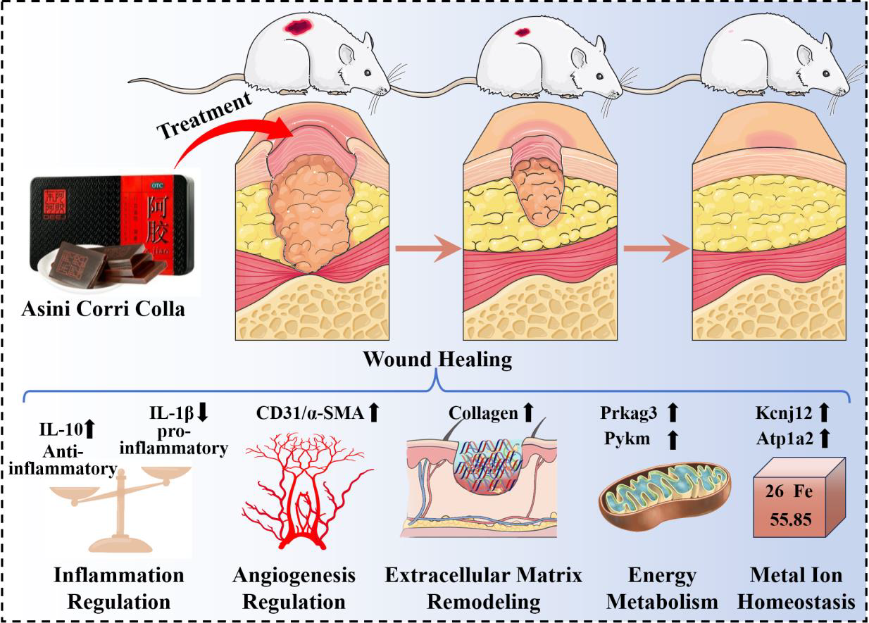
Graphical Abstract
Keywords
Asini Corii Colla, wound healing, transcriptomic
1. Introduction
Wound healing is a highly coordinated biological process involving complex interactions across multiple phases, including inflammatory response, tissue proliferation, and remodeling[1,2]. Although modern medicine has advanced significantly in wound management, the development of therapeutic strategies that effectively promote tissue regeneration, modulate inflammation, and accelerate healing remains a major challenge[3]. Traditional Chinese medicine has accumulated substantial practical experience in treating chronic wounds[4,5]. Asini Corii Colla (ACO), a precious traditional Chinese medicine with a history of nearly 3,000 years, is widely recognized for its remarkable effects in nourishing blood[6,7]. Within the framework of traditional Chinese medicine theory, it has been commonly applied in the treatment of blood deficiency patterns and various deficiency-related disorders, indicating its potential value in promoting tissue repair[8].
Previous studies have shown that enzyme-digested Asini Corii Colla accelerates wound healing and protects against oxidative stress and ultraviolet A (UVA)-induced collagen decline in cell and 3D skin equivalent models[9,10]. However, existing research has mainly relied on observational metrics such as healing rates or preliminary investigations into isolated pathways, lacking a comprehensive and systematic elucidation of its multi-target and integrative mechanisms[11]. Key scientific questions remain unanswered regarding how ACO precisely regulates the synthesis and remodeling of extracellular matrix (ECM) in the wound microenvironment, how it balances pro- and anti-inflammatory responses, and whether it modulates critical processes such as energy metabolism and angiogenesis[9,12]. This limited mechanistic understanding has hindered the modern development and broader international acceptance of ACO. Advances in high-throughput sequencing technologies now provide powerful tools for systematically elucidating the complex mechanisms of traditional Chinese medicines through transcriptomic analysis[13]. This approach allows unbiased, genome-wide acquisition of expression profiles following drug intervention[14], helping to uncover core biological pathways and molecular networks associated with ACO intervention.
Therefore, this study aims to evaluate the wound-healing efficacy of ACO and explore potential molecular associations through a series of experiments (Figure 1). While enzymatic hydrolysates of ACO have previously been reported to promote wound healing and collagen synthesis in three-dimensional skin models[9], the present study focuses on intact ACO powder and integrates in vitro, in vivo, and transcriptomic analyses. Its biosafety via in vitro cytotoxicity assays was first assessed. Subsequently, using a rat full-thickness skin defect model, we evaluated its therapeutic effects through multiple in vivo examinations, including wound closure rate, histopathological examination (Hematoxylin and Eosin (H&E) and Masson’s trichrome staining), inflammatory cytokine balance (interleukin-1β (IL-1β) and interleukin-10 (IL-10)), and angiogenesis (CD31 & α-SMA). The core of this research involves transcriptome sequencing and bioinformatic analysis of wound tissues to systematically explore the key pathways and networks by which ACO regulates inflammatory responses, energy metabolism, angiogenesis and ECM reconstruction at the gene expression level. These findings provide preliminary modern experimental evidence supporting the traditional clinical implication of ACO in wound repair and offer new perspectives for its further applications in regenerative medicine and cosmetics.
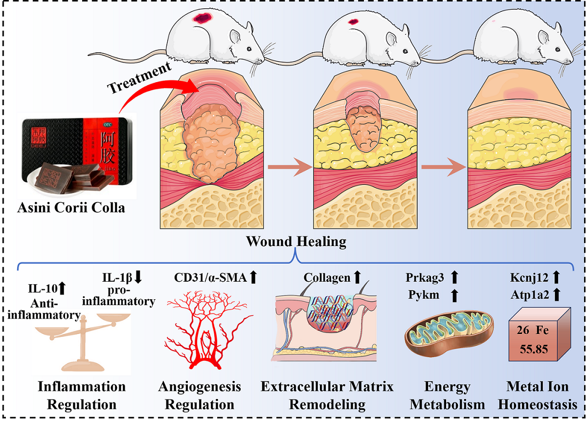
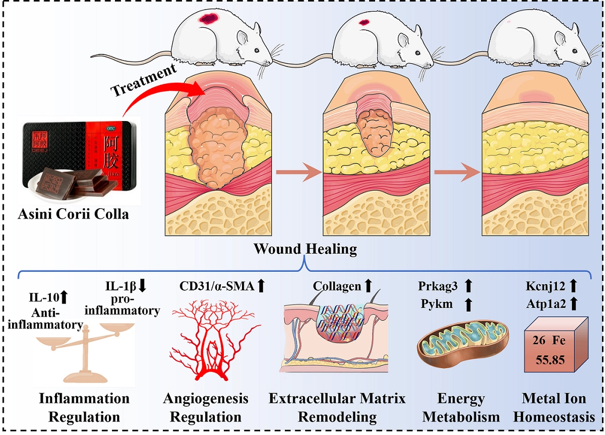
Figure 1. Schematic diagram for the possible mechanisms of ACO in promoting wound healing through coordinated regulation of multiple key biological processes. ACO: Asini Corii Colla; IL: interleukin.
2. Methods
2.1 Materials
ACO was supplied by Dong’e Ejiao Health Industry Technology Co., Ltd. All experiments were conducted using material from a single production batch (Batch No. 20240802), the same batch used in our previous investigation[10]. LC-MS/MS-based proteomic analysis of an aliquot from this batch showed that the protein fraction consisted predominantly of collagen-related proteins and collagen-derived polypeptide fragments. The complete analytical procedures and detailed proteomic data are available in the previous publication[10]. Dulbecco’s Modified Eagle Medium (DMEM), fetal bovine serum (FBS), trypsin, and penicillin-streptomycin dual-antibody solution were obtained from Gibco (USA). The Cell Counting Kit-8 (CCK-8) was purchased from Abbkine (Wuhan, China). Human skin fibroblasts (HSF) and mouse monocyte macrophage (RAW 264.7) cell lines were obtained from iCell Bioscience Inc. H&E staining kit, Masson’s trichrome staining kit, IL-1β and IL-10 immunohistochemical staining kits, as well as CD31 and α-SMA immunofluorescence staining kits, were all supplied by Wuhan Servicebio Technology Co., Ltd.
2.2 In vitro biocompatibility and functional assays
The HSF and RAW 264.7 cells were cultured in DMEM supplemented with 10% (v/v) FBS and 1% (v/v) penicillin-streptomycin at 37 °C in a humidified atmosphere with 5% CO2. A stock suspension was prepared by dispersing 1 g ACO powder in 2 mL ultrapure water preheated to 60 °C, followed by intermittent vortexing and ultrasonication until a homogeneous suspension was formed (nominal concentration: 500 mg/mL). For in vitro experiments, working concentrations (0.156-100 mg/mL) were prepared by serial dilution of the stock suspension in complete medium and then filter-sterilized through a 0.22 μm membrane. For in vivo experiments, the homogeneous suspension was used directly without filtration, and working concentrations were prepared by dilution in deionized water. All reported concentrations are nominal concentrations based on the initial amount of ACO dispersed. ACO solutions in working concentrations (0.156-100 mg/mL) were obtained by serial dilution of the ACO stock solution.
HSF and RAW 264.7 cells in the exponential growth phase were harvested and seeded into 96-well plates at cell densities of 10,000 cells/well for each cell line. After 24 hours’ incubation at 37 °C under 5% CO2 to allow cell adhesion, the medium was replaced with fresh complete medium (containing 10% FBS) supplemented with ACO working solutions at various concentrations, and the cells were cultured for another 24 hours. The medium was then removed, and the cells were washed with PBS. CCK-8 solution was added to each well, and the plates were cultured in the incubator for another 1 hour. Finally, the absorbance (OD value) of each well was measured at a wavelength of 450 nm using a microplate reader.
HSF cells were seeded in 96-well plates at a density of 5 × 10³ cells per well and cultured overnight. Cells were then treated with ACO at concentrations of 10, 20, 40, and 80 mg/mL, with untreated cells serving as the control. After incubation for 1, 3, and 5 d, 10 μL of CCK-8 reagent was added to each well and incubated for an additional 1 h. Absorbance was measured at 450 nm using a microplate reader. Cell viability was calculated relative to the untreated control group at each time point.
HSF cells were seeded in 6-well plates and cultured to 100% confluence. A straight scratch wound was created across the monolayer using a sterile 200 μL pipette tip. After washing with PBS to remove detached cells, the cells were treated with ACO at concentrations of 10, 20, 40, and 80 mg/mL in serum-free medium. Wound closure was monitored and photographed at 0, 24, and 48 h using an inverted microscope. The wound area was measured using ImageJ software.
HSF cells were serum starved for 12 h, then harvested and resuspended in serum-free medium. A total of 5 × 10⁴ cells in 200 μL of serum-free medium were seeded into the upper chamber of a Transwell insert (8 μm pore size, Corning). The lower chamber contained 600 μL of medium with 10% FBS and ACO at concentrations of 10, 20, 40, and 80 mg/mL. After 24 h of incubation, non-migrated cells on the upper surface of the insert were removed with a cotton swab. Migrated cells on the lower surface were fixed with 4% paraformaldehyde, stained with 0.1% crystal violet, and counted under a microscope in five randomly selected fields per insert.
Macrophages were treated with LPS (M1 induction) or IL-4 (M2 induction) in the presence or absence of ACO. Cells were fixed with 4% paraformaldehyde, permeabilized, and blocked. Then, cells were incubated overnight at 4 °C with primary antibodies against CD86 (M1-like marker) or CD206 (M2-like marker). After washing, cells were incubated with fluorophore-conjugated secondary antibodies, and nuclei were counterstained with DAPI. Images were captured using a fluorescence microscope.
2.3 In vivo wound regeneration evaluation of ACO
Male SD rats (≈ 200 g) obtained from the Experimental Animal Center of Southern Medical University were randomly divided into three groups using a computer-generated random number table: Vehicle control (deionized water), recombinant humanized type III collagen (RHC III, 20 mg/mL), and ACO (20 mg/mL). All experiments were performed at two time points (day 6 and day 12), with 5 rats per group at each time point. All groups received daily topical application of 100 μL of the corresponding solution per wound. The concentration of ACO used for in vivo administration (20 mg/mL) was selected based on preliminary dose response experiments in vitro (10, 20, 40, and 80 mg/mL), which showed that 20 mg/mL achieved comparable efficacy to 40 mg/mL. This concentration is also well below the cytotoxic threshold (up to 100 mg/mL) established by the extended cytotoxicity assay. Then, the allocation sequence was concealed from the investigator performing the surgeries and the outcome assessor. All rats were acclimatized for one week prior to surgery. Under isoflurane anesthesia, the dorsal hair was removed, and the skin was disinfected with 75% alcohol swabs. A 14 mm circular wound was created on each side of the dorsal skin of each rat using sterile surgical scissors. The wounds were left uncovered after each application. Since the test materials were in solution form, the daily application did not disturb the wound bed or cause mechanical detachment of the newly formed epithelium. Wound changes were photographed on days 0, 3, 6, and 12 postoperatively, and the images were analyzed using ImageJ software by two independent observers blinded to the group assignments. All animals were housed under controlled conditions (temperature 24 ± 2 °C, humidity 30%-70%, 12 h light/dark cycle) with free access to food and water. All animal experiments were conducted in compliance with the Animal Experimental Committee of Institute of Biological and Medical Engineering, Guangdong Academy of Sciences (Approval No. K2024 01 272 472).
2.4 Histological analysis
On days 6 and 12 post-surgery, tissue sections were harvested, and the paraffin-embedded tissue sections were then deparaffinized, rehydrated, and subsequently subjected to H&E and Masson’s trichrome staining. All stained sections were scanned at 20 × magnification under bright field using a fully automated digital slide scanner. Morphometric analysis was performed using ImageJ software. The wound width was determined by H&E-stained sections by measuring the distance between the two epidermal migration margins, while granulation tissue thickness was measured on H&E-stained sections as the vertical distance from the wound bed base to the surface of the granulation layer. The collagen deposition was quantified by Masson’s trichrome-stained sections. To evaluate the inflammatory response, immunohistochemical staining for IL-1β and IL-10 was also performed on regenerated tissues. For quantitative analysis, three randomly selected sections per animal were examined, and three non-overlapping fields per section were captured under a light microscope (200 × magnification). The mean integrated optical density (IOD) of positively stained areas was measured using ImageJ software. The percentage of positive staining area relative to the total tissue area was calculated for each field, and the average values were used for statistical analysis. All data were normalized to the control group (set as 100%). Additionally, angiogenesis was evaluated by immunofluorescence co-staining for CD31 and α-SMA. For quantification of angiogenesis, three randomly selected sections per animal were examined, and three non-overlapping fields per section were captured under a fluorescence microscope (200 × magnification). Vessels positive for both CD31 and α SMA were counted as mature vessels. The average number of CD31/α SMA double positive vessels per field was calculated for each animal. Additionally, at the end of the experiment, major organs (heart, liver, spleen, lung, and kidney) were harvested, fixed in 4% paraformaldehyde, embedded in paraffin, sectioned at 4 μm, and stained with hematoxylin and eosin (H&E). Histological changes were evaluated under a light microscope. All quantifications were performed by two independent observers blinded to the group assignments.
2.5 Transcriptome sequencing of wound tissue
Wound tissue samples were harvested from each group on days 6 and 12 post-operation (n = 4 per group). Total RNA was extracted using TRIzol reagent, and RNA integrity was assessed using an Agilent 2100 Bioanalyzer (RIN ≥ 7.0). Samples with RIN < 7.0, total RNA < 1 μg, visible rRNA degradation, or failed library/sequencing QC were excluded; all samples passed these criteria. cDNA libraries were constructed using the NEBNext® UltraTM II RNA Library Prep Kit and sequenced on an Illumina NovaSeq 6000 platform with a paired-end 150 bp read length, generating approximately 20-30 million reads per sample. All libraries were prepared and sequenced in a single batch.
Raw reads were filtered using Trimmomatic (v0.39) and aligned to the rat reference genome (Rnor_6.0) using STAR (v2.7.9a). Gene expression was quantified with featureCounts (v2.0.1) using Ensembl annotations. Differential expression analysis was performed using DESeq2 (v1.36.0), with genes defined as differentially expressed at false discovery rate (FDR) < 0.05 and |log2FC| ≥ 1. GO and KEGG enrichment analyses were conducted using clusterProfiler (v4.4.4), and GSEA was performed using fgsea (v1.22.0) with gene sets from MSigDB and GO, ranking metric = signal to noise, permutations = 1,000, and FDR (Benjamini-Hochberg) < 0.25 considered enriched. Principal component analysis and heatmap visualization were performed using R packages (prcomp and pheatmap). Raw sequencing data have been deposited in the NCBI Sequence Read Archive (SRA) under BioProject accession PRJNA1535550.
2.6 Statistical analysis
All data are presented as mean ± SD. Statistical analyses were performed using GraphPad Prism 9 (GraphPad Software, USA). For comparisons between two groups, Student’s t-test (two tailed) was used. For single time point comparisons among three or more groups, one way ANOVA was performed, followed by Tukey’s post hoc test. For wound closure data involving multiple groups across multiple time points, two way repeated measures ANOVA was applied, followed by Tukey’s post hoc test. For RNA seq data, the FDR was controlled using the Benjamini-Hochberg method, and genes with an adjusted p-value (p-value) < 0.05 were considered significantly differentially expressed. A p-value < 0.05 was considered statistically significant, with *, **, and *** denoting p < 0.05, p < 0.01, and p < 0.001, respectively.
3. Results
3.1 In vitro cell experiments
Dermal fibroblasts and macrophages are two pivotal cell types that regulate the skin wound healing process[15]. To evaluate the cytocompatibility of ACO on these cells, cytotoxicity assays were performed using HSF and RAW 264.7 cell models. As shown in Figure 2a,b, ACO treatment at concentrations ranging from 0.156 to 100 mg/mL showed no significant cytotoxicity in either cell type. No statistically significant differences in cell viability were detected among the experimental groups, indicating that ACO exhibits favorable cytocompatibility within 0.156-100 mg/mL range.
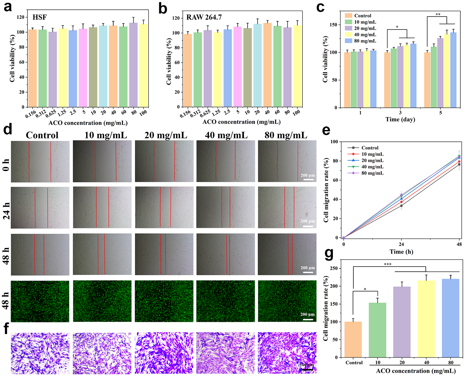
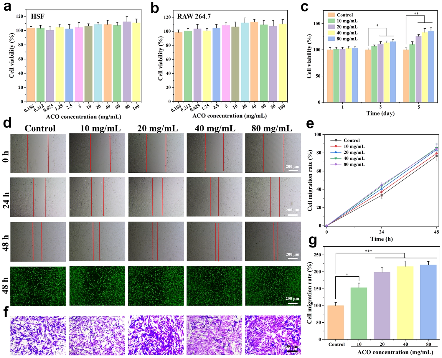
Figure 2. Cytotoxicity evaluation of ACO. (a) Cell viability of HSF and (b) RAW 264.7 after treatment with ACO at various concentrations (n = 5); (c) Bar graph of HSF cell proliferation at different concentrations of ACO (Cell viability was normalized to the respective control group at each time point and set as 100%); (d) Representative images and (e) quantitative analysis of cell migration rates obtained from scratch wound healing assay in HSF cells (n = 3); (f) Representative images and (g) quantitative analysis of cell migration obtained from Transwell migration assay (n = 3). *p < 0.05, **p < 0.01, ***p < 0.001. ACO: Asini Corii Colla; HSF: human skin fibroblasts.
To evaluate the effect of ACO on HSF cell proliferation, a time-course CCK-8 assay was performed at 1, 3, and 5 d. As shown in Figure 2c, ACO significantly promoted cell proliferation in a concentration- and time-dependent manner. At 5 d, the cell viabilities of the 10, 20, 40, and 80 mg/mL groups reached 110.18%, 125.77%, 133.10%, and 135.96% of the respective control group (p < 0.01).
The effect of ACO on cell migration was assessed using both scratch wound healing and Transwell assays. As shown in Figure 2d,e, ACO treatment significantly accelerated wound closure at both 24 and 48 h compared with the control group. The 20, 40, and 80 mg/mL groups achieved closure rates of 83.39%, 85.38% and 84.35% at 48 h, respectively, whereas the control group only reached 76.35%. Consistently, the Transwell migration assay revealed that ACO significantly increased the number of migrated cells in a concentration-dependent manner (Figure 2f,g), with the 20 mg/mL group showing approximately 1.98-fold more migrated cells than the control group. These results collectively demonstrate that ACO possesses both proliferative and promigratory activities on HSF cells.
The enhanced proliferation and migration of HSF cells upon ACO treatment suggest that ACO may facilitate skin repair by promoting the expansion and mobilization of dermal fibroblasts to the site of injury. The time-dependent CCK 8 results confirmed that ACO exerts a sustained proliferative stimulus rather than a transient effect, supporting its potential role in long term tissue regeneration. Furthermore, the scratch and Transwell assays consistently demonstrated that ACO promotes directional cell migration, which is a critical step in wound re-epithelialization and dermal remodeling. These findings indicate that ACO exhibits favorable biocompatibility, promotes cell proliferation and migration, and shows potential for wound healing applications.
To directly evaluate macrophage polarization, immunofluorescence staining for CD86 (M1-like) and CD206 (M2-like) was performed. As shown in Figure S1, LPS stimulation induced a strong CD86 signal, whereas IL-4 stimulation induced a strong CD206 signal, confirming successful polarization. In the presence of ACO, the LPS-induced CD86 signal was markedly reduced, while the IL-4-induced CD206 signal was further enhanced. These results suggest that ACO may promote a shift from M1-like toward M2-like polarization.
3.2 Construction of the rat skin wound model and morphological analysis
To evaluate the effect of ACO on skin wound healing, a full-thickness skin defect model was established in rats (Figure 3a). The dorsal wounds were monitored and photographed on days 0, 3, 6, and 12 post-surgery. As shown in Figure 3b, the ACO group exhibited a significant pro-healing effect. On day 3, wound area reduction was comparable across all groups. By day 6, however, the wound healing rates in the ACO group (83.33%) and RHC-III group (82.07%) were significantly higher than those in the control group (66.03%, p < 0.05, Figure 3c). On day 12, considerable unhealed wound areas remained in the control group (healing rate: 82.09%), whereas wounds in the ACO and RHC-III groups were nearly completely closed, with healing rates of 98.14% and 99.05%, respectively, both significantly higher than those of the control group (p < 0.05). The wound closure rate in the ACO group was comparable to that in the RHC-III group; however, no formal equivalence or superiority analysis was performed, and this comparison should be considered descriptive only.
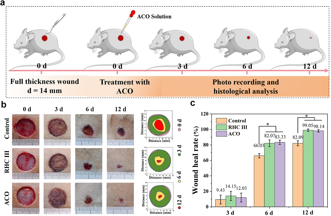
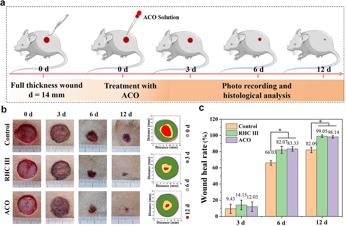
Figure 3. In vivo assessment of ACO using a rat full-thickness wound model. (a) Schematic diagram of the rat wound model establishment and treatment; (b) Representative wound photographs on days 0, 3, 6, and 12; (c) Quantification of wound healing rate (n = 5 per time point). *p < 0.05, **p < 0.01, ***p < 0.001. ACO: Asini Corii Colla; RHC III: humanized type III collagen.
3.3 Histological evaluation of wound healing by H&E and Masson’s trichrome staining
To histologically evaluate the wound-healing efficacy of ACO, regenerated skin tissues harvested on days 6 and 12 post-operation were subjected to H&E and Masson’s trichrome staining. As shown in Figure 4a, the Control, RHC-III, and ACO groups all exhibited inflammatory cell infiltration on day 6. Nevertheless, the RHC-III and ACO groups displayed more prominent fibroblast migration and proliferation, with granulation tissue partially filling the wound area. As shown in Figure 4b, the wound widths in these two groups were measured as 8.36 mm and 5.36 mm, respectively, which were significantly smaller than those of the control group (10.26 mm, p < 0.05). By day 12, the control group still exhibited marked inflammatory cell infiltration, incomplete epithelial migration toward the wound center, and disorganized tissue architecture. In contrast, the ACO group had developed more structurally intact dermal and epidermal layers, with a wound width of only 2.10 mm (Figure 4b), substantially narrower than that of the control group (5.60 mm, p < 0.05). In addition to wound width, granulation tissue thickness was also significantly increased in the ACO-treated group (Figure 4c), further supporting the pro-regenerative effect of ACO.
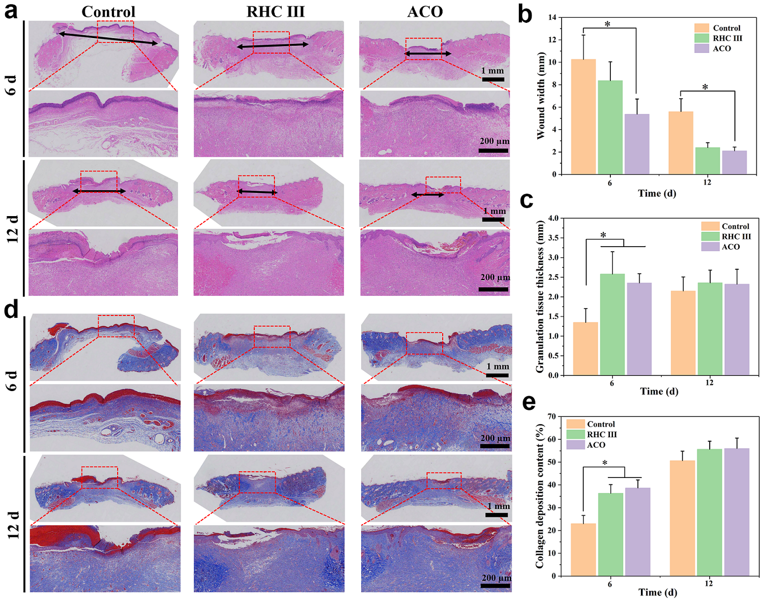
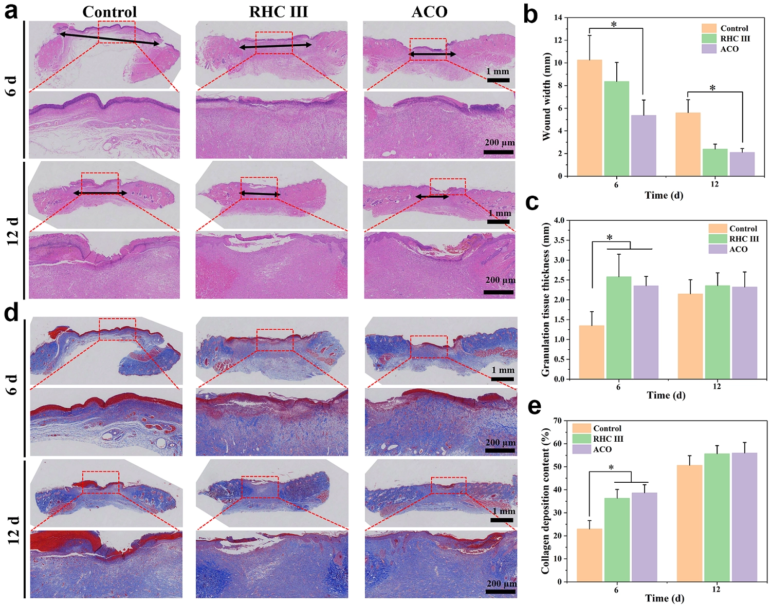
Figure 4. Histological evaluation of wound healing by H&E and Masson’s trichrome staining. (a) H&E staining images of skin tissues on days 6 and 12 of the treated wounds (black line denotes wound width); (b) Measured wound widths on days 6 and 12 (n = 5); (c) Granulation tissue thickness on days 6 and 12 (n = 5); (d) Masson’s trichrome staining images of wound tissues on days 6 and 12 post-operation; (e) Collagen deposition content on days 6 and 12 (n = 5). *p < 0.05, **p < 0.01, ***p < 0.001. ACO: Asini Corii Colla; H&E: Hematoxylin and Eosin; RHC III: humanized type III collagen.
Analysis of Masson’s trichrome staining revealed more extensive blue staining signals in the wound areas of the RHC-III, and ACO groups compared to the control group on day 6 post-operation, indicating enhanced collagen deposition (Figure 4d). As shown in Figure 4e, the collagen content in the two groups reached 36.33% and 38.66%, respectively, significantly higher than that in the control group (23%, p < 0.05). By day 12, both the RHC-III and ACO groups displayed denser and more structurally mature collagen fibers, with collagen contents of 55.66% and 56.00% (Figure 4e), respectively, which showed a trend toward higher levels (although not statistically significant) compared to the control group (50.66%). These findings indicate that ACO treatment was associated with increased collagen deposition and improved collagen fiber organization in this acute wound model. However, collagen architecture, the collagen I/collagen III (Col I/Col III) ratio, skin appendages, tensile strength, and scar formation were not evaluated. Therefore, increased collagen deposition should not be interpreted as direct evidence of superior healing quality, as excessive collagen deposition may also be associated with fibrosis or scar formation.
3.4 The modulatory effect on inflammation of ACO
Oxidative stress activates multiple intracellular signaling pathways, leading to the overexpression of chemokines and inflammatory cytokines, which upregulate the inflammatory response and delay wound healing[16]. To evaluate the modulatory effect of ACO on wound inflammation, immunohistochemical staining of regenerated tissue for IL-1β and IL-10 was performed. As shown in Figure 5a,b, on day 6 post-operation, the expression level of the pro-inflammatory cytokine IL-1β in the control group (100%) was significantly higher than that in the RHC-III group (58.86%) and the ACO group (50.87%, p < 0.05). Meanwhile, the ACO group exhibited a higher expression of the anti-inflammatory cytokine IL-10 (110.63%), suggesting an early anti-inflammatory effect of ACO (Figure 5c,d). By day 12, the expression levels of both IL-1β and IL-10 further decreased in the ACO group, reaching 27.32% and 57.86%, respectively. This decrease in IL-10 expression at the later time point is consistent with the resolution of inflammation, rather than indicating direct suppression of IL-10 by ACO. A similar trend was observed in the RHC-III group (34.42% for IL-1β and 60.97% for IL-10) on day 12. Comparisons between the two treatment groups revealed that the ACO group was more effective in restoring inflammatory balance. In contrast, the control group maintained a relatively high level of IL-10 expression (117.63%) on day 12 (Figure 5d). This observation may be consistent with ongoing immune activation in control wounds, although IL-10 alone cannot be interpreted as direct evidence of persistent inflammation. In summary, ACO effectively suppresses pro-inflammatory cytokine expression and promotes anti-inflammatory cytokine release during the early phase, thereby contributing to the modulation of the wound inflammatory microenvironment and facilitating the healing process.
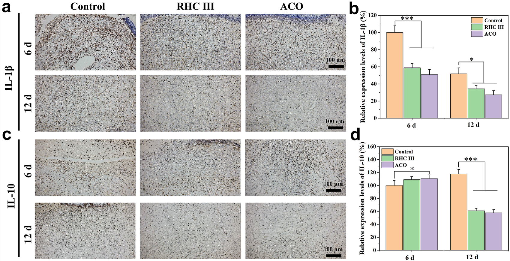
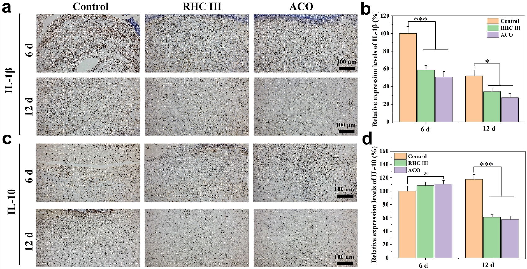
Figure 5. The modulatory effect of ACO on inflammation. (a) Representative IL-1β immunohistochemical staining images on days 6 and 12; (b) Relative expression levels of IL-1β (n = 5); (c) Representative IL-10 immunohistochemical staining images on days 6 and 12; (d) Relative expression levels of IL-10 (n = 5). *p < 0.05, **p < 0.01, ***p < 0.001. ACO: Asini Corii Colla; IL: interleukin; RHC III: humanized type III collagen.
3.5 Angiogenesis promotion effect of ACO
Neovascularization serves as a key indicator for assessing the extent of skin tissue regeneration and functional recovery, and its promotion facilitates dermal reconstruction and the formation of intact skin structure[17]. To evaluate the dynamic changes in neovascularization during wound healing, this study detected the expression of CD31 and α-SMA via immunofluorescence. As shown in Figure 6a,b, on day 6 post-operation, the number of new blood vessels in the ACO and RHC-III groups was significantly higher than that in the control group (p < 0.001), with CD31/α-SMA expression levels increasing to 148.63% and 139.86% of the control group, respectively. By day 12, physiological regression of neovessels was observed in the ACO and RHC-III groups, with CD31/α-SMA expression declining to 106.86% and 104.97%, respectively (Figure 6c,d). In contrast, the control group exhibited sustained angiogenesis attributed to incomplete wound healing. These findings collectively demonstrate that ACO effectively promotes early neovascularization in wounds, thereby improving the local supply of oxygen, nutrients, and reparative cells and accelerating the healing process.
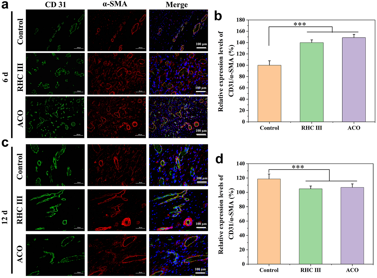
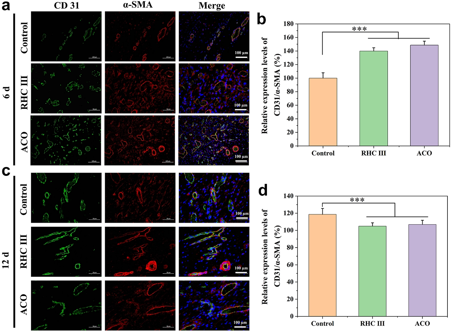
Figure 6. Promoting effect of ACO on angiogenesis. (a) CD31 (green)/α-SMA (red) immunofluorescence colocalization staining images on day 6; (b) Quantification of CD31/α-SMA double positive vessels (mature vessels) per field on day 6 (n = 5); (c) CD31 (green)/α-SMA (red) immunofluorescence colocalization staining images on day 12; (d) Quantification of CD31/α-SMA double positive vessels (mature vessels) per field on day 12 (n = 5). *p < 0.05, **p < 0.01, ***p < 0.001. ACO: Asini Corii Colla; RHC III: humanized type III collagen.
3.6 Heatmap visualization of gene expression and functional enrichment analysis in wound tissue
To comprehensively evaluate the effect of ACO intervention on skin wound healing in rats, skin tissues were collected on days 6 and 12 post-operation for transcriptome sequencing (n = 4). Heatmap analysis of gene expression profiles revealed distinct transcriptional patterns between the ACO-treated group (A6) and the control group (C6) as early as day 6 post-operation (Figure 7a). The ACO group exhibited a general upregulation trend across multiple gene clusters, suggesting that ACO may coordinately regulate a series of genes involved in tissue repair processes, including ECM remodeling, inflammation modulation, and angiogenesis. For instance, the expression of Atp6v0d2 was significantly elevated in the ACO group. This gene has been reported to promote the polarization of macrophages from the pro-inflammatory M1 phenotype toward the anti-inflammatory and pro-repair M2 phenotype, which may facilitate the transition from the inflammatory to the proliferative phase of wound healing[18]. Furthermore, the consistent upregulation of multiple regulatory genes in the ACO group suggested the potential involvement of wound healing-related signaling pathways (such as Wnt and TGF-β), which may enhance fibroblast function, collagen deposition, and neovascularization. As shown in Figure 7b, by postoperative day 12, the ACO-treated group (A12) exhibited a more mature and stable gene expression pattern compared to the control group (C12). Although the absolute number of differentially expressed genes increased from 203 to 1,225 (as detailed in Section 3.7), the overall transcriptomic profile of the ACO group exhibited tighter intra-group clustering and shifted toward a more mature and homeostatic state compared to the control group. This suggests that ACO-treated wounds had entered the tissue remodeling phase earlier, whereas the control group remained in a prolonged inflammatory and proliferative state.
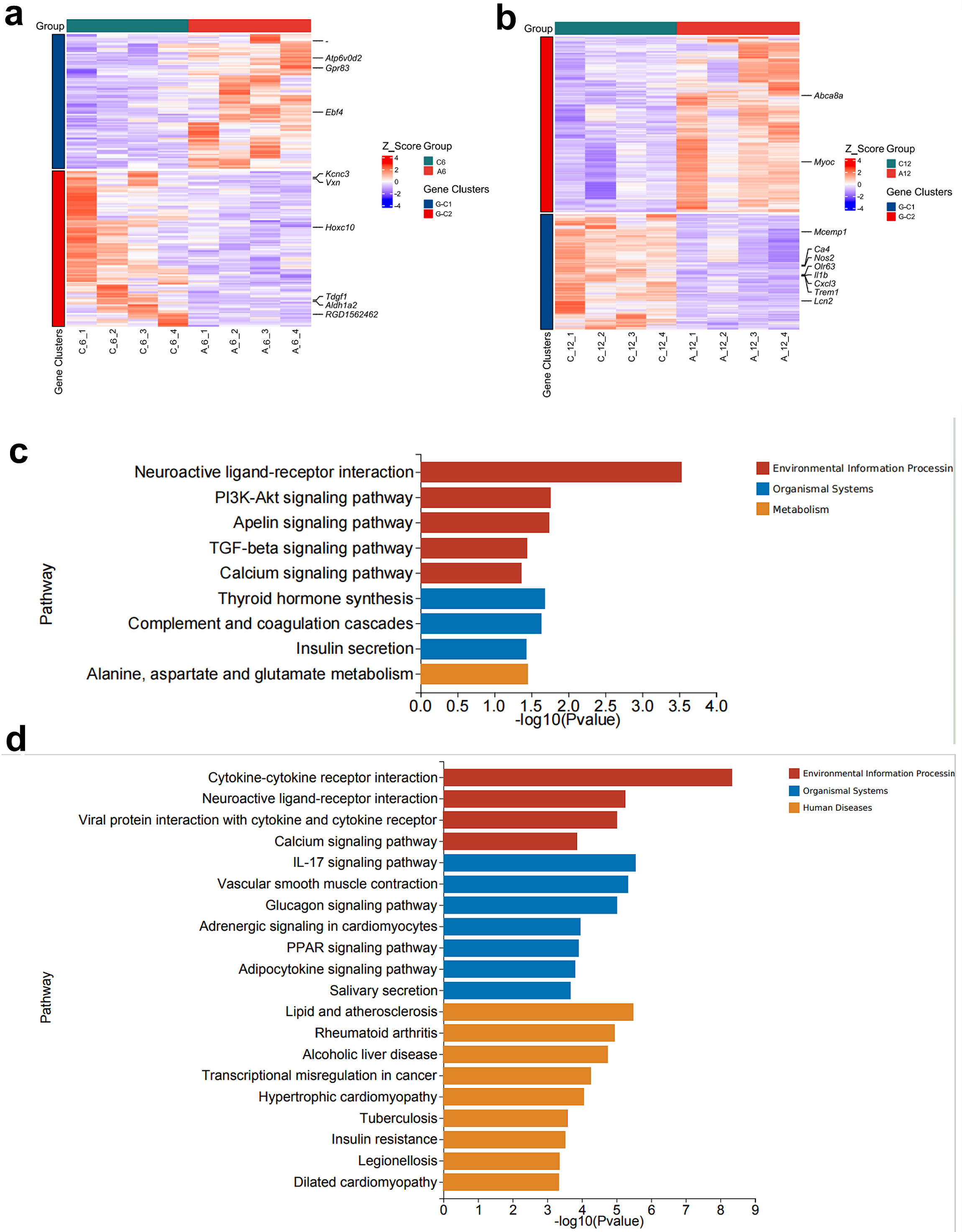
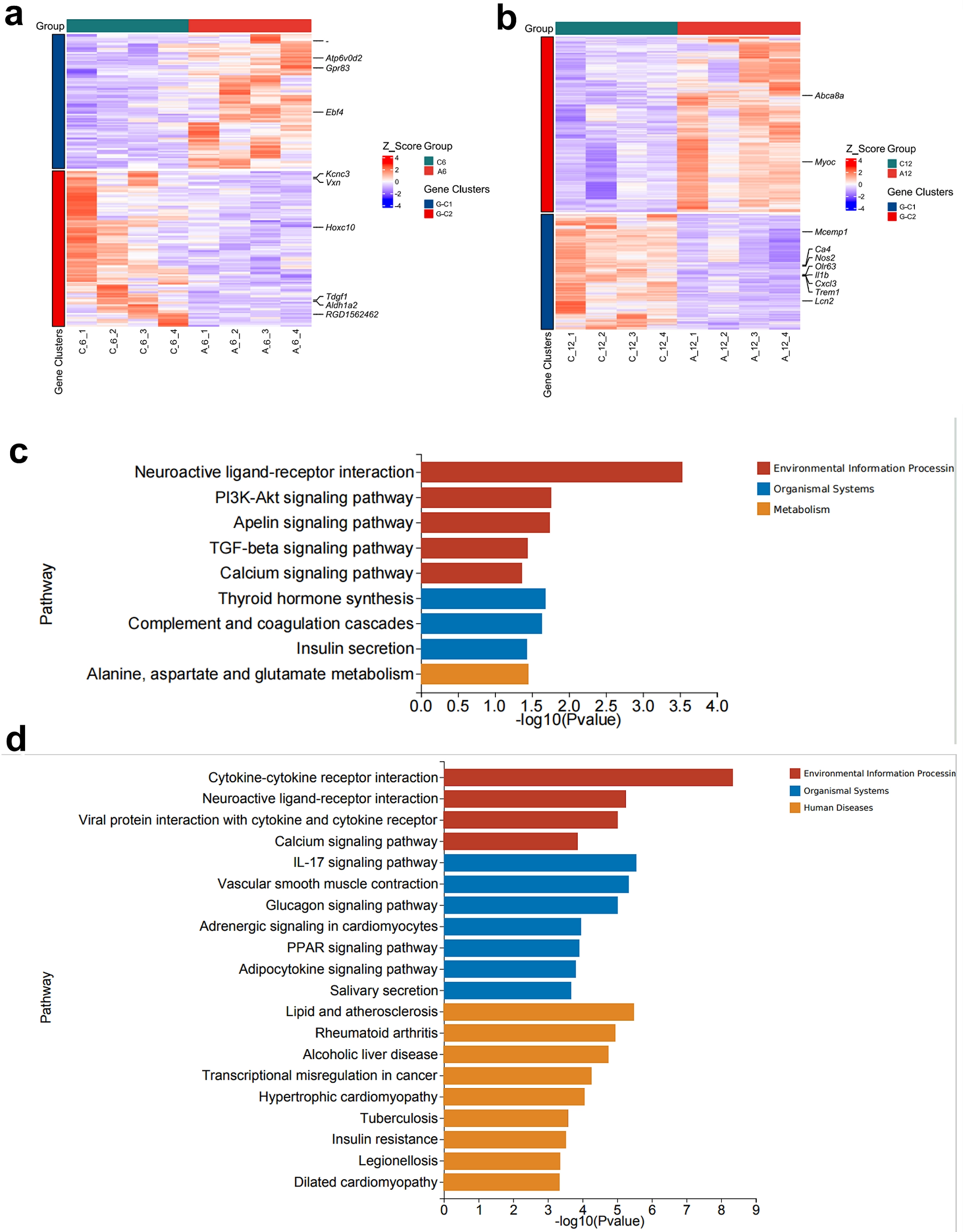
Figure 7. Heatmap visualization of gene expression and functional enrichment analysis in wound tissues. Transcriptomic profile heatmaps showing differential gene expression between the ACO-treated and control groups in wound tissues on (a) day 6 and (b) day 12 post-treatment with ACO. Pathway enrichment analysis of wound transcriptomes on (c) day 6 and (d) day 12. ACO: Asini Corii Colla.
Specifically, the pro-inflammatory gene nitric oxide synthase 2 (Nos2) remained highly upregulated in the control group, suggesting a persistent inflammatory state in the wounds, whereas its expression was significantly reduced in the ACO group, reflecting the effective control of inflammation. Concurrently, the macrophage activation marker gene triggering receptor expressed on myeloid cells 1 (Trem1) was highly expressed in the control group, indicating ongoing immune cell overactivation; the decreased Trem1 expression in the ACO group further confirmed a more timely resolution of inflammation. Collectively, these findings suggest that during the later stage of wound healing, ACO is associated with the modulation of inflammation-related genes, potentially facilitating the transition from the inflammatory to the tissue maturation phase and contributing to improved wound repair quality.
Based on transcriptomic GO/KEGG pathway enrichment analysis results (Figure 7c), during the early stage of wound healing (postoperative day 6), the ACO intervention group (A6) exhibited features of multi-pathway synergistic regulation compared to the control group (C6). For example, the Neuroactive ligand-receptor interaction pathway was significantly enriched, suggesting that ACO may modulate the inflammatory response and tissue repair microenvironment by regulating neuro-immune crosstalk (i.e., interactions between neurotransmitters and immune cell receptors). The simultaneous enrichment of the PI3K-Akt signaling pathway and TGF-β signaling pathway, which respectively regulate cell survival/proliferation and fibroblast activation/collagen synthesis, suggests that ACO treatment is associated with these pathways, which may potentially contribute to granulation tissue formation and matrix remodeling. The enrichment of the Apelin signaling pathway further suggests that ACO may be involved in angiogenesis and energy metabolism regulation, which is closely associated with early wound vascularization. Thus, during the early stage of wound healing, ACO promotes inflammation resolution, cell proliferation, and matrix remodeling through multiple mechanisms. These include synergistically regulating neuro-immune crosstalk, activating key repair signaling pathways (such as PI3K-Akt/TGF-β), and driving metabolic reprogramming, thereby providing a molecular basis for early wound healing.
By postoperative day 12, the ACO-treated group (A12) exhibited more sophisticated and systematic pathway regulatory characteristics compared to the control group (C12), indicating that ACO continues to promote tissue repair and functional recovery through multi-layered mechanisms during the late stage of wound healing (Figure 7d). Specifically, the significant enrichment of the Cytokine-cytokine receptor interaction and IL-17 signaling pathways suggests that ACO regulates cytokine networks and Th17-related immune responses to further suppress excessive inflammation and promote the restoration of immune homeostasis, creating favorable conditions for tissue maturation. The concurrent enrichment of the Neuroactive ligand-receptor interaction and Vascular smooth muscle contraction pathways indicates ACO’s potential to modulate vascular tension and blood supply through crosstalk between neurotransmitters and vasoactive mediators, thereby supporting revascularization and meeting the metabolic demands in late-stage wounds. The enrichment of the PPAR, Adipocytokine, and Glucagon signaling pathways suggests that ACO treatment is associated with the regulation of lipid metabolism, glucose metabolism, and energy balance, which may contribute to meeting the energy and biosynthetic requirements of tissue repair. The enrichment of several human disease-related pathways (such as Rheumatoid arthritis, Atherosclerosis, and Insulin resistance) does not imply that ACO induces these diseases. Rather, it may reflect molecular commonalities between the ACO-regulated gene sets and the pathological processes of these diseases, particularly in terms of inflammation resolution, metabolic balance, and ECM remodeling. These associations provide additional context for the potential role of ACO in promoting wound healing, but should be interpreted cautiously as pathway-level correlations rather than functional evidence of disease causation. Consequently, the enriched pathways encompass three major functional categories: “Environmental Information Processing”, “Organismal Systems”, and “Human Diseases”, suggesting that ACO treatment is associated with broad transcriptomic changes across immune, neural, vascular, and metabolic systems during the late stage of wound healing.
To further corroborate the transcriptomic findings, we compared the RNA-seq pathway enrichment results with independent protein-level data obtained from the same wound tissues. Specifically, the downregulation of inflammation-related pathways in the RNA-seq data was consistent with the reduced IL-1β expression and the early (day 6) increase in IL-10 expression observed by immunohistochemistry (Figure 5). Similarly, the early-stage enrichment of angiogenesis-related pathways aligned with the increased number of CD31+/α-SMA+ double-positive vessels observed on day 6 by immunofluorescence co-staining (Figure 6). Furthermore, the transcriptomic signatures of ECM remodeling and collagen metabolism were supported by the enhanced collagen deposition and improved tissue architecture observed in Masson’s trichrome-stained sections (Figure 4). These convergent lines of evidence at both the transcriptional and protein levels are consistent with the involvement of the key biological processes identified by RNA-seq analysis, although further functional studies are needed to establish causal relationships.
3.7 Integrated transcriptomics and expression changes of wound-healing related genes
Volcano plot analysis of transcriptomic data revealed distinct gene expression patterns between the ACO-treated group (A6) and the control group (C6) at day 6 of wound healing (Figure 8a). A total of 203 genes showed significant differential expression, with 94 upregulated and 109 downregulated. Notably, several key tissue repair-related genes, including Atp6v0d2 and Ebf4, were significantly upregulated in the ACO group. These genes are known to participate in macrophage polarization regulation and fibroblast differentiation during repair processes. Collectively, these differentially expressed genes demonstrate that ACO modulates the expression of multiple critical repair-related genes during the early stage of wound healing, thereby promoting tissue repair progression. Volcano plot analysis of transcriptomic data at day 12 revealed significant differences in gene expression between the ACO-treated group (A12) and the control group (C12, Figure 8b). A total of 1,225 differentially expressed genes were identified, with 739 upregulated and 486 downregulated. Notably, key inflammation-related genes including Cxcl13, Lcn2, and Trem1 were significantly downregulated, while ECM-associated genes such as Myoc and Slc15a2 showed marked upregulation. These coordinated gene expression changes suggest that during the later phase of wound healing, ACO treatment is associated with the suppression of inflammatory responses and the promotion of tissue remodeling, indicating the transition from the inflammatory phase to the proliferative phase and facilitating wound maturation. PCA revealed distinct spatiotemporal segregation in global gene expression profiles between the ACO-treated and control groups (Figure 8c). At the early healing stage (day 6), the ACO group (A6) exhibited clear separation from the control group (C6) along the first principal component, indicating that ACO intervention induces significant transcriptomic reprogramming shortly after treatment initiation. By day 12, the ACO group (A12) remained well-separated from the control group (C12), with both groups demonstrating tighter intra-group clustering. This pattern suggests improved biological consistency among replicates and implies that ACO promotes a more synchronized and coordinated repair progression during the later stage of wound healing.
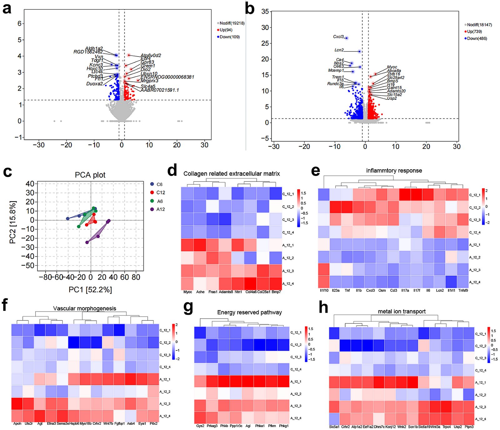
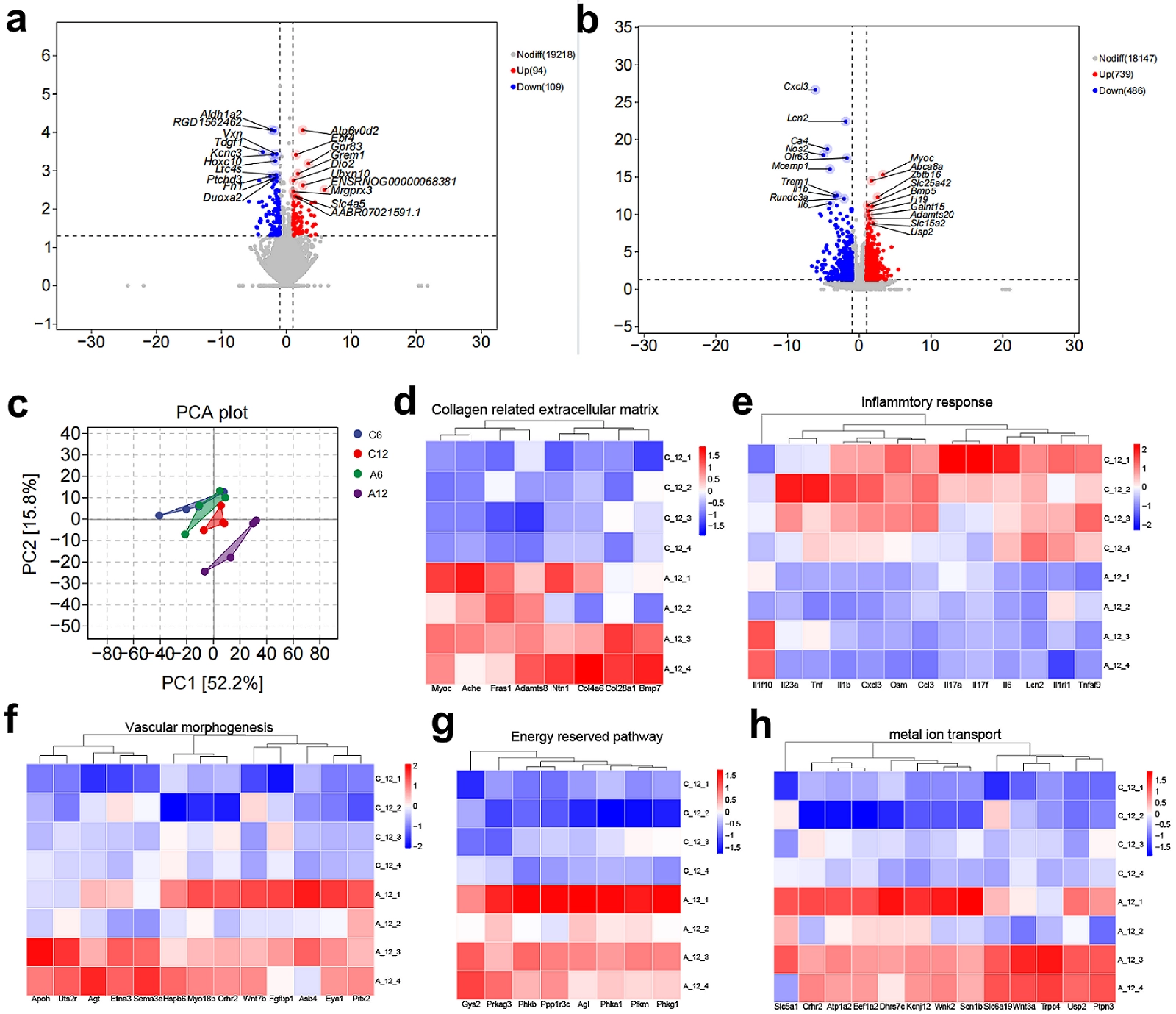
Figure 8. Integrated transcriptomic analysis and expression profiles of wound-healing-related genes. Volcano plots displaying differential gene expression between the ACO-treated and control groups in wound tissues on (a) day 6 and (b) day 12; (c) PCA of transcriptomic profiles reveals distinct segregation among the sequencing groups; Heatmaps of gene expression in the (d) collagen-related ECM pathway, (e) inflammatory response pathway, (f) vascular morphogenesis pathway, (g) energy reserve-related pathway, and (h) metal transport pathway in day 12 wound tissues. ACO: Asini Corii Colla; ECM: extracellular matrix.
Pathway analysis further revealed that ACO exerts multi-dimensional and synergistic regulatory effects during the later stage of wound healing (Figure 8d-h). In pathways related to collagen and ECM, ACO significantly upregulated key genes including Myoc, Col4a6, and Fras1, indicating enhanced collagen synthesis and cross-linking that accelerate dermal reconstruction and ECM maturation (Figure 8d). Concurrently, inflammatory response genes such as Il1b, Cxcl3, and Tnf were markedly downregulated in the ACO group, demonstrating its capacity to sustainably suppress excessive inflammation and promote the restoration of wound microenvironment homeostasis (Figure 8e). Regarding vascular morphogenesis, elevated expression of Wnt7b, Apoh, and Agt suggests that ACO coordinately promotes angiogenesis and vascular maturation to meet the hemodynamic demands of tissue repair (Figure 8f). Moreover, ACO enhanced the expression of energy reserve-related genes (e.g., Pykm, Prkag3) and metal ion transporters (e.g., Kcnj12, Atp2a2), supporting cellular migration and proliferation through maintaining ion homeostasis and energy supply (Figure 8g,h). In summary, during the advanced wound healing phase, ACO coordinately regulates four crucial biological processes: ECM remodeling, inflammation resolution, angiogenesis, and energy metabolism, collectively driving the wound repair process toward structural and functional maturation.
3.8 Comparative GSEA of transcriptomic profiles between groups
GSEA revealed temporally distinct regulatory patterns of ACO during wound healing (Figure 9). At the early stage (day 6), the ACO group showed significant enrichment in the intermediate filament cytoskeleton pathway (Figure 9a). Notably, the inflammatory regulation pathway was negatively enriched in the ACO group compared with the control group (NES: -1.48, p-value < 0.01, Adjusted p-value < 0.5), suggesting a possible association between ACO treatment and suppression of inflammatory responses (Figure 9b). By the later healing phase (day 12), the ACO group exhibited enhanced activation of the myofibril assembly pathway, suggesting a potential association with myofibroblast differentiation that may facilitate wound contraction (Figure 9c). Additionally, GSEA revealed a significant negative enrichment of the angiogenesis pathway in the ACO group (NES: -1.56, Adjusted p-value < 0.001; Figure 9d). This indicates that angiogenesis-related gene expression was downregulated in ACO-treated wounds compared with the control group by day 12. This finding is consistent with our histological observation of timely physiological vascular regression in the ACO group, whereas the unhealed control wounds exhibited sustained pathological angiogenesis. These findings collectively suggest that ACO may regulate wound healing progression from the inflammatory to the proliferative and remodeling phases through stage-specific modulations.
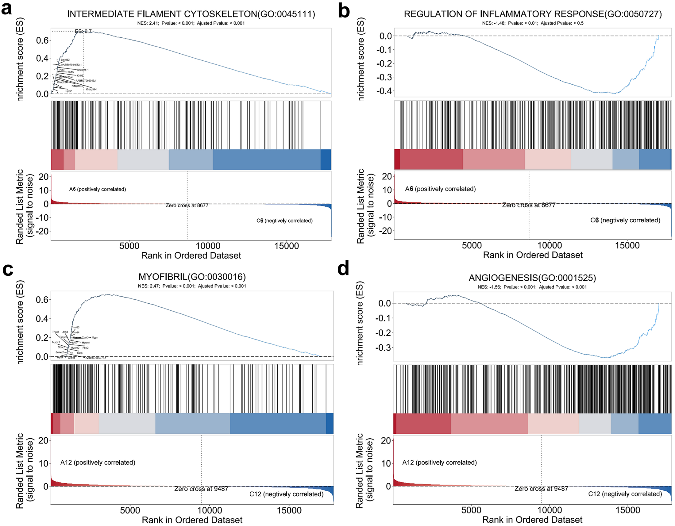
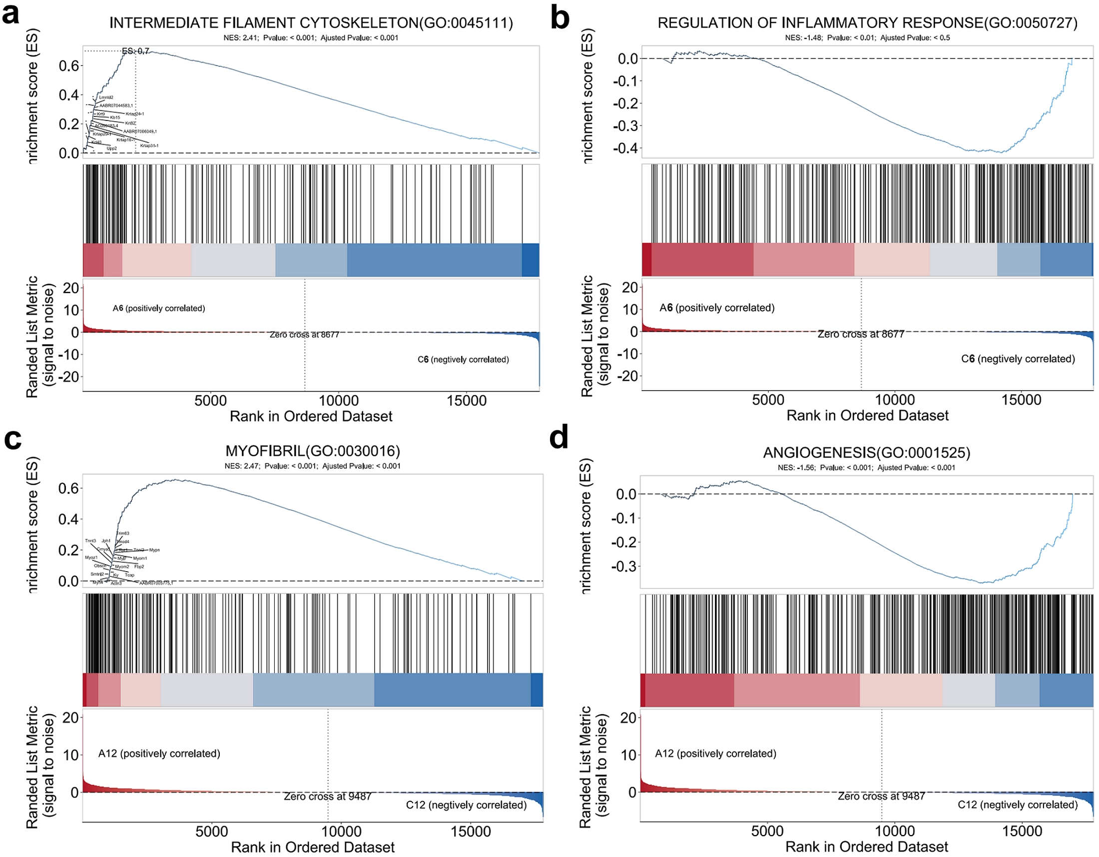
Figure 9. Comparative GSEA of transcriptomic profiles between the ACO-treated and control groups. (a) GSEA of the intermediate filament cytoskeleton pathway in wound tissues on day 6; (b) GSEA of the inflammatory response regulatory pathway in wound tissues on day 6; (c) GSEA of the myofibril assembly pathway in wound tissues on day 12; (d) GSEA of the angiogenesis pathway in wound tissues on day 12. ACO: Asini Corii Colla.
3.9 In vivo biocompatibility
To evaluate the systemic safety of ACO, H&E staining of major organs (heart, liver, spleen, lung, and kidney) was performed. As shown in Figure S2, no obvious histopathological abnormalities, inflammatory cell infiltration, or tissue damage were observed in the ACO-treated groups compared with the control group, indicating good in vivo biocompatibility. In addition, ACO is a traditional food and medicine with a long history of safe consumption, and the absence of organ toxicity in the present study further supports its favorable safety profile.
4. Discussion
This study evaluated the biocompatibility, therapeutic efficacy, and transcriptomic associations of ACO in skin wound healing. While previous studies have reported the wound-healing and collagen-promoting effects of enzymatic ACO hydrolysates in three-dimensional skin models[9], the present work provides an integrated in vitro, in vivo, and transcriptomic analysis of intact ACO powder. In vitro assays demonstrated that ACO exhibited no significant cytotoxicity to HSF and RAW 264.7 cells within the concentration range of 0.156-100 mg/mL, indicating favorable cytocompatibility. Further functional assays revealed that ACO significantly promoted HSF cell proliferation and migration in a concentration-dependent manner, as evidenced by time course CCK-8, scratch wound healing, and Transwell migration assays. In a rat full-thickness skin defect model, ACO significantly accelerated wound closure, achieving a healing rate of 98.14% on day 12 post-operation, which was comparable to that of the RHC-III group (99.05%) in this acute model and higher than that of the control group (82.09%). No formal equivalence or superiority analysis was performed, and this comparison should be considered descriptive only. Histological analysis further confirmed that ACO treatment promoted fibroblast migration and proliferation, re-epithelialization, and collagen deposition, while modulating the spatiotemporal expression of inflammatory cytokines. Transcriptomic and GSEA analyses revealed that ACO treatment was associated with gene signatures related to ECM remodeling, inflammation resolution, angiogenesis, and energy metabolism at different time points.
Regarding collagen deposition and ECM remodeling, Masson’s trichrome staining showed that collagen content in the ACO group reached 56.00% on postoperative day 12, showing a trend toward higher collagen content than the control group (50.66%), although the difference was not statistically significant; collagen fibers in the ACO group also appeared more densely packed and mature. Transcriptomic data indicated that ACO upregulated key ECM-related genes (e.g., Myoc, Col4a6, and Fras1) and enriched the TGF-β signaling pathway, and these data are consistent with the possible involvement of TGF-β/Smad signaling in ACO-mediated collagen synthesis and cross-linking during dermal remodeling[19]. With respect to inflammatory regulation, ACO significantly reduced the expression of the pro-inflammatory cytokine IL-1β (to 50.87% of the control group) and increased the expression of the anti-inflammatory cytokine IL-10 (to 110.63%) on day 6 post-operation. By day 12, both IL-1β and IL-10 levels in the ACO group had further decreased, whereas the control group showed a relatively high IL-10 level at day 12, which may reflect ongoing immune activation, although IL-10 alone cannot be interpreted as direct evidence of persistent inflammation. At the transcriptomic level, ACO treatment was associated with the upregulation of the macrophage polarization-related gene Atp6v0d2 and the downregulation of pro-inflammatory genes Nos2 and Trem1. GSEA showed negative enrichment of the inflammatory response regulation pathway in the ACO group (NES = -1.48), consistent with suppression of inflammatory responses. These results suggest that ACO treatment is associated with a timely resolution of inflammation and may help avoid excessive or prolonged inflammatory responses. In terms of angiogenesis, ACO increased the number of CD31/α-SMA-positive newly formed blood vessels by approximately 40-50% on day 6 post-operation, followed by physiological regression by day 12, exhibiting a pattern of “early promotion and late regression”, which may be associated with the prevention of aberrant vascular overgrowth. At the molecular level, ACO treatment was associated with the enrichment of the Apelin signaling pathway and Vascular smooth muscle contraction related pathways, as well as with the upregulated expression of pro-angiogenic genes such as Wnt7b and Apoh[20,21].
To summarize the key pathways identified in this study, collagen remodeling was mainly associated with the TGF-β/Smad signaling pathway and ECM-related genes such as Myoc, Col4a6, and Fras1, which was supported by increased collagen deposition in Masson’s trichrome staining. Angiogenesis was associated with the Apelin signaling pathway and genes including Wnt7b, Apoh, and Agt, and was independently supported by increased CD31/α SMA mature vessels. Inflammation regulation involved Cytokine cytokine receptor interaction and IL-17 signaling, with Atp6v0d2 upregulated and Nos2, Trem1, Il1b, Cxcl3, and Tnf downregulated, consistent with reduced IL-1β and increased IL-10 observed by immunohistochemistry. Although qPCR and Western blot validation were not performed, these independent protein level data provide preliminary support for the proposed pathways, which should be further validated in future studies.
Time course transcriptomic analysis further indicated multi stage, multi pathway associations in the ACO-treated group. At the early stage of wound healing (day 6 post-operation), ACO induced 203 differentially expressed genes, with significant enrichment of pathways including neuroactive ligand-receptor interaction, PI3K-Akt, TGF-β, and Apelin signaling, which coordinately initiate cell survival, ECM synthesis, and angiogenesis. At the late stage (day 12 post-operation), the number of differentially expressed genes increased to 1,225, with enriched pathways expanding to cytokine-cytokine receptor interaction, IL-17 signaling, PPAR signaling, Adipocytokine signaling, and Glucagon signaling, covering immune modulation, metabolic reprogramming, and vascular function maintenance. GSEA also showed that ACO enriched the “intermediate filament cytoskeleton” pathway at the early stage (promoting cell migration) and the “myofibril assembly” pathway at the late stage (promoting myofibroblast contraction and wound closure), with early positive enrichment and later negative enrichment of angiogenesis-related pathways, consistent with the histological observation of early neovascularization followed by physiological vascular regression. These findings suggest that ACO treatment is associated with multiple biological processes rather than a single target. In this acute wound model, ACO showed wound closure rates comparable to those of RHC III.
To better position the present findings, we compared this study with representative studies on ACO and other wound-healing interventions. However, this comparison is limited by several factors. First, animal models differ across studies: most previous ACO studies have used wounds healing experiment in healthy rodents or three-dimensional skin equivalents, whereas chronic wound models such as diabetic, infected, or ischemic wounds remain largely unexplored[9]. Second, formulations and doses vary considerably, ranging from enzymatic ACO hydrolysates to ACO-containing dressings or composite formulations, making direct comparison difficult[9,22]; in contrast, the present study used intact ACO powder at a defined concentration (20 mg/mL) applied topically once daily. Third, healing endpoints differ: earlier studies have mainly reported wound closure rates and histological collagen deposition, whereas the present study additionally assessed inflammatory cytokines, angiogenesis, and transcriptomic profiles. Fourth, mechanistic evidence remains limited across studies; most previous reports have relied on phenotypic endpoints without pathway-level validation, and the present transcriptomic findings are similarly descriptive and correlative. Finally, safety and batch-consistency data remain limited, and the translational maturity of ACO-based products varies. These differences highlight the need for standardized formulations, disease-relevant models, and functional validation before clinical translation can be considered.
Several limitations of this study should be acknowledged. First, the use of the same production batch across the previous proteomic characterization, the previous anti-photoaging study, and the present investigation strengthens the comparability of the findings. The previously reported effects of ACO on skin-cell proliferation and migration, collagen synthesis, UV-induced cellular senescence, extracellular matrix degradation, and skin photoaging provide directly relevant biological context for the present study[10]. However, these pharmacological and mechanistic findings do not replace comprehensive chemical characterization of the experimental material. A limitation of the present study is that all experiments were conducted using a single production batch of ACO. Although this approach minimized compositional variability within the current study and enabled direct comparison with our previous investigations, it does not permit assessment of variability among independent commercial batches. Therefore, the present findings should not be interpreted as evidence of batch-to-batch consistency. In addition, ACO is a complex mixture, and its specific bioactive components and direct molecular targets remain to be identified. Future studies should evaluate multiple independent production batches using quantitative amino acid analysis, collagen-related markers, chromatographic or mass-spectrometric peptide fingerprinting, and biological potency assays. Further chromatography-mass spectrometry-based fractionation and characterization, together with target-identification approaches, will also be required to identify the active constituents of ACO and elucidate their direct molecular targets. Second, although transcriptomics suggested multiple signaling pathways, the phosphorylation levels of key nodes (e.g., PI3K, Akt, Smad2/3) and the upstream/downstream relationships need to be validated by western blotting and other experimental approaches. Third, the animal model used in this study was an acute full thickness wound in healthy rats, which does not recapitulate the pathophysiological features of clinically challenging wounds, such as diabetic ulcers, ischemic wounds, infected wounds, or chronic inflammatory conditions. Therefore, our findings should be interpreted as proof-of-concept evidence in a standardized experimental setting. The therapeutic efficacy of ACO under clinically relevant comorbidities remains to be validated using appropriate disease specific animal models (e.g., streptozotocin-induced diabetic rats, ischemic flap models, or infected wound models). Until such studies are conducted, the translational potential of ACO for advanced wound management should be considered preliminary. It should also be noted that the full thickness wound model was established without a silicone splinting ring. Although all groups were subjected to identical conditions, making group wise comparisons valid, the wound closure data may partially reflect wound contraction rather than true re-epithelialization. To address this, we performed histological quantification of wound width (reflecting re-epithelialization), granulation tissue thickness (reflecting tissue regeneration), and collagen deposition. However, increased collagen deposition detected by Masson’s trichrome staining should not be automatically interpreted as superior healing quality, because excessive collagen deposition may also indicate fibrosis or scar formation. Collagen architecture, the Col I/Col III ratio, skin appendages, tensile strength, and scar formation were not evaluated and should be assessed in future work. Furthermore, the comparison with RHC III is limited to this acute full thickness wound model in healthy rats and should not be extended to claims of clinical equivalence or superiority. Because this model does not reproduce the pathophysiological features of diabetic, infected, or ischemic wounds, the present findings should be considered proof-of-concept evidence in acute wounds, and validation in disease specific models is required before clinical translation can be considered. Additionally, the RNA seq data presented in this study are primarily descriptive and correlative in nature. While the identified pathways and gene signatures are supported by independent protein level evidence (IL-1β/IL-10 IHC, CD31/α SMA IF, and Masson’s trichrome staining), they should be interpreted as mechanistic hypotheses rather than conclusively established mechanisms. Furthermore, by day 12, ACO-treated wounds were nearly closed while control wounds remained largely open; therefore, transcriptomic differences at this time point may partly reflect differences in tissue composition and healing stage rather than direct ACO-induced temporal regulation. A formal treatment × time interaction analysis was not performed, and this should be considered a limitation. Future studies employing gene specific interventions (e.g., siRNA, knockout models, or pharmacological inhibition) and matched healing stages are warranted to further validate the functional roles of the identified pathways. Finally, the optimal dosing regimen (formulation, concentration, frequency) of ACO requires systematic optimization. Overall, in an acute full-thickness wound model in healthy rats, ACO safely and effectively promoted wound healing, with transcriptomic data suggesting potential multi-stage regulation of inflammation, angiogenesis, ECM remodeling, and energy metabolism. These findings should be considered proof-of-concept evidence in acute wounds. The therapeutic potential of ACO in clinically challenging chronic wounds (e.g., diabetic, infected, or ischemic wounds) remains to be validated in disease-specific models before clinical translation can be considered.
5. Conclusion
This study investigated the promoting effect of ACO on skin wound healing and explored potential molecular associations. The experimental results demonstrated that ACO exhibited favorable biocompatibility with human dermal fibroblasts and macrophages within the concentration range of 0.156-100 mg/mL. In an animal model, ACO significantly promoted fibroblast migration and proliferation, while enhancing collagen deposition and maturation. In terms of inflammatory regulation, ACO effectively suppressed the expression of the pro-inflammatory cytokine IL-1β and increased the level of anti-inflammatory IL-10 during the early healing phase, thereby facilitating timely inflammation resolution and restoring wound microenvironment homeostasis in later stages. Concurrently, ACO upregulated the expression of CD31 and α-SMA to promote early angiogenesis, improving local blood supply and nutrient delivery. These findings suggest that ACO has potential in accelerating acute full-thickness skin defect healing. Transcriptomic analysis further indicated temporally distinct associations of ACO at the gene expression level. At early stages, ACO treatment was associated with enrichment of pathways including intermediate filament cytoskeleton assembly, Neuroactive ligand-receptor interactions, and PI3K-Akt/TGF-β signaling, whereas at later stages it was associated with pathways related to myofibril assembly, angiogenesis, and energy metabolism. In summary, in an acute full-thickness wound model in healthy rats, ACO was associated with improved wound healing, and transcriptomic data suggested potential associations with ECM remodeling, inflammatory response, angiogenesis, and energy metabolism. These findings should be considered proof-of-concept evidence in acute wounds and provide mechanistic hypotheses for further investigation.
Supplementary materials
The supplementary material for this article is available at: Supplementary materials.
Authors contribution
Li C, Jiang Y: Conceptualization, writing-review & editing, investigation, methodology.
Xie X: Funding acquisition, supervision, conceptualization, resources.
Zhang J, Chen Z: Investigation, methodology, conceptualization, writing-original draft.
Li Y, Yang J: Methodology, visualization, software.
Huang Z: Conceptualization, investigation, methodology, data curation.
Jia L, Lu Z: Writing-review & editing, methodology, formal analysis.
Fu M: Conceptualization, writing-original draft, writing-review & editing, investigation, methodology, data curation.
Guo J: Funding acquisition, supervision, conceptualization, writing-review & editing, resources.
Conflicts of interest
Jinshan Guo is a Youth Editorial Board Member of BME Horizon. Cong Li, Xiao Xie, Jianling Zhang, and Zhijie Chen are employees of Dong’e Ejiao Health Industry Technology Co., Ltd. The other authors declare no conflicts of interest.
Ethical approval
All animal experiments were conducted in compliance with the Animal Experimental Committee of Institute of Biological and Medical Engineering, Guangdong Academy of Sciences (Approval No. K2024-01-272-472).
Consent to participate
Not applicable.
Consent for publication
Not applicable.
Availability of data and materials
The data supporting the findings of this study can be obtained from the authors.
Funding
This work was supported by the Research and Application Project on the Skincare Effects of Asini Corii colla (Grant No. DEEJ-YB-2024-10-190) from Dong’e Ejiao Health Industry Technology Co., Ltd.
Copyright
© The Author(s) 2026.
References
-
3. Fu M, Zhao Y, Wang Y, Li Y, Wu M, Liu Q, et al. On-demand removable self-healing and pH-responsive europium-releasing adhesive dressing enables inflammatory microenvironment modulation and angiogenesis for diabetic wound healing. Small. 2023;19(3):2205489.[DOI]
-
4. Wang H, Shi S, Wang S. Can highly cited herbs in ancient Traditional Chinese medicine formulas and modern publications predict therapeutic targets for diabetes mellitus? J Ethnopharmacol. 2018;213:101-110.[DOI]
-
6. Zhang H, Liu S, Feng Q, Liu H, Wu J, Duan X, et al. Ejiao alleviates sarcopenia by AKT/mTOR/Foxo3a and AMPK pathways in an aging rat model induced by D-Galactose combined with restricted movement. J Ethnopharmacol. 2026;356:120819.[DOI]
-
10. Xie X, Chen Z, Zhang Z, Chen Z, Li C, Zhang J, et al. Asini Corii Colla exhibits anti-photoaging efficacy via activating collagen synthesis and maintaining redox & extracellular matrix homeostasis. Eng Regen. 2026;7:176-187.[DOI]
-
11. Wang Y, Chen G, Wang Q, Zhou Z, Zhao W, Wong SK, et al. A comprehensive review of traditional Chinese medicine Ejiao (colla corii asini): Chemical composition, quality control, and pharmacological actions. Nat Prod Commun. 2025.[DOI]
-
12. Yu XJ, Kong LM, Wang B, Zhai CQ, Lao YZ, Zhang LJ, et al. Asini Corii Colla (Ejiao) as a health-care food and traditional Chinese medicine: A review of its chemical composition, pharmacological activity, quality control, modern applications. J Funct Foods. 2025;125:106678.[DOI]
-
13. Li Y, Zhang X, Wang Y, Yang X. Functional genomics in medicinal plants: Achievements and future challenges. Med Plant Biol. 2025;4(1):e033.[DOI]
-
14. Iwata M, Sawada R, Iwata H, Kotera M, Yamanishi Y. Elucidating the modes of action for bioactive compounds in a cell-specific manner by large-scale chemically-induced transcriptomics. Sci Rep. 2017;7(1):40164.[DOI]
-
19. Schiller M, Javelaud D, Mauviel A. TGF-β-induced SMAD signaling and gene regulation: Consequences for extracellular matrix remodeling and wound healing. J Dermatol Sci. 2004;35(2):83-92.[DOI]
-
20. Robillard S, Lemay A, Lamontagne A, Lemay N, Pruneau V, Théroux L, et al. The apelinergic system accelerates wound healing in both type 1 and 2 diabetic mice. Sci Rep. 2026.[DOI]
-
21. Geng Y, Hua H, Xia Y, Zhou J, He J, Xu XY, et al. miR-199a-5p modulates choroidal neovascularization by regulating Wnt7b/Wnt/β-catenin signaling pathway. J Mol Histol. 2024;55(3):359-370.[DOI]
Copyright
© The Author(s) 2026. This is an Open Access article licensed under a Creative Commons Attribution 4.0 International License (https://creativecommons.org/licenses/by/4.0/), which permits unrestricted use, sharing, adaptation, distribution and reproduction in any medium or format, for any purpose, even commercially, as long as you give appropriate credit to the original author(s) and the source, provide a link to the Creative Commons license, and indicate if changes were made.
Publisher’s Note
Science Exploration remains a neutral stance on jurisdictional claims in published
maps
and institutional affiliations. The views expressed in this article are solely those
of
the author(s) and do not reflect the opinions of the Editors or the publisher.
Share And Cite


Science Exploration Style
Li C, Xie X, Jiang Y, Zhang J, Chen Z, Li Y, et al. Transcriptomic profiling of Asini Corii Colla in wound healing: Putative molecular signatures and mechanistic hypotheses. BME Horiz. 2027;5:202634. https://doi.org/10.70401/bmeh.2026.0040
Tips
Copy completed.
Submit a Manuscript
Author Instructions
Cite this Article
Article Metrics
0
View
0
Download
Cited
Article Updates

 Get citation
Science Exploration Style

Li C, Xie X, Jiang Y, Zhang J, Chen Z, Li Y, et al. Transcriptomic profiling of Asini Corii Colla in wound healing: Putative molecular signatures and mechanistic hypotheses. BME Horiz. 2027;5:202634. https://doi.org/10.70401/bmeh.2026.0040
copy
Share Link
copy