Lifestyle-associated reprogramming of neutrophil immunity: From hematopoiesis to functional remodeling

Xiaotian Chen

Jing Wang
*
*Correspondence to:
Jing Wang, Shanghai Institute of Immunology, Department of Immunology and Microbiology, Shanghai Jiao Tong University School of Medicine, Shanghai 200025, China.
E-mail: jingwang@shsmu.edu.cn
Myeloid Cells. 2026;1:202609. 10.70401/mc.2026.0009
Received: April 30, 2026Accepted: July 27, 2026Published: July 27, 2026
Abstract
Myeloid cells are central components of innate immunity and are highly responsive to environmental and lifestyle-associated cues. Among them, neutrophils are the most abundant circulating leukocytes in humans and have rapid turnover, traditionally leading to the view that they act mainly as short-lived effector cells. However, emerging evidence indicates that neutrophil function can be durably reshaped through upstream reprogramming of hematopoietic stem and progenitor cells and the bone marrow niche, resulting in trained-immunity-like changes in newly generated neutrophils. In this review, we discuss how lifestyle-associated factors shape neutrophil-associated immunity within the broader context of myeloid immune regulation, with a particular focus on long-term neutrophil functional reprogramming. We summarize current evidence that lifestyle and metabolic factors shape neutrophil production, mobilization, tissue recruitment, and effector programs. We further highlight that lifestyle induced neutrophil reprogramming is context dependent and may be either adaptive or maladaptive. Finally, we propose future directions for translating lifestyle-based immune modulation into strategies that limit chronic inflammation while preserving antimicrobial defense.
Keywords
Myeloid immunity, neutrophils, trained immunity, lifestyle factors, hematopoietic stem and progenitor cells, neutrophil reprogramming
1. Introduction
The immune system functions to protect the host against invading pathogens while continuously surveilling cellular integrity throughout the body. The majority of immune cells originate from hematopoietic stem cells (HSCs)[1], with the exception of certain tissue-resident populations. Based on hematopoietic lineage commitment, immune cells are broadly classified into lymphoid cells, derived from common lymphoid progenitors (CLPs), and myeloid cells, derived from common myeloid progenitors (CMPs)[2].
Traditionally, myeloid cells are characterized by their rapid response to stimuli, lack of antigen specificity, and absence of immunological memory, and are therefore considered central components of the innate immune system. In contrast, lymphoid cells rely on V(D)J recombination and somatic hypermutation to generate antigen-specific receptors or antibodies, thereby mediating adaptive immunity and forming long-lived immunological memory[3]. Recent studies have demonstrated that myeloid cells are not functionally static; instead, exposure to specific stimuli can induce long-term alterations in their functional state and output. This phenomenon, termed trained immunity or innate immune memory, is characterized by sustained changes in the quantity and function of myeloid cells following prior stimulation. Importantly, these stimuli are often shaped by environmental exposures and are closely linked to individual lifestyle factors[4-6].
Among myeloid cells, neutrophils represent the most abundant leukocyte population in human peripheral blood and exhibit the most rapid turnover among immune cells in both humans and mice[7,8]. Despite their short lifespan, emerging evidence suggests that neutrophils can participate in trained immunity like processes, challenging the traditional view of these cells as purely transient effectors[9,10].
In this review, we discuss how lifestyle-associated environmental factors shape neutrophil immunity, particularly long-term neutrophil functional reprogramming through hematopoietic and systemic regulatory pathways.
2. Neutrophils or Granulocyte Heterogeneity
Granulocytic cells represent a major branch of the myeloid compartment and are a key component of innate immunity. A defining feature of immune system organization is its cellular heterogeneity, whereby distinct immune cell subsets exhibit specialized functions depending on their tissue localization and microenvironmental context. Among myeloid cells, neutrophils, eosinophils, and basophils are classically grouped as granulocytes because of their abundant cytoplasmic granules and their developmental relationship within the granulocytic branch of myelopoiesis[11]. Following lineage commitment to granulocyte-monocyte progenitors (GMPs), neutrophil differentiation is orchestrated by a coordinated transcriptional network[12], in which factors such as C/EBPα[13], Gfi1[14], and PU.1[15] contribute to lineage specification and maturation. The developmental trajectory and transcriptional regulation of neutrophils have been extensively characterized in previous studies and reviews[11,16-19], and will not be discussed in detail here. In addition to neutrophils, GMPs can give rise to other granulocytic lineages, including eosinophils and basophils, through
Neutrophils also display substantial heterogeneity across circulation and tissues. Upon infiltration into organs, neutrophils can acquire distinct phenotypic and functional states that differ between homeostatic and disease conditions[18]. Historically, this diversity was underestimated, largely because neutrophils are cells with a limited lifespan and mature neutrophils exhibit relatively low transcriptional activity. As a result, earlier single-cell transcriptomic approaches often underrepresented neutrophils and failed to fully resolve the spectrum of neutrophil states[22]. With recent advances in single-cell technologies, accumulating studies have revealed that both circulating and tissue-infiltrating neutrophils exhibit distinct transcriptional programs, giving rise to diverse neutrophil subsets with tissue- and context-dependent functions. These subsets may participate not only in antimicrobial defense, but also in inflammation resolution, tissue remodeling, tumor progression, and metabolic regulation. Because neutrophil heterogeneity has been comprehensively discussed in several excellent reviews and landmark studies[23-25], it will only be briefly introduced here. Instead, this review focuses on how lifestyle-associated cues may reshape neutrophil states and contribute to
3. Trained Immunity
Myeloid cell heterogeneity varies substantially across tissues and disease contexts. For short-lived cells such as neutrophils, which are continuously generated from hematopoietic stem and progenitor cells (HSPCs), tissue-specific neutrophil states are largely shaped by local niches after recruitment into organs[2,18]. Although tissue-infiltrating neutrophils can exhibit prolonged survival under specific inflammatory conditions, their terminally differentiated and non-proliferative nature prevents them from becoming
Recent studies have shown that, after exposure to defined microbial, inflammatory, or sterile stimuli, short-lived myeloid cells can display sustained changes in gene expression and function, particularly in inflammatory and antimicrobial pathways[4,5]. These
Beyond hematopoietic propagation, inflammatory memory may also be retained within non-hematopoietic tissue niches, including epithelial stem cells[36,37]. This concept is supported by studies showing that inflammatory memory can be distributed within tissue niches and that sterile injury-induced innate immune memory may generate distal organ consequences[33]. In neutrophil-dominated inflammation, leukotriene B4 (LTB4)-dependent neutrophil swarming further illustrates how the activation state of one neutrophil population can amplify or reshape the behavior of neighboring neutrophils[38,39].
In the context of neutrophils, this concept is particularly important. Because mature neutrophils are short-lived and have limited proliferative capacity, long-term neutrophil functional alteration is most likely established upstream, at the level of HSPCs or neutrophil progenitors, rather than within mature neutrophils themselves[9,40]. Thus, trained immunity provides a framework to explain how transient environmental exposures can durably reshape neutrophil output, abundance, and functional responsiveness[10]. The molecular mechanisms underlying trained immunity have been extensively reviewed elsewhere and are not the primary focus of this review. Instead, we focus on the upstream factors that induce or modulate trained immunity. In contrast to prototypical stimuli (e.g., bacillus Calmette-Guérin (BCG) or β-glucan) that directly activate innate immune cells, lifestyle exposures exert long-lasting effects on myeloid immunity primarily through indirect systemic modulation, including metabolic, endocrine, and neural pathways. Consequently, elucidating the complex mechanisms behind lifestyle-induced immune adaptation requires an integrative systemic biology approach[4,41-45] (Figure 1). In the following sections, we discuss how representative lifestyle factors shape
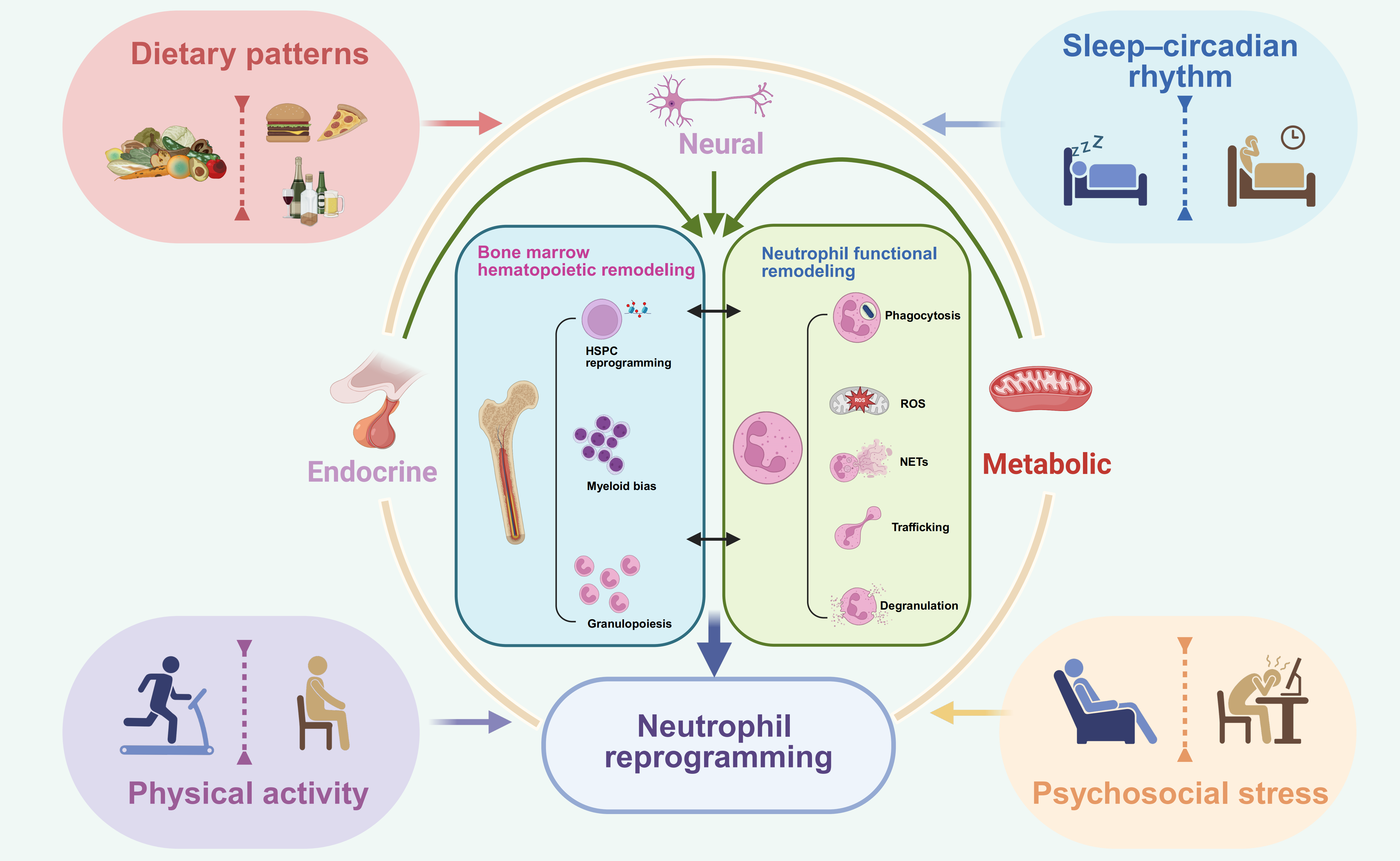
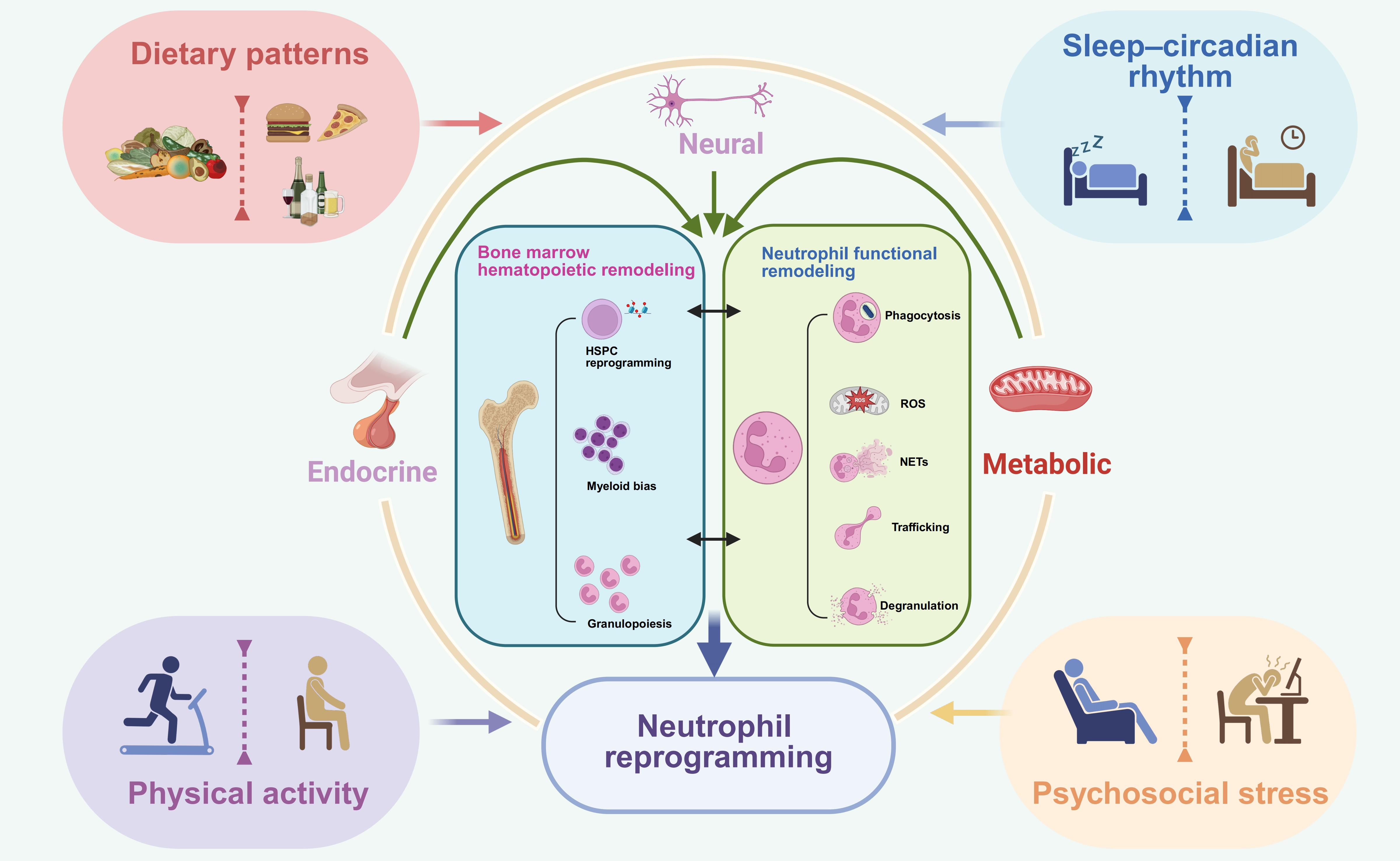
Figure 1. Lifestyle-associated systemic regulation of neutrophil reprogramming. Lifestyle factors, including dietary patterns, sleep–circadian rhythm, physical activity, and psychosocial stress, regulate neutrophil biology through interconnected neural, endocrine, and metabolic pathways. These systemic signals converge on the bone marrow to reshape hematopoietic stem and progenitor cell reprogramming, myeloid bias, and granulopoiesis, while also modulating neutrophil functional remodeling, including phagocytosis, reactive oxygen species production, neutrophil extracellular trap formation, trafficking, and degranulation. Together, these processes contribute to neutrophil reprogramming, which may lead to context-dependent adaptive or maladaptive immune outcomes. Created in BioRender. Wang, J. (2026) https://BioRender.com/ljq19fm. HSPC: hematopoietic stem and progenitor cell;
Distinguishing neutrophil reprogramming from bona fide trained immunity is essential. Neutrophil heterogeneity can arise from several distinct processes, including developmental maturation, tissue adaptation, acute priming, and long-lasting innate immune memory[46]. Developmental programming reflects stage-dependent changes during granulopoiesis, whereas tissue adaptation refers to niche-induced phenotypic and functional states acquired after neutrophil recruitment into organs. Priming usually represents a relatively rapid and reversible increase in responsiveness driven by ongoing inflammatory, metabolic, or microbial cues. In contrast, trained immunity should be reserved for durable functional changes that persist after the initial stimulus has waned and alter the response to a secondary challenge[5,6], ideally accompanied by stable epigenetic, metabolic, or transcriptional remodeling at the level of hematopoietic stem and progenitor cells or lineage-committed progenitors. Therefore, in this review, we use “trained immunity like reprogramming” when lifestyle-induced neutrophil changes suggest long-lasting remodeling but direct evidence for bona fide trained immunity remains incomplete. Moreover, lifestyle-associated chronic inflammatory or metabolic exposures may also drive neutrophils toward an exhausted-like state, in which pathogenic inflammatory features, such as enhanced swarming, increased integrin αM (CD11b) expression, and LTB4-related signaling, coexist with immunosuppressive features such as elevated programmed death-ligand 1 (PD-L1) expression, thereby promoting tissue damage while compromising host defense[47,48].
4. Diet
Diet represents a central determinant of human physiology and a major interface between lifestyle, metabolism, and immunity. Differences in nutrient composition and energy intake can reshape systemic metabolism and inflammatory tone, thereby influencing hematopoiesis, myeloid cell function, and the development of trained immunity[49]. In addition to nutrient composition, specific dietary behaviors, such as intermittent fasting and alcohol-related dietary exposure, can alter systemic energy metabolism and impose repeated metabolic stress on the immune system. Over time, these dietary cues may generate durable immune reprogramming and contribute to trained-immunity-like myeloid and neutrophil remodeling.
4.1 High-fat diet
High-fat diet (HFD), a major feature of Western-style dietary patterns, represents a chronic metabolic stimulus capable of reshaping myeloid immunity. Unlike acute microbial stimuli, HFD imposes sustained changes in lipid availability, systemic metabolism, and inflammatory tone[50]. This makes HFD an important lifestyle-associated driver of myeloid and neutrophil reprogramming.
At the hematopoietic level, HFD and obesity can directly alter stem and progenitor cell activity. Diet-induced obesity increases
HFD-associated metabolic stress also remodels mature neutrophil effector functions in a context-dependent manner. On one hand, specific lipid species may enhance selected neutrophil activities. Diets enriched in polyunsaturated fatty acids (PUFAs) can promote neutrophil chemotaxis, delay neutrophil death, and increase neutrophil accumulation in the spleen[52]. In septic Staphylococcus aureus infection, dietary PUFAs improve neutrophil function, increase host survival, and reduce bacterial burden[53]. On the other hand, chronic HFD can impair neutrophil phagocytosis, bacterial killing, and neutrophil-mediated support of hematopoietic regeneration, indicating that persistent metabolic overload may drive neutrophil dysfunction rather than simple activation[54]. Moreover, during infectious or inflammatory challenges, HFD can enhance NETs formation, as observed in influenza pneumonia in BALB/c mice. Although NETs may contribute to pathogen containment, excessive NET formation can also amplify tissue injury and inflammatory pathology[55]. HFD can also accelerate neutrophil aging and promote a pro-inflammatory neutrophil phenotype, further exacerbating metabolic dysfunction and systemic inflammation[56].
The consequences of HFD-induced neutrophil reprogramming are particularly evident in chronic inflammatory diseases. Atherosclerosis has traditionally been linked to macrophage lipid uptake, foam cell formation, and plaque inflammation; however, neutrophils are also recruited into atherosclerotic lesions and contribute to local inflammatory amplification. Notably, recent evidence shows that alternating HFD can aggravate atherosclerosis more strongly than continuous HFD. Mechanistically, alternating HFD reduces Runx1 expression in GMPs, enhances inflammatory signaling, and promotes IL-1β production in bone marrow cells upon subsequent HFD challenge. This response increases neutrophil mobilization and recruitment into atherosclerotic plaques, thereby exacerbating local inflammation and disease progression[57].
Together, these studies indicate that HFD does not simply activate neutrophils; rather, it resets their developmental and functional set points in a context-dependent manner. By acting on mature neutrophils and their progenitors, HFD reshapes neutrophil abundance, trafficking, effector function, NET formation, and aging. This persistent remodeling is highly consistent with the concept of trained immunity. However, the outcomes of HFD-induced neutrophil reprogramming are heterogeneous: they may enhance host defense in certain acute infections, but more often promote maladaptive inflammation, tissue damage, and disease progression in chronic metabolic and cardiovascular disorders.
4.2 Specific dietary metabolites driving HFD-associated neutrophil reprogramming
In the context of HFD-induced neutrophil reprogramming, specific lipid species and diet-associated metabolites may act as direct instructive signals. Saturated fatty acids (SFAs), such as palmitic acid, can directly modulate neutrophil function. In neutrophils from dairy cows with fatty liver, palmitic acid induces autophagy activation, leading to granule-dependent degradation of the adhesion molecule CD11b/CD18, also known as αMβ2 integrin. This process impairs neutrophil adhesion and migration, suggesting that lipid overload can reshape neutrophil function through an autophagy–granule homeostasis axis[58]. At the systemic level, different classes of dietary fatty acids appear to exert selective effects on neutrophil production and mobilization. Diets enriched in SFAs promote neutrophil trafficking from the bone marrow to the peripheral blood through the CXCL2–CXCR2 axis, accompanied by increased bone marrow inflammation and neutrophil apoptosis[59]. In contrast, monounsaturated and polyunsaturated fatty acids do not induce the same effect, indicating that the composition of dietary fat is a critical determinant of neutrophil output and mobilization.
In addition to dietary lipid species, gut microbiota-derived metabolites also regulate neutrophil function. Short-chain fatty acids, including acetate, butyrate, and propionate, can shift neutrophils toward a less activated or aging-like phenotype, suppressing neutrophil migration, antiviral effector molecule release, and NET formation, thereby impairing antiviral responses[60]. Moreover, HFD can reshape the gut microbiota to enhance leucine production, which activates mechanistic target of rapamycin complex 1 (mTORC1) signaling and promotes the differentiation of myeloid progenitors toward polymorphonuclear myeloid-derived suppressor cells (PMN-MDSCs)[61]. This finding supports the existence of a gut–bone marrow metabolic axis through which HFD-associated microbial metabolites can alter neutrophil-lineage fate and promote disease progression. Together, these findings suggest that HFD-associated neutrophil reprogramming is not driven by dietary fat as a single uniform stimulus. Instead, distinct lipid species, fatty acid composition, gut microbiota-derived metabolites, and amino acid-related metabolic pathways can differentially regulate HSPCs, myeloid progenitors, and mature neutrophils.
4.3 Diabetes-associated metabolic stress
Although diabetes is a clinical disease entity, from the perspective of lifestyle-associated immune regulation it can also be viewed as a chronic metabolic state shaped by long-term dietary habits, obesity, insulin resistance, and systemic metabolic imbalance. In diabetes, systemic metabolic disturbance increases circulating myeloid cell numbers, largely through enhanced HSPC activity. Mechanistically, bone marrow endothelial cells under diabetic conditions produce less C-X-C motif chemokine ligand 12 (CXCL12), thereby reducing HSPC retention signals and promoting HSPC activation, myelopoiesis, and peripheral leukocytosis[62]. This
Diabetes also impairs myeloid immune function beyond neutrophils. During antiviral responses, although antigen-specific antibodies are essential for viral control, efficient antigen presentation by professional antigen-presenting cells is equally critical. In diabetic conditions, altered glucose metabolism in lung dendritic cells increases acetyl-coenzyme A (acetyl-CoA) availability and downstream histone acetylation, leading to defective antigen-presenting function and increased susceptibility to pulmonary viral infection[63]. These findings support the broader concept that hyperglycemia and diabetes-associated metabolic rewiring can reshape myeloid cell function through both metabolic and epigenetic mechanisms.
In neutrophils, diabetes most prominently affects NET formation and inflammatory priming. Diabetic neutrophils are maintained in a chronically primed state and display an increased propensity to undergo NETosis. This process is associated with increased PAD4 expression, which promotes chromatin decondensation and NET release, ultimately contributing to tissue injury and impaired wound healing[64]. Further mechanistic studies show that high glucose can directly induce metabolic reprogramming in neutrophils, resulting in constitutive NET formation while simultaneously reducing responsiveness to subsequent inflammatory stimulation, such as lipopolysaccharide[65]. This phenotype reflects a maladaptive resetting of neutrophil functional plasticity, characterized by elevated basal activation but impaired inducible responses. Consistently, immunometabolic dysregulation in type 2 diabetes is associated with altered neutrophil functional plasticity, mitochondrial dysfunction, and compromised responses during sepsis, suggesting that diabetes impairs neutrophil host-defense capacity under acute infectious stress[66].
4.4 Fasting and feeding pattern-dependent regulation of neutrophil and myeloid remodeling
Beyond dietary composition, feeding patterns have emerged as an additional layer of nutritional regulation with profound immunological consequences[67]. Caloric restriction and intermittent or time-restricted fasting are increasingly considered effective strategies to counteract obesity-associated metabolic dysfunction. In contrast to high-fat diet induced myeloid expansion, long-term time-restricted feeding can reduce the elevated circulating levels of monocytes and neutrophils in obesity[68]. Although this intervention does not appear to markedly suppress bone marrow progenitor proliferation, it reduces Cebpa expression and thereby attenuates myeloid differentiation from HSPCs.
Fasting also regulates myeloid cells through neuroendocrine and trafficking mechanisms. Intermittent fasting activates the hypothalamic–pituitary–adrenal (HPA) axis and promotes glucocorticoid release, which drives circulating monocytes to re-enter the bone marrow through a CXCR4-dependent pathway[69]. However, this fasting-induced redistribution is not necessarily protective in all contexts. Upon refeeding, rapid myeloid cell mobilization can amplify inflammatory responses during infection, leading to worsened host outcomes and increased infection-associated mortality. At the metabolic level, fasting shifts systemic energy utilization and promotes compensatory ketogenesis. Among ketone bodies, β-hydroxybutyrate has been shown to suppress activation of the NLRP3 inflammasome, thereby reducing NLRP3-dependent production of IL-1β and IL-18 in monocytes and limiting inflammatory disease progression[70]. This provides a mechanistic link between fasting-induced metabolic remodeling and the suppression of inflammatory myeloid programs.
However, the immunological effects of fasting are highly dependent on fasting duration and intensity. In contrast to long-term or repeated time-restricted feeding, short-term intensive fasting can produce the opposite phenotype. After 72 hours of water-only fasting in humans, circulating CD45+ immune cells are increased, with a particularly marked expansion of neutrophils[71]. Transcriptomic and proteomic analyses further indicate that these neutrophils display enhanced degranulation and
4.5 Alcohol-associated dietary exposure and neutrophil reprogramming
Alcohol intake represents another lifestyle-associated metabolic exposure that can reshape neutrophil function. Both acute and chronic alcohol exposure have been reported to disrupt NET formation, producing a biphasic pattern of NET dysregulation[72]. Alcohol can promote spontaneous or aberrant NETosis, thereby amplifying sterile inflammation and contributing to tissue injury in
5. Sleep and Circadian Rhythms
In addition to dietary and metabolic cues, sleep and circadian rhythms represent fundamental lifestyle-associated regulators of immune function. Sleep occupies a substantial proportion of daily life and is tightly coupled to the 24-hour light–dark cycle, which coordinates behavior, metabolism, endocrine activity, and immune surveillance. This temporal organization allows the immune system to anticipate recurring environmental challenges and adjust host defense accordingly. In this section, we discuss how circadian timing and sleep-related rhythms shape neutrophil biology and contribute to trained-immunity-like immune reprogramming.
5.1 Circadian rhythms
Multiple innate leukocyte populations, including neutrophils, monocytes, and macrophages, exhibit daily oscillations in circulating numbers, tissue recruitment, and effector functions[76]. These rhythmic changes suggest that innate immune responses are temporally organized to match periods of increased environmental exposure and host-defense demand.
At the mechanistic level, neutrophil rhythms are closely linked to diurnal trafficking between the bone marrow and peripheral tissues. Newly released neutrophils enter the bloodstream in a time-dependent manner, while aged circulating neutrophils, characterized by a CD62Llo CXCR4hi phenotype, migrate back to the bone marrow for clearance[77]. This rhythmic neutrophil clearance modulates the hematopoietic niche through macrophage- and liver X receptor-dependent mechanisms and contributes to the rhythmic release of hematopoietic progenitors into the circulation. Thus, neutrophils are not only regulated by circadian hematopoietic rhythms, but can also feed back to shape the bone marrow niche.
Circadian timing also directly influences neutrophil antimicrobial activity. Recent work identified a neutrophil-intrinsic circadian timer that optimizes bactericidal function during the light phase[78]. Mechanistically, Per2 promotes neutrophil reactive oxygen species (ROS) production and bacterial killing by enhancing infection-responsive hmgb1a expression, whereas Cry1a restrains this response. A conserved cis-regulatory element containing BMAL1 and nuclear factor kappa B (NF-κB) binding motifs gates hmgb1a induction to the light phase, thereby linking environmental light cues to time-of-day variation in neutrophil bactericidal activity. Together, these findings indicate that neutrophil-intrinsic and systemic circadian rhythms coordinate neutrophil production, aging, bone marrow homing, tissue recruitment, and antimicrobial activity. This temporal regulation provides an additional layer of neutrophil functional plasticity and establishes an important framework for understanding how disrupted sleep, altered light exposure, or circadian misalignment may reshape neutrophil-associated trained immunity.
5.2 Sleep quality and duration
Whereas circadian clocks operate at the cellular level, sleep represents an organismal rhythmic state that is closely intertwined with, but not identical to, cell-intrinsic circadian regulation. Sleep influences systemic metabolism, hormonal secretion, autonomic nervous activity, and inflammatory tone, thereby providing a major physiological pathway through which lifestyle can regulate innate immunity[79]. In this context, sleep quality and sleep duration may shape myeloid cell production, neutrophil function, and trained immunity like immune reprogramming.
Adequate sleep appears to support protective innate immune responses. In mice, sleep increases the number of classical monocytes in peripheral blood and spleen and modulates the expression of the clock gene Arntl in monocytes[80]. Sleep also enhances the ROS producing capacity of monocytes and neutrophils, which is associated with improved survival in systemic bacterial infection models. These findings suggest that physiological sleep can enhance selected myeloid effector functions and improve host defense.
However, disrupted sleep can have the opposite effect. Sleep fragmentation increases circulating classical monocytes and neutrophils and enhances the proliferative activity of HSPCs. Mechanistically, fragmented sleep reduces hypothalamic hypocretin production, which acts on bone marrow pre-neutrophils to increase colony-stimulating factor 1 (CSF1) production, thereby stimulating myelopoiesis. This expansion of circulating monocytes and neutrophils promotes atherosclerotic plaque growth and
Compared with experimentally induced sleep fragmentation, insufficient sleep and sleep deprivation are more common in modern society, often driven by occupational stress, social pressure, and altered work schedules. Sleep deprivation has been associated with increased risk and poorer outcomes in cardiovascular, metabolic, malignant, neurodegenerative, and immune-related diseases[79]. In humans, even one week of sleep restriction, defined as less than six hours of sleep per night, can impair neutrophil function by reducing reduced nicotinamide adenine dinucleotide phosphate (NADPH) oxidase activity and phagocytic capacity, potentially increasing susceptibility to infection[82]. In chronically sleep-deprived populations, such as resident physicians, neutrophils also show reduced NET-forming capacity, accompanied by lower activity of key NET-associated proteins, including neutrophil elastase and myeloperoxidase. Notably, these defects can recover after restoration of normal sleep, suggesting that sleep-dependent neutrophil dysfunction may be reversible[83].
Together, these studies indicate that sleep acts as a systemic regulator of myeloid immunity. Physiological sleep supports selected antimicrobial functions of neutrophils and monocytes, whereas fragmented or insufficient sleep promotes HSPC activation, myeloid bias, neutrophil activation, and impaired antimicrobial effector responses. Thus, sleep disruption may contribute to maladaptive trained-immunity-like myeloid remodeling, characterized by persistent hematopoietic reprogramming, elevated inflammatory tone, and compromised neutrophil host-defense capacity.
6. Physical Activity
Beyond nutrient intake and sleep–wake cycles, habitual physical activity and sedentary behavior represent important lifestyle factors that shape innate immune homeostasis[84]. Unlike many detrimental lifestyle exposures discussed above, regular exercise is generally considered a beneficial intervention that may counteract metabolic and inflammatory dysfunction. Nevertheless, exercise also acts as a systemic physiological stimulus that reshapes energy metabolism, autonomic activity, endocrine signaling, and inflammatory tone. Through these pathways, physical activity can influence hematopoiesis, myeloid cell output, and neutrophil functional states. In the following sections, we discuss how acute high-intensity exercise and long-term exercise training differentially regulate
6.1 Acute high-intensity exercise
Acute exercise can rapidly reshape peripheral neutrophil abundance, lifespan, and effector function. During strenuous exercise, such as marathon running, serum granulocyte colony-stimulating factor (G-CSF) levels increase, promoting neutrophil mobilization and expansion in the peripheral blood. G-CSF also delays neutrophil apoptosis, thereby prolonging neutrophil survival after acute exercise[85]. However, the effect of acute exercise on neutrophil survival is highly dependent on exercise intensity and redox status. Severe acute exercise can increase oxidative stress and mitochondrial ROS production in neutrophils, thereby accelerating apoptosis. In contrast, long-term moderate exercise training increases neutrophil glutathione levels, maintains a more reduced intracellular environment, protects mitochondrial integrity, and delays apoptosis, with some protective effects persisting even after detraining[86]. Acute exercise can also enhance neutrophil effector function. In sedentary individuals, a single bout of exercise increases neutrophil phagocytic activity against Candida albicans, and this enhancement can persist for an extended period after exercise[87]. Notably, this effect appears to be independent of adrenocorticotropic hormone (ACTH) and cortisol levels, suggesting that acute exercise induced neutrophil activation is not solely mediated by classical stress hormone responses.
Beyond mature neutrophils, acute exercise can also influence HSPC dynamics. Although G-CSF is known to mobilize neutrophils and can act on hematopoietic stem cells, exercise-induced HSPC mobilization appears to depend strongly on exercise intensity. Vigorous exercise, but not lower-intensity exercise, mobilizes CD34+ hematopoietic stem and progenitor cells into the peripheral blood. This response is not primarily driven by G-CSF, but instead involves β2-adrenergic receptor signaling and hemodynamic shear stress[88]. These observations are consistent with broader evidence that adrenergic signals regulate the bone marrow niche and HSPC function. For example, degeneration of adrenergic nerves in the bone marrow impairs β3-adrenergic signaling, alters the hematopoietic niche, and promotes HSPC aging[89]. Overall, acute exercise induces a short-lived but robust immune perturbation. Whether this response contributes to long-term trained immunity like remodeling likely depends on repeated exposure, recovery, baseline fitness, and the balance between beneficial functional activation and excessive oxidative stress.
6.2 Long-term exercise training
In contrast to acute exercise, long-term exercise training appears to regulate neutrophil biology primarily by reshaping neutrophil function and the hematopoietic capacity of their progenitors, rather than simply altering the peripheral neutrophil pool. Mechanistically, exercise reduces leptin production, which is sensed by leptin receptor–positive bone marrow stromal cells and leads to increased expression of quiescence-promoting niche factors, including CXCL12, SCF, VCAM1, and ANGPT1. This niche remodeling limits HSPC proliferation and reduces inflammatory myeloid cell production[44]. Notably, although leptin levels return to baseline after cessation of exercise, exercise-induced changes in leukocyte output as well as in the HSPC epigenome and transcriptome can persist for several weeks, suggesting that long-term exercise may leave a durable imprint on hematopoietic function. Earlier studies also support the idea that exercise training can alter hematopoietic stem cell quantity and functional properties, including their proliferative and differentiation potential[90]. In addition, endurance exercise has been reported to promote medullary hematopoiesis, further indicating that the bone marrow is an important target of exercise-induced systemic adaptation[91]. Although different models have reported seemingly divergent effects of exercise on HSPC activity, these findings collectively suggest that exercise can reshape hematopoietic output in a context-dependent manner.
At the neutrophil level, long-term exercise does not simply induce broad activation; rather, it appears to selectively optimize neutrophil function. For example, in older women, adaptation to resistance training is associated with increased neutrophil phagocytic activity without a parallel increase in oxidative burst, suggesting improved antimicrobial capacity without excessive oxidative inflammation[92]. In disease-associated settings, such as obesity and cancer, exercise may also counteract pathological myeloid remodeling. For instance, in obesity-associated colon cancer, exercise reduces proliferative signaling in marrow-derived myeloid cells, thereby limiting abnormal myeloid cell accumulation and inflammatory reprogramming[93].
Together, these studies indicate that long-term exercise regulates neutrophil-associated immunity at both the hematopoietic and effector-cell levels. On one hand, exercise reshapes HSPC fate and reduces inflammatory myeloid output; on the other hand, it
7. Psychosocial Stressors
Psychosocial stress is increasingly recognized as a major lifestyle-associated regulator of immune function. In contrast to acute stress, chronic psychological stress imposes sustained neuroendocrine and autonomic signals through the HPA axis, sympathetic nervous system, and neuroimmune humoral pathways[45,94-96]. These signals can reshape systemic inflammatory tone, hematopoiesis, and peripheral myeloid-cell function. Chronic stress and social isolation are closely related but distinct psychosocial exposures that may reprogram neutrophil-associated immunity through partially overlapping mechanisms. Here, we discuss their effects on hematopoiesis, myeloid differentiation, and neutrophil inflammatory states.
7.1 Chronic stress
Previous work has shown that chronic variable stress activates hematopoietic stem cells, driving them from a relatively quiescent state into increased proliferation and thereby enhancing the production of inflammatory myeloid cells, including neutrophils and monocytes[45]. Mechanistically, stress-induced sympathetic activation promotes noradrenaline release in the bone marrow niche, which signals through β3-adrenergic receptors on stromal cells to reduce CXCL12 expression. This weakens HSC-retention and quiescence signals, leading to increased HSC proliferation, myeloid output, and inflammatory leukocytosis. This pathway is conceptually related to the exercise-regulated bone marrow niche discussed above, but chronic stress drives a pro-inflammatory rather than homeostasis promoting hematopoietic program. Stress-associated hormonal signals can further bias hematopoietic differentiation. Recent evidence indicates that vasopressin promotes aberrant myeloid differentiation of HSCs in stress related depression. In this setting, vasopressin can stimulate neutrophils to return to the bone marrow and augment myelopoiesis through an IL-36G–IL-1RL2 axis, thereby establishing a feed-forward loop between neutrophils, HSCs, and neuroinflammation. The resulting myeloid output, including ELANE high neutrophil associated programs, may contribute to brain inflammation and depressive like pathology[96]. In addition, psychosocial stress can exert systemic effects on hematopoiesis and peripheral leukocyte composition in a sex-dependent manner, indicating that stress-induced myeloid reprogramming is shaped by host biological context[97].
At the peripheral effector level, chronic stress further reshapes neutrophil inflammatory function. In cancer models, chronic stress promotes glucocorticoid-dependent neutrophil activation and NET formation, thereby altering the tissue microenvironment and enhancing metastatic progression. Neutrophil depletion, neutrophil-specific glucocorticoid receptor deletion, or NET digestion can reduce stress-induced metastasis, supporting a causal role for neutrophils and NETs in this process[95]. Similarly, in models of cerebral amyloid angiopathy, chronic stress promotes NET formation and exacerbates vascular and tissue injury, further linking
7.2 Social isolation
Humans and mice are both social species, and social isolation represents a distinct form of chronic psychosocial stress that can reshape innate immunity. Compared with general psychological stress, perceived social isolation is particularly associated with sustained neuroimmune activation and inflammatory remodeling of the leukocyte compartment. Studies have shown that social stress activates the sympathetic nervous system and enhances β-adrenergic signaling, thereby promoting myelopoiesis and upregulating inflammatory gene expression in circulating leukocytes[99]. This finding suggests that social adversity can influence immune function not only by acutely redistributing leukocytes, but also by altering hematopoietic output.
Persistent social stress can further mobilize HSCs from the bone marrow to the spleen, establishing sustained extramedullary myelopoiesis and maintaining a long-term supply of inflammatory myeloid cells[100]. At the transcriptomic level, perceived social isolation induces a coordinated remodeling of leukocyte gene expression characterized by enrichment of myeloid differentiation programs and activation of inflammatory pathways, a pattern referred to as a myeloid differentiation architecture of the leukocyte transcriptome dynamics[101]. Together, these findings indicate that social isolation can establish a persistent myeloid-biased inflammatory state through neuroimmune regulation of hematopoiesis.
Although the available evidence is not neutrophil-specific, social isolation-induced myeloid bias may indirectly influence neutrophil output because neutrophils represent a major product of stress-enhanced myelopoiesis. By increasing inflammatory myeloid production, altering leukocyte gene expression, and sustaining extramedullary hematopoiesis, social isolation may lower the activation threshold of neutrophils and promote a long-lasting pro-inflammatory innate immune state. In turn,
8. Other Lifestyle Factors
In addition to the major lifestyle factors discussed above, other modifiable exposures may also contribute to neutrophil-associated immune reprogramming. Here, we briefly discuss oral health, particularly periodontitis, and cigarette smoking as examples of chronic inflammatory inputs that can reshape myeloid and neutrophil function.
Oral health is a lifestyle-modifiable state shaped by hygiene habits, diet, smoking, socioeconomic factors, and access to dental care. Periodontitis represents a chronic inflammatory disease of the oral mucosa and has long been epidemiologically and mechanistically linked to cardiovascular diseases, including myocardial infarction. One possible mechanism is that periodontal inflammation disrupts the oral epithelial barrier, allowing oral pathobionts or their products to enter the circulation and influence distal organs. Recent work showed that oral pathobionts can aggravate myocardial infarction by mobilizing B2 cells, supporting the concept that oral inflammation can trigger systemic immune responses beyond the local periodontal niche[102]. However, this study did not directly examine neutrophil reprogramming. Neutrophil-specific evidence is provided by a mouse study showing that long-term periodontitis biased bone marrow granulocytic progenitors toward the generation of Siglec-F+ neutrophils[103]. These neutrophils accumulated in infarcted myocardium, promoted collagen deposition, activated fibroblasts, and thereby exacerbated myocardial fibrosis after myocardial infarction.
Cigarette smoking represents a chronic environmental exposure that can exert sustained effects on both the hematopoietic niche and peripheral neutrophil function. At the bone marrow level, cigarette smoke can directly alter the hematopoietic stem cell niche, impairing the maintenance and differentiation of hematopoietic stem cells and disrupting hematopoietic homeostasis. Experimental evidence indicates that cigarette smoke exposure reduces both hematopoietic stem cells and mesenchymal stromal cells, suggesting that smoking may compromise the cellular architecture required for normal hematopoiesis[104]. Earlier studies also showed that cigarette smoking is associated with marked changes in bone marrow structure and cellular composition, including altered hematopoietic activity and shifts in cell populations[105]. Furthermore, smoking-related bone marrow abnormalities have been described as “dysmyelopoiesis,” characterized by disordered myeloid differentiation and maturation, indicating that smoking may induce aberrant myeloid reprogramming at its hematopoietic source[106].
At the neutrophil functional level, smoking can alter responsiveness to inflammatory stimuli. Neutrophils from smokers exhibit increased expression of formyl-methionyl-leucyl-phenylalanine (fMLP) receptors, which may enhance their sensitivity to chemoattractant signals[107]. This receptor-level sensitization suggests that neutrophils in smokers may exist in a primed state with a lowered activation threshold. Together, these findings indicate that cigarette smoking can remodel neutrophil-associated immunity at both the hematopoietic and effector-cell levels, providing another example of chronic exposure–induced, trained-immunity-like inflammatory reprogramming.
9. Therapeutic neutrophil rejuvenation and resolution of inflammation
The concept of neutrophil rejuvenation provides a translational extension of lifestyle-induced neutrophil reprogramming. Chronic inflammatory or metabolic exposures may drive neutrophils toward aged, exhausted, or dysfunctional states characterized by excessive inflammatory activity, impaired antimicrobial responses, defective resolution, or increased tissue-damaging effector programs[48,56,64,65]. Therefore, therapeutic strategies should not aim simply to deplete or broadly suppress neutrophils, but rather to restore balanced neutrophil function. Recent genetic and pharmacological studies suggest that targeting senescent-neutrophil accumulation[108], aberrant swarming[38], dysregulated NET formation or clearance[73], inflammatory lipid mediators[38,109], metabolic dysfunction[110], and bone marrow output may promote resolution of inflammation while preserving host defense[109]. In this context, regular exercise, sleep restoration, and rational dietary modulation may represent potential non-pharmacological approaches to neutrophil rejuvenation[43,44,68], although their direct effects on neutrophil rejuvenation require further investigation. Smoking cessation and improved oral health may provide additional benefits by reducing chronic inflammatory exposures, although their direct effects on neutrophil rejuvenation require further investigation. Combining lifestyle-based interventions with targeted modulation of neutrophil inflammatory programs may provide new opportunities to reduce chronic tissue damage without compromising antimicrobial immunity.
10. Conclusion
Taken together, current evidence suggests that, in many experimental settings, long-term lifestyle-associated alterations in neutrophil function may largely reflect epigenetic and transcriptional reprogramming of upstream hematopoietic compartments rather than being maintained within mature neutrophils themselves. Thus, the durable effects of lifestyle exposures on neutrophils likely arise from a reset of the hematopoietic production program, which continuously generates new neutrophils with biased functional properties. However, the precise cellular locus of this reprogramming remains unclear. It is not yet known whether these epigenetic alterations are established primarily in hematopoietic stem cells, multipotent progenitors, lineage-committed myeloid progenitors, or more restricted granulocytic precursors. In addition, the temporal stability of such changes remains poorly defined, including whether they persist after exposure cessation or require repeated reinforcement. Importantly, most of the current evidence supporting this framework derives from experimental animal models, whereas direct evidence in humans remains limited. Long-term longitudinal and cohort-based studies in humans, combined with high-resolution molecular profiling of hematopoietic progenitor compartments, will be essential to define the cellular origin, durability, and clinical relevance of lifestyle-induced neutrophil reprogramming. From a translational perspective, lifestyle-induced neutrophil reprogramming can be viewed both as a risk mechanism for chronic inflammation and disease progression and as a potentially modifiable immunological target. Interventions such as regular exercise, sleep restoration, smoking cessation, improved oral health, and rational dietary modulation may help reset hematopoietic and neutrophil functional states, thereby reducing pathological inflammation while preserving antimicrobial defense. The key challenge for future research is not simply to enhance or suppress neutrophils, but to identify and correct harmful reprogramming while maintaining or restoring beneficial host-protective responses.
Acknowledgments
We thank all the members of the Jing Wang Laboratory at the Shanghai Institute of Immunology.
Authors contribution
Chen X: Conceptualization, writing-original draft.
Wang J: Conceptualization, writing-review & editing.
Conflicts of interest
The authors declare no conflicts of interest.
Ethical approval
Not applicable.
Consent to participate
Not applicable.
Consent for publication
Not applicable.
Availability of data and materials
Not applicable.
Funding
This work is supported by National Natural Science Foundation of China (82371760 and W2512034 to J.W.) and The Noncommunicable Chronic Diseases-National Science and Technology Major Program (2023ZD0500402 to J.W.).
Copyright
© The Author(s) 2026.
References
-
3. Lam N, Lee Y, Farber DL. A guide to adaptive immune memory. Nat Rev Immunol. 2024;24(11):810-829.[DOI]
-
4. Hajishengallis G, Netea MG, Chavakis T. Trained immunity in chronic inflammatory diseases and cancer. Nat Rev Immunol. 2025;25(7):497-514.[DOI]
-
5. Mhlanga MM, Fanucchi S, Ozturk M, Divangahi M. Cellular and molecular mechanisms of innate memory responses. Annu Rev Immunol. 2025;43:615-640.[DOI]
-
8. Herro R, Grimes HL. The diverse roles of neutrophils from protection to pathogenesis. Nat Immunol. 2024;25(12):2209-2219.[DOI]
-
11. Kwok I, Becht E, Xia Y, Ng M, Teh YC, Tan L, et al. Combinatorial single-cell analyses of granulocyte-monocyte progenitor heterogeneity reveals an early uni-potent neutrophil progenitor. Immunity. 2020;53(2):303-318.e5.[DOI]
-
13. Wang X, Scott E, Sawyers CL, Friedman AD. C/EBPalpha bypasses granulocyte colony-stimulating factor signals to rapidly induce PU.1 gene expression, stimulate granulocytic differentiation, and limit proliferation in 32D cl3 myeloblasts. Blood. 1999;94(2):560-571.[PubMed]
-
15. DeKoter RP, Walsh JC, Singh H. PU.1 regulates both cytokine—dependent proliferation and differentiation of granulocyte/macrophage progenitors. EMBO J. 1998;17(15):4456-4468.[DOI]
-
16. Dinh HQ, Eggert T, Meyer MA, Zhu YP, Olingy CE, Llewellyn R, et al. Coexpression of CD71 and CD117 identifies an early unipotent neutrophil progenitor population in human bone marrow. Immunity. 2020;53(2):319-334.e6.[DOI]
-
23. Ballesteros I, Hidalgo A. The neutrophil collective. Cell. 2025;188(25):7019-7035.[DOI]
-
24. Cerezo-Wallis D, Rubio-Ponce A, Richter M, Pitino E, Kwok I, Marteletto G, et al. Architecture of the neutrophil compartment. Nature. 2026;649(8098):1003-1012.[DOI]
-
29. Honda M, Kubes P. Neutrophils and neutrophil extracellular traps in the liver and gastrointestinal system. Nat Rev Gastroenterol Hepatol. 2018;15(4):206-221.[DOI]
-
35. Mitroulis I, Ruppova K, Wang B, Chen LS, Grzybek M, Grinenko T, et al. Modulation of myelopoiesis progenitors is an integral component of trained immunity. Cell. 2018;172(1-2):147-161.e12.[DOI]
-
37. Naik S, Larsen SB, Gomez NC, Alaverdyan K, Sendoel A, Yuan S, et al. Inflammatory memory sensitizes skin epithelial stem cells to tissue damage. Nature. 2017;550(7677):475-480.[DOI]
-
46. Divangahi M, Aaby P, Khader SA, Barreiro LB, Bekkering S, Chavakis T, et al. Trained immunity, tolerance, priming and differentiation: Distinct immunological processes. Nat Immunol. 2021;22(1):2-6.[DOI]
-
47. Gong Z, Li Q, Shi J, Li P, Hua L, Shultz LD, et al. Immunosuppressive reprogramming of neutrophils by lung mesenchymal cells promotes breast cancer metastasis. Sci Immunol. 2023;8(80):eadd5204.[DOI]
-
53. Svahn SL, Grahnemo L, Pálsdóttir V, Nookaew I, Wendt K, Gabrielsson B, et al. Dietary polyunsaturated fatty acids increase survival and decrease bacterial load during septic Staphylococcus aureus infection and improve neutrophil function in mice. Infect Immun. 2015;83(2):514-521.
-
60. Carrillo-Salinas FJ, Parthasarathy S, Moreno de Lara L, Borchers A, Ochsenbauer C, Panda A, et al. Short-chain fatty acids impair neutrophil antiviral function in an age-dependent manner. Cells. 2022;11(16):2515.[DOI]
-
65. Joshi MB, Ahamed R, Hegde M, Nair AS, Ramachandra L, Satyamoorthy K. Glucose induces metabolic reprogramming in neutrophils during type 2 diabetes to form constitutive extracellular traps and decreased responsiveness to lipopolysaccharides. Biochim Biophys Acta Mol Basis Dis. 2020;1866(12):165940.
-
66. Ganesh K, Gomes SM, Vasishta S, Poojary G, Thimmappa PY, Peesapati S, et al. Immuno-metabolic dysregulation in type 2 diabetes is associated with altered neutrophil functional plasticity, mitochondrial dysfunction, and compromised responses in sepsis. J Transl Med. 2026;24(1):156.
-
72. Khan MAS, Song BJ, Wang X, Iqbal S, Szabo G, Chang SL. Neutrophil extracellular traps (NETs) and NETosis in alcohol-associated diseases: A systematic review. Alcohol Clin Exp Res. 2025;49(4):697-711.[DOI]
-
76. Scheiermann C, Kunisaki Y, Frenette PS. Circadian control of the immune system. Nat Rev Immunol. 2013;13(3):190-198.[DOI]
-
79. Garbarino S, Lanteri P, Bragazzi NL, Magnavita N, Scoditti E. Role of sleep deprivation in immune-related disease risk and outcomes. Commun Biol. 2021;4:1304.[DOI]
-
84. Lavie CJ, Ozemek C, Carbone S, Katzmarzyk PT, Blair SN. Sedentary behavior, exercise, and cardiovascular health. Circ Res. 2019;124(5):799-815.[DOI]
-
89. Maryanovich M, Zahalka AH, Pierce H, Pinho S, Nakahara F, Asada N, et al. Adrenergic nerve degeneration in bone marrow drives aging of the hematopoietic stem cell niche. Nat Med. 2018;24(6):782-791.[DOI]
-
95. He XY, Gao Y, Ng D, Michalopoulou E, George S, Adrover JM, et al. Chronic stress increases metastasis via neutrophil-mediated changes to the microenvironment. Cancer Cell. 2024;42(3):474-486.e12.[DOI]
-
105. Terashima T, Wiggs B, English D, Hogg JC, van Eeden SF. The effect of cigarette smoking on the bone marrow. Am J Respir Crit Care Med. 1997;155(3):1021-1026.[DOI]
Copyright
© The Author(s) 2026. This is an Open Access article licensed under a Creative Commons Attribution 4.0 International License (https://creativecommons.org/licenses/by/4.0/), which permits unrestricted use, sharing, adaptation, distribution and reproduction in any medium or format, for any purpose, even commercially, as long as you give appropriate credit to the original author(s) and the source, provide a link to the Creative Commons license, and indicate if changes were made.
Publisher’s Note
Science Exploration remains a neutral stance on jurisdictional claims in published
maps
and institutional affiliations. The views expressed in this article are solely those
of
the author(s) and do not reflect the opinions of the Editors or the publisher.
Share And Cite


Science Exploration Style
Chen X, Wang J. Lifestyle-associated reprogramming of neutrophil immunity: From hematopoiesis to functional remodeling. Myeloid Cells. 2026;1:202609. https://doi.org/10.70401/mc.2026.0009
Tips
Copy completed.
Submit a Manuscript
Author Instructions
Cite this Article
Article Metrics
0
View
0
Download
Cited
Article Updates

- Abstract
- Keywords
- 1. Introduction
- 2. Neutrophils or Granulocyte Heterogeneity
- 3. Trained Immunity
- 4. Diet
- 5. Sleep and Circadian Rhythms
- 6. Physical Activity
- 7. Psychosocial Stressors
- 8. Other Lifestyle Factors
- 9. Therapeutic neutrophil rejuvenation and resolution of inflammation
- 10. Conclusion
- Acknowledgments
- Authors contribution
- Conflicts of interest
- Ethical approval
- Consent to participate
- Consent for publication
- Availability of data and materials
- Funding
- References
- Copyright
 Get citation
Science Exploration Style

Chen X, Wang J. Lifestyle-associated reprogramming of neutrophil immunity: From hematopoiesis to functional remodeling. Myeloid Cells. 2026;1:202609. https://doi.org/10.70401/mc.2026.0009
copy
Share Link
copy